予防 cardiology における技術的ルネサンス – 人工知能、定量冠動脈コンピュータ断層撮影
はじめに
心血管医学は、主に受動的で症状対症療法的なモデルから、早期発見と個別化されたリスク評価に基づく予防的枠組みへと、実質的なパラダイムシフトを遂げている。この移行の中心にあるのは、人工知能(AI)と冠動脈の統合である コンピュータ断層撮影 血管造影(CCTA)、特にその開発を通じて 動脈硬化 イメージング定量化CT(AI-QCT)。このアプローチは、従来の診断ツールでは非閉塞性の脂質リッチな冠動脈を確実検出できないという、心臓病学における長年の限界に対処するものである。 歯垢, 、それにもかかわらず 病変 の大半を占めている責任がある 急性冠症候群 そして 心臓突然死 イベント (1).
この限界の臨床的意義は大きい。疫学および病理学的研究は、一貫して大部分が 心筋梗塞 破裂前には血流制限を引き起こしていなかったプラークに由来する(2)。そのため 心血管疾患 世界的な主要な死亡原因であり続けており、年間死亡者数は2030年までに2,000万人を超えると予測されているため、臨床イベントが発生する前に高リスクの冠動脈アテローム硬化を特定するための改良された方法がますます必要とされている(3)。.

心血管リスク層別化の進化と動脈硬化の生物学
従来の心血管リスク層別化は、血清脂質レベルなどの間接的なマーカーに依存している、, 血圧, 血糖コントロール, 喫煙 集団ベースの調査に組み込まれたステータスおよび人口統計学的変数 リスク計算ツール. これらのモデルは集団レベルでは有用であることが証明されているが、個々の患者、特に一見して「正常な」患者に適用する場合には、しばしば精度を欠く。“ 危険因子 それにもかかわらず、顕著な潜在性疾患を隠し持っているプロファイル(4)。.
動脈硬化は、内腔の閉塞によってのみ定義される障害ではなく、動脈壁の慢性炎症性疾患である。プラークは血管内に発達する intima and consists of varying proportions of lipid, fibrous tissue, inflammatory cells, and calcium. Conventional diagnostic tests—including exercise stress testing, electrocardiography, and invasive coronary angiography—primarily detect disease once it produces hemodynamically significant stenosis, typically defined as luminal narrowing of 70% or greater (5).
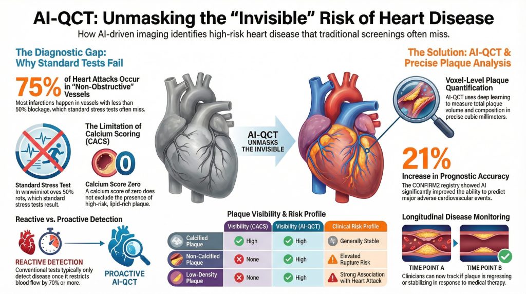
Insight 1: Autopsy and angiographic studies demonstrate that approximately 70–75% of myocardial infarctions occur in vessels with less than 50% 狭窄 prior to the index event, indicating that most patients who experience acute coronary syndromes would not have been identified as high risk by standard stress testing alone (2).
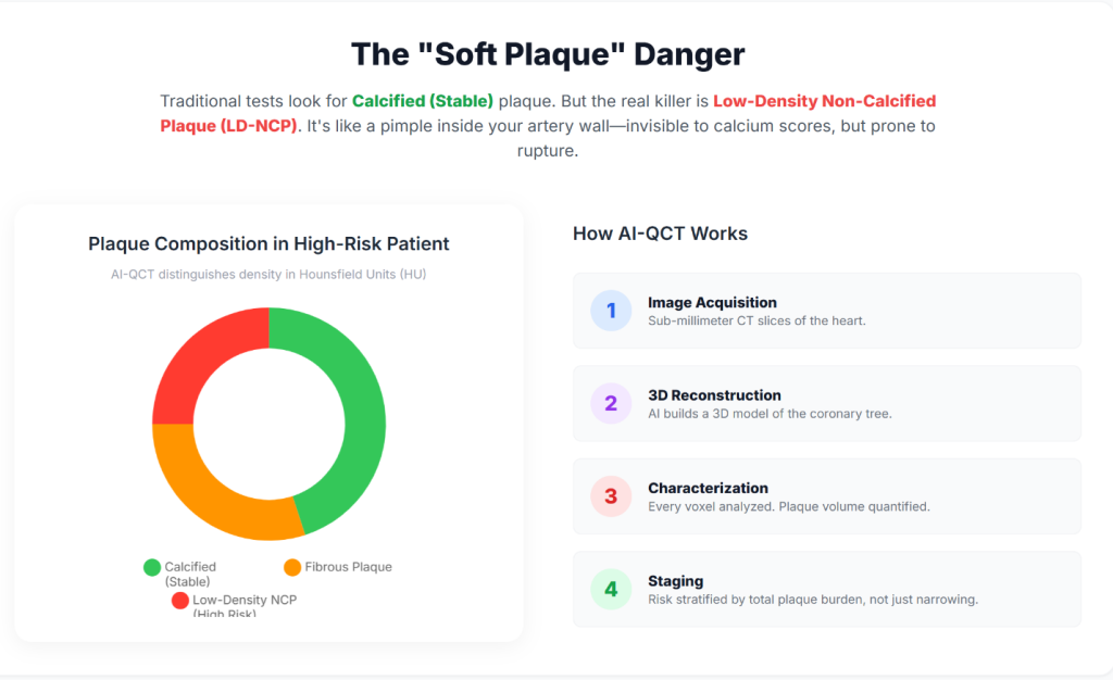
Insight 2: Coronary 動脈 calcium scoring (CACS) was developed to improve detection of subclinical disease by identifying 石灰化プラーク. However, CACS does not detect 非石灰化プラーク, and a カルシウムスコア of zero does not exclude the presence of lipid-rich, rupture-prone lesions (6).
Insight 3: AI-QCT enables non-invasive quantification of total coronary plaque burden, including low-density non-calcified plaque, which has been shown to carry a higher association with future cardiovascular events than stenosis severity alone (7).
| Plaque Characteristic | Visibility (CACS) | Visibility (Standard CCTA) | Visibility (AI-QCT) | Clinical Risk Profile |
| Calcified plaque | 高 | 高 | 高 | Generally stable; marker of chronic disease |
| Non-calcified plaque | None | Qualitative / limited | Quantitative / high | Elevated rupture risk |
| Low-density plaque | None | Variable | 高 | Strong association with ACS |
| Total plaque volume | None | Indirect estimate | Precise (mm³) | Strong predictor of future events |
Technical Architecture of AI-Enabled Coronary Analysis
AI-QCT platforms perform voxel-level analysis of the coronary tree rather than relying on visual interpretation alone. These systems employ deep-learning architectures—including convolutional neural networks (CNNs), three-dimensional U-Net segmentation models, and VGG-derived classifiers—trained on large, curated datasets that link imaging features with invasive validation and long-term clinical outcomes (8).
Insight 4: AI-QCT begins with acquisition of high-resolution CCTA. Contemporary multi-detector CT systems, including 64- and 640-slice scanners, provide the temporal and 空間解像度 necessary to minimize motion artifact and enable accurate segmentation of coronary anatomy (5).
Insight 5: Automated algorithms identify the coronary ルーメン and vessel wall and classify plaque components according to Hounsfield unit attenuation values, allowing differentiation of lipid-rich, fibrous, and calcified tissue (9).
Insight 6: AI-derived ischemia indices integrate plaque morphology, lesion length, vessel remodeling, and luminal geometry to estimate flow limitation, providing a non-invasive correlate of invasive fractional flow reserve (FFR) (10).
Clinical Validation and Peer-Reviewed Evidence
AI-QCT has been evaluated in multiple prospective trials, registries, and comparative studies published in leading cardiovascular and imaging journals, including Journal of the American College of Cardiology, Circulation: Cardiovascular Imaging, 、および European Heart Journal – Cardiovascular Imaging (11).
Landmark Trials and Registries
Insight 7: その CREDENCE trial demonstrated close agreement between AI-assisted CCTA analysis and invasive quantitative coronary angiography and FFR, supporting the diagnostic accuracy of AI-QCT for identifying hemodynamically significant disease (12).
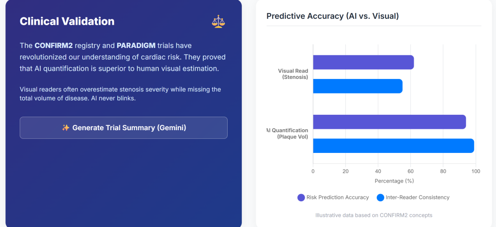
Insight 8: その CONFIRM2 registry, encompassing tens of thousands of patients across multiple countries, showed that AI-quantified plaque features identify a wide gradient of cardiovascular risk among patients with non-obstructive 冠動脈疾患 (7).
Insight 9: その CERTAIN study demonstrated that AI-QCT significantly improved diagnostic certainty compared with standard interpretation, resulting in more appropriate medical therapy and fewer unnecessary downstream tests (1).
| Study | N | Key Finding | P-value | Clinical Implication |
| CONFIRM2 | 6,550 | AUC for MACE improved from 0.62 to 0.75 | <0.001 | Improved prognostic accuracy |
| CREDENCE | 513 | High concordance with invasive FFR | <0.01 | Viable non-invasive ischemia assessment |
| PACIFIC-1 | 208 | AI exceeded expert readers | <0.05 | Reduced inter-observer variability |
| PROMISE (subset) | 4,347 | 41% stenoses reclassified | — | Reduced false positives |
Preventive Cardiology Implementation and Longitudinal Disease Monitoring
Integration of AI-QCT into preventive cardiology workflows shifts the clinical focus from episodic evaluation toward longitudinal monitoring of atherosclerotic disease.
Insight 10: Quantification of total plaque burden enables objective assessment of disease progression or regression over time, facilitating a treat-to-target approach guided by serial imaging (7).
Insight 11: AI-based comparison tools permit evaluation of changes in plaque composition, including stabilization or regression of 脂質富化プラーク in response to medical therapy (13).
Hardware Advances: Photon-Counting Computed Tomography
The performance of AI-QCT is dependent on the quality of input imaging data, which continues to improve with advances in CT hardware.
Insight 12: Photon-counting CT (PCCT) systems directly convert X-ray photons into electrical signals, reducing electronic noise and calcium blooming artifacts compared with conventional energy-integrating detectors (14).
Insight 13: PCCT provides higher spatial resolution with lower radiation and contrast dose, expanding eligibility for coronary CT imaging to patients with high 心拍数, elevated ボディ質量指数, or extensive coronary calcification (15).
Economic and Health System Implications
The adoption of AI-enabled cardiac imaging aligns with broader healthcare trends toward value-based care and prevention.
Insight 14: Market analyses project rapid growth of AI applications in cardiology, driven by improved diagnostic efficiency and potential reductions in downstream costs associated with acute coronary events (16).
Insight 15: Early identification of high-risk plaque phenotypes may reduce myocardial infarction rates and associated healthcare expenditures by enabling earlier and more targeted intervention (17).
Conclusions and Future Directions
Preventive cardiology is increasingly transitioning toward a biologically informed model that emphasizes plaque burden and composition rather than stenosis severity alone. AI-QCT provides a reproducible, quantitative framework for assessing coronary atherosclerosis before clinical events occur.
Insight 16: Future developments are likely to include integration of agentic AI systems, longitudinal digital heart models, and multimodal data streams incorporating genomic and metabolic information (18).
Insight 17: As AI assumes a greater role in image interpretation and quantification, clinicians may increasingly focus on higher-level clinical decision-making, risk communication, and individualized therapy optimization (19).
参考文献
- Nurmohamed NS, Cole JH, Budoff MJ, et al. Impact of atherosclerosis imaging-quantitative computed tomography on diagnostic certainty, downstream testing, coronary revascularization, and medical therapy: the CERTAIN study. Eur Heart J Cardiovasc Imaging. 2024;25(6):857-866. doi:10.1093/ehjci/jeae029
- Falk E, Shah PK, Fuster V. Coronary plaque disruption. Circulation. 1995;92(3):657-671. doi:10.1161/01.cir.92.3.657
- Global Burden of Cardiovascular Diseases and Risks 2023 Collaborators. Global, Regional, and National Burden of Cardiovascular Diseases and Risk Factors in 204 Countries and Territories, 1990-2023. J Am Coll Cardiol. 2025;86(22):2167-2243. doi:10.1016/j.jacc.2025.08.015
- Lloyd-Jones DM. Cardiovascular risk prediction: basic concepts, current status, and future directions. Circulation. 2010;121(15):1768-1777. doi:10.1161/CIRCULATIONAHA.109.849166
- Douglas PS, Hoffmann U, Patel MR, et al. Outcomes of anatomical versus functional testing for coronary artery disease. N Engl J Med. 2015;372(14):1291-1300. doi:10.1056/NEJMoa1415516
- Greenland P, Blaha MJ, Budoff MJ, Erbel R, Watson KE. Coronary Calcium Score and Cardiovascular Risk. J Am Coll Cardiol. 2018;72(4):434-447. doi:10.1016/j.jacc.2018.05.027
- Feuchtner GM, Lacaita PG, Bax JJ, et al. AI-Quantitative CT Coronary Plaque Features Associate With a Higher Relative Risk in Women: CONFIRM2 Registry. Circ Cardiovasc Imaging. 2025;18(6):e018235. doi:10.1161/CIRCIMAGING.125.018235
- Oikonomou EK, Khera R. Machine learning in precision diabetes care and cardiovascular risk prediction. Cardiovasc Diabetol. 2023;22(1):259. Published 2023 Sep 25. doi:10.1186/s12933-023-01985-3
- Koo BK, Yang S, Jung JW, et al. Artificial Intelligence-Enabled Quantitative Coronary Plaque and Hemodynamic Analysis for Predicting Acute Coronary Syndrome. JACC Cardiovasc Imaging. 2024;17(9):1062-1076. doi:10.1016/j.jcmg.2024.03.015
- Nørgaard BL, Leipsic J, Gaur S, et al. Diagnostic performance of noninvasive fractional flow reserve derived from coronary computed tomography angiography in suspected coronary artery disease: the NXT trial (Analysis of Coronary Blood Flow Using CT Angiography: Next Steps). J Am Coll Cardiol. 2014;63(12):1145-1155. doi:10.1016/j.jacc.2013.11.043
- Beerkens FJ, Tang GHL, Kini AS, et al. Transcatheter Aortic Valve Replacement Beyond Severe Aortic Stenosis: JACC State-of-the-Art Review. J Am Coll Cardiol. 2025;85(9):944-964. doi:10.1016/j.jacc.2024.11.051
- Griffin WF, Choi AD, Riess JS, et al. AI Evaluation of Stenosis on Coronary CTA, Comparison With Quantitative Coronary Angiography and Fractional Flow Reserve: A CREDENCE Trial Substudy. JACC Cardiovasc Imaging. 2023;16(2):193-205. doi:10.1016/j.jcmg.2021.10.020
- Zaman S, Wasfy JH, Kapil V, et al. The Lancet Commission on rethinking coronary artery disease: moving from ischaemia to atheroma. Lancet. 2025;405(10486):1264-1312. doi:10.1016/S0140-6736(25)00055-8
- Willemink MJ, Persson M, Pourmorteza A, Pelc NJ, Fleischmann D. Photon-counting CT: Technical Principles and Clinical Prospects. Radiology. 2018;289(2):293-312. doi:10.1148/radiol.2018172656
- Schiebler ML, Jinzaki M, Yanagawa M, et al. Future Applications of Cardiothoracic CT. Radiology. 2025;315(3):e240085. doi:10.1148/radiol.240085
- Windecker S, Gilard M, Achenbach S, et al. Device innovation in cardiovascular medicine: a report from the European Society of Cardiology Cardiovascular Round Table. Eur Heart J. 2024;45(13):1104-1115. doi:10.1093/eurheartj/ehae069
- Undas A, Musiał J, Ząbczyk M. Antiphospholipid antibodies and atherosclerotic vascular disease: recent advances. Rheumatol Int. 2025;45(12):279. Published 2025 Nov 29. doi:10.1007/s00296-025-06050-8
- Topol EJ. High-performance medicine: the convergence of human and artificial intelligence. Nat Med. 2019;25(1):44-56. doi:10.1038/s41591-018-0300-7
- Shetty NS, Gaonkar M, Pampana A, et al. Association of Pathogenic/Likely Pathogenic Inherited Cardiomyopathy Variants With Heart Failure: A TOPMed Multiancestry Analysis. Mayo Clin Proc. 2025;100(11):1948-1955. doi:10.1016/j.mayocp.2025.01.021

