
De Technologische Renaissance in de Preventieve Cardiologie – Kunstmatige Intelligentie, Kwantitatieve Coronaire Tomografie
Inleiding
De cardiovasculaire geneeskunde ondergaat een ingrijpende paradigmaverschuiving: er vindt een verschuiving plaats van een overwegend reactief, op symptomen gebaseerd model naar een preventief kader dat is gebaseerd op vroege opsporing en geïndividualiseerde risicobeoordeling. Centraal in deze transitie staat de integratie van kunstmatige intelligentie (AI) met coronaire computertomografie angiografie (CCTA), met name door de ontwikkeling van aderverkalking beeldvorming–kwantitatieve computertomografie (AI-QCT). Deze benadering adresseert een al lang bestaande beperking in de cardiologie: het onvermogen van traditionele diagnostische hulpmiddelen om betrouwbaar niet-obstructieve, lipiderijke kransslagaders te detecteren tandplak, ondanks zulke laesies verantwoordelijk zijn voor het grootste deel van acute coronaire syndromen en plotselinge hartdood evenementen (1).
De klinische relevantie van deze beperking is aanzienlijk. Uit epidemiologische en pathologische onderzoeken blijkt keer op keer dat de meeste myocardinfarcten ontstaan uit plaques die vóór de ruptuur de doorstroming niet beperkten (2). Zoals hart- en vaatziekten blijft de belangrijkste doodsoorzaak wereldwijd, waarbij wordt verwacht dat het aantal sterfgevallen per jaar tegen 2030 de 20 miljoen zal overschrijden, zijn verbeterde methoden om hoogrisico coronaire atherosclerose te identificeren voordat klinische gebeurtenissen optreden steeds noodzakelijker (3).

Evolutie van CardIale Risicostratificatie en de Biologie van Atherosclerose
Traditionele cardiovasculaire risicostratificatie is afhankelijk van indirecte merkers zoals serumlipidenwaarden, bloeddruk, glycemische controle, roken status en demografische variabelen opgenomen in populatiegebaseerde risicocalculators. Hoewel deze modellen op populatieniveau hun nut hebben bewezen, schieten ze vaak tekort in nauwkeurigheid wanneer ze worden toegepast op individuele patiënten, met name bij patiënten die ogenschijnlijk “normaal” zijn” risicofactor patiënten die niettemin een aanzienlijke subklinische aandoening hebben (4).
Atherosclerose is een chronische ontstekingsziekte van de vaatwand in plaats van een aandoening die uitsluitend wordt gekenmerkt door luminale obstructie. Plaque ontwikkelt zich in de intima en bestaat uit wisselende verhoudingen van lipiden, weeëweefsel, ontstekingscellen en calcium. Conventionele diagnostische tests — waaronder inspannings-ecg's, elektrocardiografie en invasieve coronairangiografie — detecteren de ziekte voornamelijk zodra deze optreedt hemodynamisch significante stenose, doorgaans gedefinieerd als een vernauwing van het luminaal van 70% of meer (5).
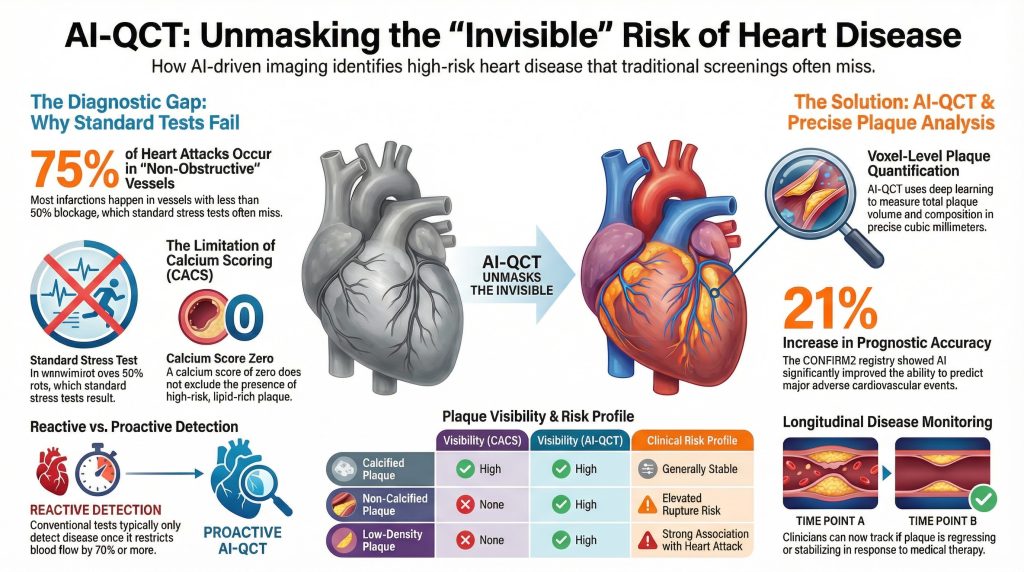
Inzicht 1: Uit autopsie- en angiografisch onderzoek blijkt dat ongeveer 70–75% van de hartinfarcten zich voordoet in bloedvaten met een diameter van minder dan 50% stenose vóór de indexgebeurtenis, wat erop wijst dat de meeste patiënten met acute coronaire syndromen niet als hoogrisicopatiënten zouden zijn aangemerkt op basis van alleen standaard inspanningstesten (2).
Inzicht 2: kransslagader- slagader calcium scoring (CACS) was developed to improve detection of subclinical disease by identifying verkalkte plaque. However, CACS does not detect niet-verkalkte plaque, and a calciumscore of zero does not exclude the presence of lipid-rich, rupture-prone lesions (6).
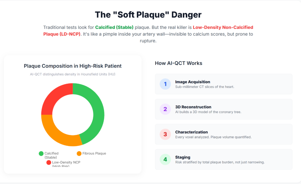
Insight 3: AI-QCT enables non-invasive quantification of total coronary plaquebelasting, inclusief low-density non-calcified plaque, which has been shown to carry a higher association with future cardiovascular events than stenosis severity alone (7).
| Plaque-kenmerk | Visibility (CACS) | Visibility (Standard CCTA) | Visibility (AI-QCT) | Clinical Risk Profile |
| Verkalkte plaque | Hoog | Hoog | Hoog | Generally stable; marker of chronic disease |
| Niet-verkalkte plaque | None | Qualitative / limited | Quantitative / high | Elevated rupture risk |
| Low-density plaque | None | Variabele | Hoog | Strong association with ACS |
| Totaal plaquevolume | None | Indirect estimate | Precise (mm³) | Strong predictor of future events |
Technical Architecture of AI-Enabled Coronary Analysis
AI-QCT platforms perform voxel-level analysis of the coronary tree rather than relying on visual interpretation alone. These systems employ deep-learning architectures—including convolutional neural networks (CNNs), three-dimensional U-Net segmentation models, and VGG-derived classifiers—trained on large, curated datasets that link imaging features with invasive validation and long-term clinical outcomes (8).
Insight 4: AI-QCT begins with acquisition of high-resolution CCTA. Contemporary multi-detector CT systems, including 64- and 640-slice scanners, provide the temporal and ruimtelijke resolutie necessary to minimize motion artifact and enable accurate segmentation of coronary anatomy (5).
Insight 5: Automated algorithms identify the coronary lumen and vessel wall and classify plaque components according to Hounsfield unit attenuation values, allowing differentiation of lipid-rich, fibrous, and calcified tissue (9).
Insight 6: AI-derived ischemie indices integrate plaque morphology, lesion length, vessel remodeling, and luminal geometry to estimate flow limitation, providing a non-invasive correlate of invasive fractionale stromingsreserve (FFR) (10).
Clinical Validation and Peer-Reviewed Evidence
AI-QCT has been evaluated in multiple prospective trials, registries, and comparative studies published in leading cardiovascular and imaging journals, including Journal of the American College of Cardiology, Circulation: Cardiovascular Imaging, en European Heart Journal – Cardiovascular Imaging (11).
Landmark Trials and Registries
Insight 7: De CREDENCE trial demonstrated close agreement between AI-assisted CCTA analysis and invasive kwantitatieve coronaire angiografie and FFR, supporting the diagnostic accuracy of AI-QCT for identifying hemodynamically significant disease (12).
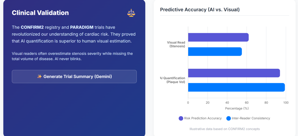
Insight 8: De CONFIRM2-register, encompassing tens of thousands of patients across multiple countries, showed that AI-quantified plaque features identify a wide gradient of cardiovascular risk among patients with non-obstructive kransslagaderziekte (7).
Insight 9: De BEPAALDE studie demonstrated that AI-QCT significantly improved diagnostic certainty compared with standard interpretation, resulting in more appropriate medical therapy and fewer unnecessary downstream tests (1).
| Studie | N | Belangrijkste bevinding | P-value | Clinical Implication |
| CONFIRM2 | 6,550 | AUC for MACE improved from 0.62 to 0.75 | <0.001 | Improved prognostic accuracy |
| CREDENCE | 513 | High concordance with invasive FFR | <0.01 | Viable non-invasive ischemia assessment |
| PACIFIC-1 | 208 | AI exceeded expert readers | <0.05 | Reduced inter-observer variability |
| PROMISE (subset) | 4,347 | 41% stenoses reclassified | — | Reduced false positives |
Preventive Cardiology Implementation and Longitudinal Disease Monitoring
Integration of AI-QCT into preventive cardiology workflows shifts the clinical focus from episodic evaluation toward longitudinal monitoring of atherosclerotic disease.
Insight 10: Quantification of total plaque burden enables objective assessment of disease progression or regression over time, facilitating a treat-to-target approach guided by serial imaging (7).
Insight 11: AI-based comparison tools permit evaluation of changes in plaque composition, including stabilization or regression of lipiderijke plaque in response to medical therapy (13).
Hardware Advances: Photon-Counting Computed Tomography
The performance of AI-QCT is dependent on the quality of input imaging data, which continues to improve with advances in CT hardware.
Insight 12: Foton-teldende CT systems directly convert X-ray photons into electrical signals, reducing electronic noise and calcium blooming artifacts compared with conventional energy-integrating detectors (14).
Insight 13: PCCT provides higher spatial resolution with lower radiation and contrast dose, expanding eligibility for coronary CT imaging to patients with high hartfrequenties, elevated body-massindex, or extensive coronary verkalking (15).
Economic and Health System Implications
The adoption of AI-enabled cardiac imaging aligns with broader healthcare trends toward value-based care and prevention.
Insight 14: Market analyses project rapid growth of AI applications in cardiology, driven by improved diagnostic efficiency and potential reductions in downstream costs associated with acute coronary events (16).
Insight 15: Early identification of high-risk plaque phenotypes may reduce myocardial infarction rates and associated healthcare expenditures by enabling earlier and more targeted intervention (17).
Conclusions and Future Directions
Preventive cardiology is increasingly transitioning toward a biologically informed model that emphasizes plaque burden and composition rather than stenosis severity alone. AI-QCT provides a reproducible, quantitative framework for assessing coronary atherosclerosis before clinical events occur.
Insight 16: Future developments are likely to include integration of agentic AI systems, longitudinal digital heart models, and multimodal data streams incorporating genomic and metabolic information (18).
Insight 17: As AI assumes a greater role in image interpretation and quantification, clinicians may increasingly focus on higher-level clinical decision-making, risk communication, and individualized therapy optimization (19).
Referenties
- Nurmohamed NS, Cole JH, Budoff MJ, et al. Impact of atherosclerosis imaging-quantitative computed tomography on diagnostic certainty, downstream testing, coronary revascularization, and medical therapy: the CERTAIN study. Eur Heart J Cardiovasc Imaging. 2024;25(6):857-866. doi:10.1093/ehjci/jeae029
- Falk E, Shah PK, Fuster V. Coronary plaque disruption. Circulation. 1995;92(3):657-671. doi:10.1161/01.cir.92.3.657
- Global Burden of Cardiovascular Diseases and Risks 2023 Collaborators. Global, Regional, and National Burden of Cardiovascular Diseases and Risk Factors in 204 Countries and Territories, 1990-2023. J Am Coll Cardiol. 2025;86(22):2167-2243. doi:10.1016/j.jacc.2025.08.015
- Lloyd-Jones DM. Cardiovascular risk prediction: basic concepts, current status, and future directions. Circulation. 2010;121(15):1768-1777. doi:10.1161/CIRCULATIONAHA.109.849166
- Douglas PS, Hoffmann U, Patel MR, et al. Outcomes of anatomical versus functional testing for coronary artery disease. N Engl J Med. 2015;372(14):1291-1300. doi:10.1056/NEJMoa1415516
- Greenland P, Blaha MJ, Budoff MJ, Erbel R, Watson KE. Coronary Calcium Score and Cardiovascular Risk. J Am Coll Cardiol. 2018;72(4):434-447. doi:10.1016/j.jacc.2018.05.027
- Feuchtner GM, Lacaita PG, Bax JJ, et al. AI-Quantitative CT Coronary Plaque Features Associate With a Higher Relative Risk in Women: CONFIRM2 Registry. Circ Cardiovasc Imaging. 2025;18(6):e018235. doi:10.1161/CIRCIMAGING.125.018235
- Oikonomou EK, Khera R. Machine learning in precision diabetes care and cardiovascular risk prediction. Cardiovasc Diabetol. 2023;22(1):259. Published 2023 Sep 25. doi:10.1186/s12933-023-01985-3
- Koo BK, Yang S, Jung JW, et al. Artificial Intelligence-Enabled Quantitative Coronary Plaque and Hemodynamic Analysis for Predicting Acute Coronary Syndrome. JACC Cardiovasc Imaging. 2024;17(9):1062-1076. doi:10.1016/j.jcmg.2024.03.015
- Nørgaard BL, Leipsic J, Gaur S, et al. Diagnostic performance of noninvasive fractional flow reserve derived from coronary computed tomography angiography in suspected coronary artery disease: the NXT trial (Analysis of Coronary Blood Flow Using CT Angiography: Next Steps). J Am Coll Cardiol. 2014;63(12):1145-1155. doi:10.1016/j.jacc.2013.11.043
- Beerkens FJ, Tang GHL, Kini AS, et al. Transcatheter Aortic Valve Replacement Beyond Severe Aortic Stenosis: JACC State-of-the-Art Review. J Am Coll Cardiol. 2025;85(9):944-964. doi:10.1016/j.jacc.2024.11.051
- Griffin WF, Choi AD, Riess JS, et al. AI Evaluation of Stenosis on Coronary CTA, Comparison With Quantitative Coronary Angiography and Fractional Flow Reserve: A CREDENCE Trial Substudy. JACC Cardiovasc Imaging. 2023;16(2):193-205. doi:10.1016/j.jcmg.2021.10.020
- Zaman S, Wasfy JH, Kapil V, et al. The Lancet Commission on rethinking coronary artery disease: moving from ischaemia to atheroma. Lancet. 2025;405(10486):1264-1312. doi:10.1016/S0140-6736(25)00055-8
- Willemink MJ, Persson M, Pourmorteza A, Pelc NJ, Fleischmann D. Photon-counting CT: Technical Principles and Clinical Prospects. Radiology. 2018;289(2):293-312. doi:10.1148/radiol.2018172656
- Schiebler ML, Jinzaki M, Yanagawa M, et al. Future Applications of Cardiothoracic CT. Radiology. 2025;315(3):e240085. doi:10.1148/radiol.240085
- Windecker S, Gilard M, Achenbach S, et al. Device innovation in cardiovascular medicine: a report from the European Society of Cardiology Cardiovascular Round Table. Eur Heart J. 2024;45(13):1104-1115. doi:10.1093/eurheartj/ehae069
- Undas A, Musiał J, Ząbczyk M. Antiphospholipid antibodies and atherosclerotic vascular disease: recent advances. Rheumatol Int. 2025;45(12):279. Published 2025 Nov 29. doi:10.1007/s00296-025-06050-8
- Topol EJ. High-performance medicine: the convergence of human and artificial intelligence. Nat Med. 2019;25(1):44-56. doi:10.1038/s41591-018-0300-7
- Shetty NS, Gaonkar M, Pampana A, et al. Association of Pathogenic/Likely Pathogenic Inherited Cardiomyopathy Variants With Heart Failure: A TOPMed Multiancestry Analysis. Mayo Clin Proc. 2025;100(11):1948-1955. doi:10.1016/j.mayocp.2025.01.021

