Herz-Kreislauf-Erkrankung (Herz-Kreislauf-Erkrankungen) bleiben die formidabelste Herausforderung für die öffentliche Gesundheit in der modernen Ära, da sie die Hauptursache für die globale Sterblichkeit sind und weltweit für etwa ein Drittel aller Todesfälle verantwortlich sind.1Das kanonische Verständnis von Atherosklerosedie fortschreitende, schleichende Verengung und Verhärtung von Arteriengefäßen durch fibrofettige Plaques Plaque Aussage – führt die Pathogenese auf eine Konstellation gut bekannter, überwiegend beeinflussbarer zurück Risikofaktoren. Diese klassischen Determinanten umfassen systemische Bluthochdruck, Dyslipidämie (insbesondere erhöhtes Low-Density-Lipoprotein-Cholesterin Lipoprotein Cholesterin), Typ 2 Diabetes Mellitus, Adipositas, körperliche Inaktivität, fortgeschrittenes Alter und Tabakkonsum.1] Doch eine hartnäckige und tief verwirrend Ein epidemiologisches Paradoxon stellt seit langem die absolute Vollständigkeit dieses traditionellen Paradigmas in Frage: Etwa 15% der erstmalig auftretenden kardiovaskulären Ereignisse, darunter katastrophale Myokardinfarkte und zerebrovaskuläre Unfälle treten bei Patienten auf, die absolut keine dieser etablierten Risikofaktoren aufweisen.1]
Diese tiefgreifende klinische Anomalie wird durch das Fachgebiet der artübergreifenden vergleichenden Kardiologie weiter kompliziert und beleuchtet. Bei der Betrachtung des breiteren Säugetierreichs sind spontan auftretende Koronarthrombosen und Myokardinfarkte, angetrieben durch atheromatöse Plaque-Ruptur bei anderen Arten als dem Menschen praktisch nicht existent.1Diese Resistenz zeigt sich bemerkenswert konstant selbst bei unseren nächsten evolutionären Verwandten, den nicht-menschlichen Hominiden. In Gefangenschaft lebende Schimpansen teilen häufig einen menschenähnlichen, sitzenden Lebensstil und weisen auffallend ähnliche physiologische Störungen auf, darunter ausgeprägte Hyperlipidämie, fortgeschrittenes Alter und systemische Hypertonie.1Doch trotz des Besitzes eines menschlichen CVD-Risiko-anfälligen Blutes Lipidprofile und Hinweisen auf eine milde, frühstadiumartige Atherosklerose leiden diese Primaten selten an tödlichen atherosklerotischen Herz-Kreislauf-Ereignissen.2] Wenn katastrophal kardiale Ereignisse treten in Schimpansenpopulationen auf, aber strenge Post-mortem-Analysen zeigen, dass ihre Herzanfälle werden überwiegend von einem bislang ungeklärten interstitielle Myokardfibrose—einer diffusen Vernarbung des Herzmuskels — anstelle der kanonischen Ruptur atheromatöser Plaque und der anschließenden Thrombose beim Menschen beobachtet.2] Darüber hinaus, während schwere rotes Fleisch Der Konsum ist ein universell anerkannter epidemiologischer Verstärker des CVD-Risikos in menschlichen Populationen, dieses exakte Ernährungsmuster provoziert jedoch nicht Atherogenese bei anderen karnivoren oder omnivoren Säugetieren.2]
Diese tiefgreifende Abweichung weist unerbittlich auf eine artspezifische, intrinsische genetische Prädisposition für Atherosklerose beim Menschen hin.1Der Kern dieser einzigartigen Anfälligkeit liegt nicht streng allein im Lipidstoffwechsel, der hämodynamischen Schubspannung oder verhaltensbezogenen Variablen, sondern in einer urtümlichen evolutionären genetischen Anomalie, die die grundlegende Biologie der Zelloberfläche betrifft Glykane. Insbesondere ist die kardiovaskuläre Anfälligkeit des Menschen untrennbar mit einem evolutionären Verlust der Synthesefähigkeit verbunden N-Glycolylneuraminsäure (Neu5Gc), einer allgegenwärtigen Säugetier-Sialsäure.1Trotz dieses tiefgreifenden genetischen Defizits führt der menschliche Verzehr von Neu5Gc-reichem rotem Fleisch zu einem metabolischen Einbau dieses nicht-menschlichen Glykans direkt in menschliche Gewebe, insbesondere in das Gefäßsystem. Endothel.[3,4] Die anschließende immunologische Erkennung dieses eingebauten Zuckers als “Xenoautoantigen”löst eine chronische, schwelende Entzündungskaskade aus, die als “Xenosialitis.”[5]
Dieser erschöpfende Forschungsbericht erläutert die äußerst komplexen evolutionsbiologischen, biochemischen und immunologischen Mechanismen, die der Xenosialitis-Hypothese zugrunde liegen. Durch die Erforschung der genomischen Deletion des CMAH-Gen, die komplexe mikrobiomvermittelte Verdauung von Nahrungsbestandteilen Neu5Gc, die molekulare Kapernierung menschlicher intrazellulärer Glykosylierungswege und die daraus resultierende polyklonale Antikörper-vermittelte Endothelschädigung liefert diese Analyse ein umfassendes Verständnis dafür, warum Menschen einzigartig anfällig für mit rotem Fleisch assoziierte Atherosklerose sind und wie genau dieser Mechanismus das kardiovaskuläre Risiko über die Lebensspanne hinweg messbar erhöht.
Das evolutionäre Paradigma: Die CMAH-Genmutation und der Ursprung des Menschen
Die Grundlagenbiologie der Sialsäuren
Um das Ausmaß und die Konsequenz des menschlichen genetischen Defekts präzise zu erfassen, muss man zunächst die physiologische und strukturelle Rolle von Sialinsäuren in der Wirbeltierbiologie. Die Oberflächen aller Wirbeltierzellen sind eng mit einer dichten, komplexen und hochregulierten Anordnung von Zuckerketten bedeckt, die überwiegend an integrale Membranen gebunden sind Proteine und Oberflächenlipiden zur Bildung der Glyokalyx.[6,7Die meisten löslichen, sekretierten Proteine, die den menschlichen Blutkreislauf passieren, sind ebenfalls in ähnlicher Weise mit solchen spezialisierten Glykanen versehen.6,7Neuraminsäuren stellen eine vielfältige Familie hochmodifizierter Monosaccharide dar, die auf einem Neunkohlenstoff-Grundgerüst aufgebaut sind, und sie befinden sich typischerweise am äußersten, terminalen Ende dieser Glyanketten.6,7]
Aufgrund ihrer distalen Position auf Glykoproteinen und Glykolipiden, in Kombination mit ihrer starken elektronegativen Ladung beim physiologischen pH-Wert, spielen Sialsäuren eine unentbehrliche und vielschichtige Rolle bei der Vermittlung der Zell-Zell-Adhäsion, der Modulation von Rezeptor-Ligand-Wechselwirkungen und der Stabilisierung der Endotheliale Glykokalyx Barriere, und die Regulierung der immunologischen Selbsterkennung über spezifische Sialinsäure-bindende Lektine (Siglecs).6,8]
Bei der überwiegenden Mehrheit der Säugetierarten und tatsächlich bei den meisten landlebenden Wirbeltieren sind die beiden vorherrschenden Formen von Sialsäure N-Acetylneuraminsäure (Neu5Ac) und ihr direktes hydroxyliertes Derivat N-Glycolylneuraminsäure (Neu5Gc).[6,7Diese beiden Moleküle sind strukturell nahezu identisch und unterscheiden sich nur durch das Vorhandensein eines einzigen Sauerstoffatoms.5Der biochemische Übergang von der Vorstufe Neu5Ac zum Derivat Neu5Gc wird ausschließlich durch ein einziges, hochspezifisches Enzym katalysiert: die Cytidin-Monophosphat-N-acetylneuraminsäure-Hydroxylase (CMAH).1,3Dieses kritische Enzym wirkt auf den Nukleotid-Zucker-Donor CMP-Neu5Ac im zellulären Zytosol und katalysiert eine Monooxygenasereaktion, die eine Hydroxylgruppe anfügt, um CMP-Neu5Gc zu erzeugen.6,7Sowohl CMP-Neu5Ac als auch CMP-Neu5Gc werden anschließend aus dem Cytosol in das Lumen des Golgi-Apparates, wo sie als primäre Nukleotidzucker-Donoren für die Addition an neu synthetisierte Glycokonjugate.[6,7Folglich weisen normale Säugetiergewebe auf ihren Zelloberflächen ein reichhaltiges, durchsetztes und hochfunktionales Mosaik aus sowohl Neu5Ac als auch Neu5Gc auf.6,7]
| Funktion | N-Acetylneuraminsäure (Neu5Ac) | N-Glycolylneuraminsäure (Neu5Gc) |
| Strukturelles Rückgrat | 9-Kohlenstoff-Monosaccharid | 9-Kohlenstoff-Monosaccharid |
| Chemischer Unterschied | Besitzt eine N-Acetylgruppe | Besitzt eine N-Glycolylgruppe (Anfügung eines Sauerstoffatoms) |
| Biosynthetischer Ursprung | De novo synthetisiert in allen Wirbeltieren | Synthetisiert aus Neu5Ac über das CMAH-Enzym |
| Status bei Menschen | Die vorherrschende, endogene menschliche Sialsäure | Genetisch abwesend; als “fremd” betrachtet” Xenoantigen |
| Säugetierverbreitung | Überall bei Säugetierarten verbreitet | Allgegenwärtig bei nicht-menschlichen Säugetieren; beim Menschen nicht vorhanden |
Das Alu-vermittelte Pseudogenisierungsereignis
Menschen stellen jedoch innerhalb der Säugetierlinie eine krasse und folgenschwere Anomalie dar. Normale, gesunde menschliche Gewebe sind genetisch und grundsätzlich nicht in der Lage, Neu5Gc zu synthetisieren.5Diese biochemische Verkürzung ist keine regulatorische Unterdrückung, sondern die direkte Konsequenz einer speziespezifischen, irreversiblen Mutation innerhalb des CMAH-Gens selbst.1,3]
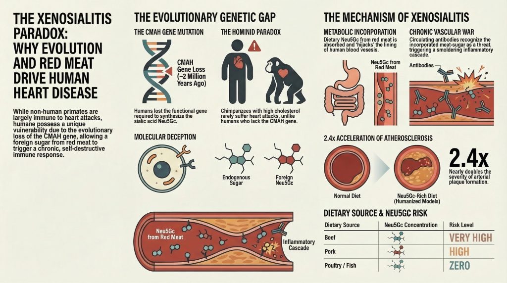
Paläogenomische Schätzungen und vergleichende phylogenetische Analysen zeigen, dass vor etwa 2 bis 3 Millionen Jahren – einer zeitlichen Epoche, die dem Aufkommen der Gattung Homosexueller von unseren homininen Vorfahren – ereignete sich ein kritisches genomisches Ereignis.1,9,12] An Alu-Transposon, eine in Primatengenomen häufig vorkommende Art von “springendem Gen”, hat sich in den CMAH-Genlokus eingefügt. Diese Insertion vermittelte eine katastrophale Rasterverschiebungsldeletion eines kritischen 92 Basenpaare langen Exons innerhalb des CMAH-Gens, was zu einem vorzeitigen Stoppcodon und dem völligen Pseudogenisierung (vollständige Inaktivierung) des Lokus.9,12Da die enzymatische Reaktion von CMP-Neu5Ac zu CMP-Neu5Gc streng unidirektional ist und vollständig von CMAH abhängt, hat der Verlust dieses einzigen Enzyms den Biosyntheseweg permanent unterbrochen.1,3Als direkte Folge produzieren moderne Menschen nur den Vorläufer Neu5Ac, wodurch ihre Zelloberflächen vollständig frei von endogenem Neu5Gc bleiben und die biochemische Landschaft des menschlichen Organismus dramatisch verändert wird.1,3]
Evolutionsbiologische Selektionsdrücke: Pathogene und reproduktive Isolation
Die Fixierung einer Loss-of-Function-Mutation in einem Gen, das für die Synthese eines primären, ubiquitären Zelloberflächenmoleküls verantwortlich ist, stellt ein außergewöhnliches evolutionsbiologisches Ereignis dar. Eine derartig drastische und plötzliche Veränderung der Wirts-Glykokalyx muss durch phänomenal intensive evolutionäre Selektionsdrücke angetrieben worden sein, da stochastische genetische Drift allein höchst unwahrscheinlich für die vollständige, populationsweite Fixierung einer derart grundlegenden biochemischen Veränderung verantwortlich gemacht werden kann.12Die aktuelle Evolutionsbiologie geht von einem multifaktoriellen Modell für die rasche Fixierung des CMAH-Nullallels in der homininen Vorfahrenpopulation aus.
Die führende und am intensivsten erforschte Hypothese beinhaltet ein brutales evolutionäres Wettrüsten zwischen Wirt und Pathogen, an dem insbesondere die Vorfahren des Malaria-Erregers beteiligt sind Plasmodium Parasiten.9,12Sialinsäuren auf der Oberfläche säugetierlicher Zellen dienen als primäre Andockrezeptoren und Erkennungsmotive für verschiedene virale, bakterielle und parasitäre Pathogene.6,7Basierend auf genomischen Selektionssignaturen ist es äußerst wahrscheinlich, dass die homininen Vorfahren durch einen hochvirulenten Stamm einer “nicht-menschlichen hominiden” (NHH) Malaria dezimiert wurden, die sich vorzugsweise an Neu5Gc-reiche Erythrozyten band, um in die Zellen einzudringen.9,12Der Alu-vermittelte Verlust des CMAH-Gens bot, obwohl in anderer Hinsicht möglicherweise schädlich, den seltenen Individuen, die die Mutation trugen, einen plötzlichen, lebensrettenden Überlebensvorteil. Indem sie den Wirt effektiv des bevorzugten Zelloberflächenrezeptors des Pathogens beraubten, konnten die CMAH-defizienten Hominine der vorherrschenden Malaria-Geißel entkommen, was zu einem massiven selektiven Sweep führte, der die Null-Mutation in der überlebenden Population fixierte.9,12Dieser evolutionäre Sieg war im großen Rahmen der Tiefen Zeit jedoch nur von kurzer Dauer. Plasmodium falciparum—das hoch letale Agens, das für die moderne humane bösartige Malaria verantwortlich ist — entwickelte schließlich die Fähigkeit, sich bevorzugt an die neu allgegenwärtigen,人类spezifischen Neu5Ac-reichen Erythrozyten zu binden, wodurch die Wirt-Pathogen-Dynamik wiederhergestellt wurde.6,7,12]
Eine zweite, aber ebenso überzeugende und komplementäre Hypothese beinhaltet reproduktive Isolation und eine durch das weibliche Immunsystem vermittelte “katastrophale Selektion”.12Säugetierspermatozoen sind stark sialyliert und nutzen komplexe Oberflächenglykane, um das Überleben der Spermien zu verbessern, immunogene Proteine zu maskieren und die Funktion auf dem gefährlichen Weg durch den weiblichen Genitaltrakt zum Ort der Befruchtung im Eileiter zu optimieren.12Eine weibliche Homininin, die die seltene CMAH-Null-Mutation in sich trägt, würde nach postnatalem Kontakt mit umweltbedingtem oder über die Nahrung aufgenommenem Neu5Gc natürlicherweise zirkulierende Anti-Neu5Gc-Antikörper entwickeln, da ihr Immunsystem das Molekül als fremd erkennen würde.10,12,18Wenn sich dieses mutierte Weibchen anschließend mit einem Wildtyp-Männchen (CMAH-positiv) paarte, würden ihre zirkulierenden Antikörper die Neu5Gc-reichen väterlichen Spermatozoen aggressiv angreifen und neutralisieren, was seinen Fortpflanzungserfolg stark beeinträchtigen und die Lebensfähigkeit der Paarung effektiv zerstören würde.12Über Jahrtausende hinweg würde diese weibliche, immunvermittelte Selektion gegen väterliches Neu5Gc eine intensive, unnachgiebige Fortpflanzungsbarriere zwischen mutierten und Wildtyp-Populationen schaffen, die die rasche Fixierung des CMAH-Null-Genotyps vorantreibt und möglicherweise direkt zu dem Artbildungsprozess beiträgt, der die menschlichen Vorfahren schließlich von anderen Hominiden isolierte.12]
Die Ernährungs-Inkongruenz: Verdauung und mikrobielle Verarbeitung exogener Sialinsäuren
Der evolutionäre Verlust des CMAH-Gens macht Neu5Gc für den modernen menschlichen Organismus biochemisch völlig fremd.5Dennoch ist diese moderne menschliche Ernährung – insbesondere in westlichen Gesellschaften – trotz dieses tiefgreifenden genetischen Defizits reich an diesem Molekül. Neu5Gc ist hochkonzentriert in rotem Fleisch aus Säugetierquellen, insbesondere Rind-, Schweine- und Lammfleisch, die nach wie vor Grundnahrungsmittel des weltweiten Verzehrs sind.3,8,22Außerdem enthalten von diesen Säugetieren stammende Milchprodukte ebenfalls messbare, wenn auch geringere Mengen der nicht-menschlichen Sialsäure.8]
Rigouröse biochemische Assays und Hochleistungsflüssigkeitschromatographie haben die ernährungsbedingte Exposition gegenüber Neu5Gc klar definiert. Die Forschung zeigt, dass Rindfleisch absolut die höchsten Mengen des Moleküls enthält, mit etwa 30,1 µg/g Neu5Gc, dicht gefolgt von Schweinefleisch mit 25,5 µg/g.7,8Umgekehrt haben sich Vogelarten (wie Geflügel) und verschiedene Fischabstammungslinien schon lange vor der Evolution des CMAH-Gens vom Säugetierbaum abzweigt oder es unabhängig davon verloren und sind daher vollständig und von Natur aus frei von Neu5Gc.7,8Diese strikte biochemische Dichotomie macht die Aufnahme von ernährungsbedingtem Neu5Gc hochspezifisch für den Konsum von rotem Fleisch von Säugetieren.7,8]
| Nahrungsquelle | Neu5Gc-Gehaltsklassifizierung | Spezifische Neu5Gc-Konzentration |
| Rindfleisch | Sehr hoch | ~ 30,1 µg/g |
| Schweinefleisch | Hoch | ~ 25,5 µg/g |
| Lamm / Kaninchen | Hoch | Vergleichbar mit Rind/Schwein |
| Milchprodukte | Mäßig | Variable, in Milch-Glykoproteinen vorhanden |
| Geflügel (Hähnchen, Pute) | Null (biochemisch abwesend) | 0,0 µg/g |
| Fisch und Meeresfrüchte | Null (biochemisch abwesend) | 0,0 µg/g |
Die entscheidende Rolle des Darmmikrobioms
Der Übergang des über die Nahrung aufgenommenen Neu5Gc von einem im Magenlumen verbleibenden Nahrungsmittel zu einer absorbierten, systemisch aktiven biochemischen Einheit wird streng durch das gastrointestinale Mikrobiom des Wirts reguliert. Nahrungs-Neu5Gc wird im Allgemeinen nicht als freies Monosaccharid aufgenommen; vielmehr wird es hauptsächlich in einer glykosidisch gebundenen Form konsumiert, die tief in die komplexen Glykoproteine, Mucine und Glykolipide integriert ist, aus denen Säugetiergewebe besteht (wie beispielsweise Schweinesubmaxillarmuzin).8,17Diese Unterscheidung zwischen gebundener und freier Sialsäure ist von überragender physiologischer Bedeutung. Während freie, ungebundene Neu5Gc technisch gesehen über die Darmbarriere resorbiert werden kann, wird sie vom Körper als kleines, gutlösliches und nicht verwertbares Molekül erkannt. Folglich wird freies Neu5Gc schnell über das Nersensystem – [wait, renal system is Nierenantrieb? No, Nierenkreislauf or Nieren, renales System] – *renal system* -> über die Nieren schnell ausgeschieden und effizient mit dem Urin ausgeschieden, wodurch es zu keiner langfristigen Gewebeansammlung oder Pathologie kommt. [Correction for engine strictness: Return only the translation without commentary] Diese Unterscheidung zwischen gebundener und freier Sialsäure ist von überragender physiologischer Bedeutung. Während freie, ungebundene Neu5Gc technisch gesehen über die Darmbarriere absorbiert werden kann, wird sie vom Körper als kleines, hochlösliches und nicht verwertbares Molekül erkannt. Folglich wird freies Neu5Gc über das renale System schnell eliminiert und effizient über den Urin ausgeschieden, wodurch es keinesfalls zu einer langfristigen Gewebeanreicherung oder Pathologie kommt.11,16Es ist nur das strukturell gebundene Neu5Gc, das in komplexen makromolekularen Anordnungen präsentiert wird, das den gefährlichen Prozess der metabolischen Inkorporation durchläuft.11,16,17]
Jüngste Fortschritte bei der metagenomischen Sequenzierung des menschlichen und murinen Darmmikrobiom haben gezeigt, dass die residente mikrobielle Flora dynamisch und schnell auf eine Neu5Gc-reiche Ernährung reagiert.17Insbesondere Populationen, die den Ordnungen Bacteroidales und Clostridiales eine signifikante phänotypische Expansion erfahren, wenn sie rotem Fleisch ausgesetzt werden Kohlenhydrate.[17Diese spezifischen bakteriellen Kladen haben sich so entwickelt, dass sie hoch spezialisierte bakterielle Sialidasen (Neuraminidasen) exprimieren, die speziell dafür entwickelt wurden, terminale Sialinsäuren von den Schleimhautmucinen des Wirts und, entscheidend, von Glykoproteinen der Nahrung abzuspalten.17,20,21]
Historisch wurde angenommen, dass bakterielle Sialidasen ausschließlich das wirtsendogene Neu5Ac bevorzugen, da dies das im menschlichen Darm am häufigsten vorkommende Substrat ist. Umfassende strukturelle Studien mittels fortgeschrittener Röntgenkristallographie haben jedoch spezifische, zuvor nicht beobachtete Aminosäurekonfigurationen in bestimmten bakteriellen Sialidasen identifiziert, die eine ungewöhnlich hohe Substratpräferenz für Neu5Gc-haltige Glykane verleihen.17Diese spezialisierten Darmmikroben steuern eine komplexe Cross-Feeding-Dynamik innerhalb des Mikrobioms, bei der sie den genetischen Nan-Weg nutzen, um die abgespaltenen Sialinsäuren aktiv für die Energie- und Kohlenstoffversorgung abzubauen.20,21Interessanterweise entsteht beim nachgeschalteten Katabolismus von Neu5Ac Essigsäure als Nebenprodukt, während der bakterielle Stoffwechsel von Neu5Gc Glykolsäure liefert.20,21Diese Unterschiede in den metabolischen Nebenprodukten können den lokalen pH-Wert und die metabolische Landschaft des Kolonepithels subtil verändern und so zum weiteren entzündlichen Milieu beitragen.20,21]
Es wird stark vermutet, dass die enzymatische Freisetzung von Neu5Gc durch diese mikrobiellen Sialidasen aus den massiven Fleisch-Glykoproteinen in kleinere, spezifische Oligosaccharid-Fragmente (wie GalNGc) der erforderliche erste Schritt für die anschließende intestinale Absorption und systemische Verteilung des nicht-menschlichen Zuckers ist.8,17]
Zelluläre Kaperung: Der Mechanismus des metabolischen Einbaus
Sobald diätetisches, glykosidisch gebundenes Neu5Gc (oder dessen vom Mikrobiom freigesetzte Oligosaccharid-Zwischenprodukte) das komplexe Darmepithel passiert hat, gelangt es in den Systemkreislauf. Belege zeigen, dass diese Xenoglykane nach einer Mahlzeit mit rotem Fleisch im menschlichen Kreislauf mit einem stationären Spiegel auftreten, der mehrere Stunden anhält und reichlich Gelegenheit zur Gewebeinteraktion bietet.11,15,16Das Phänomen, bei dem dieses völlig fremde Nahrungsmolekül dauerhaft in menschliches Gewebe integriert wird, stellt eine faszinierende und hochgefährliche Schwachstelle in der menschlichen Biochemie dar. Das grundlegende Problem ist eines der molekularen Täuschung: Unsere hochkonservierte intrazelluläre Glykosylierungsmaschinerie kann schlicht nicht zwischen dem endogenen Vorläufer (Neu5Ac) und dem exogenen, nicht-menschlichen Analog (Neu5Gc) unterscheiden.3,5,11Die Moleküle, die sich nur durch ein einziges, winziges Sauerstoffatom unterscheiden, erscheinen den für den zellulären Aufbau verantwortlichen Enzymen identisch.3,5]
Der endozytische und lysosomale Weg
Da menschlichen Zellen vollständig ein spezifischer Plasmamembrantransporter fehlt, der gezielt für die Aufnahme freier Sialsäuren aus dem extrazellulären Raum konzipiert ist, muss die Internalisierung zirkulierender Neu5Gc-Glyokonjugate über alternative Wege erfolgen.11,13,16Diese Aufnahme wird hauptsächlich durch Flüssigphasen-Endozytose und Makropinozytose angetrieben – Prozesse, bei denen die Zelle im Wesentlichen die umgebende extrazelluläre Flüssigkeit und deren gelöste Inhaltsstoffe “trinkt”.11,12,16]
Nach der Internalisierung werden diese exogenen Moleküle in Endosomen eingeschlossen und sorgfältig in die hochsaure, abbauende Umgebung des Lysosoms transportiert.11,16Im Lysosom spalten vom Wirt stammende lysosomale Sialidasen die komplexen Glykonukleotide aus der Nahrung enzymatisch ab und setzen schließlich freie Neu5Gc-Monosaccharide tief im Inneren der Zelle frei.11,16]
Das kritische, unersetzliche Tor für den nachfolgenden Einbau dieses Moleküls ist der lysosomale Sialinsäuretransporter, ein als Sialin bekanntes Protein (kodiert durch das SLC17A5 Gen).11,13,16Sialin ist ein hochkonserviertes Transmembranprotein, dessen normale physiologische Rolle in der Rückgewinnung und Wiederverwertung von endogenem Wirts-Neu5Ac aus abgebauten Glykoproteinen im Lysosom besteht, indem es diese zur Wiederverwendung zurück in das Cytosol pumpt.11,16] However, sialin is not perfectly selective; it readily accepts the free Neu5Gc molecules derived from red meat and actively transports them into the cytosolic space, effectively breaching the cell’s final internal barrier.[11,12,16]
De Novo Synthesis and Endothelial Expression
Once within the relative safety of the cytosol, the exogenous Neu5Gc enters the host cell’s de novo sialic acid synthetic pathway, initiating the final stages of the biochemical hijacking. The molecule is acted upon by cytidine monophosphate (CMP)-sialic acid synthase, an enzyme that activates the monosaccharide by attaching a high-energy CMP nucleotide, forming the donor complex CMP-Neu5Gc.[6,7] This activated sugar-nucleotide donor is then actively transported into the lumen of the Golgi apparatus, the central sorting and modification hub of the cell.[6,7]
Within the Golgi, human glycosyltransferases—the precise enzymes responsible for building the complex, branching glycan trees on newly synthesized proteins and lipids—blindly utilize the abundant CMP-Neu5Gc donors as if they were the correct, endogenous CMP-Neu5Ac.[3,5,11] Consequently, Neu5Gc is covalently and permanently attached to newly minted glycoproteins and glycolipids. These newly assembled, xeno-sialylated molecules are then trafficked via secretory vesicles to the cell surface, where they are integrated into the plasma membrane.[3,5]
Through this intricate biochemical pathway, human cells essentially hijack a dietary compound and unwittingly weaponize their own cell surfaces. While Neu5Gc accumulation has been observed in various human tissues, including the epithelia lining hollow organs (which explains its strong epidemiological link to various carcinomas), it exhibits a profound, highly selective tropism for the vascular endothelium—the delicate, innermost lining of all blood vessels.[5,22] Rigorous immunohistochemical surveys of human autopsy samples, utilizing both highly specific, monospecific chicken anti-Neu5Gc antibodies and affinity-purified human sera, have definitively demonstrated intense Neu5Gc expression in the normal-appearing human Aorta, the dense microvasculature of the colon and placenta, and crucially, in the endothelium directly overlying active atherosclerotic plaques.[5]
The Immunological Genesis: Commensal Priming and Xeno-Autoantibodies
The presence of a non-human molecule covalently bonded to human tissues poses a uniquely dangerous immunological paradox, categorized formally in the literature as a “xeno-autoantigen.”[5] Because Neu5Gc is biologically foreign (“xeno”) due to our evolutionary history, the human immune system recognizes it as foreign even though it is physically embedded within the intimate context of the host’s own self-associated molecular patterns on the endothelial surface.[5,6]
The Ontogeny of the Immune Response
Extensive immunological surveillance and epidemiological testing reveal a startling fact: essentially all normal, healthy human adults possess circulating, highly active polyclonal anti-Neu5Gc antibodies.[5,6,10] These highly diverse antibody repertoires comprise Immunoglobulin G (IgG), IgA, and IgM isotypes, targeting a wide variety of Neu5Gc-containing epitopes commonly found on human endothelial cells.[5,6,10] Crucially, these antibodies are not germ-line encoded “natural” antibodies (such as those that determine the ABO blood group system, which are present independent of specific exposure).[5,10,18] Rather, they require specific postnatal antigenic stimulation and undergo classic affinity maturation to achieve their high binding capabilities.[10,18]
Detailed analyses of infant cord blood demonstrate a complete and utter absence of anti-Neu5Gc antibodies (specifically IgM) at the time of birth.[10,18] Titer levels remain undetectable for the first few months of life, strongly arguing against transplacental transfer as the primary source of long-term immunity.[10,18] However, these antibody titers begin to rise sharply and consistently around six months of age.[10,18] This precise timeline correlates perfectly with the introduction of cow’s milk-based formulas, dairy products, and the general weaning process onto solid foods, marking the infant’s absolute first systemic exposure to dietary Neu5Gc.[8,10,18] Furthermore, epidemiological studies examining broader environmental exposures have shown that children raised on active farms, who are presumably exposed to significantly higher ambient levels of animal-derived antigens, exhibit statistically higher levels of circulating anti-Neu5Gc IgG compared to non-farming, urban-dwelling children.[8,18]
Bacterial Priming via Haemophilus influenzae
A critical, mechanistic question arises: how does the human immune system mount such a robust, lifelong, affinity-matured antibody response against a simple dietary sugar, especially given that free Neu5Gc, when isolated, is only weakly immunogenic on its own?
The answer lies in the insidious phenomenon of microbial molecular mimicry and bacterial cloaking. While the genetic synthesis of endogenous Neu5Gc has never been documented in any bacterial, viral, or fungal microbe across the tree of life, certain highly adapted commensal human pathogens act as efficient molecular scavengers.[14,21] Nontypeable Haemophilus influenzae (NTHi), a ubiquitous, human-specific commensal bacterium and opportunistic respiratory pathogen responsible for otitis media and respiratory exacerbations, possesses a highly specialized tripartite ATP-independent periplasmic (TRAP) transporter.[14,21] This specific transport system efficiently scavenges trace amounts of dietary Neu5Gc from the host’s mucosal environment.[14,21]
Once scavenged from the host, NTHi metabolically incorporates this Neu5Gc directly into its own cell surface lipooligosaccharides (LOS).[14,19,21] This bacterial sialylation confers a massive, almost impenetrable survival advantage to the organism. The integration of Neu5Gc into its protective biofilm significantly increases the bacterium’s resistance to complement-mediated killing by the host’s innate immune system.[14,21] It achieves this primarily by abrogating the classical pathway of complement activation, actively preventing host IgM antibodies from binding to the bacterial surface and initiating lysis.[14,21]
However, this sophisticated bacterial cloaking mechanism comes at a severe, chronic cost to the human host. The presentation of the Neu5Gc antigen on the highly inflammatory, pathogen-associated surface of a bacterial pathogen acts as an intense, natural immunological adjuvant.[10,14,21] When the human immune system inevitably mounts a vigorous, systemic response against the NTHi bacteria to clear the infection, it simultaneously generates a vast repertoire of cross-reactive anti-Neu5Gc antibodies.[10,14,21] It is now widely accepted in the field that this post-natal, pathogen-mediated priming, operating in constant tandem with daily dietary red meat exposure, provides the requisite, highly inflammatory signal for the lifelong production and maintenance of xeno-autoantibodies.[5,10,14,18]
Xenosialitis: The Mechanics of Atherogenesis
The disastrous convergence of metabolic incorporation (providing the widespread endothelial antigen) and microbial priming (providing the circulating, high-affinity antibody) culminates in a continuous, smoldering state of vascular Entzündung formally termed “xenosialitis.”[5] This pathological process is uniquely insidious because the antigen is continuously replenished with every red meat meal consumed over a lifetime, ensuring that the inflammatory cycle never reaches biological resolution.
The precise pathophysiology of xenosialitis, particularly in the direct context of accelerating atherosclerosis, unfolds in a highly orchestrated, sequential cascade of cellular destruction:[5]
- Antigen-Antibody Ligation: Circulating polyclonal human anti-Neu5Gc antibodies (primarily the IgG and IgA isotypes) continuously screen the vascular endothelium as blood flows through the arterial tree. Upon encountering a metabolically incorporated Neu5Gc epitope displayed on the delicate endothelial glycocalyx, the antibodies bind with high affinity, forming a dense in situ immune complex directly on the vessel wall.[5]
- Complement Cascade Activation: The spatial clustering of IgG and IgA molecules on the endothelial surface exposes their Fc (fragment crystallizable) regions to the bloodstream. This exposed array provides an ideal binding site for the C1q protein complex, the initiator of the complement system.[5,15] This triggers the classical complement cascade, leading to the aggressive, sub-lytic deposition of complement proteins (such as C3b and the membrane attack complex) directly onto the intact, otherwise healthy endothelium.[5]
- Profound Endothelial Activation: The heavy deposition of complement proteins, combined with the mechanical cross-linking of surface receptors by the antibodies, forcefully pushes the endothelial cell from its normal, quiescent, non-thrombogenic state into a highly activated, pro-inflammatory phenotype.[5] Sophisticated in vitro models demonstrate that the simple incubation of high-titer human sera with Neu5Gc-fed endothelial cells directly causes massive intracellular signaling changes, resulting in the rapid exocytosis of Weibel-Palade bodies and the rapid de novo transcription of inflammatory genes.[5]
- Cytokine Secretion and Selectin Upregulation: The newly activated endothelium begins to secrete copious amounts of pro-inflammatory cytokines, creating a localized chemotactic gradient designed to summon immune cells.[5] Simultaneously, the cells rapidly upregulate the surface expression of critical adhesion molecules, specifically E-selectin and P-selectin.[5] P-selectin, in particular, is a potent, well-recognized mediator of recurrent thromboembolism and cardiovascular disorders, operating to facilitate the initial, rolling tethering of circulating leukocytes to the vessel wall.[23]
- Leukocyte Extravasation and Plaque Formation: The heavy presence of selectins and the disrupted glycocalyx leads to Neu5Gc-dependent monocyte binding.[5] The circulating monocytes roll along the activated endothelium, firmly adhere via integrin interactions, and actively diapedese (squeeze) into the subendothelial space (the tunica Intima). Once trapped inside the arterial wall, these hyperactive monocytes transform into resident macrophages, begin to aggressively engulf oxidized low-density lipoproteins (LDL), and swell into massive foam cells. The accumulation and subsequent apoptotic death of these foam cells forms the foundational, highly thrombogenic necrotic core of the atherosclerotic plaque.[5]
- Synergistic Amplification Loops: This destructive cascade does not operate in a vacuum; it is further amplified by systemic inflammatory states. The presence of Tumor Necrosis Factor-alpha (TNF-α), a ubiquitous, highly potent pro-inflammatory cytokine typically upregulated during flares of general vascular damage or Metabolisches Syndrom, selectively and powerfully enhances the reactivity of human anti-Neu5Gc antibodies, creating a compounding, positive feedback loop of vascular destruction.[5] Additionally, heme iron, highly concentrated in red meat, exerts its own independent pro-inflammatory and oxidative effects that compound the endothelial distress initiated by the glycans.[22,23]
| Stage of Pathogenesis | Cellular/Molecular Actor | Action and Consequence |
| 1. Target Identification | Endothelial Neu5Gc & Circulating IgG/IgA | Formation of in situ immune complexes on the otherwise healthy arterial wall. |
| 2. Cascade Initiation | Complement Protein C1q | Binding to antibody Fc regions, triggering classical complement deposition. |
| 3. Cellular Shift | Endothelial Cell | Transition from a quiescent state to an activated, pro-inflammatory phenotype. |
| 4. Surface Alteration | E-selectin & P-selectin | Massive upregulation on the cell surface, acting as tethering hooks for immune cells. |
| 5. Infiltration | Monocytes / Macrophages | Tethering, diapedesis into the tunica intima, and subsequent transformation into lipid-laden foam cells. |
| 6. Plaque Maturation | Foam Cells & Cytokines | Accumulation creates the necrotic core; cytokine release (TNF-α) amplifies further antibody reactivity. |
Quantifying the Risk: Incontestable Evidence from Humanized Mouse Models
To rigorously isolate, verify, and quantify the specific contribution of the CMAH evolutionary loss to cardiovascular disease—expressly separating it from the myriad confounding lifestyle factors inherent to modern human existence (such as Rauchen, sedentarism, and refined sugar consumption)—researchers engineered a profound, highly specific transgenic animal model: the human-like Cmah^-/- LDL-Rezeptor^-/- mouse.[1]
Wild-type laboratory mice (Cmah^+/+) naturally and abundantly produce Neu5Gc and, like virtually all other mammals, do not suffer from xenosialitis.[1] To accurately recreate the human condition, researchers utilized genetic engineering to knock out both the Cmah gene (perfectly mimicking the human evolutionary loss 2 to 3 million years ago) and the LDL-Rezeptor (Low-Density Lipoprotein Receptor) gene. The LDL-Rezeptor knockout is a standard procedure required to induce baseline susceptibility to diet-driven atherosclerosis in mice, allowing the researchers to measure variations in plaque formation over reasonable experimental timeframes.[1]
The experimental data derived from these models yielded two distinct, highly significant quantifications of disease progression, beautifully separating the risk into two components: the intrinsic risk multiplier (the genetic penalty of being human) and the extrinsic risk multiplier (the dietary penalty of consuming red meat).
The Intrinsic Risk (The Baseline Human Penalty)
In the meticulously controlled first phase of the experiment, researchers fed both the wild-type control Cmah^+/+ mice and the human-like Cmah^-/- knockout mice an identical, highly controlled high-fat diet that was entirely devoid of all sialic acids (a Sias-free diet).[1] The results were staggering: even in the absolute, complete absence of dietary Neu5Gc, the human-like Cmah^-/- mice suffered a highly significant 1.9-fold increase in atherogenesis compared directly to the wild-type controls.[1]
This near-doubling of plaque formation in a controlled environment isolated the intrinsic biological cost of the CMAH mutation.[1] Extensive histological and biochemical analyses revealed that the loss of the gene fundamentally altered the baseline physiology of the organism. The Cmah^-/- mice exhibited highly elevated macrophage cytokine expression, rendering their white blood cells inherently hyperactive, hypersensitive, and prone to inflammatory overreactions even without a specific antigen trigger.[1] Furthermore, the mutation directly induced enhanced hyperglycemia and a strong metabolic tendency toward diabetes, significantly elevating their HOMA-IR (Homeostatic Model Assessment for Insulin Resistance) scores.[1] This baseline, intrinsic systemic dysfunction elegantly explains why even strict human vegetarians and vegans—who consume absolutely zero red meat and often lack traditional risk factors like high cholesterol—remain significantly more prone to unexplained heart attacks and Schlaganfälle than captive chimpanzees with poor diets.[1,2] We are genetically wired for higher inflammation.
The Extrinsic Risk (The Red Meat Penalty)
In the critical second phase of the study, researchers sought to replicate the full, compounding mechanism of human red meat consumption and xenosialitis. They first immunized the human-like Cmah^-/- mice with specialized Neu5Gc-bearing antigens (utilizing complete Freund’s adjuvant) to generate circulating, affinity-matured anti-Neu5Gc antibodies, perfectly mimicking the human immune profile developed after weaning.[1] Subsequently, these immunized, humanized mice were fed a high-fat diet specifically enriched with high levels of Neu5Gc (mimicking a heavy, sustained red meat diet).[1]
The pathological results were dramatic and unequivocal. The immunized, Neu5Gc-fed mice suffered a massive 2.4-fold increase in the severity of atherosclerosis compared to control mice fed either a Neu5Ac-rich or a completely Sias-free high-fat diet.[1] Rigorous quantification of the aortic atherosclerosis was conducted: mice were euthanized, perfused, and their aortas dissected and pinned flat. Staining with Sudan IV to measure total lipid burden, alongside serial cryosections of the aortic sinus stained with Masson’s trichrome, revealed substantially more advanced Läsionen, highly expansive necrotic core areas, and dense, pathological collagen Faser deposition.[1]
Critically, this aggressive 2.4-fold acceleration of plaque formation could not be explained by downstream changes in blood lipoprotein levels, Gesamtcholesterin parameters, or Glucose metrics.[1] The blood lipids remained virtually identical between the experimental cohorts; the massive cardiovascular destruction was entirely, explicitly driven by the localized, antibody-mediated inflammatory destruction of the endothelium—the pure, unadulterated manifestation of xenosialitis.[1]
Clinical Correlates and Broader Systemic Pathologies
The insidious nature of Neu5Gc integration and subsequent xenosialitis extends far beyond the microscopic parameters of isolated mouse models; it manifests broadly in observable clinical phenomena and contributes to a wide spectrum of human systemic pathologies. The mechanism is not isolated to the major Herzkranzgefäße but affects the entire vascular tree.
Diagnostic Indicators: Frank’s Sign and Microvascular Degradation
The systemic, ubiquitous nature of the endothelial damage caused by xenosialitis may actually be reflected in highly visible, external microvascular beds. Clinically, the presence of a diagonal earlobe crease (DELC)—known eponymously in medical literature as Frank’s sign—has been firmly and repeatedly established as an independent predictor of Koronare Herzkrankheit and peripheral vascular disease.[25,26] This correlation holds true entirely independent of commonly known risk factors such as hypertension, diabetes, hyperlipidemia, and smoking.[25,26]
The underlying etiology of Frank’s sign is heavily hypothesized to be a parallel process of systemic microvascular disease and the accelerated, age-related weakening of elastin and collagen fibers occurring simultaneously in both the vascular beds of the earlobe and the coronary arteries.[25,26] Given the wide systemic distribution of Neu5Gc in the human glycocalyx, particularly in dense microvasculature, the chronic, low-grade inflammation of xenosialitis likely accelerates this precise mechanism of microvascular degradation.[5] The continuous immune complex formation and complement deposition degrade the delicate collagen-elastin ratio, providing a highly plausible, unifying biochemical link to this visible, macroscopic diagnostic marker.[25,26]
Circulating Antibodies and Acute Systemic Vasculitis
The pathogenic potential of anti-Neu5Gc antibodies is further evidenced in acute, devastating human vascular diseases. Elevated titers of anti-Neu5Gc antibodies are prominently and consistently observed in Kawasaki disease, an acute, febrile, and often severe childhood systemic vasculitis that primarily affects the coronary arteries.[24] Notably, higher circulating antibody levels are detected in patients exhibiting morphologically normal coronary arteries compared to those suffering from severe, late-stage complications like massive aneurysms or dilated coronaries.[24] This seemingly inverse correlation suggests a consumptive pathogenesis: during acute, massive inflammatory phases, large volumes of circulating anti-Neu5Gc antibodies may be rapidly consumed, binding heavily and sequestering at the sites of active vascular damage in the coronary walls. This effectively lowers the measurable circulating titer in the blood draw while the antibodies actively drive the pathology deep within the arterial wall.[24]
Furthermore, elevated levels of IgA class antibodies specifically directed against dietary antigens have been directly identified in the sera of severely atherosclerotic subjects compared to highly selected, healthy controls.[27] This finding underscores the intimate, inseparable link between gut-derived dietary antigens, systemic immune hypersensitivity, and the ultimate arterial plaque burden.[27] The mechanism is not confined to cardiovascular disease; the incorporation of Neu5Gc is also heavily implicated in cancer biology. Because Neu5Gc preferentially accumulates in malignant tissues due to rapid tumor growth rates, increased rates of micropinocytosis, and the hypoxic upregulation of the sialin transporter, xenosialitis provides a chronic inflammatory tumor microenvironment that directly promotes carcinogenesis, angiogenesis, and metastasis.[8,22]
The Imminent Threat of Viral Cross-Reactivity
An emerging, highly critical area of study involves the interaction of the pre-existing xenosialitis paradigm with severe, acute viral infections. When humans mount massive, desperate antibody responses to enveloped viral pathogens (such as SARS-CoV-2 or Influenza A), the resulting neutralizing antiviral antibodies may inadvertently exacerbate underlying vascular disease.[15] Because enveloped viruses utilize the host’s own cellular glycosylation machinery to synthesize their viral envelopes, the newly formed virions become coated in whatever sialic acids the host cell is expressing.[15] In a human consuming red meat, these viral envelopes will be laced with Neu5Gc.[15]
Consequently, the massive wave of neutralizing antiviral antibodies produced during the infection may indiscriminately cross-react with xenosialylated (Neu5Gc-containing) epitopes scattered across the host’s own healthy endothelial tissues.[15] This massive, systemic antibody cross-reactivity has the potential to supercharge the pre-existing state of xenosialitis.[15] The resulting hyper-inflammatory storm leads to severe, disseminated coagulopathies, fatal cytokine storms, and profound, widespread endothelial damage.[15] This hypothesis elegantly explains why baseline cardiovascular health and dietary habits are such profound, overriding determinants of mortality during acute viral respiratory syndromes, as the viral infection essentially ignites the latent powder keg of diet-induced xenosialitis.[15]
Therapeutic Interventions and Future Scientific Horizons
Understanding the intricate mechanics of Neu5Gc integration not only resolves a massive evolutionary and epidemiological puzzle but also opens highly novel, actionable therapeutic avenues. These interventions aim to actively mitigate cardiovascular risk beyond the current paradigm of traditional statin therapy, platelet inhibition, and Blutdruck management.
Dietary Sialic Acid Interventions and Competitive Inhibition
The most immediate, biologically sound intervention is the direct modulation of the dietary sialic acid profile. Experimental data unequivocally demonstrates that intervening in the biochemical pathway can rapidly halt or even reverse the atherosclerotic acceleration.[4] In the heavily validated Cmah^-/- human-like mouse model, simply switching the subjects from a Neu5Gc-rich high-fat diet to a Neu5Gc-free high-fat diet actively protected against accelerated atherogenesis.[4]
Remarkably, adding a massive, five-fold excess of Neu5Ac (the safe, endogenous human sialic acid) to a high-fat diet effectively outcompeted the dangerous Neu5Gc for cellular uptake and utilization, effectively mitigating the inflammatory risk.[4] Furthermore, feeding a Neu5Ac-enriched diet intrinsically protected against atherosclerosis in LDL-Rezeptor^-/- mice even in the absolute absence of dietary Neu5Gc, provided the host possessed the human-like Cmah-null genetic background, suggesting that flooding the system with the “correct” sugar actively stabilizes the human endothelium.[4]
Sources of highly concentrated, bioavailable Neu5Ac are currently being aggressively investigated. Collocalia mucoid extracts, commonly known as edible bird’s nest, are extraordinarily rich in natural Neu5Ac.[4] Biochemical digestion studies show that proper culinary preparation, specifically controlling the stewing temperature, maximizes the breakdown of the high-molecular-weight glycopeptides within the nest into highly absorbable, low-molecular-weight free Neu5Ac fragments.[4] This optimizes its bioavailability, allowing it to enter the bloodstream in high concentrations to serve as a competitive inhibitor against dietary Neu5Gc.[4]
Microbiome Engineering and Industrial Enzymatic Cleavage
A second, highly innovative therapeutic horizon involves the direct manipulation of the food supply using the very biochemical mechanisms evolved by the gut microbiome. The recent discovery of specific bacterial sialidases (derived from Bacteroidales und Clostridiales) that exhibit an overwhelming, highly specific enzymatic preference for cleaving Neu5Gc over Neu5Ac presents a massive industrial and medical opportunity.[17]
These powerful enzymes, capable of efficiently liberating bound Neu5Gc from complex red meat glycoproteins, could be utilized in the industrial processing of meat products prior to retail sale.[17] By pre-treating mammalian red meat with these specific recombinant bacterial sialidases, the meat could be effectively “stripped” of bound Neu5Gc.[17] Because free, unbound Neu5Gc is poorly absorbed by the gut and is rapidly, safely cleared by the renal system through the urine, converting the dangerous, bound Neu5Gc into harmless, free Neu5Gc directly in the food matrix could drastically reduce the risk of inflammatory diseases.[11,16] This approach would effectively sever the biochemical chain that leads to xenosialitis without requiring total population-level dietary abstinence from red meat.[17,20]
Conclusion
The profound intersection of evolutionary biology, intracellular glycosylation mechanics, immunology, and modern dietary habits provides a comprehensive, molecularly precise explanation for the uniquely human predisposition to cardiovascular disease. The human cardiovascular system operates under a permanent, intrinsic genetic penalty—the indelible legacy of the Alu-mediated CMAH pseudogenization that occurred millions of years ago. While this dramatic mutation likely saved our hominin ancestors from catastrophic, malaria-driven parasitic extinction, it left the modern human glycocalyx permanently altered and highly vulnerable.
The regular consumption of mammalian red meat introduces massive, unnatural quantities of the “lost” sialic acid, Neu5Gc, directly into the human gastrointestinal tract. There, specialized bacterial sialidases liberate the molecule, allowing it to be absorbed. Through a tragic case of molecular mimicry, the human cellular machinery indiscriminately incorporates this foreign glycan via the sialin transporter, covalently presenting it on the luminal surface of the vascular endothelium. Primed by early-life dietary exposure and continuous molecular scavenging by commensal pathogens like Nontypeable Haemophilus influenzae, the human immune system recognizes the altered endothelium as a foreign threat. The resulting relentless, lifelong barrage of polyclonal IgG and IgA antibodies triggers classical complement deposition, massive selectin expression, monocyte extravasation, and continuous, smoldering plaque formation.
This highly validated mechanism, termed xenosialitis, beautifully and completely explains the 15% of clinical atherosclerosis cases occurring in patients utterly devoid of traditional risk factors. It flawlessly accounts for the massive 2.4-fold acceleration of atherogenesis seen in controlled animal models upon red meat consumption, and it clarifies why chimpanzees—our closest evolutionary relatives with identical lipid profiles—remain immune to coronary atherosclerosis. Moving forward, interventions targeting competitive Neu5Ac supplementation and the industrial enzymatic cleavage of Neu5Gc from the food supply offer the unprecedented promise of nullifying this ancient evolutionary trade-off, fundamentally altering the landscape of cardiovascular preventative medicine and redefining our understanding of diet-induced pathology.
Referenzen
- Kawanishi K, Dhar C, Do R, Varki N, Gordts PLSM, Varki A. Human species-specific loss of CMP-N-acetylneuraminic acid hydroxylase enhances atherosclerosis via intrinsic and extrinsic mechanisms. Proc Natl Acad Sci U S A. 2019;116(32):16036-16045. doi:10.1073/pnas.1902902116
- Baldessari A, Snyder J, Ahrens J, Murnane R. Fatal myocardial fibrosis in an aged chimpanzee (Pan troglodytes).
- Banda K, Gregg CJ, Chow R, Varki NM, Varki A. Metabolism of vertebrate amino sugars with N-glycolyl groups: mechanisms underlying gastrointestinal incorporation of the non-human sialic acid xeno-autoantigen N-glycolylneuraminic acid.
- Kawanishi K, Coker JK, Grunddal KV, et al. Dietary Neu5Ac Intervention Protects Against Atherosclerosis Associated With Human-Like Neu5Gc Loss-Brief Report.
- Dhar C, Sasmal A, Varki A. From “Serum Sickness” to “Xenosialitis”: Past, Present, and Future Significance of the Non-human Sialic Acid Neu5Gc.
- Lewis AL, Chen X, Schnaar RL, Varki A. Sialic Acids and Other Nonulosonic Acids. In: Varki A, Cummings RD, Esko JD, et al., eds.
- Varki A. Sialic acids in human health and disease.
- Altman MO, Gagneux P. Absence of Neu5Gc and Presence of Anti-Neu5Gc Antibodies in Humans-An Evolutionary Perspective. Front Immunol. 2019;10:789. Published 2019 Apr 30. doi:10.3389/fimmu.2019.00789
- Varki A. Colloquium paper: uniquely human evolution of sialic acid genetics and biology. Proc Natl Acad Sci U S A. 2010;107 Suppl 2(Suppl 2):8939-8946. doi:10.1073/pnas.0914634107
- Padler-Karavani V, Yu H, Cao H, et al. Diversity in specificity, abundance, and composition of anti-Neu5Gc antibodies in normal humans: potential implications for disease. Glycobiology. 2008;18(10):818-830. doi:10.1093/glycob/cwn072
- Bardor M, Nguyen DH, Diaz S, Varki A. Mechanism of uptake and incorporation of the non-human sialic acid N-glycolylneuraminic acid into human cells. J Biol Chem. 2005;280(6):4228-4237. doi:10.1074/jbc.M412040200
- Varki A. Multiple changes in sialic acid biology during human evolution. Glycoconj J. 2009;26(3):231-245. doi:10.1007/s10719-008-9183-z
- Dubois L, Pietrancosta N, Cabaye A, et al. Amino Acids Bearing Aromatic or Heteroaromatic Substituents as a New Class of Ligands for the Lysosomal Sialic Acid Transporter Sialin. J Med Chem. 2020;63(15):8231-8249. doi:10.1021/acs.jmedchem.9b02119
- Apicella MA. Nontypeable Haemophilus influenzae: the role of N-acetyl-5-neuraminic acid in biology. Front Cell Infect Microbiol. 2012;2:19. Published 2012 Mar 13. doi:10.3389/fcimb.2012.00019
- Taylor RE, Gregg CJ, Padler-Karavani V, et al. Novel mechanism for the generation of human xeno-autoantibodies against the nonhuman sialic acid N-glycolylneuraminic acid. J Exp Med. 2010;207(8):1637-1646. doi:10.1084/jem.20100575
- Verheijen FW, Verbeek E, Aula N, et al. A new gene, encoding an anion transporter, is mutated in sialic acid storage diseases. Nat Genet. 1999;23(4):462-465. doi:10.1038/70585
- Zaramela LS, Martino C, Alisson-Silva F, et al. Gut bacteria responding to dietary change encode sialidases that exhibit preference for red meat-associated carbohydrates. Nat Microbiol. 2019;4(12):2082-2089. doi:10.1038/s41564-019-0564-9
- Padler-Karavani V, Hurtado-Ziola N, Pu M, et al. Human xeno-autoantibodies against a non-human sialic acid serve as novel serum biomarkers and immunotherapeutics in cancer. Cancer Res. 2011;71(9):3352-3363. doi:10.1158/0008-5472.CAN-10-4102
- Vimr ER, Kalivoda KA, Deszo EL, Steenbergen SM. Diversity of microbial sialic acid metabolism. Microbiol Mol Biol Rev. 2004;68(1):132-153. doi:10.1128/MMBR.68.1.132-153.2004
- Tailford LE, Crost EH, Kavanaugh D, Juge N. Mucin glycan foraging in the human gut microbiome. Front Genet. 2015;6:81. Published 2015 Mar 19. doi:10.3389/fgene.2015.00081
- Samraj AN, Pearce OM, Läubli H, et al. A red meat-derived glycan promotes inflammation and cancer progression. Proc Natl Acad Sci U S A. 2015;112(2):542-547. doi:10.1073/pnas.1417508112
- Hull SC, Mszar R, Ostfeld RJ, et al. Diet and Prevention of Cardiovascular Disease and Cancer: JACC: CardioOncology State-of-the-Art Review. JACC CardioOncol. 2025;7(6):649-667. doi:10.1016/j.jaccao.2025.07.008
- Evrengül H, Dursunoğlu D, Kaftan A, et al. Bilateral diagonal earlobe crease and coronary artery disease: a significant association. Dermatology. 2004;209(4):271-275. doi:10.1159/000080847
- Padler-Karavani V, Tremoulet AH, Yu H, Chen X, Burns JC, Varki A. A simple method for assessment of human anti-Neu5Gc antibodies applied to Kawasaki disease. PLoS One. 2013;8(3):e58443. doi:10.1371/journal.pone.0058443
- Shmilovich H, Cheng VY, Rajani R, et al. Relation of diagonal ear lobe crease to the presence, extent, and severity of coronary artery disease determined by coronary computed tomography angiography. Am J Cardiol. 2012;109(9):1283-1287. doi:10.1016/j.amjcard.2011.12.024
- Gakovic B, Neskovic SA, Vranic I, et al. The relationship of diagonal earlobe crease (Frank’s sign) and obstructive coronary artery disease in patients undergoing coronary angiography. Wien Klin Wochenschr. 2023;135(23-24):667-673. doi:10.1007/s00508-023-02297-y
- Muscari A, Volta U, Bonazzi C, et al. Association of serum IgA antibodies to milk antigens with severe atherosclerosis. Atherosclerosis. 1989;77(2-3):251-256. doi:10.1016/0021-9150(89)90088-9

