Cardiovascular disease (CVD) remains the most formidable public health challenge of the modern era, standing as the leading cause of global mortality and accounting for approximately one-third of all deaths worldwide.[1] The canonical understanding of atherosclerosis—the progressive, insidious narrowing and hardening of arterial vessels via fibrofatty plaque deposition—attributes the pathogenesis to a constellation of well-established, predominantly modifiable risk factors. These classical determinants include systemic hypertension, dyslipidemia (specifically elevated low-density lipoprotein cholesterol), type 2 diabetes mellitus, obesity, physical inactivity, advancing age, and tobacco consumption.[1] However, a persistent and deeply confounding epidemiological paradox has long challenged the absolute comprehensiveness of this traditional paradigm: approximately 15% of first-time cardiovascular disease events, including catastrophic myocardial infarctions and cerebrovascular accidents, occur in patients who present with a complete and absolute absence of these established risk factors.[1]
This profound clinical anomaly is further complicated and illuminated by the field of cross-species comparative cardiology. When observing the broader mammalian kingdom, naturally occurring coronary thromboses and myocardial infarctions driven by atheromatous plaque rupture are virtually non-existent in species other than humans.[1] This resistance holds remarkably true even in our closest evolutionary relatives, the non-human hominids. Captive chimpanzees frequently share human-like sedentary lifestyles and display strikingly analogous physiological derangements, including profound hyperlipidemia, advanced age, and systemic hypertension.[1] Yet, despite possessing human-like CVD-risk-prone blood lipid profiles and evidence of mild, early-stage atherosclerosis, these primates rarely suffer from terminal atherosclerotic CVD events.[2] When catastrophic cardiac events do occur in chimpanzee populations, rigorous post-mortem analyses reveal that their heart attacks are overwhelmingly driven by an as-yet unexplained interstitial myocardial fibrosis—a diffuse scarring of the heart muscle—rather than the canonical atheromatous plaque rupture and subsequent thrombosis seen in human patients.[2] Furthermore, while heavy red meat consumption is a universally recognized epidemiological amplifier of CVD risk in human populations, this exact dietary pattern does not provoke atherogenesis in other carnivorous or omnivorous mammals.[2]
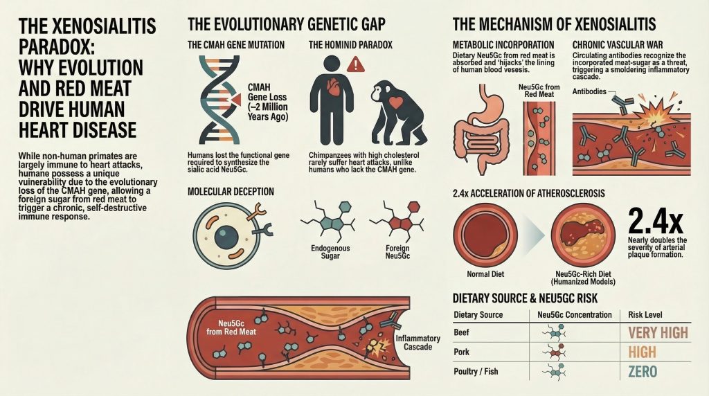
This profound divergence points inexorably toward a species-specific, intrinsic genetic predisposition to atherosclerosis in humans.[1] The crux of this unique vulnerability lies not strictly in lipid metabolism, hemodynamic shear stress, or behavioral variables alone, but in an ancient evolutionary genetic anomaly involving the fundamental biology of cell-surface glycans. Specifically, human cardiovascular susceptibility is inextricably linked to an evolutionary loss of the ability to synthesize N-glycolylneuraminic acid (Neu5Gc), a ubiquitous mammalian sialic acid.[1] Despite this profound genetic deficiency, human consumption of Neu5Gc-rich red meat results in the metabolic incorporation of this non-human glycan directly into human tissues, most notably the vascular endothelium.[3,4] The subsequent immunological recognition of this incorporated sugar as a “xeno-autoantigen” triggers a chronic, smoldering inflammatory cascade termed “xenosialitis.”[5]
This exhaustive research report elucidates the highly intricate evolutionary, biochemical, and immunological mechanisms underpinning the xenosialitis hypothesis. By exploring the genomic deletion of the CMAH gene, the complex microbiome-mediated digestion of dietary Neu5Gc, the molecular hijacking of human intracellular glycosylation pathways, and the resultant polyclonal antibody-mediated endothelial destruction, this analysis provides a comprehensive understanding of why humans are uniquely susceptible to red meat-associated atherosclerosis, and precisely how this mechanism quantifiably escalates cardiovascular risk across the lifespan.
The Evolutionary Paradigm: The CMAH Gene Mutation and Human Origins
The Fundamental Biology of Sialic Acids
To accurately comprehend the magnitude and consequence of the human genetic defect, one must first establish the physiological and structural role of sialic acids within vertebrate biology. The surfaces of all vertebrate cells are intimately decorated with a dense, complex, and highly regulated array of sugar chains, which are predominantly attached to integral membrane proteins and surface lipids to form the glycocalyx.[6,7] Most soluble secreted proteins traversing the human circulation are also similarly decorated with such specialized glycans.[6,7] Sialic acids represent a diverse family of highly modified monosaccharides built upon a nine-carbon backbone, and they are typically found capping the outermost, terminal ends of these glycan chains.[6,7]
Due to their distal location on glycoproteins and glycolipids, combined with their strong electronegative charge at physiological pH, sialic acids play indispensable and multifaceted roles in mediating cell-to-cell adhesion, modulating receptor-ligand interactions, stabilizing the endothelial glycocalyx barrier, and regulating immune self-recognition via specific sialic acid-binding lectins (Siglecs).[6,8]
In the vast majority of mammalian species, and indeed across most terrestrial vertebrates, the two predominant forms of sialic acid are N-acetylneuraminic acid (Neu5Ac) and its direct hydroxylated derivative, N-glycolylneuraminic acid (Neu5Gc).[6,7] These two molecules are structurally nearly identical, differing by the presence of only a single oxygen atom.[5] The biochemical transition from the precursor Neu5Ac to the derivative Neu5Gc is catalyzed exclusively by a single, highly specific enzyme: cytidine monophosphate-N-acetylneuraminic acid hydroxylase (CMAH).[1,3] This critical enzyme acts upon the nucleotide-sugar donor CMP-Neu5Ac within the cellular cytosol, facilitating a mono-oxygenase reaction that adds a hydroxyl group to generate CMP-Neu5Gc.[6,7] Both CMP-Neu5Ac and CMP-Neu5Gc are subsequently transported from the cytosol into the lumen of the Golgi apparatus, where they serve as the primary nucleotide-sugar donors for addition to newly synthesized glycoconjugates.[6,7] Consequently, normal mammalian tissues exhibit a rich, interspersed, and highly functional mosaic of both Neu5Ac and Neu5Gc on their cellular surfaces.[6,7]
| Feature | N-acetylneuraminic acid (Neu5Ac) | N-glycolylneuraminic acid (Neu5Gc) |
| Structural Backbone | 9-carbon monosaccharide | 9-carbon monosaccharide |
| Chemical Difference | Possesses an N-acetyl group | Possesses an N-glycolyl group (addition of one oxygen atom) |
| Biosynthetic Origin | Synthesized de novo in all vertebrates | Synthesized from Neu5Ac via the CMAH enzyme |
| Status in Humans | The predominant, endogenous human sialic acid | Genetically absent; considered a “foreign” xeno-antigen |
| Mammalian Distribution | Ubiquitous across mammalian species | Ubiquitous across non-human mammals; absent in humans |
The Alu-Mediated Pseudogenization Event
Humans, however, represent a stark and consequential anomaly within the mammalian lineage. Normal, healthy human tissues are genetically and fundamentally incapable of synthesizing Neu5Gc.[5] This biochemical truncation is not a regulatory suppression, but the direct consequence of a species-specific, irreversible mutation within the CMAH gene itself.[1,3]
Paleogenomic estimates and comparative phylogenetic analyses indicate that approximately 2 to 3 million years ago—a temporal epoch corresponding to the emergence of the genus Homo from our hominin ancestors—a critical genomic event occurred.[1,9,12] An Alu transposable element, a type of “jumping gene” common in primate genomes, inserted itself into the CMAH genetic locus. This insertion mediated a catastrophic frame-shift deletion of a critical 92-base-pair exon within the CMAH gene, resulting in a premature stop codon and the total pseudogenization (complete inactivation) of the locus.[9,12] Because the enzymatic reaction from CMP-Neu5Ac to CMP-Neu5Gc is strictly unidirectional and reliant entirely on CMAH, the loss of this single enzyme permanently severed the biosynthetic pathway.[1,3] As a direct result, modern humans produce only the precursor, Neu5Ac, leaving their cellular surfaces completely devoid of endogenous Neu5Gc and dramatically altering the biochemical landscape of the human organism.[1,3]
Evolutionary Selection Pressures: Pathogens and Reproductive Isolation
The fixation of a loss-of-function mutation in a gene responsible for the synthesis of a primary, ubiquitous cell-surface molecule is an extraordinary evolutionary event. Such a drastic and sudden alteration in the host glycocalyx must have been driven by phenomenally intense evolutionary selection pressures, as stochastic genetic drift alone is highly unlikely to account for the complete, population-wide fixation of such a fundamental biochemical change.[12] Current evolutionary biology posits a multi-factorial model for the rapid fixation of the CMAH null allele in the ancestral hominin population.
The leading, and most aggressively researched, hypothesis involves a brutal host-pathogen evolutionary arms race, specifically involving the ancestors of the malaria-causing Plasmodium parasites.[9,12] Mammalian cell-surface sialic acids serve as the primary docking receptors and recognition motifs for various viral, bacterial, and parasitic pathogens.[6,7] It is highly probable, based on genomic signatures of selection, that ancestral hominins were decimated by a highly virulent strain of “non-human hominid” (NHH) malaria that preferentially bound to Neu5Gc-rich erythrocytes to gain cellular entry.[9,12] The Alu-mediated loss of the CMAH gene, while potentially deleterious in other respects, provided a sudden, life-saving survival advantage to the rare individuals carrying the mutation. By effectively stripping the host of the pathogen’s preferred cell-surface receptor, the CMAH-deficient hominins were able to escape the prevailing malarial scourge, leading to a massive selective sweep that fixed the null mutation across the surviving population.[9,12] This evolutionary victory, however, was temporary in the grand scope of deep time. Plasmodium falciparum—the highly lethal agent responsible for modern human malignant malaria—eventually evolved the capacity to bind preferentially to the newly ubiquitous, human-specific Neu5Ac-rich erythrocytes, re-establishing the host-pathogen dynamic.[6,7,12]
A secondary, yet equally compelling and complementary hypothesis involves reproductive isolation and “catastrophic selection” mediated by the female immune system.[12] Mammalian spermatozoa are heavily sialylated, utilizing complex surface glycans to enhance sperm survival, mask immunogenic proteins, and optimize function along the perilous journey through the female reproductive tract to the site of fertilization in the oviduct.[12] A female hominin carrying the rare CMAH-null mutation would naturally develop circulating anti-Neu5Gc antibodies upon post-natal exposure to environmental or dietary Neu5Gc, as her immune system would recognize the molecule as non-self.[10,12,18] If this mutant female subsequently mated with a wild-type (CMAH-positive) male, her circulating antibodies would aggressively target and neutralize the Neu5Gc-rich paternal sperm, severely depressing his reproductive success and effectively destroying the viability of the pairing.[12] Over thousands of generations, this female immune-mediated selection against paternal Neu5Gc would create an intense, unyielding reproductive barrier between mutant and wild-type populations, driving the rapid fixation of the CMAH-null genotype and potentially contributing directly to the speciation event that ultimately isolated human ancestors from other hominids.[12]
The Dietary Mismatch: Digestion and Microbial Processing of Exogenous Sialic Acids
The evolutionary loss of the CMAH gene renders Neu5Gc biochemically completely foreign to the modern human organism.[5] Yet, despite this profound genetic deficiency, modern human diets—particularly in Westernized societies—are replete with this molecule. Neu5Gc is highly concentrated in red meats derived from mammalian sources, specifically beef, pork, and lamb, which remain staples of global consumption.[3,8,22] Furthermore, dairy products derived from these mammals also contain measurable, though lesser, quantities of the non-human sialic acid.[8]
Rigorous biochemical assays and high-performance liquid chromatography have clearly defined the dietary landscape of Neu5Gc exposure. Research demonstrates that beef contains the absolute highest levels of the molecule, harboring approximately 30.1 µg/g of Neu5Gc, followed closely by pork at 25.5 µg/g.[7,8] Conversely, avian species (such as poultry) and diverse fish lineages diverged from the mammalian tree long before the evolution of the CMAH gene, or independently lost it, and are therefore completely and natively devoid of Neu5Gc.[7,8] This strict biochemical dichotomy makes dietary Neu5Gc exposure highly specific to mammalian red meat consumption.[7,8]
| Dietary Source | Neu5Gc Content Classification | Specific Neu5Gc Concentration |
| Beef | Very High | ~ 30.1 µg/g |
| Pork | High | ~ 25.5 µg/g |
| Lamb / Rabbit | High | Comparable to Beef/Pork |
| Dairy Products | Moderate | Variable, present in milk glycoproteins |
| Poultry (Chicken, Turkey) | Zero (Biochemically Absent) | 0.0 µg/g |
| Fish and Seafood | Zero (Biochemically Absent) | 0.0 µg/g |
The Critical Role of the Gut Microbiome
The transition of dietary Neu5Gc from a consumed foodstuff resting in the gastric lumen to an absorbed, systemically active biochemical entity is tightly regulated by the host gastrointestinal microbiome. Dietary Neu5Gc is not generally ingested as a free monosaccharide; rather, it is consumed primarily in a glycosidically bound form, integrated deeply into the complex glycoproteins, mucins, and glycolipids that comprise mammalian tissue (such as porcine submaxillary mucin).[8,17] This distinction between bound and free sialic acid is of paramount physiological importance. While free, un-bound Neu5Gc can technically be absorbed across the intestinal barrier, it is recognized by the body as a small, highly soluble, and non-utilizable molecule. Consequently, free Neu5Gc is rapidly cleared by the renal system and excreted efficiently in the urine, completely failing to cause long-term tissue accumulation or pathology.[11,16] It is only the structurally bound Neu5Gc, presented in complex macromolecular arrays, that undergoes the dangerous process of metabolic incorporation.[11,16,17]
Recent advances in metagenomic sequencing of the human and murine gut microbiome have revealed that the resident microbial flora dynamically and rapidly responds to a Neu5Gc-rich diet.[17] Specifically, populations belonging to the orders Bacteroidales and Clostridiales undergo significant phenotypic expansion when exposed to red meat-associated carbohydrates.[17] These specific bacterial clades have evolved to express highly specialized bacterial sialidases (neuraminidases) designed specifically to cleave terminal sialic acids from host mucosal mucins and, crucially, from dietary glycoproteins.[17,20,21]
Historically, it was assumed that bacterial sialidases exclusively preferred the host-endogenous Neu5Ac, as this is the most abundant substrate in the human gut. However, profound structural studies utilizing advanced X-ray crystallography have identified specific, previously unobserved amino acid configurations within certain bacterial sialidases that confer an unusually high-affinity substrate preference for Neu5Gc-containing glycans.[17] These specialized gut microbes orchestrate a complex cross-feeding dynamic within the microbiome, wherein they utilize the Nan genetic pathway to actively catabolize the cleaved sialic acids for energy and carbon sourcing.[20,21] Interestingly, the downstream catabolism of Neu5Ac produces acetic acid as a byproduct, whereas the bacterial metabolism of Neu5Gc yields glycolic acid.[20,21] These differences in metabolic byproducts may subtly alter the local pH and metabolic landscape of the colonic epithelium, contributing to the broader inflammatory milieu.[20,21]
The enzymatic liberation of Neu5Gc by these microbial sialidases from the massive meat glycoproteins into smaller, specific oligosaccharide fragments (such as GalNGc) is heavily suspected to be the requisite first step for the subsequent intestinal absorption and systemic distribution of the non-human sugar.[8,17]
Cellular Hijacking: The Mechanism of Metabolic Incorporation
Once dietary, glycosidically-bound Neu5Gc (or its microbiome-liberated oligosaccharide intermediates) transverses the complex intestinal epithelium, it enters the systemic circulation. Evidence demonstrates that following a red meat meal, these xeno-glycans appear in the human circulation at a steady-state level that persists for several hours, providing ample opportunity for tissue interaction.[11,15,16] The phenomenon by which this completely foreign dietary molecule is permanently integrated into human tissues represents a fascinating and highly dangerous vulnerability in human biochemistry. The fundamental issue is one of molecular deception: our highly conserved intracellular glycosylation machinery simply cannot distinguish between the endogenous precursor (Neu5Ac) and the exogenous, non-human analog (Neu5Gc).[3,5,11] The molecules, differing by only a single, tiny oxygen atom, appear identical to the enzymes responsible for cellular construction.[3,5]
The Endocytic and Lysosomal Pathway
Because human cells completely lack a dedicated plasma membrane transporter specifically designed for the uptake of free sialic acids from the extracellular space, the internalization of circulating Neu5Gc-glycoconjugates must occur via alternative routes.[11,13,16] This uptake is driven primarily by fluid-phase endocytosis and macropinocytosis—processes where the cell essentially “drinks” the surrounding extracellular fluid and its dissolved contents.[11,12,16]
Upon internalization, these exogenous molecules are encapsulated in endosomes and carefully trafficked to the highly acidic, degradative environment of the lysosome.[11,16] Within the lysosome, host-derived lysosomal sialidases enzymatically cleave the complex dietary glycoconjugates, finally releasing free Neu5Gc monosaccharides deep within the cell.[11,16]
The critical, irreplaceable gateway for the subsequent incorporation of this molecule is the lysosomal sialic acid transporter, a protein known as sialin (encoded by the SLC17A5 gene).[11,13,16] Sialin is a highly conserved transmembrane protein whose normal physiological role is the salvage and recycling of endogenous host Neu5Ac from degraded glycoproteins in the lysosome, pumping them back into the cytosol for reuse.[11,16] However, sialin is not perfectly selective; it readily accepts the free Neu5Gc molecules derived from red meat and actively transports them into the cytosolic space, effectively breaching the cell’s final internal barrier.[11,12,16]
De Novo Synthesis and Endothelial Expression
Once within the relative safety of the cytosol, the exogenous Neu5Gc enters the host cell’s de novo sialic acid synthetic pathway, initiating the final stages of the biochemical hijacking. The molecule is acted upon by cytidine monophosphate (CMP)-sialic acid synthase, an enzyme that activates the monosaccharide by attaching a high-energy CMP nucleotide, forming the donor complex CMP-Neu5Gc.[6,7] This activated sugar-nucleotide donor is then actively transported into the lumen of the Golgi apparatus, the central sorting and modification hub of the cell.[6,7]
Within the Golgi, human glycosyltransferases—the precise enzymes responsible for building the complex, branching glycan trees on newly synthesized proteins and lipids—blindly utilize the abundant CMP-Neu5Gc donors as if they were the correct, endogenous CMP-Neu5Ac.[3,5,11] Consequently, Neu5Gc is covalently and permanently attached to newly minted glycoproteins and glycolipids. These newly assembled, xeno-sialylated molecules are then trafficked via secretory vesicles to the cell surface, where they are integrated into the plasma membrane.[3,5]
Through this intricate biochemical pathway, human cells essentially hijack a dietary compound and unwittingly weaponize their own cell surfaces. While Neu5Gc accumulation has been observed in various human tissues, including the epithelia lining hollow organs (which explains its strong epidemiological link to various carcinomas), it exhibits a profound, highly selective tropism for the vascular endothelium—the delicate, innermost lining of all blood vessels.[5,22] Rigorous immunohistochemical surveys of human autopsy samples, utilizing both highly specific, monospecific chicken anti-Neu5Gc antibodies and affinity-purified human sera, have definitively demonstrated intense Neu5Gc expression in the normal-appearing human aorta, the dense microvasculature of the colon and placenta, and crucially, in the endothelium directly overlying active atherosclerotic plaques.[5]
The Immunological Genesis: Commensal Priming and Xeno-Autoantibodies
The presence of a non-human molecule covalently bonded to human tissues poses a uniquely dangerous immunological paradox, categorized formally in the literature as a “xeno-autoantigen.”[5] Because Neu5Gc is biologically foreign (“xeno”) due to our evolutionary history, the human immune system recognizes it as foreign even though it is physically embedded within the intimate context of the host’s own self-associated molecular patterns on the endothelial surface.[5,6]
The Ontogeny of the Immune Response
Extensive immunological surveillance and epidemiological testing reveal a startling fact: essentially all normal, healthy human adults possess circulating, highly active polyclonal anti-Neu5Gc antibodies.[5,6,10] These highly diverse antibody repertoires comprise Immunoglobulin G (IgG), IgA, and IgM isotypes, targeting a wide variety of Neu5Gc-containing epitopes commonly found on human endothelial cells.[5,6,10] Crucially, these antibodies are not germ-line encoded “natural” antibodies (such as those that determine the ABO blood group system, which are present independent of specific exposure).[5,10,18] Rather, they require specific postnatal antigenic stimulation and undergo classic affinity maturation to achieve their high binding capabilities.[10,18]
Detailed analyses of infant cord blood demonstrate a complete and utter absence of anti-Neu5Gc antibodies (specifically IgM) at the time of birth.[10,18] Titer levels remain undetectable for the first few months of life, strongly arguing against transplacental transfer as the primary source of long-term immunity.[10,18] However, these antibody titers begin to rise sharply and consistently around six months of age.[10,18] This precise timeline correlates perfectly with the introduction of cow’s milk-based formulas, dairy products, and the general weaning process onto solid foods, marking the infant’s absolute first systemic exposure to dietary Neu5Gc.[8,10,18] Furthermore, epidemiological studies examining broader environmental exposures have shown that children raised on active farms, who are presumably exposed to significantly higher ambient levels of animal-derived antigens, exhibit statistically higher levels of circulating anti-Neu5Gc IgG compared to non-farming, urban-dwelling children.[8,18]
Bacterial Priming via Haemophilus influenzae
A critical, mechanistic question arises: how does the human immune system mount such a robust, lifelong, affinity-matured antibody response against a simple dietary sugar, especially given that free Neu5Gc, when isolated, is only weakly immunogenic on its own?
The answer lies in the insidious phenomenon of microbial molecular mimicry and bacterial cloaking. While the genetic synthesis of endogenous Neu5Gc has never been documented in any bacterial, viral, or fungal microbe across the tree of life, certain highly adapted commensal human pathogens act as efficient molecular scavengers.[14,21] Nontypeable Haemophilus influenzae (NTHi), a ubiquitous, human-specific commensal bacterium and opportunistic respiratory pathogen responsible for otitis media and respiratory exacerbations, possesses a highly specialized tripartite ATP-independent periplasmic (TRAP) transporter.[14,21] This specific transport system efficiently scavenges trace amounts of dietary Neu5Gc from the host’s mucosal environment.[14,21]
Once scavenged from the host, NTHi metabolically incorporates this Neu5Gc directly into its own cell surface lipooligosaccharides (LOS).[14,19,21] This bacterial sialylation confers a massive, almost impenetrable survival advantage to the organism. The integration of Neu5Gc into its protective biofilm significantly increases the bacterium’s resistance to complement-mediated killing by the host’s innate immune system.[14,21] It achieves this primarily by abrogating the classical pathway of complement activation, actively preventing host IgM antibodies from binding to the bacterial surface and initiating lysis.[14,21]
However, this sophisticated bacterial cloaking mechanism comes at a severe, chronic cost to the human host. The presentation of the Neu5Gc antigen on the highly inflammatory, pathogen-associated surface of a bacterial pathogen acts as an intense, natural immunological adjuvant.[10,14,21] When the human immune system inevitably mounts a vigorous, systemic response against the NTHi bacteria to clear the infection, it simultaneously generates a vast repertoire of cross-reactive anti-Neu5Gc antibodies.[10,14,21] It is now widely accepted in the field that this post-natal, pathogen-mediated priming, operating in constant tandem with daily dietary red meat exposure, provides the requisite, highly inflammatory signal for the lifelong production and maintenance of xeno-autoantibodies.[5,10,14,18]
Xenosialitis: The Mechanics of Atherogenesis
The disastrous convergence of metabolic incorporation (providing the widespread endothelial antigen) and microbial priming (providing the circulating, high-affinity antibody) culminates in a continuous, smoldering state of vascular inflammation formally termed “xenosialitis.”[5] This pathological process is uniquely insidious because the antigen is continuously replenished with every red meat meal consumed over a lifetime, ensuring that the inflammatory cycle never reaches biological resolution.
The precise pathophysiology of xenosialitis, particularly in the direct context of accelerating atherosclerosis, unfolds in a highly orchestrated, sequential cascade of cellular destruction:[5]
- Antigen-Antibody Ligation: Circulating polyclonal human anti-Neu5Gc antibodies (primarily the IgG and IgA isotypes) continuously screen the vascular endothelium as blood flows through the arterial tree. Upon encountering a metabolically incorporated Neu5Gc epitope displayed on the delicate endothelial glycocalyx, the antibodies bind with high affinity, forming a dense in situ immune complex directly on the vessel wall.[5]
- Complement Cascade Activation: The spatial clustering of IgG and IgA molecules on the endothelial surface exposes their Fc (fragment crystallizable) regions to the bloodstream. This exposed array provides an ideal binding site for the C1q protein complex, the initiator of the complement system.[5,15] This triggers the classical complement cascade, leading to the aggressive, sub-lytic deposition of complement proteins (such as C3b and the membrane attack complex) directly onto the intact, otherwise healthy endothelium.[5]
- Profound Endothelial Activation: The heavy deposition of complement proteins, combined with the mechanical cross-linking of surface receptors by the antibodies, forcefully pushes the endothelial cell from its normal, quiescent, non-thrombogenic state into a highly activated, pro-inflammatory phenotype.[5] Sophisticated in vitro models demonstrate that the simple incubation of high-titer human sera with Neu5Gc-fed endothelial cells directly causes massive intracellular signaling changes, resulting in the rapid exocytosis of Weibel-Palade bodies and the rapid de novo transcription of inflammatory genes.[5]
- Cytokine Secretion and Selectin Upregulation: The newly activated endothelium begins to secrete copious amounts of pro-inflammatory cytokines, creating a localized chemotactic gradient designed to summon immune cells.[5] Simultaneously, the cells rapidly upregulate the surface expression of critical adhesion molecules, specifically E-selectin and P-selectin.[5] P-selectin, in particular, is a potent, well-recognized mediator of recurrent thromboembolism and cardiovascular disorders, operating to facilitate the initial, rolling tethering of circulating leukocytes to the vessel wall.[23]
- Leukocyte Extravasation and Plaque Formation: The heavy presence of selectins and the disrupted glycocalyx leads to Neu5Gc-dependent monocyte binding.[5] The circulating monocytes roll along the activated endothelium, firmly adhere via integrin interactions, and actively diapedese (squeeze) into the subendothelial space (the tunica intima). Once trapped inside the arterial wall, these hyperactive monocytes transform into resident macrophages, begin to aggressively engulf oxidized low-density lipoproteins (LDL), and swell into massive foam cells. The accumulation and subsequent apoptotic death of these foam cells forms the foundational, highly thrombogenic necrotic core of the atherosclerotic plaque.[5]
- Synergistic Amplification Loops: This destructive cascade does not operate in a vacuum; it is further amplified by systemic inflammatory states. The presence of Tumor Necrosis Factor-alpha (TNF-α), a ubiquitous, highly potent pro-inflammatory cytokine typically upregulated during flares of general vascular damage or metabolic syndrome, selectively and powerfully enhances the reactivity of human anti-Neu5Gc antibodies, creating a compounding, positive feedback loop of vascular destruction.[5] Additionally, heme iron, highly concentrated in red meat, exerts its own independent pro-inflammatory and oxidative effects that compound the endothelial distress initiated by the glycans.[22,23]
| Stage of Pathogenesis | Cellular/Molecular Actor | Action and Consequence |
| 1. Target Identification | Endothelial Neu5Gc & Circulating IgG/IgA | Formation of in situ immune complexes on the otherwise healthy arterial wall. |
| 2. Cascade Initiation | Complement Protein C1q | Binding to antibody Fc regions, triggering classical complement deposition. |
| 3. Cellular Shift | Endothelial Cell | Transition from a quiescent state to an activated, pro-inflammatory phenotype. |
| 4. Surface Alteration | E-selectin & P-selectin | Massive upregulation on the cell surface, acting as tethering hooks for immune cells. |
| 5. Infiltration | Monocytes / Macrophages | Tethering, diapedesis into the tunica intima, and subsequent transformation into lipid-laden foam cells. |
| 6. Plaque Maturation | Foam Cells & Cytokines | Accumulation creates the necrotic core; cytokine release (TNF-α) amplifies further antibody reactivity. |
Quantifying the Risk: Incontestable Evidence from Humanized Mouse Models
To rigorously isolate, verify, and quantify the specific contribution of the CMAH evolutionary loss to cardiovascular disease—expressly separating it from the myriad confounding lifestyle factors inherent to modern human existence (such as smoking, sedentarism, and refined sugar consumption)—researchers engineered a profound, highly specific transgenic animal model: the human-like Cmah^-/- Ldlr^-/- mouse.[1]
Wild-type laboratory mice (Cmah^+/+) naturally and abundantly produce Neu5Gc and, like virtually all other mammals, do not suffer from xenosialitis.[1] To accurately recreate the human condition, researchers utilized genetic engineering to knock out both the Cmah gene (perfectly mimicking the human evolutionary loss 2 to 3 million years ago) and the Ldlr (Low-Density Lipoprotein Receptor) gene. The Ldlr knockout is a standard procedure required to induce baseline susceptibility to diet-driven atherosclerosis in mice, allowing the researchers to measure variations in plaque formation over reasonable experimental timeframes.[1]
The experimental data derived from these models yielded two distinct, highly significant quantifications of disease progression, beautifully separating the risk into two components: the intrinsic risk multiplier (the genetic penalty of being human) and the extrinsic risk multiplier (the dietary penalty of consuming red meat).
The Intrinsic Risk (The Baseline Human Penalty)
In the meticulously controlled first phase of the experiment, researchers fed both the wild-type control Cmah^+/+ mice and the human-like Cmah^-/- knockout mice an identical, highly controlled high-fat diet that was entirely devoid of all sialic acids (a Sias-free diet).[1] The results were staggering: even in the absolute, complete absence of dietary Neu5Gc, the human-like Cmah^-/- mice suffered a highly significant 1.9-fold increase in atherogenesis compared directly to the wild-type controls.[1]
This near-doubling of plaque formation in a controlled environment isolated the intrinsic biological cost of the CMAH mutation.[1] Extensive histological and biochemical analyses revealed that the loss of the gene fundamentally altered the baseline physiology of the organism. The Cmah^-/- mice exhibited highly elevated macrophage cytokine expression, rendering their white blood cells inherently hyperactive, hypersensitive, and prone to inflammatory overreactions even without a specific antigen trigger.[1] Furthermore, the mutation directly induced enhanced hyperglycemia and a strong metabolic tendency toward diabetes, significantly elevating their HOMA-IR (Homeostatic Model Assessment for Insulin Resistance) scores.[1] This baseline, intrinsic systemic dysfunction elegantly explains why even strict human vegetarians and vegans—who consume absolutely zero red meat and often lack traditional risk factors like high cholesterol—remain significantly more prone to unexplained heart attacks and strokes than captive chimpanzees with poor diets.[1,2] We are genetically wired for higher inflammation.
The Extrinsic Risk (The Red Meat Penalty)
In the critical second phase of the study, researchers sought to replicate the full, compounding mechanism of human red meat consumption and xenosialitis. They first immunized the human-like Cmah^-/- mice with specialized Neu5Gc-bearing antigens (utilizing complete Freund’s adjuvant) to generate circulating, affinity-matured anti-Neu5Gc antibodies, perfectly mimicking the human immune profile developed after weaning.[1] Subsequently, these immunized, humanized mice were fed a high-fat diet specifically enriched with high levels of Neu5Gc (mimicking a heavy, sustained red meat diet).[1]
The pathological results were dramatic and unequivocal. The immunized, Neu5Gc-fed mice suffered a massive 2.4-fold increase in the severity of atherosclerosis compared to control mice fed either a Neu5Ac-rich or a completely Sias-free high-fat diet.[1] Rigorous quantification of the aortic atherosclerosis was conducted: mice were euthanized, perfused, and their aortas dissected and pinned flat. Staining with Sudan IV to measure total lipid burden, alongside serial cryosections of the aortic sinus stained with Masson’s trichrome, revealed substantially more advanced lesions, highly expansive necrotic core areas, and dense, pathological collagen fiber deposition.[1]
Critically, this aggressive 2.4-fold acceleration of plaque formation could not be explained by downstream changes in blood lipoprotein levels, total cholesterol parameters, or glucose metrics.[1] The blood lipids remained virtually identical between the experimental cohorts; the massive cardiovascular destruction was entirely, explicitly driven by the localized, antibody-mediated inflammatory destruction of the endothelium—the pure, unadulterated manifestation of xenosialitis.[1]
Clinical Correlates and Broader Systemic Pathologies
The insidious nature of Neu5Gc integration and subsequent xenosialitis extends far beyond the microscopic parameters of isolated mouse models; it manifests broadly in observable clinical phenomena and contributes to a wide spectrum of human systemic pathologies. The mechanism is not isolated to the major coronary arteries but affects the entire vascular tree.
Diagnostic Indicators: Frank’s Sign and Microvascular Degradation
The systemic, ubiquitous nature of the endothelial damage caused by xenosialitis may actually be reflected in highly visible, external microvascular beds. Clinically, the presence of a diagonal earlobe crease (DELC)—known eponymously in medical literature as Frank’s sign—has been firmly and repeatedly established as an independent predictor of coronary artery disease and peripheral vascular disease.[25,26] This correlation holds true entirely independent of commonly known risk factors such as hypertension, diabetes, hyperlipidemia, and smoking.[25,26]
The underlying etiology of Frank’s sign is heavily hypothesized to be a parallel process of systemic microvascular disease and the accelerated, age-related weakening of elastin and collagen fibers occurring simultaneously in both the vascular beds of the earlobe and the coronary arteries.[25,26] Given the wide systemic distribution of Neu5Gc in the human glycocalyx, particularly in dense microvasculature, the chronic, low-grade inflammation of xenosialitis likely accelerates this precise mechanism of microvascular degradation.[5] The continuous immune complex formation and complement deposition degrade the delicate collagen-elastin ratio, providing a highly plausible, unifying biochemical link to this visible, macroscopic diagnostic marker.[25,26]
Circulating Antibodies and Acute Systemic Vasculitis
The pathogenic potential of anti-Neu5Gc antibodies is further evidenced in acute, devastating human vascular diseases. Elevated titers of anti-Neu5Gc antibodies are prominently and consistently observed in Kawasaki disease, an acute, febrile, and often severe childhood systemic vasculitis that primarily affects the coronary arteries.[24] Notably, higher circulating antibody levels are detected in patients exhibiting morphologically normal coronary arteries compared to those suffering from severe, late-stage complications like massive aneurysms or dilated coronaries.[24] This seemingly inverse correlation suggests a consumptive pathogenesis: during acute, massive inflammatory phases, large volumes of circulating anti-Neu5Gc antibodies may be rapidly consumed, binding heavily and sequestering at the sites of active vascular damage in the coronary walls. This effectively lowers the measurable circulating titer in the blood draw while the antibodies actively drive the pathology deep within the arterial wall.[24]
Furthermore, elevated levels of IgA class antibodies specifically directed against dietary antigens have been directly identified in the sera of severely atherosclerotic subjects compared to highly selected, healthy controls.[27] This finding underscores the intimate, inseparable link between gut-derived dietary antigens, systemic immune hypersensitivity, and the ultimate arterial plaque burden.[27] The mechanism is not confined to cardiovascular disease; the incorporation of Neu5Gc is also heavily implicated in cancer biology. Because Neu5Gc preferentially accumulates in malignant tissues due to rapid tumor growth rates, increased rates of micropinocytosis, and the hypoxic upregulation of the sialin transporter, xenosialitis provides a chronic inflammatory tumor microenvironment that directly promotes carcinogenesis, angiogenesis, and metastasis.[8,22]
The Imminent Threat of Viral Cross-Reactivity
An emerging, highly critical area of study involves the interaction of the pre-existing xenosialitis paradigm with severe, acute viral infections. When humans mount massive, desperate antibody responses to enveloped viral pathogens (such as SARS-CoV-2 or Influenza A), the resulting neutralizing antiviral antibodies may inadvertently exacerbate underlying vascular disease.[15] Because enveloped viruses utilize the host’s own cellular glycosylation machinery to synthesize their viral envelopes, the newly formed virions become coated in whatever sialic acids the host cell is expressing.[15] In a human consuming red meat, these viral envelopes will be laced with Neu5Gc.[15]
Consequently, the massive wave of neutralizing antiviral antibodies produced during the infection may indiscriminately cross-react with xenosialylated (Neu5Gc-containing) epitopes scattered across the host’s own healthy endothelial tissues.[15] This massive, systemic antibody cross-reactivity has the potential to supercharge the pre-existing state of xenosialitis.[15] The resulting hyper-inflammatory storm leads to severe, disseminated coagulopathies, fatal cytokine storms, and profound, widespread endothelial damage.[15] This hypothesis elegantly explains why baseline cardiovascular health and dietary habits are such profound, overriding determinants of mortality during acute viral respiratory syndromes, as the viral infection essentially ignites the latent powder keg of diet-induced xenosialitis.[15]
Therapeutic Interventions and Future Scientific Horizons
Understanding the intricate mechanics of Neu5Gc integration not only resolves a massive evolutionary and epidemiological puzzle but also opens highly novel, actionable therapeutic avenues. These interventions aim to actively mitigate cardiovascular risk beyond the current paradigm of traditional statin therapy, platelet inhibition, and blood pressure management.
Dietary Sialic Acid Interventions and Competitive Inhibition
The most immediate, biologically sound intervention is the direct modulation of the dietary sialic acid profile. Experimental data unequivocally demonstrates that intervening in the biochemical pathway can rapidly halt or even reverse the atherosclerotic acceleration.[4] In the heavily validated Cmah^-/- human-like mouse model, simply switching the subjects from a Neu5Gc-rich high-fat diet to a Neu5Gc-free high-fat diet actively protected against accelerated atherogenesis.[4]
Remarkably, adding a massive, five-fold excess of Neu5Ac (the safe, endogenous human sialic acid) to a high-fat diet effectively outcompeted the dangerous Neu5Gc for cellular uptake and utilization, effectively mitigating the inflammatory risk.[4] Furthermore, feeding a Neu5Ac-enriched diet intrinsically protected against atherosclerosis in Ldlr^-/- mice even in the absolute absence of dietary Neu5Gc, provided the host possessed the human-like Cmah-null genetic background, suggesting that flooding the system with the “correct” sugar actively stabilizes the human endothelium.[4]
Sources of highly concentrated, bioavailable Neu5Ac are currently being aggressively investigated. Collocalia mucoid extracts, commonly known as edible bird’s nest, are extraordinarily rich in natural Neu5Ac.[4] Biochemical digestion studies show that proper culinary preparation, specifically controlling the stewing temperature, maximizes the breakdown of the high-molecular-weight glycopeptides within the nest into highly absorbable, low-molecular-weight free Neu5Ac fragments.[4] This optimizes its bioavailability, allowing it to enter the bloodstream in high concentrations to serve as a competitive inhibitor against dietary Neu5Gc.[4]
Microbiome Engineering and Industrial Enzymatic Cleavage
A second, highly innovative therapeutic horizon involves the direct manipulation of the food supply using the very biochemical mechanisms evolved by the gut microbiome. The recent discovery of specific bacterial sialidases (derived from Bacteroidales and Clostridiales) that exhibit an overwhelming, highly specific enzymatic preference for cleaving Neu5Gc over Neu5Ac presents a massive industrial and medical opportunity.[17]
These powerful enzymes, capable of efficiently liberating bound Neu5Gc from complex red meat glycoproteins, could be utilized in the industrial processing of meat products prior to retail sale.[17] By pre-treating mammalian red meat with these specific recombinant bacterial sialidases, the meat could be effectively “stripped” of bound Neu5Gc.[17] Because free, unbound Neu5Gc is poorly absorbed by the gut and is rapidly, safely cleared by the renal system through the urine, converting the dangerous, bound Neu5Gc into harmless, free Neu5Gc directly in the food matrix could drastically reduce the risk of inflammatory diseases.[11,16] This approach would effectively sever the biochemical chain that leads to xenosialitis without requiring total population-level dietary abstinence from red meat.[17,20]
Conclusion
The profound intersection of evolutionary biology, intracellular glycosylation mechanics, immunology, and modern dietary habits provides a comprehensive, molecularly precise explanation for the uniquely human predisposition to cardiovascular disease. The human cardiovascular system operates under a permanent, intrinsic genetic penalty—the indelible legacy of the Alu-mediated CMAH pseudogenization that occurred millions of years ago. While this dramatic mutation likely saved our hominin ancestors from catastrophic, malaria-driven parasitic extinction, it left the modern human glycocalyx permanently altered and highly vulnerable.
The regular consumption of mammalian red meat introduces massive, unnatural quantities of the “lost” sialic acid, Neu5Gc, directly into the human gastrointestinal tract. There, specialized bacterial sialidases liberate the molecule, allowing it to be absorbed. Through a tragic case of molecular mimicry, the human cellular machinery indiscriminately incorporates this foreign glycan via the sialin transporter, covalently presenting it on the luminal surface of the vascular endothelium. Primed by early-life dietary exposure and continuous molecular scavenging by commensal pathogens like Nontypeable Haemophilus influenzae, the human immune system recognizes the altered endothelium as a foreign threat. The resulting relentless, lifelong barrage of polyclonal IgG and IgA antibodies triggers classical complement deposition, massive selectin expression, monocyte extravasation, and continuous, smoldering plaque formation.
This highly validated mechanism, termed xenosialitis, beautifully and completely explains the 15% of clinical atherosclerosis cases occurring in patients utterly devoid of traditional risk factors. It flawlessly accounts for the massive 2.4-fold acceleration of atherogenesis seen in controlled animal models upon red meat consumption, and it clarifies why chimpanzees—our closest evolutionary relatives with identical lipid profiles—remain immune to coronary atherosclerosis. Moving forward, interventions targeting competitive Neu5Ac supplementation and the industrial enzymatic cleavage of Neu5Gc from the food supply offer the unprecedented promise of nullifying this ancient evolutionary trade-off, fundamentally altering the landscape of cardiovascular preventative medicine and redefining our understanding of diet-induced pathology.
References
- Kawanishi K, Dhar C, Do R, Varki N, Gordts PLSM, Varki A. Human species-specific loss of CMP-N-acetylneuraminic acid hydroxylase enhances atherosclerosis via intrinsic and extrinsic mechanisms. Proc Natl Acad Sci U S A. 2019;116(32):16036-16045. doi:10.1073/pnas.1902902116
- Baldessari A, Snyder J, Ahrens J, Murnane R. Fatal myocardial fibrosis in an aged chimpanzee (Pan troglodytes).
- Banda K, Gregg CJ, Chow R, Varki NM, Varki A. Metabolism of vertebrate amino sugars with N-glycolyl groups: mechanisms underlying gastrointestinal incorporation of the non-human sialic acid xeno-autoantigen N-glycolylneuraminic acid.
- Kawanishi K, Coker JK, Grunddal KV, et al. Dietary Neu5Ac Intervention Protects Against Atherosclerosis Associated With Human-Like Neu5Gc Loss-Brief Report.
- Dhar C, Sasmal A, Varki A. From “Serum Sickness” to “Xenosialitis”: Past, Present, and Future Significance of the Non-human Sialic Acid Neu5Gc.
- Lewis AL, Chen X, Schnaar RL, Varki A. Sialic Acids and Other Nonulosonic Acids. In: Varki A, Cummings RD, Esko JD, et al., eds.
- Varki A. Sialic acids in human health and disease.
- Altman MO, Gagneux P. Absence of Neu5Gc and Presence of Anti-Neu5Gc Antibodies in Humans-An Evolutionary Perspective. Front Immunol. 2019;10:789. Published 2019 Apr 30. doi:10.3389/fimmu.2019.00789
- Varki A. Colloquium paper: uniquely human evolution of sialic acid genetics and biology. Proc Natl Acad Sci U S A. 2010;107 Suppl 2(Suppl 2):8939-8946. doi:10.1073/pnas.0914634107
- Padler-Karavani V, Yu H, Cao H, et al. Diversity in specificity, abundance, and composition of anti-Neu5Gc antibodies in normal humans: potential implications for disease. Glycobiology. 2008;18(10):818-830. doi:10.1093/glycob/cwn072
- Bardor M, Nguyen DH, Diaz S, Varki A. Mechanism of uptake and incorporation of the non-human sialic acid N-glycolylneuraminic acid into human cells. J Biol Chem. 2005;280(6):4228-4237. doi:10.1074/jbc.M412040200
- Varki A. Multiple changes in sialic acid biology during human evolution. Glycoconj J. 2009;26(3):231-245. doi:10.1007/s10719-008-9183-z
- Dubois L, Pietrancosta N, Cabaye A, et al. Amino Acids Bearing Aromatic or Heteroaromatic Substituents as a New Class of Ligands for the Lysosomal Sialic Acid Transporter Sialin. J Med Chem. 2020;63(15):8231-8249. doi:10.1021/acs.jmedchem.9b02119
- Apicella MA. Nontypeable Haemophilus influenzae: the role of N-acetyl-5-neuraminic acid in biology. Front Cell Infect Microbiol. 2012;2:19. Published 2012 Mar 13. doi:10.3389/fcimb.2012.00019
- Taylor RE, Gregg CJ, Padler-Karavani V, et al. Novel mechanism for the generation of human xeno-autoantibodies against the nonhuman sialic acid N-glycolylneuraminic acid. J Exp Med. 2010;207(8):1637-1646. doi:10.1084/jem.20100575
- Verheijen FW, Verbeek E, Aula N, et al. A new gene, encoding an anion transporter, is mutated in sialic acid storage diseases. Nat Genet. 1999;23(4):462-465. doi:10.1038/70585
- Zaramela LS, Martino C, Alisson-Silva F, et al. Gut bacteria responding to dietary change encode sialidases that exhibit preference for red meat-associated carbohydrates. Nat Microbiol. 2019;4(12):2082-2089. doi:10.1038/s41564-019-0564-9
- Padler-Karavani V, Hurtado-Ziola N, Pu M, et al. Human xeno-autoantibodies against a non-human sialic acid serve as novel serum biomarkers and immunotherapeutics in cancer. Cancer Res. 2011;71(9):3352-3363. doi:10.1158/0008-5472.CAN-10-4102
- Vimr ER, Kalivoda KA, Deszo EL, Steenbergen SM. Diversity of microbial sialic acid metabolism. Microbiol Mol Biol Rev. 2004;68(1):132-153. doi:10.1128/MMBR.68.1.132-153.2004
- Tailford LE, Crost EH, Kavanaugh D, Juge N. Mucin glycan foraging in the human gut microbiome. Front Genet. 2015;6:81. Published 2015 Mar 19. doi:10.3389/fgene.2015.00081
- Samraj AN, Pearce OM, Läubli H, et al. A red meat-derived glycan promotes inflammation and cancer progression. Proc Natl Acad Sci U S A. 2015;112(2):542-547. doi:10.1073/pnas.1417508112
- Hull SC, Mszar R, Ostfeld RJ, et al. Diet and Prevention of Cardiovascular Disease and Cancer: JACC: CardioOncology State-of-the-Art Review. JACC CardioOncol. 2025;7(6):649-667. doi:10.1016/j.jaccao.2025.07.008
- Evrengül H, Dursunoğlu D, Kaftan A, et al. Bilateral diagonal earlobe crease and coronary artery disease: a significant association. Dermatology. 2004;209(4):271-275. doi:10.1159/000080847
- Padler-Karavani V, Tremoulet AH, Yu H, Chen X, Burns JC, Varki A. A simple method for assessment of human anti-Neu5Gc antibodies applied to Kawasaki disease. PLoS One. 2013;8(3):e58443. doi:10.1371/journal.pone.0058443
- Shmilovich H, Cheng VY, Rajani R, et al. Relation of diagonal ear lobe crease to the presence, extent, and severity of coronary artery disease determined by coronary computed tomography angiography. Am J Cardiol. 2012;109(9):1283-1287. doi:10.1016/j.amjcard.2011.12.024
- Gakovic B, Neskovic SA, Vranic I, et al. The relationship of diagonal earlobe crease (Frank’s sign) and obstructive coronary artery disease in patients undergoing coronary angiography. Wien Klin Wochenschr. 2023;135(23-24):667-673. doi:10.1007/s00508-023-02297-y
- Muscari A, Volta U, Bonazzi C, et al. Association of serum IgA antibodies to milk antigens with severe atherosclerosis. Atherosclerosis. 1989;77(2-3):251-256. doi:10.1016/0021-9150(89)90088-9

