
Age-Associated Atherosclerosis: Mechanisms of Vascular Aging, Cellular Senescence, and Multimodal Mitigation
Narrative Review
Abstract
Atherosclerotic cardiovascular disease rises sharply with advancing age, but chronological age is not a single causal exposure. This narrative review examines how cumulative apolipoprotein B-containing lipoprotein exposure interacts with age-associated vascular remodeling, cellular senescence, mitochondrial dysfunction, impaired mitochondrial quality control, and clonal hematopoiesis. Evidence is separated explicitly into randomized human, human observational/histologic/genetic, and preclinical tiers. Human data support age-associated extracellular-matrix remodeling, arterial stiffening, senescence-related phenotypes, and clonal hematopoiesis as contributors to vascular risk, whereas several mitochondrial and senotherapeutic mechanisms remain predominantly preclinical. Clinical mitigation remains multimodal: randomized evidence supports lipid lowering in secondary prevention, blood-pressure control in older adults, and selected dietary and anti-inflammatory strategies in defined populations, while dedicated evidence for statin initiation in primary prevention beyond age 75 remains limited. Senolytics, mitophagy enhancers, and other geroprotective strategies have not demonstrated clinical atherosclerotic outcome benefit in humans. Vascular aging adds mechanisms to, rather than replaces, cumulative lipoprotein-mediated atherogenesis, and evidence strength varies substantially across the proposed pathways.
Keywords: atherosclerosis; vascular aging; cellular senescence; clonal hematopoiesis; mitochondrial dysfunction; older adults; cardiovascular prevention.
Review approach and evidence grading
This is a narrative rather than a systematic review. Sources were selected to represent the principal mechanistic and clinical literatures bearing on age-associated atherosclerosis, and quantitative claims were checked against primary publications when available rather than accepted from secondary or review-level citation.
Claims are drawn from three tiers, kept separable throughout: (i) randomized trial evidence in humans; (ii) human observational, histologic, and genetic-epidemiological evidence; and (iii) mechanistic and preclinical evidence in cell and animal models. Tier (iii) findings are labeled preclinical wherever they appear and are not clinically actionable. Human observational associations are not interpreted as establishing causal mediation unless supported by intervention, genetic, or convergent experimental evidence. Evidence-tier labels within tables refer to the sources cited in that row, not to the field generally.
This narrative review involved no original enrollment of human participants or animals and no generation of identifiable participant-level data.
1. Epidemiological kinetics and late-life disease burden
Atherosclerosis is a progressive inflammatory disease of the arterial wall that initiates early in life, and the incidence of clinically manifest atherosclerotic events rises steeply with advancing age [2]. Age-associated changes in the vessel wall — matrix remodeling, senescence, mitochondrial dysfunction and impaired mitochondrial quality control, and clonal hematopoiesis among them — have been implicated in atherogenesis and, in experimental models and human observational studies, in features associated with plaque vulnerability. This does not establish that the longitudinal rate of human plaque accumulation itself increases non-linearly with chronological age; that proposition has not been demonstrated, and clinically meaningful thresholds for coronary plaque progression remain unsettled.
Age is among the strongest contributors to absolute predicted ASCVD risk in contemporary risk models [32]. This is a statement about weight within risk models, not a demonstration that age outranks cumulative lipid exposure as a causal driver; the two are not commensurable, because age partly integrates exposure time. Fatty streaks and diffuse intimal thickening develop in adolescence and early adulthood, whereas clinically obstructive, unstable, and calcified lesions become increasingly prevalent beyond the sixth decade [2].
The contribution of additional age-associated mechanisms may become increasingly relevant across the lifespan [2]. In young and middle-aged adults, atherogenesis requires cumulative exposure to apolipoprotein B-containing lipoproteins [6] and is driven importantly by that exposure together with smoking, blood pressure, diabetes, and other causal factors, against a background of relatively intact vascular repair [2]. The relative contribution of these exposures cannot be ranked from the data assembled here. In older adults, the lipid-driven process continues and is superimposed on cell-autonomous vascular senescence, hematopoietic somatic mutation, impaired autophagic quality control, and extracellular-matrix degradation [2]. The distinction is one of added mechanism, not substituted mechanism.
The age bands used in this review — 20-39, 40-59, 60-79, and 80 years and older — are an illustrative organizing framework rather than empirically derived biological thresholds. No inflection point in the mechanisms discussed has been mapped to a specific chronological age in humans; the bands are a device for exposition and correspond broadly to commonly reported surveillance strata.
1.0 What “acceleration” denotes in this review
Because the term is used inconsistently, accelerated atherosclerosis is defined operationally here as atherosclerotic burden, vascular dysfunction, a biological-aging signature, or clinical ASCVD occurring earlier, to a greater degree, or progressing more rapidly than expected for chronological age and measured conventional risk-factor exposure.
These are alternative operational phenotypes of accelerated vascular and atherosclerotic aging rather than manifestations of a single validated construct. They are not interchangeable, are measured by different instruments, and a person may satisfy one and not another. Vascular functional impairment and biological-aging signatures are not measures of atherosclerotic burden; they are grouped here only because the same terminology is applied to them in the literature. Throughout this review, vascular aging denotes age-associated structural and functional change in the artery wall irrespective of plaque; atherosclerotic aging denotes the subset of that change bearing on plaque formation or composition; and accelerated atherosclerosis is reserved for the operational phenotypes defined above.
Table 1. Alternative operational phenotypes grouped under “accelerated atherosclerosis.” These are distinct constructs with distinct measurement bases; the table is not a staging system, and no threshold is clinically validated for individual decision-making.
| Phenotype | Typical measurement basis | What an abnormal result establishes | Principal limitation |
| Excess anatomic burden for age | Coronary artery calcium score, carotid plaque area or number, CCTA plaque volume | Burden exceeds an age- and sex-referenced distribution; some reference sets are additionally stratified by race or ethnicity | Percentile position is not a progression rate; reference distributions are cohort-specific |
| Accelerated structural progression | Serial IMT, serial CAC, serial CCTA plaque volume, primarily in research settings | Change over time exceeds that observed in an appropriate reference population or a prespecified research threshold | Requires two standardized measurements; change may approach or fall below the resolution of an individual examination |
| Vascular functional impairment | Pulse wave velocity, flow-mediated dilation, central pressure augmentation | Arterial stiffening or endothelial dysfunction exceeding an age reference | Functional measures correlate imperfectly with plaque burden |
| Biological-aging signature | Epigenetic and proteomic age estimators, telomere length, senescence markers | A biological-aging estimator indicates greater age-associated molecular change than expected for chronological age | Research measures; no validated treatment threshold |
| Premature clinical ASCVD | Event occurring at an unusually young age according to the study or guideline definition used | Clinical disease earlier than population expectation | An event is a discrete endpoint, not a rate |
1.1 Prevalence and incidence — with attention to case definition
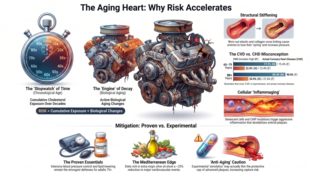
Prevalence figures for “cardiovascular disease” in older adults are routinely quoted without the definitional caveat that drives them. In American Heart Association surveillance, the composite CVD category includes hypertension. Using NHANES 2015-2018 data as reported in AHA surveillance, CVD prevalence at ages 60-79 years is 77.5% in males and 75.4% in females, rising to 89.4% and 90.8%, respectively, at age 80 years and older [3]. Narrowing the case definition to coronary heart disease in the same data yields substantially lower figures: 22.0% of males and 13.4% of females at ages 60-79, and 33.9% and 21.6% at age 80 and older [3]. Because this review concerns atherosclerosis rather than blood pressure, the coronary heart disease figures are more directly relevant; widely circulated estimates near 86-90% should be understood as hypertension-inclusive.
The 2015-2018 NHANES cycle is used because it provides the sex- and age-stratified comparison required here. More recent surveillance is available in the 2026 Statistical Update [4]. Age-specific rates of first cardiovascular events likewise rise markedly across later decades of life [4]. Absolute rates are sex-, calendar-period-, ascertainment-, and case-definition-specific, so no single set of point estimates is quoted here.
1.2 The “lipid paradox” in the very old
An epidemiological nuance in octogenarians and nonagenarians is attenuation, and sometimes inversion, of traditional risk-factor associations — the so-called lipid paradox, described in early form three decades ago [5] and still debated [34]. Three explanations dominate and are not mutually exclusive: reverse causation and confounding by illness, because frailty, occult malignancy, chronic infection, heart failure, and protein-energy wasting can lower cholesterol [5]; survivorship selection, because those most susceptible to LDL-driven atherogenesis may disproportionately have experienced events or died before the ninth decade; and competing risk, because a rising hazard of non-atherosclerotic death changes observed cause-specific and cumulative-incidence relationships.
Attenuation of an observational association in the very old is not evidence against LDL causality. Genetic evidence supports LDL causality across the life course, and randomized statin evidence demonstrates reduction in major vascular events among older adults, particularly those with established vascular disease [6,7]. Absolute ASCVD burden remains highest in older adults [3,4], reflecting age-associated vascular remodeling superimposed on decades of lipoprotein exposure [2].
1.3 Risk models and biological thresholds
Contemporary prediction frameworks incorporate age as a continuous covariate and do not specify a discrete biological age threshold. This is a property of model structure rather than a finding about vascular biology: the absence of a threshold term in a prediction equation is not evidence that no biological inflection exists, and a strong age coefficient is not evidence that one does.
Table 2. Age-stratified disease burden and illustrative mechanisms. Prevalence values are drawn from AHA surveillance [3] and are shown only for the strata that source reports directly; an em dash indicates that no directly reported age- and sex-stratified value was available. Age assignments are illustrative and do not indicate biological onset, exclusivity, or a validated staging system.
| Age cohort (years) | CVD prevalence, hypertension-inclusive [3] | CHD prevalence [3] | Mechanisms highlighted in this age band (Sec.) | Common ASCVD manifestations |
| 20-39 | — | — | Endothelial dysfunction, diffuse intimal thickening, early lipid retention (2.1) | Silent fatty streaks, early lipid-rich lesions |
| 40-59 | — | — | The above, plus cumulative apoB exposure, VSMC migration and phenotype switching (1, 2.2) | Stable angina, acute coronary syndromes |
| 60-79 | 77.5% M / 75.4% F | 22.0% M / 13.4% F | The above, plus vascular senescence, stiffening, mitochondrial ROS, calcification (2.1-2.4) | Myocardial infarction, ischemic stroke, claudication |
| 80+ | 89.4% M / 90.8% F | 33.9% M / 21.6% F | The above, plus rising CHIP prevalence, cumulative elastin and matrix remodeling, and age-associated impairment of vascular repair (2.5-2.6) | Complex multivessel CAD; ischemic stroke; peripheral or multiterritory atherosclerotic disease |
2. Intrinsic and extrinsic mechanisms of vascular aging
2.1 Extracellular matrix remodeling, glycation, and hemodynamic shear stress
Aging alters the mechanical properties of central elastic arteries, establishing a biophysical feedback loop proposed to promote atherogenesis [8]. Chronic pulsatile stress triggers elastolysis, elastase up-regulation, and fraying of elastin fibrils, with compensatory collagen deposition and cross-linking [8]. Cumulative glycemic exposure contributes to formation and accumulation of advanced glycation end-products (AGEs) on long-lived structural proteins; these modifications are subject to slow turnover rather than being strictly irreversible [8]. AGE cross-linking of collagen impairs compliance [8], while AGE-RAGE engagement activates endothelial NF-kB signaling and amplifies inflammatory and adhesion-molecule expression [61]. AGE accumulation is one contributor among several to age-related arterial stiffening.
The hemodynamic consequence is stiffening, manifest as rising carotid-femoral pulse wave velocity and central systolic pressure, with diastolic pressure typically falling at the population level after the sixth decade and pulse pressure widening [8].
Regions exposed to low or oscillatory endothelial shear stress (ESS) — the tangential frictional force of flowing blood, expressed in dyn/cm2 — are preferentially atheroprone [9]. Reported physiological and low-ESS ranges vary by vessel, methodology, species, and temporal averaging and should be treated as experimental reference ranges rather than universal thresholds. Values of roughly 15-70 dyn/cm2 have been reported for relatively straight segments under pulsatile unidirectional flow, whereas disturbed-flow regions at bifurcations, curvatures, and outer branch walls may show time-averaged magnitudes below approximately 10-12 dyn/cm2 or oscillatory ESS with a near-zero time average [9]. Low ESS down-regulates eNOS, up-regulates VCAM-1 and ICAM-1, and facilitates lipoprotein retention and monocyte transendothelial migration [9].
An aging-specific qualification is important. Aging dilates and stiffens arteries [8], and low-ESS regions are atheroprone [9], but the composite claim that aging enlarges low-ESS territory has not been clearly demonstrated in aging-specific human hemodynamic studies. Age-related geometric and mechanical change may expand low-ESS zones; testing this directly in age-stratified coronary CT angiography cohorts with computational fluid dynamics would be informative.
2.2 Local renin-angiotensin signaling in the aged arterial wall
Two facts are commonly conflated in secondary literature and are separated here. First, arterial-wall angiotensin II (Ang-II) is locally regulated and can differ substantially from circulating levels [10]. Tissue and plasma measurements are not methodologically equivalent, so this should be understood as compartmental separation rather than as a simple concentration ratio. That separation is present across adult life and is not itself an age-related increase.
Second, multiple components of the local renin-angiotensin system — including angiotensinogen, ACE and chymase activity, Ang-II abundance, and AT1-receptor signaling — have been reported to increase in aged arterial tissue across animal models and human tissue studies, whereas plasma renin activity and circulating Ang-II tend to fall with age [10,11]. The intramural age-associated increase should not be conflated with the tissue-to-plasma compartmental difference.
Local Ang-II acts as a mitogen and pressor agent, driving vascular smooth-muscle cell (VSMC) hypertrophy, synthetic phenotype switching, migration, and matrix metalloproteinase secretion, particularly MMP-2 [10,11].
In 16-year longitudinal follow-up of the Malmö Diet and Cancer cohort, mean common carotid intima-media thickness progressed at 0.011 mm/year in men and 0.010 mm/year in women, with faster progression at the bifurcation (0.036 and 0.030 mm/year, respectively) [12]. Approximately 0.01 mm/year, or 0.1 mm per decade for the common carotid, is therefore a reasonable summary of these longitudinal data. Cross-sectional age-reference studies report larger between-age differences [8,13], but cross-sectional and longitudinal quantities should not be arithmetically converted into one another. Carotid IMT also incorporates non-atherosclerotic medial hypertrophy and adaptive wall thickening and is therefore a structural readout rather than a direct measure of plaque accumulation.
2.3 Cellular senescence and the senescence-associated secretory phenotype
DNA damage, telomere attrition, and chronic oxidative stress up-regulate the cyclin-dependent kinase inhibitors p16INK4a and p21CIP1/WAF1, producing durable replicative arrest in vascular endothelial cells and VSMCs [1,14]. In human atherectomy and endarterectomy specimens, cells displaying senescence-associated beta-galactosidase positivity together with shortened telomeres concentrate at atherosclerotic sites and are largely absent from adjacent non-diseased tissue [14]. Neither marker is individually specific for senescence, so these findings identify a senescence-associated phenotype rather than definitively identifying senescent cells.
Senescent cells develop a pro-inflammatory senescence-associated secretory phenotype (SASP), including secretion of IL-6, IL-1beta, TNF-alpha, MCP-1/CCL2, and matrix-degrading enzymes such as MMP-2 and MMP-9 [1,15]. Human observational data show senescence-associated markers co-localizing with advanced lesion regions and intraplaque calcification [14-16]. Animal studies show that genetic or pharmacological senescent-cell targeting can reduce SASP-related signaling, matrix degradation, calcification, or vascular dysfunction [15,45]. Fibrous-cap thinning and impaired re-endothelialization are mechanistically coherent consequences, but causal sequences are not established in humans [15].
Senescence in plaque is not uniformly deleterious. VSMCs are essential for fibrous-cap matrix and stability, whereas VSMC senescence impairs proliferative and matrix-producing repair [15]. Accordingly, effects of senescent-cell clearance are stage- and model-dependent. In Ldlr-/- mice, p16-directed genetic clearance reduced plaque burden and increased cap thickness, while navitoclax reduced aortic plaque burden [57]. In Apoe-/- mice, p16-driven clearance did not reduce plaque burden, cap thickness, or necrotic core area and increased apoptotic cells and inflammation, while navitoclax reduced lesion size without increasing cap thickness [58]. In Apoe-/- mice with advanced lesions and persistent hyperlipidemia, navitoclax reduced smooth-muscle-derived cells within the fibrous cap, decreased cap thickness, and increased mortality [59]. Thus, pharmacological senolysis in advanced experimental atherosclerosis can deplete reparative cap cells and compromise plaque stability, materially qualifying therapeutic inference.
2.4 Mitochondrial dysfunction, mitophagy, and vascular inflammaging
In murine vascular-aging models, aged arterial tissue exhibits declining mtDNA copy number and reduced expression of the copy-number regulators TFAM, PGC-1alpha, and the mtDNA helicase Twinkle [17]. These changes track with reduced mitochondrial respiration, decreased carotid compliance and distensibility, and rising aortic pulse wave velocity [17]. Transgenic Twinkle overexpression increases mtDNA copy number, improves arterial respiration, and delays vascular aging in that model, whereas polymerase-gamma-mutant mice with degraded mtDNA integrity age faster [17]. In ApoE-/- mice, related mitochondrial rescue reduces necrotic-core area and increases relative fibrous-cap thickness [18]. This evidence base is predominantly murine; equivalent longitudinal human vascular Twinkle data do not exist.
Damaged mitochondria are cleared through mitophagy. In the canonical PINK1/Parkin pathway, depolarization stabilizes PINK1 on the outer membrane and recruits Parkin, which ubiquitylates outer-membrane proteins including MFN1, MFN2, and VDAC1 and targets the organelle for autophagic-lysosomal degradation [19]. Parkin-independent pathways also exist.
The relationship between aging and mitophagic flux in the vessel wall is more complicated than a simple decline. In aged murine aorta, Parkin protein and mitophagy markers are increased alongside elevated IL-6 and impaired respiration, consistent with compensatory or stalled mitophagy responding to a rising burden of damaged organelles rather than simple pathway loss [19]. Increased pathway markers do not establish increased successful flux. Global autophagic capacity declines with age in many tissues, and available data are consistent with clearance becoming insufficient relative to damaged-organelle burden in the aged artery [50]. Persistent damaged mitochondria can generate mitochondrial reactive oxygen species and release mitochondrial damage-associated molecular patterns, including unmethylated CpG mtDNA, into the cytosol [19].
Cytosolic mitochondrial damage-associated molecular patterns engage NLRP3 inflammasome and TLR9/MyD88 signaling. In the murine model in which this pathway was demonstrated, this signaling contributes to a feed-forward IL-6-associated inflammatory loop linking mitochondrial dysfunction to increased atherogenesis [19]. An equivalent longitudinal causal sequence has not been demonstrated in humans.
2.5 Clonal hematopoiesis of indeterminate potential (CHIP)
Clonal hematopoiesis of indeterminate potential (CHIP) denotes age-related acquisition of somatic mutations in leukemia-associated driver genes within hematopoietic stem and progenitor cells, traditionally operationalized by detection of a driver mutation at variant allele fraction (VAF) >=2% in the absence of persistent unexplained cytopenia and without a diagnosable hematologic neoplasm [20]. When persistent otherwise unexplained cytopenia coexists with clonality, current classifications designate clonal cytopenia of undetermined significance (CCUS), which carries a different malignant-progression risk [21]. The cardiovascular literature summarized here concerns CHIP, not CCUS. Prevalence rises from <1% below age 40 to approximately 10% among adults older than 70 in early sequencing cohorts, with higher estimates when deep sequencing detects clones below the 2% threshold [20]. The 2% VAF cutoff is an assay and reporting convention, not a biological boundary.
Dominant drivers include DNMT3A, TET2, and ASXL1, with JAK2 V617F less common but functionally distinctive [20,22]. In Tet2-deficient murine macrophages, loss of function augments IL-1beta and IL-6 signaling in association with NLRP3 inflammasome activation [23].
Effect sizes require precision. CHIP carriers had approximately 1.9- to 2.0-fold the risk of incident coronary heart disease in nested case-control analyses of the BioImage and Malmö Diet and Cancer cohorts and approximately 4-fold higher odds of early-onset myocardial infarction in ATVB and PROMIS [22]. The often-quoted ~12-fold figure applies specifically to JAK2 V617F carriers in the original cohort estimate, whereas DNMT3A, TET2, and ASXL1 were associated with roughly 1.7- to 2.0-fold risk [22]. It is gene-specific, not clone-size-specific, and should not be restated as “VAF above 10% confers a 12-fold increase.” Larger clones (VAF >=10%) are separately associated with greater cardiovascular and all-cause risk in observational cohorts, and CHIP carriers show higher coronary artery calcium scores than noncarriers [20,22].
Biological causality is supported in mice: in hyperlipidemic Ldlr-/- animals, reconstitution with Tet2-deficient bone marrow increases lesion size and necrotic-core burden [23]. These experiments establish that the mechanism can operate, not the magnitude or universality of a causal effect in humans.
In an exploratory genomic substudy of CANTOS, 338 sequenced participants (8.6%) carried clonal hematopoiesis; TET2 carriers appeared to derive greater MACE reduction from canakinumab than noncarriers [24]. This is hypothesis-generating subgroup evidence, not a demonstrated treatment-effect modifier.
Counter-evidence remains important. In a pooled analysis of 63,700 participants from five randomized cardiovascular outcome trials, CHIP carriage was not significantly associated with major cardiovascular events (adjusted HR 1.07, 95% CI 0.99-1.16) [25]. In the PESA cohort, baseline clonal hematopoiesis was associated with subsequent de novo femoral atherosclerosis over approximately six years, whereas atherosclerotic burden did not measurably accelerate mutant-clone expansion over the same period [26]. These findings support a predominant direction from clonal hematopoiesis toward atherosclerosis while not excluding reciprocal effects in other settings. CHIP remains biologically compelling and experimentally supported; its independent prognostic value in intensively treated secondary-prevention populations is less certain than community-cohort literature implies.
2.6 Endothelial progenitor cells and proteotoxic stress
Circulating cell populations historically classified as endothelial progenitor cells show age-associated reductions in number or function in some assays, but findings depend on assay and surface-marker phenotype [1]. “Endothelial progenitor cell” does not denote a single defined lineage. Many populations given that label are hematopoietic or angiogenic cells rather than bona fide endothelial progenitors, and endothelial colony-forming cells are not equivalent to CD34/KDR flow-cytometric or culture-based assays. The association between reduced progenitor measures and vascular dysfunction in older adults is reasonably consistent; the inference that progenitor failure causes impaired repair is not established.
Experimental and observational evidence also suggests that aging can compromise vascular proteostasis: declining ubiquitin-proteasome activity and impaired chaperone function may permit accumulation of misfolded and aggregated proteins in endothelial cells and VSMCs, promoting proteotoxic stress, unfolded-protein responses, and ER-stress-mediated apoptosis [1]. Individual steps are better established in nonvascular tissue than in the human arterial wall, so this sequence is presented as a mechanistic model rather than a demonstrated human pathway.
Table 3. Mechanisms of vascular aging, mediators, and atherosclerosis-related phenotype or effect.
| Biological mechanism | Primary molecular mediators | Direct vascular consequence | Atherosclerosis-related phenotype or effect | Evidence tier (as cited) |
| Matrix degradation and AGE cross-linking [8,9] | Collagen cross-linking, AGE-RAGE, elastase, MMPs | Stiffening, rising PWV and systolic BP, reduced compliance | Lipoprotein retention and monocyte entry in low-ESS regions (<10-12 dyn/cm2 is a commonly used research definition; method-dependent) | Human observational + mechanistic |
| Local Ang-II signaling [10,11] | Locally regulated intramural Ang-II; ACE, chymase, AT1 | VSMC hypertrophy, synthetic switching, MMP-2 activation | Age-associated arterial remodeling; carotid IMT increases longitudinally by ~0.01 mm/yr in population data [12] | Animal + human tissue |
| Cellular senescence and SASP [1,14-16] | p16INK4a, p21CIP1, SA-beta-gal, MMP-2/9, IL-6, IL-1beta | Replicative arrest, ECM degradation, paracrine spread | Co-localization with calcified and advanced lesion regions in human tissue; causal effects primarily in animal models, with cap effects model- and stage-dependent | Human histology [14] + animal [15,45] |
| Mitochondrial dysfunction and dysregulated or insufficient mitophagic quality control [17-19,50] | Reduced mtDNA copy number, TFAM, PGC-1alpha, Twinkle; PINK1/Parkin dysregulation; mtDAMPs | Elevated mROS, NLRP3 and TLR9/MyD88 activation | Feed-forward IL-6/mROS signaling in animal models; reduced compliance; necrotic core [18] | Animal primary; human inferred |
| CHIP [20,22-24] | TET2, DNMT3A, ASXL1, JAK2 V617F (VAF >=2%) | Driver-dependent myeloid skewing, monocyte hyper-reactivity, IL-1beta/IL-6 excess | ~2x CHD; ~4x early-onset MI; JAK2 V617F ~12x CHD risk in the original cohort estimate; higher CAC; lesion growth in mice | Human epidemiology + causal animal model + longitudinal human imaging; prognostic magnitude heterogeneous in treated trial populations [25,26] |
| Progenitor decline and proteotoxicity [1] | Heterogeneous progenitor populations, telomere attrition, UPS dysfunction, UPR | Association with impaired endothelial repair; intracellular proteotoxicity | Barrier disruption and apoptosis (model, not demonstrated pathway) | Human observational, methodologically heterogeneous |
3. Therapeutic mitigation strategies
3.1 Nutritional therapeutics: the CORDIOPREV evidence
The randomized, single-blind CORDIOPREV trial enrolled 1,002 patients with established coronary heart disease and followed them for a median of 7 years, comparing a Mediterranean dietary pattern enriched in extra-virgin olive oil (EVOO; 35% total fat, 22% monounsaturated fatty acids, <50% carbohydrate) with a low-fat diet (28% fat, 12% monounsaturated fatty acids, >55% -omplex carbohydrate) [27,28]. The cohort had a mean age of 59.5 years (SD 8.7) and was 82.5% male [28], so extension to octogenarians is extrapolation. It. Is important to note that the low fat arm herein is substantially higher than other diets and is only slightly lower than the EVOO component.
Among 939 participants with baseline carotid imaging, the Mediterranean diet decreased common carotid IMT at 5 years (-0.027 +/- 0.008 mm; P<0.001) and maintained the reduction at 7 years (-0.031 +/- 0.008 mm; P<0.001) relative to baseline; the low-fat diet produced no change [27]. Maximum carotid plaque height was reduced in the Mediterranean arm relative to the low-fat arm across follow-up, whereas plaque number did not differ [27]. These are group-level structural changes. Common carotid IMT progression is not a validated surrogate for cardiovascular-event reduction; in the PROG-IMT individual-participant meta-analysis, IMT progression did not independently predict cardiovascular events [54].
The CORDIOPREV primary endpoint was a prespecified composite of myocardial infarction, revascularization, ischemic stroke, peripheral artery disease, and cardiovascular death [28], not the same MACE definition used in many pharmacological trials. Events occurred in 198 participants: 87 in the Mediterranean arm and 111 in the low-fat arm (28.1 versus 37.7 per 1,000 person-years; log-rank P=0.039) [28]. Multivariable-adjusted hazard ratios across model specifications ranged from 0.719 (95% CI 0.541-0.957) to 0.753 (95% CI 0.568-0.998), corresponding to approximately 25-28% lower estimated hazard [28]. The upper confidence bound in the least favorable model approaches unity, and the trial was single-center and unblinded to participants.
Long-term consumption of the EVOO-rich Mediterranean diet was also associated with slower decline in estimated glomerular filtration rate, with a larger difference among participants with type 2 diabetes or mild renal impairment [29]. This is a secondary renal-function outcome, not a demonstrated renoprotective clinical effect. MEDLIFE analyses are observational within the randomized cohort and are discussed in Section 3.4.
CORDIOPREV tested a whole dietary pattern and cannot attribute effects to isolated EVOO components. Biomarker substudies demonstrate modulation of circulating AGEs in association with IMT change but do not establish a component-specific mechanism [31]. Assertions that individual polyphenols suppress SASP secretion require separate primary experimental evidence and are not supported by the clinical trial itself.
3.2 Pharmacological interventions
The March 2026 ACC/AHA/Multisociety Dyslipidemia Guideline replaced the 2018 blood cholesterol guideline. It adopts the PREVENT equations to guide primary-prevention lipid-lowering decisions, restores LDL-C and non-HDL-C treatment goals with lower targets for higher-risk groups, expands the role of coronary artery calcium scoring for risk reclassification, recommends measuring lipoprotein(a) at least once, and states that after age 75 years LDL-C-lowering pharmacotherapy can be considered in conjunction with lifestyle interventions to reduce ASCVD risk [32]. Thus, the post-75 population does not lack contemporary guidance; what remains limited is dedicated randomized evidence specifically addressing statin initiation for primary prevention after age 75.
In the Cholesterol Treatment Trialists’ Collaboration meta-analysis of 28 trials, statin therapy produced a significant proportional reduction in major vascular events per 1.0 mmol/L LDL-C reduction in participants older than 75 with pre-existing vascular disease, with no evidence that proportional benefit is abolished by age [7]. Higher baseline event rates can translate preserved relative effects into larger absolute benefits, although realized benefit depends on competing risk, adherence, and treatment horizon [7,34].
Dedicated randomized evidence for primary prevention after age 75 remains limited. STAREE (NCT02099123) randomized 9,971 community-dwelling Australians aged >=70 years (mean 74.7; 40% >=75; 52% women; mean baseline LDL-C 126 mg/dL) without clinical cardiovascular disease, diabetes, or dementia to atorvastatin 40 mg or placebo, with co-primary endpoints of disability-free survival and major cardiovascular events [35]. Recruitment ran from July 2015 through March 2023. As of August 13, 2026, no peer-reviewed primary-outcome report had been published.
PREVENTABLE (NCT04262206) is randomizing 20,000 US adults aged >=75 years without clinically evident cardiovascular disease, disability, or dementia to atorvastatin 40 mg or placebo across approximately 100 sites. Its primary outcome is survival free of new dementia or persisting disability; cardiovascular events are secondary outcomes [36]. No peer-reviewed primary results had been published as of August 13, 2026. The endpoint asymmetry matters: PREVENTABLE’s primary endpoint is a geriatric functional composite rather than conventional MACE. The US Preventive Services Task Force continues to judge evidence insufficient to assess benefits and harms of statin initiation for primary prevention in adults aged 76 years and older [37].
A separate 2026 French pragmatic, open-label non-inferiority trial addressed discontinuation rather than initiation. Among 1,160 adults aged >=75 years who had taken a statin for at least one year for primary prevention and had no ASCVD history, three-year all-cause mortality was 7.2% after discontinuation versus 7.9% with continuation, meeting the prespecified non-inferiority criterion [55]. This finding does not establish cardiovascular-event equivalence, does not answer the initiation question, and should be interpreted in light of open-label design and lower-than-anticipated mortality.
Age-related pharmacokinetic changes are heterogeneous. Reduced total body water and lean mass, lower functional hepatic volume, and declining renal function can alter distribution and clearance [33,38]. Renal clearance contributes more to several hydrophilic statins, whereas atorvastatin and simvastatin rely predominantly on hepatic metabolism and transporter-mediated handling [38]. Older adults report statin-associated muscle symptoms more frequently in observational settings, but this does not establish greater statin-attributable muscle toxicity because multimorbidity, interacting medications, background musculoskeletal symptoms, and ascertainment differ [38]. The SAMS-CI can support structured assessment [39]. In the SAMSON n-of-1 crossover trial, most of the excess symptom burden participants attributed to statins was also observed during placebo exposure [40]. Management options include dose reduction, alternate-day dosing, agent switching, and non-statin add-on therapy [38]. In JUPITER, rosuvastatin was associated with a 28% relative increase in incident diabetes among participants with at least one major diabetes risk factor, with no significant excess among those without such risk factors; vascular events and deaths prevented exceeded incident diabetes cases in that population [41].
CANTOS demonstrated that quarterly canakinumab reduced recurrent cardiovascular events in post-myocardial-infarction patients with hsCRP >=2 mg/L independently of lipid lowering [42]. A secondary analysis associated larger on-treatment hsCRP reductions with greater event reduction, but because it stratified by post-randomization response it does not establish a predictive biomarker strategy [60]. Canakinumab increased fatal infection and is not approved for a cardiovascular indication.
LoDoCo2 showed that colchicine 0.5 mg once daily reduced its primary composite endpoint in chronic coronary disease (HR 0.69, 95% CI 0.57-0.83) [43], with a numerical excess of non-cardiovascular death that remains incompletely explained. By contrast, CLEAR SYNERGY (OASIS 9) found no reduction in cardiovascular death, recurrent myocardial infarction, stroke, or ischemia-driven revascularization after acute MI treated with PCI: 9.1% versus 9.3% with placebo (HR 0.99, 95% CI 0.85-1.16; P=0.93) [56]. The discordance does not support a uniform benefit narrative. Differences in disease phase, dose regimen, background therapy, event rates, and trial conduct have been proposed but not established as explanations.
Colchicine safety is particularly relevant in geriatric polypharmacy. Renal or hepatic impairment and concomitant strong CYP3A4 or P-glycoprotein inhibitors materially affect toxicity and dosing. Interacting drugs include clarithromycin, several azole antifungals, verapamil, diltiazem, and cyclosporine; current product labeling and the 2023 chronic coronary disease guideline should be consulted [44]. The guideline assigns low-dose colchicine a Class 2b recommendation in chronic coronary disease [44]. LoDoCo2 did not select patients by hsCRP or another inflammatory biomarker; it should therefore not be described as biomarker-directed.
3.3 Geroprotective strategies: preclinical status
All approaches in this section are preclinical or early-phase with respect to atherosclerotic endpoints. None is clinically indicated for atherosclerosis.
Dasatinib plus quercetin and the BCL-2-family inhibitor navitoclax can preferentially induce apoptosis in certain senescent-cell populations. “Senolytic” is not a mechanism with a predictable class effect: these agents differ substantially in molecular target and in the senescent-cell populations they deplete. In aged and atherosclerotic mice, chronic senolytic treatment can alleviate vasomotor dysfunction and reduce aortic calcification and intrinsic wall stiffness [45]. Effects on plaque stability, however, are stage- and model-dependent. In advanced lesions, navitoclax has reduced smooth-muscle-derived fibrous-cap cells, decreased cap thickness, and increased mortality, conflicting with earlier reports of cap thickening [57-59]. Human experience is confined to small open-label and early-phase studies in idiopathic pulmonary fibrosis [46] and diabetic kidney disease [47], with physical-function or senescent-cell-burden endpoints. No human trial has tested a senolytic against an atherosclerotic clinical endpoint.
Oral spermidine improved aortic pulse wave velocity and nitric-oxide-mediated endothelial dilation and reduced aortic AGEs, collagen I, nitrotyrosine, and superoxide in aged mice [48]. In aged hyperlipidemic mice, spermidine attenuated mitochondrial dysfunction and atherogenesis [19]. In ApoE-/- mice, spermidine reduced necrotic-core formation and lipid accumulation without changing plaque size or cellular composition, and the effect required intact smooth-muscle autophagy [49].
Metformin has been proposed as a geroprotective agent, and the Targeting Aging with Metformin (TAME) concept remains a framework for testing whether intervention on aging biology can delay multimorbidity [51]. TAME remains a design proposal rather than a completed study, and no TAME efficacy data had been published as of August 13, 2026. No clinical evidence demonstrates that metformin improves atherosclerotic outcomes through a distinct geroprotective mechanism. mTOR inhibitors attenuate mitochondrial ROS and modulate SASP in model systems, but vascular-specific evidence is thinner and rapalogs carry immunosuppressive and dyslipidemic effects that are non-trivial in older adults.
3.4 Hemodynamic and lifestyle mitigation
Psychosocial stress is associated with adverse cardiovascular outcomes through interacting behavioral, autonomic, neuroendocrine, inflammatory, and hemodynamic pathways [62]. The contribution of individual vascular-aging mechanisms in humans remains uncertain.
Blood-pressure control has direct randomized support in older adults. In HYVET, antihypertensive treatment in patients aged 80 years and older reduced stroke, heart failure, and all-cause mortality [52]. In the SPRINT subgroup aged >=75 years, intensive systolic targeting reduced major cardiovascular events and all-cause mortality, with broadly consistent findings across frailty strata [53]. Intensive treatment requires monitoring for hypotension, syncope, electrolyte abnormalities, and acute kidney injury; SPRINT did not demonstrate an excess of injurious falls [53]. Renin-angiotensin-aldosterone system inhibition additionally reduces Ang-II signaling and can favorably affect vascular remodeling and central hemodynamics [8,10], but HYVET and SPRINT do not establish that these vascular-aging mechanisms mediate clinical benefit.
In a five-year prospective analysis of 851 CORDIOPREV participants, high MEDLIFE adherence (>13 points) was associated with lower adjusted odds of developing metabolic syndrome (OR 0.37, 95% CI 0.19-0.75) and higher odds of reversing pre-existing metabolic syndrome (OR 2.08, 95% CI 1.11-3.91) versus the low-adherence group (<12 points) [30]. These are observational associations within a randomized cohort: participants were not randomized to the lifestyle index, adherence was self-reported, and residual confounding is likely. Regular aerobic activity is associated with better-preserved endothelial function in older adults [1]. Proposed mechanisms — including shear-related eNOS signaling, lower circulating inflammatory cytokines, and improved mitochondrial quality control — are supported chiefly by physiological and animal studies rather than randomized vascular-outcome data.
Table 4. Mitigation strategies by domain, mechanism, outcome, and evidence tier. Evidence-tier labels describe the sources cited in each row.
| Domain | Intervention | Targeted mechanism | Principal outcome | Evidence tier (as cited) |
| Nutritional [27-29,31] | EVOO-rich Mediterranean dietary pattern | Whole-pattern anti-inflammatory and endothelial effects; AGE modulation | CORDIOPREV adjusted HR 0.719-0.753 (~25-28% lower estimated hazard) for the prespecified cardiovascular composite; IMT-CC -0.031 mm at 7 yr (group-level structural outcome, not a validated event surrogate); slower eGFR decline as a secondary outcome | RCT (mean age 59.5 yr; extrapolation to older adults) |
| Lipid management [7,32,35-38,55] | Moderate/high-intensity statin and other LDL-lowering therapy as appropriate | HMG-CoA reductase inhibition, apoB lowering, plaque stabilization | Established secondary-prevention benefit in older adults; 2026 guideline permits consideration of LDL-C-lowering pharmacotherapy after 75; dedicated initiation trials pending; discontinuation trial did not establish cardiovascular equivalence or answer initiation | RCT/meta-analysis + current guideline |
| Anti-inflammatory [42-44,56] | Colchicine 0.5 mg daily; canakinumab (not approved for CV use) | Microtubule-dependent inflammatory signaling; IL-1beta/NLRP3-related pathways | LoDoCo2: HR 0.69 in chronic coronary disease; CLEAR SYNERGY: 9.1% vs 9.3% after MI; neither colchicine trial biomarker-selected | RCT (discordant) + guideline (Class 2b) |
| Geroprotective [15,19,45-51,57-59] | Senolytics, spermidine, metformin, rapalogs | Senescent-cell clearance, autophagy/mitophagy modulation, mTOR inhibition | Preclinical effects include reduced necrotic core, calcification, or vasomotor dysfunction; cap effects are stage- and model-dependent and conflicting; no demonstrated human atherosclerotic outcome benefit | Preclinical; early-phase human in other indications |
| Hemodynamic [8,10,52,53] | Antihypertensive therapy; ACE inhibition/ARB | BP lowering; blockade of intramural Ang-II signaling | HYVET and SPRINT >=75 subgroup: reduced clinical events and mortality | RCT + mechanistic |
| Lifestyle [1,30] | MEDLIFE adherence, aerobic exercise | Favorable endothelial shear-related signaling, eNOS up-regulation, reduced inflammaging | MEDLIFE observational associations: incident metabolic syndrome OR 0.37; reversal OR 2.08 | Observational within randomized cohort |
4. Limitations
First, this is a narrative review. Sources were selected for representativeness rather than by a prespecified search and screening protocol, and selection bias toward well-cited mechanistic literature cannot be excluded.
Second, the mechanistic architecture of vascular aging rests substantially on rodent and nonhuman-primate work. For several pathways, human evidence remains predominantly cross-sectional tissue, biomarker, or associative data rather than direct longitudinal intervention.
Third, the randomized dietary evidence anchoring Section 3.1 comes from a single-center trial with a predominantly male, middle-aged cohort, and its generalization to geriatric populations is inferential.
Fourth, the CHIP literature remains in flux. Causal animal data are strong, community- and population-based cohort associations are robust, and treated-trial-population data are comparatively null. Endpoint heterogeneity, clone composition, background therapy, statistical power, and selection into randomized trials all differ across these datasets; attributing discordance to any single explanation would be premature.
Fifth, carotid IMT is used as a structural readout, but IMT progression has not been shown to predict events independently in individual-participant meta-analysis [54]; structural regression should not be equated with event reduction.
5. Conclusions
The incidence of clinical atherosclerotic events rises steeply with advancing age [4]. Age-related vascular mechanisms may contribute to earlier clinical disease and to greater atherosclerotic burden than would be predicted from measured conventional risk factors alone — two of the operational phenotypes defined in Section 1.0. Whether the longitudinal rate of human plaque accumulation itself accelerates with chronological age remains undemonstrated. The rise in event incidence among older adults is best understood as age-associated vascular processes superimposed on cumulative apoB exposure rather than replacement of lipoprotein-mediated atherogenesis [2,6].
In the aging vessel wall, matrix degradation, AGE cross-linking, altered intramural angiotensin II signaling, mitochondrial dysfunction, impaired mitochondrial quality control, and cellular senescence constitute candidate or experimentally supported pathways capable of promoting atherogenesis or contributing to features associated with plaque vulnerability [16]. Evidence strength is not uniform: human tissue and histologic data support matrix, angiotensin-II, and senescence pathways, whereas mitochondrial and mitophagy evidence is primarily murine and lacks an equivalent longitudinal human vascular counterpart.
Age-related somatic mutation in hematopoietic stem cells, manifesting as CHIP, can promote driver-dependent inflammatory signaling and is causally sufficient to accelerate atherogenesis in experimental models. Human imaging supports a predominant direction from clonal hematopoiesis toward atherosclerosis, without excluding reciprocal contribution, while CHIP’s independent prognostic weight in intensively treated human populations remains contested [22-26].
Mitigation is multimodal. CORDIOPREV supports a Mediterranean dietary pattern in secondary prevention but was not a geriatric trial [27,28]. Statin therapy has established secondary-prevention benefit in older adults, and the 2026 multisociety guideline states that LDL-C-lowering pharmacotherapy can be considered after age 75 alongside lifestyle intervention; STAREE and PREVENTABLE are intended to inform remaining primary-prevention uncertainty [32,35,36]. Low-dose colchicine reduced events in LoDoCo2 in chronic coronary disease but was neutral in CLEAR SYNERGY after myocardial infarction [43,56]. Blood-pressure lowering has direct randomized support in adults aged 75 years and older [52,53]. Senotherapeutic and mitophagy-directed strategies remain investigational, with no demonstrated clinical atherosclerotic outcome benefit in humans [15,19,45-51,57-59].
Current management therefore remains centered on lowering exposure to apoB-containing lipoproteins, blood-pressure control, smoking avoidance and cessation, management of diabetes and related metabolic risk where applicable, dietary quality, and physical activity. Low-dose colchicine may be considered in appropriately selected patients with chronic coronary disease according to contemporary guidelines and patient-specific contraindications.
Declarations
Funding. This work received no specific grant from any funding agency in the public, commercial, or not-for-profit sectors.
Conflicts of interest. The author declares no competing interests.
Data availability. Not applicable. This article is a narrative review and reports no original participant-level data.
Ethics approval. Not applicable. This narrative review involved no original enrollment of human participants or animals and no generation of identifiable participant-level data.
Use of generative AI. The author used a large language model to assist with drafting, editing, and reference formatting. The author reviewed and verified all content, is responsible for all scientific claims and citations, and takes full responsibility for the integrity and accuracy of the submitted work.
References
- Head T, Daunert S, Goldschmidt-Clermont PJ. The aging risk and atherosclerosis: a fresh look at arterial homeostasis. Front Genet. 2017;8:216.
- Tyrrell DJ, Goldstein DR. Ageing and atherosclerosis: vascular intrinsic and extrinsic factors and potential role of IL-6. Nat Rev Cardiol. 2021;18(1):58-68.
- Virani SS, Alonso A, Aparicio HJ, Benjamin EJ, Bittencourt MS, Callaway CW, et al. Heart disease and stroke statistics-2021 update: a report from the American Heart Association. Circulation. 2021;143(8):e254-e743.
- Palaniappan LP, Allen NB, Almarzooq ZI, Anderson CAM, Arora P, Avery CL, et al. 2026 heart disease and stroke statistics: a report of US and global data from the American Heart Association. Circulation. 2026;153(9):e275-e906.
- Hazzard WR, Ettinger WH Jr. Aging and atherosclerosis: changing considerations in cardiovascular disease prevention as the barrier to immortality is approached in old age. Am J Geriatr Cardiol. 1995;4(4):16-36.
- Ference BA, Ginsberg HN, Graham I, et al. Low-density lipoproteins cause atherosclerotic cardiovascular disease: evidence from genetic, epidemiologic, and clinical studies. A consensus statement from the European Atherosclerosis Society Consensus Panel. Eur Heart J. 2017;38(32):2459-2472.
- Cholesterol Treatment Trialists’ Collaboration. Efficacy and safety of statin therapy in older people: a meta-analysis of individual participant data from 28 randomised controlled trials. Lancet. 2019;393(10170):407-415.
- Fleg JL, Strait J. Age-associated changes in cardiovascular structure and function: a fertile milieu for future disease. Heart Fail Rev. 2012;17(4-5):545-554.
- Chatzizisis YS, Coskun AU, Jonas M, Edelman ER, Feldman CL, Stone PH. Role of endothelial shear stress in the natural history of coronary atherosclerosis and vascular remodeling: molecular, cellular, and vascular behavior. J Am Coll Cardiol. 2007;49(25):2379-2393.
- Lakatta EG, Wang M, Najjar SS. Arterial aging and subclinical arterial disease are fundamentally intertwined at macroscopic and molecular levels. Med Clin North Am. 2009;93(3):583-604.
- Wang M, Takagi G, Asai K, et al. Aging increases aortic MMP-2 activity and angiotensin II in nonhuman primates. Hypertension. 2003;41(6):1308-1316.
- Rosvall M, Persson M, Östling G, et al. Risk factors for the progression of carotid intima-media thickness over a 16-year follow-up period: the Malmö Diet and Cancer Study. Atherosclerosis. 2015;239(2):615-621.
- Ebrahim S, Papacosta O, Whincup P, et al. Carotid plaque, intima-media thickness, cardiovascular risk factors, and prevalent cardiovascular disease in men and women: the British Regional Heart Study. Stroke. 1999;30(4):841-850.
- Minamino T, Miyauchi H, Yoshida T, Ishida Y, Yoshida H, Komuro I. Endothelial cell senescence in human atherosclerosis: role of telomere in endothelial dysfunction. Circulation. 2002;105(13):1541-1544.
- Molnár ÁÁ, Pásztor DT, Tarcza Z, Merkely B. Cells in atherosclerosis: focus on cellular senescence from basic science to clinical practice. Int J Mol Sci. 2023;24(24):17129.
- Chakrabarti A, Goldstein DR, Sutton NR. Age-associated arterial calcification: the current pursuit of aggravating and mitigating factors. Curr Opin Lipidol. 2020;31(5):265-272.
- Foote K, Reinhold J, Yu EPK, Figg NL, Finigan A, Murphy MP, et al. Restoring mitochondrial DNA copy number preserves mitochondrial function and delays vascular aging in mice. Aging Cell. 2018;17(4):e12773.
- Yu EPK, Reinhold J, Yu H, et al. Mitochondrial respiration is reduced in atherosclerosis, promoting necrotic core formation and reducing relative fibrous cap thickness. Arterioscler Thromb Vasc Biol. 2017;37(12):2322-2332.
- Tyrrell DJ, Blin MG, Song J, et al. Age-associated mitochondrial dysfunction accelerates atherogenesis. Circ Res. 2020;126(3):298-314.
- Jaiswal S, Fontanillas P, Flannick J, et al. Age-related clonal hematopoiesis associated with adverse outcomes. N Engl J Med. 2014;371(26):2488-2498.
- Khoury JD, Solary E, Abla O, et al. The 5th edition of the World Health Organization classification of haematolymphoid tumours: myeloid and histiocytic/dendritic neoplasms. Leukemia. 2022;36(7):1703-1719.
- Jaiswal S, Natarajan P, Silver AJ, et al. Clonal hematopoiesis and risk of atherosclerotic cardiovascular disease. N Engl J Med. 2017;377(2):111-121.
- Fuster JJ, MacLauchlan S, Zuriaga MA, et al. Clonal hematopoiesis associated with TET2 deficiency accelerates atherosclerosis development in mice. Science. 2017;355(6327):842-847.
- Svensson EC, Madar A, Campbell CD, et al. TET2-driven clonal hematopoiesis and response to canakinumab: an exploratory analysis of the CANTOS randomized clinical trial. JAMA Cardiol. 2022;7(5):521-528.
- Marston NA, Pirruccello JP, Melloni GEM, et al. Clonal hematopoiesis, cardiovascular events and treatment benefit in 63,700 individuals from five TIMI randomized trials. Nat Med. 2024;30(9):2641-2647.
- Díez-Díez M, Ramos-Neble BL, de la Barrera J, et al. Unidirectional association of clonal hematopoiesis with atherosclerosis development. Nat Med. 2024;30(10):2857-2866.
- Jimenez-Torres J, Alcala-Diaz JF, Torres-Peña JD, et al. Mediterranean diet reduces atherosclerosis progression in coronary heart disease: an analysis of the CORDIOPREV randomized controlled trial. Stroke. 2021;52(11):3440-3449.
- Delgado-Lista J, Alcala-Diaz JF, Torres-Peña JD, et al.; CORDIOPREV Investigators. Long-term secondary prevention of cardiovascular disease with a Mediterranean diet and a low-fat diet (CORDIOPREV): a randomised controlled trial. Lancet. 2022;399(10338):1876-1885.
- Podadera-Herreros A, Alcala-Diaz JF, Gutierrez-Mariscal FM, et al. Long-term consumption of a Mediterranean diet or a low-fat diet on kidney function in coronary heart disease patients: the CORDIOPREV randomized controlled trial. Clin Nutr. 2022;41(2):552-559.
- Romero-Cabrera JL, Garcia-Rios A, Sotos-Prieto M, et al. Adherence to a Mediterranean lifestyle improves metabolic status in coronary heart disease patients: a prospective analysis from the CORDIOPREV study. J Intern Med. 2023;293(5):574-588.
- Gutierrez-Mariscal FM, Lopez-Moreno A, Torres-Peña JD, et al. Modulation of circulating levels of advanced glycation end products and its impact on intima-media thickness of both common carotid arteries: CORDIOPREV randomised controlled trial. Cardiovasc Diabetol. 2024;23(1):361.
- Blumenthal RS, Morris PB, Gaudino M, Johnson HM, Anderson TS, Bittner VA, et al. 2026 ACC/AHA/AACVPR/ABC/ACPM/ADA/AGS/APhA/ASPC/NLA/PCNA guideline on the management of dyslipidemia: a report of the American College of Cardiology/American Heart Association Joint Committee on Clinical Practice Guidelines. J Am Coll Cardiol. 2026;87(19):2624-2757.
- Nguyen TN, Woodward M, Butt JH, McMurray JJV, Lip GYH, Kaski JC. Optimizing cardiovascular pharmacotherapy in older adults with frailty. Nat Rev Cardiol. 2026;23:433-443.
- Baratta F, Moscucci F, Lospinuso I, Cocomello N, Bruno G, Cangemi R, et al. Lipid-lowering therapy and cardiovascular prevention in elderly. Drugs. 2025;85(6):801-812.
- Zoungas S, Curtis A, Tonkin A, et al. Baseline characteristics of participants in STAREE: a randomized trial for primary prevention of cardiovascular disease events and prolongation of disability-free survival in older people. J Am Heart Assoc. 2024;13(23):e036357.
- Joseph J, Pajewski NM, Dolor RJ, et al.; PREVENTABLE Trial Research Group. Pragmatic evaluation of events and benefits of lipid lowering in older adults (PREVENTABLE): trial design and rationale. J Am Geriatr Soc. 2023;71(6):1701-1713.
- US Preventive Services Task Force. Statin use for the primary prevention of cardiovascular disease in adults: US Preventive Services Task Force recommendation statement. JAMA. 2022;328(8):746-753.
- Stone J, Kumar M, Orkaby AR. The role of statin therapy in older adults: best practices and unmet challenges. Expert Rev Cardiovasc Ther. 2024;22(7):301-311.
- Rosenson RS, Miller K, Bayliss M, et al. The Statin-Associated Muscle Symptom Clinical Index (SAMS-CI): revision for clinical use, content validation, and inter-rater reliability. Cardiovasc Drugs Ther. 2017;31(2):179-186.
- Wood FA, Howard JP, Finegold JA, et al. N-of-1 trial of a statin, placebo, or no treatment to assess side effects. N Engl J Med. 2020;383(22):2182-2184.
- Ridker PM, Pradhan A, MacFadyen JG, Libby P, Glynn RJ. Cardiovascular benefits and diabetes risks of statin therapy in primary prevention: an analysis from the JUPITER trial. Lancet. 2012;380(9841):565-571.
- Ridker PM, Everett BM, Thuren T, et al.; CANTOS Trial Group. Antiinflammatory therapy with canakinumab for atherosclerotic disease. N Engl J Med. 2017;377(12):1119-1131.
- Nidorf SM, Fiolet ATL, Mosterd A, et al.; LoDoCo2 Trial Investigators. Colchicine in patients with chronic coronary disease. N Engl J Med. 2020;383(19):1838-1847.
- Virani SS, Newby LK, Arnold SV, et al. 2023 AHA/ACC/ACCP/ASPC/NLA/PCNA guideline for the management of patients with chronic coronary disease. Circulation. 2023;148(9):e9-e119.
- Roos CM, Zhang B, Palmer AK, et al. Chronic senolytic treatment alleviates established vasomotor dysfunction in aged or atherosclerotic mice. Aging Cell. 2016;15(5):973-977.
- Justice JN, Nambiar AM, Tchkonia T, et al. Senolytics in idiopathic pulmonary fibrosis: results from a first-in-human, open-label, pilot study. EBioMedicine. 2019;40:554-563.
- Hickson LJ, Langhi Prata LGP, Bobart SA, et al. Senolytics decrease senescent cells in humans: preliminary report from a clinical trial of dasatinib plus quercetin in individuals with diabetic kidney disease. EBioMedicine. 2019;47:446-456.
- LaRocca TJ, Gioscia-Ryan RA, Hearon CM, Seals DR. The autophagy enhancer spermidine reverses arterial aging. Mech Ageing Dev. 2013;134(7-8):314-320.
- Michiels CF, Kurdi A, Timmermans JP, De Meyer GRY, Martinet W. Spermidine reduces lipid accumulation and necrotic core formation in atherosclerotic plaques via induction of autophagy. Atherosclerosis. 2016;251:319-327.
- Zeng Y, Buonfiglio F, Li J, Pfeiffer N, Gericke A. Mechanisms underlying vascular inflammaging: current insights and potential treatment approaches. Aging Dis. 2025;16(4):1889-1917.
- Barzilai N, Crandall JP, Kritchevsky SB, Espeland MA. Metformin as a tool to target aging. Cell Metab. 2016;23(6):1060-1065.
- Beckett NS, Peters R, Fletcher AE, et al.; HYVET Study Group. Treatment of hypertension in patients 80 years of age or older. N Engl J Med. 2008;358(18):1887-1898.
- Williamson JD, Supiano MA, Applegate WB, et al.; SPRINT Research Group. Intensive vs standard blood pressure control and cardiovascular disease outcomes in adults aged >=75 years: a randomized clinical trial. JAMA. 2016;315(24):2673-2682.
- Lorenz MW, Polak JF, Kavousi M, et al.; PROG-IMT Study Group. Carotid intima-media thickness progression to predict cardiovascular events in the general population (the PROG-IMT collaborative project): a meta-analysis of individual participant data. Lancet. 2012;379(9831):2053-2062.
- Bonnet F, Poulizac P, Rousselot N, Couffinhal T, Galli G, Berdaï D, et al.; SAGA/SITE Study Group. Discontinuation of statins for primary prevention of atherosclerotic cardiovascular disease in adults aged 75 years or older (SAGA/SITE): a multicentre, open-label, pragmatic, non-inferiority randomised trial. Lancet Healthy Longev. 2026;7:100884.
- Jolly SS, d’Entremont MA, Lee SF, et al.; CLEAR Investigators. Colchicine in acute myocardial infarction. N Engl J Med. 2025;392(7):633-642.
- Childs BG, Baker DJ, Wijshake T, Conover CA, Campisi J, van Deursen JM. Senescent intimal foam cells are deleterious at all stages of atherosclerosis. Science. 2016;354(6311):472-477.
- Garrido AM, Kaistha A, Uryga AK, et al. Efficacy and limitations of senolysis in atherosclerosis. Cardiovasc Res. 2022;118(7):1713-1727.
- Karnewar S, Karnewar V, Shankman LS, Owens GK. Treatment of advanced atherosclerotic mice with ABT-263 reduced indices of plaque stability and increased mortality. JCI Insight. 2024;9:e173863.
- Ridker PM, MacFadyen JG, Everett BM, Libby P, Thuren T, Glynn RJ; CANTOS Trial Group. Relationship of C-reactive protein reduction to cardiovascular event reduction following treatment with canakinumab: a secondary analysis from the CANTOS randomised controlled trial. Lancet. 2018;391(10118):319-328.
- Basta G, Lazzerini G, Massaro M, et al. Advanced glycation end products activate endothelium through signal-transduction receptor RAGE: a mechanism for amplification of inflammatory responses. Circulation. 2002;105(7):816-822.
- Levine GN, Cohen BE, Commodore-Mensah Y, et al. Psychological health, well-being, and the mind-heart-body connection: a scientific statement from the American Heart Association. Circulation. 2021;143(10):e763-e783.

