
The Lifecourse Continuum of Human Atherosclerosis: Onset, Prevalence, Apolipoprotein B Causality, and Dietary Modification
Abstract
Human atherosclerosis is commonly managed as a disease of aging, yet the pathological record places its earliest lesions before birth and its imaging-detectable expression in a large proportion of middle-aged adults. This paper synthesizes the fetal, pediatric, autopsy, and population-imaging literature to establish when the disease begins, how prevalent it becomes, and what separates genuine pathology from normal arterial development. We argue that adaptive intimal thickening must be distinguished categorically from lipid-retaining lesions, and that failure to draw this distinction explains much of the disagreement in the historical autopsy literature, including the apparent sixty-year decline in coronary atherosclerosis among young military decedents. We then examine the evidence identifying apolipoprotein B-containing lipoproteins as the necessary initiating substrate and the cumulative-exposure model unifying Mendelian randomization, pediatric familial hypercholesterolemia cohorts, and randomized lipid-lowering trials. Comparative evidence from subsistence-living populations and preindustrial mummified remains is used to test the claim that atherosclerosis is an inevitable consequence of aging. We review dietary strategies for lowering lifetime apoB exposure in children and adults, address the contested question of polyunsaturated vegetable oils, and state explicitly what the high prevalence of subclinical lesions does and does not imply. An appendix documents source-verification findings, including corrected figures for claims frequently mis-reported in the secondary literature.
Index Terms
apolipoprotein B, atherosclerosis, cumulative exposure, dietary fat, familial hypercholesterolemia, fetal origins, intimal thickening, low-density lipoprotein, preventive cardiology.
I. Introduction
ATHEROSCLEROSIS is commonly managed as a disease of later life. The pathological record does not support that framing. The earliest lesions appear before birth, and by the third decade a substantial fraction of the population carries lesions capable of progression [5], [6]. The disease is a lifelong continuum, not a late event.
This creates a conceptual problem. If arterial lesions become very common by later life, in what sense is atherosclerosis a disease at all? Answering that requires two things: a histological boundary between adaptation and pathology, and a causal model explaining why the process runs quickly in some populations and slowly in others.
Sections II and III establish the histological framework and the autopsy evidence, including the methodological heterogeneity that has distorted interpretation of the historical military series. Section IV develops the apolipoprotein B (apoB) cumulative-exposure model. Section V evaluates the evolutionary hypothesis. Sections VI and VII address dietary modification. Section VIII states what the ubiquity of subclinical lesions does and does not mean. Numerical claims have been verified against primary sources; discrepancies are documented in the Appendix.
II. Distinguishing Arterial Adaptation From Disease
A. The Histological Boundary
The arterial wall comprises three layers: the intima, lined by a monolayer of endothelial cells; the muscular media; and the outer adventitia. Early in postnatal life, and in nearly all adults, the intima thickens focally at branch points and bends where flow becomes disturbed. Adaptive intimal thickening is characterized predominantly by smooth muscle cells within a proteoglycan-collagen matrix and, unlike lipid-retaining atherosclerotic lesions, lacks the defining features of lipid accumulation and inflammatory foam-cell infiltration [1], [2].
The classification developed by Stary for the American Heart Association [1] and refined by Virmani and colleagues [2] arranges subsequent lesions along a progression. The intimal xanthoma, historically termed the fatty streak, is the first lesion involving lipid: apoB-containing particles cross the endothelium, are retained in the subendothelial matrix, and are engulfed by macrophages that become lipid-laden foam cells. Pathologic intimal thickening follows, characterized by acellular extracellular lipid pools in the deep intima and localized smooth muscle cell loss. The fibroatheroma is the first lesion with a true necrotic core beneath a fibrous cap. The thin-cap fibroatheroma, in which inflammatory proteolysis has reduced cap thickness below 65 micrometers, is the phenotype most associated with rupture and acute coronary thrombosis [2]. Fig. 1 summarizes this progression against the evidence base.
For purposes of this review, the operational criterion is that a vascular finding constitutes pathology when it involves retention and modification of apoB-containing lipoproteins accompanied by cellular injury or inflammatory infiltration, irrespective of whether the lesion is microscopic, asymptomatic, or reversible. Adaptive intimal thickening does not meet this criterion; the intimal xanthoma does.
B. Why the Distinction Governs Interpretation
This boundary is not merely taxonomic. A pathologist scoring any intimal thickening as atherosclerosis will report substantially higher prevalence than one scoring only lipid-retaining, inflammatory lesions. Because the historical autopsy literature spans decades of evolving criteria and preparation methods, prevalence figures from different eras are not directly commensurable without accounting for differences in definitions, preparation, and grading. Section III examines the consequences.
III. Autopsy Evidence Across the Lifespan
A. Fetal and Early Childhood Lesions
Napoli and colleagues examined 82 fetal aortas from spontaneous abortions and premature newborns dying within twelve hours of birth, mean fetal age 6.2 ± 1.3 months [3]. Specimens were grouped by maternal cholesterol status: 22 from normocholesterolemic mothers, 33 from hypercholesterolemic mothers, and 27 from mothers hypercholesterolemic only during gestation. Fatty streaks containing native and oxidized low-density lipoprotein (LDL) were present in fetal aortas, and lesion extent was greater in fetuses of hypercholesterolemic mothers. In fetuses younger than six months, fetal plasma cholesterol correlated with maternal cholesterol (R = 0.86, P = 0.001), and fetal cholesterol declined with gestational age (R = −0.88). LDL accumulation and oxidative modification preceded monocyte recruitment, consistent with a response-to-retention rather than a response-to-injury initiating sequence [3].
These are true pathological lesions by the criterion of Section II-A. They are not, however, evidence that fetal lipid accumulation inevitably progresses to clinical disease. The Fate of Early Lesions in Children (FELIC) study examined aortas from 156 normocholesterolemic children aged 1 to 13 years and found that lesions in the youngest children were smaller than those in corresponding fetuses, consistent with postnatal regression after the intrauterine exposure ends [4]. Lesion size then increased with age in both exposure groups, and progressed faster in children of hypercholesterolemic mothers despite normal cholesterol in the children themselves. The trajectory of an early lesion is therefore strongly influenced by subsequent exposure and is not fixed at its formation.
B. Adolescence and Young Adulthood
The Pathobiological Determinants of Atherosclerosis in Youth (PDAY) program examined arteries from individuals aged 15 to 34 years dying of external causes. Reported sample sizes differ across PDAY publications and are a recurrent source of confusion: the 1993 natural-history analysis reported 1,532 subjects [5], the 2000 topographic analysis 2,876 [6]. Both are correct for their respective analyses.
In the youngest PDAY stratum, aged 15 to 19 years, all thoracic and abdominal aortas and about half of right coronary arteries carried grossly visible sudanophilic lesions [5]. Lesion extent increased monotonically with age, and lesion burden was associated with post-mortem markers of non-HDL cholesterol, smoking indexed by serum thiocyanate, adiposity, and glycemia [6]. Black subjects exhibited more extensive fatty streaks than white subjects, whereas progression to raised lesions tracked conventional risk factors.
The PDAY risk score, derived from these post-mortem associations, subsequently predicted clinical outcomes in living cohorts. In the CARDIA study, each standard-deviation increase in score predicted cardiovascular events with hazard ratios of 1.74 to 2.04 over fifteen years, with C-statistics of 0.77 to 0.79 [7]. The score also predicted carotid intima-media thickness in the Cardiovascular Risk in Young Finns cohort [39]. This supports biological continuity between lesions observed at autopsy in youth and clinically manifest disease decades later.
The Bogalusa Heart Study linked prospectively measured antemortem risk factors to autopsy pathology in 204 individuals aged 2 to 39 years, of whom 93 had prior risk-factor data [8]. Lesion burden increased stepwise with the number of risk factors. With 0, 1, 2, and 3 to 4 risk factors, aortic intimal surface covered by fatty streaks was 19.1, 30.3, 37.9, and 35.0 percent; coronary fatty streak values were 1.3, 2.5, 7.9, and 11.0 percent; and coronary fibrous plaque involvement rose from 0.6 to 7.2 percent. Abbreviated citations of this study frequently report only the endpoints of the coronary series, obscuring the intermediate strata.
C. The Military Series and the Problem of Comparability
Enos and colleagues reported gross evidence of coronary disease in 77.3 percent of 300 American soldiers killed in Korea, mean age 22.1 years [9]. A subsequent series of 105 Vietnam-era casualties reported coronary atherosclerosis in 45 percent, with severe disease in 5 percent [10]. Webber and colleagues, examining 3,832 service members who died in Iraq and Afghanistan at mean age 25.9 years, reported any coronary atherosclerosis in 8.5 percent, severe in 2.3 percent, moderate in 4.7 percent, and minimal in 1.5 percent [11]. A 2021 analysis of special operations personnel found coronary or aortic atherosclerosis in 17.4 percent, with severe coronary disease in 5.1 percent [12].
The decline from 77.3 to 8.5 percent across six decades may reflect genuine change in exposure, methodological difference between series, or both [11]. It cannot be apportioned between them from these autopsy series alone, because case selection, preparation, and grading criteria differ across them. The Enos series used gross visual assessment of unfixed, collapsed arteries with a disease threshold extending from fibrous intimal thickening to occlusive plaque; by contemporary criteria that threshold may have captured findings that would now be classified as adaptive intimal thickening rather than atherosclerosis. PDAY used standardized fixation in 10 percent neutral buffered formalin with central-laboratory Sudan IV staining and independent grading by three pathologists [5], and the modern military series applied different severity thresholds again. Webber and colleagues attributed the decline to either real or artifactual phenomena [11]. The conclusion that coronary atherosclerosis has nearly disappeared among young adults is not supported at the magnitude the raw comparison suggests.
D. Middle and Later Life
In the PESA cohort of asymptomatic adults aged 40 to 54 years, subclinical atherosclerosis was detected in 63 percent across all territories examined [13]. In SCAPIS, comprising more than 25,000 Swedish adults aged 50 to 64 without known coronary disease, coronary computed tomographic angiography detected atherosclerosis in 42.1 percent, with significant stenosis in 5.2 percent; 5.5 percent of individuals with a coronary calcium score of zero nonetheless had detectable coronary atherosclerosis [14]. Intravascular ultrasound of coronary arteries from healthy heart transplant donors demonstrated atherosclerosis in 17 percent of donors under 20 years, 60 percent of those in their fourth decade, and 85 percent of those aged 50 and older [15]. Table I summarizes the principal studies.
Two distinctions must be preserved. First, any atherosclerosis, obstructive stenosis, and clinically manifest disease are not interchangeable endpoints: SCAPIS found 5.2 percent significant stenosis in a population in which 42.1 percent had detectable plaque [14]. Second, prevalence depends on modality, and calcium scoring in particular is specific but insensitive for early and noncalcified disease [14]. The substantially higher obstructive-stenosis prevalence estimates identified during source verification could not be reconciled with the population imaging literature reviewed here (Appendix, item 10).
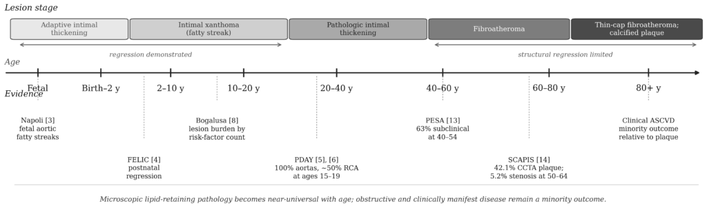
Fig. 1. Lesion stage progression and supporting evidence across the human lifespan. Adaptive intimal thickening (upper band, left) is a physiological response to hemodynamic stress and is not disease [1], [2]. Pathology begins with the intimal xanthoma, in which apoB-containing lipoproteins are retained and modified [2], [16]. Regression has been demonstrated for early lipid-retaining lesions [4]. Bracketed numerals refer to the reference list. The age axis is compressed and not linear.
TABLE I
Principal Autopsy and Imaging Studies of Atherosclerosis Onset and Prevalence
| Study | Design and country | Sample and age | Method | Principal finding |
| Napoli et al. [3] | Fetal autopsy series; Italy | 82 aortas; mean fetal age 6.2 mo | Histology; immunostaining for apoB, oxLDL, CD68 | Fatty streaks present in fetal aortas; extent greater with maternal hypercholesterolemia; lipid retention and oxidation preceded monocyte recruitment |
| FELIC [4] | Child autopsy series; Italy | 156 children aged 1–13 y | Computer-assisted morphometry | Lesions smaller in youngest children than in corresponding fetuses, indicating postnatal regression; faster progression with maternal hypercholesterolemia |
| PDAY [5], [6] | Multicenter autopsy program; USA | 1,532 (1993); 2,876 (2000); ages 15–34 | Standardized formalin fixation; Sudan IV; central grading | All aortas and about half of right coronary arteries had lesions at ages 15–19; burden tracked non-HDL cholesterol, smoking, adiposity, glycemia |
| Bogalusa [8] | Cohort linked to autopsy; USA | 204 autopsies aged 2–39; 93 with antemortem data | Gross staining; morphometry | Stepwise increase in lesion burden with number of childhood risk factors; coronary fibrous plaque 0.6% to 7.2% |
| Enos et al. [9] | Combat casualty autopsy; Korea | 300 males; mean age 22.1 y | Gross visual assessment; unfixed arteries | Coronary disease in 77.3%; grading threshold included fibrous intimal thickening and may therefore yield higher prevalence than contemporary lesion definitions |
| Webber et al. [11] | Casualty autopsy registry; USA | 3,832; mean age 25.9 y | Standardized autopsy grading | Any coronary atherosclerosis 8.5%; severe 2.3%; historical decline may reflect both secular and methodological differences |
| Tuzcu et al. [15] | Transplant donor imaging; USA | Donor hearts across adult age range | Intravascular ultrasound | Atherosclerosis in 17% of donors under 20 y, 60% in fourth decade, 85% at 50 y and older |
| PESA [13] | Asymptomatic cohort; Spain | Adults aged 40–54 y | Multiterritorial ultrasound and calcium scoring | Subclinical atherosclerosis in 63% in at least one territory |
| SCAPIS [14] | Population cohort; Sweden | >25,000 adults aged 50–64 y | Coronary CT angiography | Coronary atherosclerosis 42.1%; significant stenosis 5.2%; 5.5% of those with calcium score zero had plaque |
IV. Apolipoprotein B and Cumulative Exposure
A. The Initiating Substrate
Every atherogenic lipoprotein particle — LDL, its precursors VLDL and IDL, and lipoprotein(a) — carries a single molecule of apolipoprotein B. Measurement of LDL cholesterol quantifies the cholesterol mass transported; measurement of apoB quantifies particle number. Because atherosclerosis is initiated by arterial retention of apoB-containing particles, particle number rather than cholesterol cargo is the mechanistically relevant exposure variable [16], [41].
We state the causal claim precisely. Arterial retention of apoB-containing lipoproteins is a necessary initiating event in the prevailing causal model of atherosclerosis [16], [41]; local vascular biology — endothelial permeability, proteoglycan composition, disturbed flow, and inflammatory signaling — determines where and when retention occurs. This formulation preserves the causal argument while acknowledging that lesion localization is not explained by particle concentration alone. It is consistent with the observation that lesions form preferentially at branch points and bends, where adaptive intimal thickening and disturbed flow coincide.
Three independent evidence streams converge on this conclusion: mechanistic studies of retention and oxidative modification within the arterial wall [41]; genetic studies showing that lifelong lower exposure to apoB-containing lipoproteins is associated with substantially lower coronary risk [42]; and randomized trials of pharmacological LDL reduction demonstrating event reduction proportional to absolute LDL lowering [43]. These streams are synthesized in [16] and [17].
B. The Cumulative-Exposure Model
Because retained particles accumulate over time, plaque burden reflects the integral of apoB concentration over years of exposure, expressed as cholesterol-years or apoB-years by explicit analogy with pack-years of cigarette exposure [17]; the empirical support for that model is set out in [18] and [19] below. Pooled cohort analysis shows that cumulative LDL-C exposure during young adulthood and middle age predicts later cardiovascular events independently of midlife LDL-C level [18]. In CARDIA, among 4,366 participants followed from ages 18 to 40, each standard-deviation increase in cumulative apoB exposure was associated with a hazard ratio of 1.53 (95 percent confidence interval 1.36 to 1.72) for incident atherosclerotic cardiovascular disease after age 40, attenuating to approximately 1.30 after covariate adjustment [19].
The model supports an empirical inflection rather than a fixed threshold: in the same cohort, hazard increased above a usual apoB exposure of about 75 mg/dL per year across ages 18 to 40 [19]. This is a cohort-derived value, not a biological constant, and the authors present it as a potential clinical target requiring validation. The larger round numbers frequently quoted in secondary literature — 5,000 to 6,000 mg/dL-years of cumulative LDL, or 1,300 to 1,500 mg/dL-years of apoB — are modeling heuristics without direct empirical derivation. The associated milestones, namely that homozygous familial hypercholesterolemia crosses such a threshold in childhood, heterozygous familial hypercholesterolemia in early adulthood, and average Western exposure near midlife, are illustrative consequences of the model and broadly consistent with observed epidemiology; the specific ages are not measured values.
C. Evidence From Early Intervention
The twenty-year follow-up of statin therapy initiated in childhood for familial hypercholesterolemia provides one of the strongest available human demonstrations of the exposure model [20]. Treated patients exhibited carotid intima-media thickness progression comparable to unaffected siblings. At age 39, cumulative incidence of cardiovascular events was 1 percent among treated patients versus 26 percent among their affected parents, and cardiovascular mortality was 0 versus 7 percent.
This result is often summarized as a twenty-five-fold reduction in adult coronary events. That framing overstates the inferential strength of the design: the comparison is between treated offspring and their untreated parents, a non-randomized cross-generational contrast confounded by secular improvement in cardiovascular care. The findings strongly support benefit from early treatment, but the cross-generational comparison should not be interpreted as a causal fold-reduction estimate.
V. The Evolutionary Hypothesis
The material in this section is hypothesis rather than established explanation, and is presented at a lower evidentiary tier than Sections III and IV. It is included because it bears on the interpretive question of whether atherosclerosis is inevitable, but no part of the causal argument depends on it.
A. Antagonistic Pleiotropy
The apoB transport system serves essential functions in cholesterol transport and hepatic lipid export [41]. A role in innate host defense has also been described: apoB binds and sequesters a Staphylococcus aureus quorum-sensing peptide, limiting expression of the virulence genes required for invasive infection [44]. One proposed evolutionary interpretation of the late-life costs of such a system invokes antagonistic pleiotropy, under which selection favors variants maximizing fitness during the reproductive period even when those variants impose costs over a post-reproductive interval where selection pressure is attenuated [45]. Such traits have been hypothesized to confer advantages under infectious and nutritional pressures, becoming maladaptive under sustained apoB elevation across an extended modern lifespan. This account is coherent and consistent with the comparative data below, but it has not been tested directly in humans and should not be treated as demonstrated.
B. Comparative Population Evidence
The Tsimane, a forager-horticulturalist population of the Bolivian Amazon, provide striking evidence that low lifetime exposure is compatible with near-absence of coronary calcification in old age. Among 705 adults aged 40 years and older, 85 percent had a coronary artery calcium score of zero; among those older than 75 years, 65 percent had a score of zero and 8 percent had a score of 100 or greater [21]. This is a five-fold lower prevalence of significant coronary calcification than the MESA reference population, observed despite substantial infectious and inflammatory burden [21].
O’Keefe and colleagues [22] have argued that physiologically normal LDL cholesterol for humans lies between 50 and 70 mg/dL, drawing comparisons with values reported in human neonates, wild non-human primates, and the least-exposed human populations. On this view, conventional laboratory reference intervals describe a population with pathological exposure rather than a healthy reference state. That argument is advanced in a narrative review and opinion article rather than original research, and several of its comparative lipid values are asserted with limited primary documentation. It is cited here as a synthesis of a position, not as primary evidence, and the underlying comparative values warrant independent verification.
Countervailing evidence comes from the Horus study, which performed whole-body computed tomography on 137 mummified remains from four preindustrial cultures spanning more than four millennia [23]. Arterial calcification was identified in 47 of 137 remains, 34 percent, in all four populations including the Unangan hunter-gatherer group. Mean age at death was higher among affected than unaffected individuals, reproducing the age association seen in living populations.
These findings are reconcilable. Interpretation is limited by potential selection of preserved remains, uncertain representativeness of the sampled populations, and incomplete information regarding historical environmental exposures. Calcification detected by computed tomography is a late marker insensitive to early disease. The defensible synthesis is that atherosclerosis is an inherent potential of the human artery whose rate and clinical expression are strongly modifiable, with lifetime exposure to apoB-containing lipoproteins a major modifiable determinant.
VI. Dietary Modification of Lifetime Exposure
A. Patterns With Established LDL Effect
The dietary portfolio approach, combining viscous soluble fiber, plant sterols, soy protein, and tree nuts within a plant-based pattern, produces LDL reduction approaching that of a low-dose first-generation statin [24]. The Mediterranean pattern, tested in PREDIMED, a large dietary intervention trial whose 2018 report reanalyzed the data after documented departures from the randomization protocol at some sites, reduced cardiovascular events, with benefit observed despite high total fat intake from olive oil and nuts rather than through fat restriction [25]. Vegetarian and vegan patterns lower LDL cholesterol relative to omnivorous comparators in randomized trials [46].
B. Pediatric Safety
Two randomized trials address the principal objection to lipid-lowering diets in children, namely impairment of growth or neurodevelopment. The Dietary Intervention Study in Children randomized 663 children aged 8 to 10 years with elevated LDL to a reduced-fat, reduced-cholesterol pattern for a mean of seven years, observing LDL reduction with no adverse effect on height, growth velocity, iron status, or sexual maturation [26]. The Special Turku Coronary Risk Factor Intervention Project initiated dietary counseling at seven months of age and reported cholesterol reduction without impairment of growth [27]; a separate analysis within the same cohort found no adverse effect on neurologic development at age five [40]. Pediatric guidelines nonetheless caution against fat restriction in infancy. The current U.S. integrated pediatric guideline recommends human milk or iron-fortified formula through the first year without fat modification, and for toddlers aged 12 to 24 months with a family history of obesity, cardiovascular disease, or hypercholesterolemia, transition to reduced-fat milk only after discussion with the pediatric care provider and only within an overall diet supplying about 30 percent of calories from fat [47].
C. Very-Low-Fat and Added-Oil-Restricted Patterns
Patterns restricting total fat and eliminating added oils, including oils incorporated into prepared foods, rest on a different evidentiary basis. The Lifestyle Heart Trial reported angiographic regression of coronary stenosis under a very-low-fat plant-based diet [28], but delivered the diet within a bundle including exercise, stress management, and smoking cessation, so the independent contribution of oil restriction cannot be isolated; supporting clinical work in this tradition includes uncontrolled case-series evidence such as [29]. Their cardiovascular plausibility rests principally on favorable effects on established risk factors, including LDL-C and apoB. The position exceeds the evidence when it asserts that dietary oils are uniquely harmful beyond their energy density, or that very-low-fat patterns outperform higher-fat Mediterranean patterns for hard endpoints; the available randomized cardiovascular-outcome evidence does not establish that hierarchy.
VII. Polyunsaturated Vegetable Oils
Substitution of polyunsaturated for saturated fat remains a prominent contemporary dietary debate about how to lower lifetime apoB exposure, and is addressed here for that reason. The supporting evidence is substantial. The Cochrane review of saturated fat reduction included 15 randomized trials and 56,675 participants; in the 12 trials contributing 53,758 participants to the combined cardiovascular events analysis, reducing saturated fat produced a 17 percent relative reduction (risk ratio 0.83; 95 percent confidence interval 0.70 to 0.98; GRADE moderate-quality evidence). The review concluded that replacing saturated fat with polyunsaturated fat or with carbohydrate each appeared useful, that subgrouping did not suggest significant differences between replacement with polyunsaturated fat and replacement with carbohydrate, and that data on replacement with monounsaturated fat and protein were very limited [30]. The American Heart Association Presidential Advisory estimated, in its synthesis of those trials, a reduction in cardiovascular disease of approximately 30 percent [31]. A pooled analysis of 30 prospective cohorts found higher circulating linoleic acid associated with lower incident disease and mortality [32], and controlled feeding trials have not demonstrated significant increases in C-reactive protein, interleukin-6, or tumor necrosis factor alpha with increased linoleic acid intake [33], [34].
Two lines of evidence raise concern. A single high-fat meal transiently impairs brachial artery flow-mediated dilation, resolving within about six hours [35]; the effect is not specific to seed oils, since the same investigators observed it following olive oil meals [36]. Separately, reanalyses of recovered data from two historical trials found higher mortality in the intervention arms: the Sydney Diet Heart Study reported all-cause mortality of 17.6 versus 11.8 percent [37], and the Minnesota Coronary Experiment found greater cholesterol reduction associated with higher mortality [38]. Both recovered-data analyses carry important design and data limitations, including incomplete recovery of original records [37], [38]. Trans fat exposure from period-typical margarines is a plausible contributor but was not measured between groups and remains debated, including by the authors of the reanalyses; it should not be presented as an established explanation. The Sydney intervention used safflower oil, which contains no omega-3 fatty acids.
The two bodies of evidence are asymmetric. The apoB-lowering mechanism is supported by mechanistic, human genetic, and randomized outcome evidence [16], [41]-[43]; the postprandial signal rests on a transient surrogate that has not been shown to predict hard outcomes when elicited by a meal. The trial reanalyses are genuinely discordant and should not be dismissed, but their methodological limitations render them insufficient, in isolation, to outweigh the aggregate randomized and prospective evidence. Taken together, the evidence supports dietary patterns that reduce lifetime apoB exposure and does not establish polyunsaturated vegetable oils as a primary vascular hazard [30]-[34], [37], [38].
VIII. What “Everyone Has Atherosclerosis” Means
The claim that atherosclerosis is universal is frequently deployed to argue that it is therefore normal. The evidence assembled here supports a more specific statement.
Lipid-retaining arterial lesions become very common with advancing age. The stronger claim that nearly all adults eventually develop microscopic lipid-retaining pathology is not established by the imaging cohorts cited here and would require direct age-spanning histopathologic evidence. Such lesions should not be conflated with obstructive coronary disease, plaque rupture, or clinical cardiovascular events, which remain minority outcomes relative to the prevalence of plaque. SCAPIS found detectable coronary atherosclerosis in 42.1 percent of adults aged 50 to 64 but significant stenosis in 5.2 percent [14]. Lifetime exposure to apoB-containing particles is a major determinant of lesion initiation and progression, although local vascular biology and other risk factors influence lesion fate.
Statistical ubiquity is therefore not evidence of physiological normality. Hypertension and insulin resistance are also common in aging Western populations without thereby constituting healthy reference states. The Tsimane findings argue against coronary calcification being an unavoidable consequence of human aging [21], although no single observational cohort is definitive. The FELIC observation of postnatal lesion regression indicates that early lesions are not committed to progression [4].
IX. Conclusion
The capacity for atherosclerosis is intrinsic to the human artery. Sustained exposure to apoB-containing lipoproteins promotes intimal lipid retention over time [16]; fetal aortic histology demonstrates that this can begin before birth [3], and the preindustrial mummy record shows that it is not uniquely modern [23]. In this limited sense the susceptibility is universal; the resulting lipid-retaining lesion is nonetheless pathological.
The rate of progression and its clinical expression are not fixed. The Tsimane population demonstrates that advanced age can occur with remarkably low coronary artery calcium burden [21], and individuals with familial hypercholesterolemia treated from childhood exhibit vascular trajectories approximating unaffected siblings [20]. Because cumulative exposure to apoB-containing particles is a major causal and modifiable determinant of disease burden [16]-[19], the high prevalence of atherosclerosis in later life is an argument for early and sustained exposure reduction rather than for therapeutic fatalism.
Appendix: Source Verification Record
The following documents discrepancies, unverifiable claims, and overstatements identified during verification. Items are retained because several affected claims circulate widely in secondary literature.
1) Napoli et al., fetal aortas. Sample composition, mean fetal age, and both correlation coefficients confirmed. Not confirmed: whether the primary report gives lesion prevalence percentages (commonly cited as 30 percent versus up to 90 percent by maternal status) or only lesion area; the abstract emphasizes area and extent. Correction to an earlier verification note: the lesion-composition values of 17.3 percent (native LDL only), 13.3 percent (oxidized LDL without macrophages), and 58.6 percent (both) are stated in the published abstract and are not suspect. They are percentages of immunopositive atherogenic sites, not of fetuses, which is the denominator most often lost in secondary citation. They are therefore omitted from prevalence statements in this paper.
2) FELIC lesion regression magnitude. Postnatal regression is reported in the primary study and is stated qualitatively here. The specific figure of 64 percent smaller lesions in children under three years, frequently quoted in secondary sources, was not verified against the primary text and is not asserted.
3) PDAY sample size. The discrepant values of 1,532 and 2,876 are both correct, corresponding to the 1993 natural-history and 2000 topographic analyses. The exact intimal-surface pairs sometimes quoted (2.2 to 12.2 percent in white men; 3.1 to 15.4 percent in black men) were not verified against source tables and are not reported numerically.
4) Bogalusa risk-factor gradient. All four-value series confirmed as reported in Section III-B. Abbreviated citations reporting only the endpoints of the coronary fatty streak series omit the intermediate strata.
5) Enos severity breakdown. The overall 77.3 percent figure and mean age are confirmed. The severity breakdown sometimes quoted (35 percent fibrous thickening, 26 percent narrowing of 10 to 49 percent, 15 percent severe) is consistent with secondary sources but was not verified against the 1953 report and is not stated in the body.
6) Adaptive intimal thickening in the Korean War series. The claim that the Enos series classified adaptive intimal thickening as atherosclerosis is an inference from the reported grading criteria, not a statement by the original investigators. It is presented in Section III-C as a methodological caveat.
7) Tsimane calcium prevalence. Correct values are 85 percent with calcium score zero at ages 40 and above, 65 percent at ages 75 and above, and 8 percent with score of 100 or greater in the older stratum. Secondary tables reporting 15 percent and 35 percent Tsimane prevalence in calcified-plaque columns are inversions of the calcium-score-zero data and are erroneous.
8) Familial hypercholesterolemia follow-up. Event rates of 1 versus 26 percent and mortality of 0 versus 7 percent are confirmed. The derived twenty-five-fold reduction framing is not a valid causal effect estimate, for the reasons given in Section IV-C.
9) ApoB-years thresholds. The cumulative-exposure concept is well established, and a cohort-derived inflection near 75 mg/dL per year of usual apoB exposure is reported in CARDIA [19]. The larger round thresholds circulating in secondary literature (5,000 to 6,000 mg/dL-years LDL; 1,300 to 1,500 mg/dL-years apoB) could not be traced to a primary derivation and are identified as heuristics in Section IV-B. An earlier draft of this paper attributed a hazard ratio of 1.05 per 100 mg/dL-years to this literature; that figure could not be located in any primary source and has been replaced with the reported effect estimates.
10) Late-life prevalence figures. PESA and SCAPIS values are confirmed. Not independently verified: carotid plaque of 87 percent at ages 63 to 65 in the ACE 1950 cohort; pooled ARIC and MESA figures of 11 percent with calcium score zero at ages 75 and above and calcium present in 95 percent at ages 90 and above; and 11 percent noncalcified plaque prevalence among symptomatic patients with calcium score zero. Overstated and not used: obstructive stenosis prevalences of 45 percent at ages 60 to 80 and 65 percent above age 80, microscopic disease approaching 100 percent, and symptomatic disease of 15 to 25 percent.
11) Stary lesion frequencies in infancy and puberty. Commonly cited values (isolated macrophage foam cells in 45 percent of infants within the first eight months; 65 percent with fatty-streak lesions and 8 percent at preatheroma or atheroma stage in the 12 to 14 year stratum) are consistent with the published series but were not verified against original tables and are not stated numerically.
12) O’Keefe comparative lipid values. Reference [22] is a narrative review and opinion article, not original research. Its reported lipid values for hunter-gatherer populations and wild primates are asserted with limited primary citation and were not independently verified. It is cited in Section V-B as a synthesis of a position rather than as primary evidence.
13) Adaptive intimal thickening prevalence in newborns. The repeated figure of up to 30 percent of newborns could not be traced to a primary source and is not asserted.
14) Pediatric fat restriction threshold. Earlier drafts left unresolved whether fat restriction is contraindicated below 12 months or below two years of age. The apparent conflict reflects two provisions of the same guideline: no fat modification during the first year, and reduced-fat milk permitted between 12 and 24 months for selected children only, within a diet still supplying about 30 percent of calories from fat [47]. Both are now stated in Section VI-B.
Acknowledgment and Disclosures
The author is the founder and sole operator of Curing Heart Disease, LLC, which operates a cardiovascular health education platform and derives revenue from that activity. No external funding was received for this work, and no industry sponsor participated in its design, analysis, or drafting. The author reports no other financial relationships relevant to the subject matter.
This paper is a narrative synthesis of published literature. It involved no new human or animal subjects research and required no ethics approval. It is intended for scientific and professional audiences and does not constitute medical advice.
References
- Stary HC, Chandler AB, Dinsmore RE, et al. A definition of advanced types of atherosclerotic lesions and a histological classification of atherosclerosis. A report from the Committee on Vascular Lesions of the Council on Arteriosclerosis, American Heart Association. Circulation. 1995;92(5):1355-1374. doi:10.1161/01.cir.92.5.1355
- Virmani R, Kolodgie FD, Burke AP, Farb A, Schwartz SM. Lessons from sudden coronary death: a comprehensive morphological classification scheme for atherosclerotic lesions. Arterioscler Thromb Vasc Biol. 2000;20(5):1262-1275. doi:10.1161/01.atv.20.5.1262
- Napoli C, D’Armiento FP, Mancini FP, et al. Fatty streak formation occurs in human fetal aortas and is greatly enhanced by maternal hypercholesterolemia. Intimal accumulation of low density lipoprotein and its oxidation precede monocyte recruitment into early atherosclerotic lesions. J Clin Invest. 1997;100(11):2680-2690. doi:10.1172/JCI119813
- Napoli C, Glass CK, Witztum JL, Deutsch R, D’Armiento FP, Palinski W. Influence of maternal hypercholesterolaemia during pregnancy on progression of early atherosclerotic lesions in childhood: Fate of Early Lesions in Children (FELIC) study. Lancet. 1999;354(9186):1234-1241. doi:10.1016/S0140-6736(99)02131-5
- Natural history of aortic and coronary atherosclerotic lesions in youth. Findings from the PDAY Study. Pathobiological Determinants of Atherosclerosis in Youth (PDAY) Research Group. Arterioscler Thromb. 1993;13(9):1291-1298. doi:10.1161/01.atv.13.9.1291
- McGill HC Jr, McMahan CA, Herderick EE, et al. Effects of coronary heart disease risk factors on atherosclerosis of selected regions of the aorta and right coronary artery. PDAY Research Group. Pathobiological Determinants of Atherosclerosis in Youth. Arterioscler Thromb Vasc Biol. 2000;20(3):836-845. doi:10.1161/01.atv.20.3.836
- Gidding SS, Colangelo LA, Nwabuo CC, et al. PDAY risk score predicts cardiovascular events in young adults: the CARDIA study. Eur Heart J. 2022;43(30):2892-2900. doi:10.1093/eurheartj/ehac009
- Berenson GS, Srinivasan SR, Bao W, Newman WP 3rd, Tracy RE, Wattigney WA. Association between multiple cardiovascular risk factors and atherosclerosis in children and young adults. The Bogalusa Heart Study. N Engl J Med. 1998;338(23):1650-1656. doi:10.1056/NEJM199806043382302
- Enos WF, Holmes RH, Beyer J. Coronary disease among United States soldiers killed in action in Korea; preliminary report. J Am Med Assoc. 1953;152(12):1090-1093. doi:10.1001/jama.1953.03690120006002
- McNamara JJ, Molot MA, Stremple JF, Cutting RT. Coronary artery disease in combat casualties in Vietnam. JAMA. 1971;216(7):1185-1187. doi:10.1001/jama.1971.03180330061012
- Webber BJ, Seguin PG, Burnett DG, Clark LL, Otto JL. Prevalence of and risk factors for autopsy-determined atherosclerosis among US service members, 2001-2011. JAMA. 2012;308(24):2577-2583. doi:10.1001/jama.2012.70830
- Kotwal RS, Mazuchowski EL, Howard JT, et al. Autopsy-Determined Atherosclerosis in Elite US Military Special Operations Forces. J Spec Oper Med. 2021;21(2):19-24. doi:10.55460/RLN9-UQ2X
- Fernández-Friera L, Peñalvo JL, Fernández-Ortiz A, et al. Prevalence, Vascular Distribution, and Multiterritorial Extent of Subclinical Atherosclerosis in a Middle-Aged Cohort: The PESA (Progression of Early Subclinical Atherosclerosis) Study. Circulation. 2015;131(24):2104-2113. doi:10.1161/CIRCULATIONAHA.114.014310
- Bergström G, Persson M, Adiels M, et al. Prevalence of Subclinical Coronary Artery Atherosclerosis in the General Population. Circulation. 2021;144(12):916-929. doi:10.1161/CIRCULATIONAHA.121.055340
- Tuzcu EM, Kapadia SR, Tutar E, et al. High prevalence of coronary atherosclerosis in asymptomatic teenagers and young adults: evidence from intravascular ultrasound. Circulation. 2001;103(22):2705-2710. doi:10.1161/01.cir.103.22.2705
- Ference BA, Ginsberg HN, Graham I, et al. Low-density lipoproteins cause atherosclerotic cardiovascular disease. 1. Evidence from genetic, epidemiologic, and clinical studies. A consensus statement from the European Atherosclerosis Society Consensus Panel. Eur Heart J. 2017;38(32):2459-2472. doi:10.1093/eurheartj/ehx144
- Ference BA, Braunwald E, Catapano AL. The LDL cumulative exposure hypothesis: evidence and practical applications. Nat Rev Cardiol. 2024;21(10):701-716. doi:10.1038/s41569-024-01039-5
- Zhang Y, Pletcher MJ, Vittinghoff E, et al. Association Between Cumulative Low-Density Lipoprotein Cholesterol Exposure During Young Adulthood and Middle Age and Risk of Cardiovascular Events. JAMA Cardiol. 2021;6(12):1406-1413. doi:10.1001/jamacardio.2021.3508
- Zheutlin AR, Handoo F, Luebbe S, et al. Cumulative exposure to atherogenic lipoprotein particles in young adults and subsequent incident atherosclerotic cardiovascular disease. Eur Heart J. 2025;46(41):4302-4312. doi:10.1093/eurheartj/ehaf472
- Luirink IK, Wiegman A, Kusters DM, et al. 20-Year Follow-up of Statins in Children with Familial Hypercholesterolemia. N Engl J Med. 2019;381(16):1547-1556. doi:10.1056/NEJMoa1816454
- Kaplan H, Thompson RC, Trumble BC, et al. Coronary atherosclerosis in indigenous South American Tsimane: a cross-sectional cohort study. Lancet. 2017;389(10080):1730-1739. doi:10.1016/S0140-6736(17)30752-3
- O’Keefe JH Jr, Cordain L, Harris WH, Moe RM, Vogel R. Optimal low-density lipoprotein is 50 to 70 mg/dl: lower is better and physiologically normal. J Am Coll Cardiol. 2004;43(11):2142-2146. doi:10.1016/j.jacc.2004.03.046
- Thompson RC, Allam AH, Lombardi GP, et al. Atherosclerosis across 4000 years of human history: the Horus study of four ancient populations. Lancet. 2013;381(9873):1211-1222. doi:10.1016/S0140-6736(13)60598-X
- Jenkins DJ, Kendall CW, Marchie A, et al. Effects of a dietary portfolio of cholesterol-lowering foods vs lovastatin on serum lipids and C-reactive protein. JAMA. 2003;290(4):502-510. doi:10.1001/jama.290.4.502
- Estruch R, Ros E, Salas-Salvadó J, et al. Primary Prevention of Cardiovascular Disease with a Mediterranean Diet Supplemented with Extra-Virgin Olive Oil or Nuts. N Engl J Med. 2018;378(25):e34. doi:10.1056/NEJMoa1800389
- Obarzanek E, Kimm SY, Barton BA, et al. Long-term safety and efficacy of a cholesterol-lowering diet in children with elevated low-density lipoprotein cholesterol: seven-year results of the Dietary Intervention Study in Children (DISC). Pediatrics. 2001;107(2):256-264. doi:10.1542/peds.107.2.256
- Niinikoski H, Lagström H, Jokinen E, et al. Impact of repeated dietary counseling between infancy and 14 years of age on dietary intakes and serum lipids and lipoproteins: the STRIP study. Circulation. 2007;116(9):1032-1040. doi:10.1161/CIRCULATIONAHA.107.699447
- Ornish D, Brown SE, Scherwitz LW, et al. Can lifestyle changes reverse coronary heart disease? The Lifestyle Heart Trial. Lancet. 1990;336(8708):129-133. doi:10.1016/0140-6736(90)91656-u
- Esselstyn CB Jr, Gendy G, Doyle J, Golubic M, Roizen MF. A way to reverse CAD? J Fam Pract. 2014;63(7):356-364b.
- Hooper L, Martin N, Jimoh OF, Kirk C, Foster E, Abdelhamid AS. Reduction in saturated fat intake for cardiovascular disease. Cochrane Database Syst Rev. 2020;8(8):CD011737. Published 2020 Aug 21. doi:10.1002/14651858.CD011737.pub3
- Sacks FM, Lichtenstein AH, Wu JHY, et al. Dietary Fats and Cardiovascular Disease: A Presidential Advisory From the American Heart Association. Circulation. 2017;136(3):e1-e23. doi:10.1161/CIR.0000000000000510
- Marklund M, Wu JHY, Imamura F, et al. Biomarkers of Dietary Omega-6 Fatty Acids and Incident Cardiovascular Disease and Mortality. Circulation. 2019;139(21):2422-2436. doi:10.1161/CIRCULATIONAHA.118.038908
- Johnson GH, Fritsche K. Effect of dietary linoleic acid on markers of inflammation in healthy persons: a systematic review of randomized controlled trials. J Acad Nutr Diet. 2012;112(7):1029-1041.e10415. doi:10.1016/j.jand.2012.03.029
- Su H, Liu R, Chang M, Huang J, Wang X. Dietary linoleic acid intake and blood inflammatory markers: a systematic review and meta-analysis of randomized controlled trials. Food Funct. 2017;8(9):3091-3103. doi:10.1039/c7fo00433h
- Vogel RA, Corretti MC, Plotnick GD. Effect of a single high-fat meal on endothelial function in healthy subjects. Am J Cardiol. 1997;79(3):350-354. doi:10.1016/s0002-9149(96)00760-6
- Vogel RA, Corretti MC, Plotnick GD. The postprandial effect of components of the Mediterranean diet on endothelial function. J Am Coll Cardiol. 2000;36(5):1455-1460. doi:10.1016/s0735-1097(00)00896-2
- Ramsden CE, Zamora D, Leelarthaepin B, et al. Use of dietary linoleic acid for secondary prevention of coronary heart disease and death: evaluation of recovered data from the Sydney Diet Heart Study and updated meta-analysis. BMJ. 2013;346:e8707. Published 2013 Feb 4. doi:10.1136/bmj.e8707
- Ramsden CE, Zamora D, Majchrzak-Hong S, et al. Re-evaluation of the traditional diet-heart hypothesis: analysis of recovered data from Minnesota Coronary Experiment (1968-73). BMJ. 2016;353:i1246. Published 2016 Apr 12. doi:10.1136/bmj.i1246
- McMahan CA, Gidding SS, Viikari JS, et al. Association of Pathobiologic Determinants of Atherosclerosis in Youth risk score and 15-year change in risk score with carotid artery intima-media thickness in young adults (from the Cardiovascular Risk in Young Finns Study). Am J Cardiol. 2007;100(7):1124-1129. doi:10.1016/j.amjcard.2007.05.035
- Rask-Nissilä L, Jokinen E, Terho P, et al. Effects of diet on the neurologic development of children at 5 years of age: the STRIP project. J Pediatr. 2002;140(3):328-333. doi:10.1067/mpd.2002.122393
- Boren J, Chapman MJ, Krauss RM, et al. Low-density lipoproteins cause atherosclerotic cardiovascular disease: pathophysiological, genetic, and therapeutic insights: a consensus statement from the European Atherosclerosis Society Consensus Panel. Eur Heart J. 2020;41(24):2313-2330. doi:10.1093/eurheartj/ehz962
- Ference BA, Yoo W, Alesh I, et al. Effect of long-term exposure to lower low-density lipoprotein cholesterol beginning early in life on the risk of coronary heart disease: a Mendelian randomization analysis. J Am Coll Cardiol. 2012;60(25):2631-2639. doi:10.1016/j.jacc.2012.09.017
- Cholesterol Treatment Trialists’ (CTT) Collaboration. Efficacy and safety of more intensive lowering of LDL cholesterol: a meta-analysis of data from 170,000 participants in 26 randomised trials. Lancet. 2010;376(9753):1670-1681. doi:10.1016/S0140-6736(10)61350-5
- Peterson MM, Mack JL, Hall PR, et al. Apolipoprotein B is an innate barrier against invasive Staphylococcus aureus infection. Cell Host Microbe. 2008;4(6):555-566. doi:10.1016/j.chom.2008.10.001
- Williams GC. Pleiotropy, natural selection, and the evolution of senescence. Evolution. 1957;11(4):398-411. doi:10.1111/j.1558-5646.1957.tb02911.x
- Wang F, Zheng J, Yang B, Jiang J, Fu Y, Li D. Effects of vegetarian diets on blood lipids: a systematic review and meta-analysis of randomized controlled trials. J Am Heart Assoc. 2015;4(10):e002408. doi:10.1161/JAHA.115.002408
- Expert Panel on Integrated Guidelines for Cardiovascular Health and Risk Reduction in Children and Adolescents; National Heart, Lung, and Blood Institute. Expert panel on integrated guidelines for cardiovascular health and risk reduction in children and adolescents: summary report. Pediatrics. 2011;128(Suppl 5):S213-S256. doi:10.1542/peds.2009-2107C

