
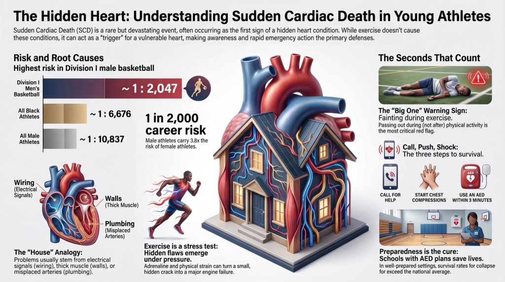
Sudden Cardiac Death in Young People
Epidemiology, Disease Mechanisms, Diagnosis, and Prevention
1. Scope and Definitions
Sudden cardiac death is conventionally defined as unexpected death from a cardiac cause occurring within one hour of symptom onset in a witnessed event, or within 24 hours of the person last being seen alive and well in an unwitnessed one. Sudden cardiac arrest (SCA) describes the same event when circulation is restored; the distinction between arrest and death is determined largely by the speed and quality of the response, which is why the two are best considered a single clinical entity with divergent outcomes rather than separate phenomena [1].
This review concerns people under approximately 35 years of age. The age boundary is not arbitrary. Above it, coronary atherosclerosis progressively dominates the etiologic picture, and the clinical problem converges with that of adult ischemic heart disease. Below it, the causes are predominantly genetic, congenital, or acquired non-atherosclerotic conditions, and the diagnostic and preventive strategies differ accordingly.
The population most studied is competitive athletes, for three reasons: they are systematically enumerated, they undergo medical evaluation that generates data, and their deaths are reported. This creates a substantial ascertainment asymmetry. Sudden death in non-athletic young people is more common in absolute terms but far less well characterized, and much of what follows is derived from athletic cohorts and applies to the general young population with unquantified precision.
2. Epidemiology
2.1 Incidence and the problem of measurement
Historical incidence estimates were degraded by methodological limitations that systematically biased them downward: reliance on media reports and voluntary registries, imprecise denominators derived from participation estimates rather than enumerated cohorts, wide and heterogeneous age ranges, and failure to distinguish exertional from non-exertional events [2,3].
A 20-year multi-database surveillance study of National Collegiate Athletic Association athletes addressed most of these limitations. Covering 2002–2022 and 9,106,516 athlete-years, it identified 143 adjudicated SCD cases among 1,102 total deaths, using four independent case-ascertainment strategies [4,5]. No single source captured more than 82% of cases — a finding that helps explain why earlier single-source estimates were low.
The resulting overall incidence was 1 per 63,682 athlete-years (95% CI 1:54,065–1:75,010) [4].
For comparison, Denmark, where SCD reporting in athletes is mandatory, reports an annual incidence of approximately 1.2 per 100,000 among competitive athletes for events occurring during or within one hour of exertion. Differences in case definition, exertional attribution, and population make direct comparison across national datasets unreliable, and apparent discrepancies between series more often reflect ascertainment methodology than true differences in risk.
2.2 Risk stratification by sex, race, and sport
Risk is not distributed evenly. The NCAA surveillance data demonstrate substantial and consistent gradients.
Table 1. Incidence of sudden cardiac death by demographic and athletic stratum, NCAA athletes, 2002–2022 [4]
| Stratum | Incidence (per athlete-year) | 95% CI | 4-year career risk | Ratio vs. overall |
| Division I men’s basketball, White | 1:5,848 | 1:2,498–1:13,691 | ~1:1,462 | 10.9 |
| Division I men’s basketball, Black | 1:7,696 | wide, overlapping | ~1:1,924 | 8.3 |
| Division I men’s basketball, overall | 1:8,188 | — | ~1:2,047 | 7.8 |
| Basketball, all divisions | 1:19,164 | — | ~1:4,791 | 3.3 |
| American-style football, all divisions | 1:31,743 | — | ~1:7,936 | 2.0 |
| All Black athletes | 1:26,704 | 1:20,417–1:34,925 | ~1:6,676 | 2.4 |
| All male athletes | 1:43,348 | 1:36,228–1:51,867 | ~1:10,837 | 1.5 |
| All White athletes | 1:74,581 | 1:60,247–1:92,326 | ~1:18,645 | 0.85 |
| All female athletes | 1:164,504 | 1:110,552–1:244,787 | ~1:41,126 | 0.39 |
| Overall cohort | 1:63,682 | 1:54,065–1:75,010 | ~1:15,921 | 1.00 (reference) |
Career risk is the simple four-fold annualized approximation used in the source publication [4].
Sex. Male athletes experience approximately 3.8 times the incidence of female athletes (commonly rounded to fourfold in secondary sources) [4]. The explanation is incompletely established but likely multifactorial: greater left ventricular mass and wall thickness for a given body size, differences in autonomic response and repolarization, higher prevalence of some arrhythmogenic substrates, and differences in the intensity and type of athletic exposure [6]. Notably, the sex difference is smaller for the channelopathies — particularly long QT syndrome, where female sex is a risk factor for events after puberty — than for the structural cardiomyopathies.
Race. Black athletes experience approximately 2.8 times the incidence of White athletes [4]. This finding requires careful interpretation. Race in this literature is a social variable functioning as an imperfect proxy for a mixture of unmeasured factors: differential participation by sport and position, body size, hypertension prevalence, sickle cell trait, socioeconomic access to care, structural determinants of health, and possibly ancestry-associated cardiac phenotypes. Contemporary guidance explicitly frames race as a sociopolitical construct in this context and calls for the capture of social determinants of health in future research [1]. The practical implication is that “Black race” identifies elevated risk without explaining it, and is a poor foundation for clinical algorithms.
Sport. Basketball carries the highest risk of any studied discipline, and does so independently: after multivariable adjustment for sex and race, basketball participation remains associated with elevated risk (odds ratio 2.75, 95% CI 1.73–4.34) [4]. American-style football contributes the largest absolute number of cases by virtue of roster size. Soccer is likewise identified among the higher-incidence disciplines. The common features of these sports — high-dynamic, stop-start exertion with repeated maximal efforts and abrupt autonomic transitions — are plausibly mechanistic rather than incidental, though the hypothesis has not been directly tested [7,8].
One frequently repeated error deserves correction. Within Division I men’s basketball, the point estimate for White players (1:5,848) exceeded that for Black players (1:7,696) [4]. Secondary sources commonly invert this. Both estimates rest on very small event counts with wide, overlapping confidence intervals, and the ordering within basketball should not be treated as established in either direction.
2.3 Temporal trend
SCD incidence among NCAA athletes declined by approximately 29% per five-year interval across the study period (five-year incidence rate ratio 0.71, 95% CI 0.61–0.82), while non-cardiovascular mortality in the same population remained unchanged (IRR 0.98, 95% CI 0.94–1.04) [4].
The specificity of the decline to cardiovascular death is suggestive, but the study was not designed to identify a mechanism and its authors decline to attribute the trend to any single intervention. Plausible contributors include broader ECG use at well-resourced programs, improved emergency action planning and automated external defibrillator deployment, better recognition of exertional warning symptoms, and secular changes in ascertainment. Disentangling these remains an open problem, and the trend should not be cited as evidence for the efficacy of any particular preventive strategy.
3. Etiology: The Spectrum of Disease
3.1 Distribution of causes
Among the 118 of 143 NCAA cases with sufficient information for adjudication [4]:
- Autopsy-negative sudden unexplained death (AN-SUD): 19.5%
- Idiopathic left ventricular hypertrophy or possible cardiomyopathy: 16.9%
- Hypertrophic cardiomyopathy: 12.7%
- Remainder: congenital coronary artery anomalies, arrhythmogenic cardiomyopathy, myocarditis, aortic dissection, and other causes
This distribution differs materially from the historical picture, in which hypertrophic cardiomyopathy was described as the single dominant cause [2,3]. The shift reflects improved forensic rigor rather than a change in disease biology: standardized expert adjudication reclassifies as idiopathic or unexplained many cases that less systematic review attributed to HCM on the basis of borderline wall thickness alone.
Across the broader literature, the proportion of cases in which autopsy fails to identify a structural cause ranges from approximately 10% to 42%, with variation driven by the thoroughness of the examination, the expertise of the pathologist, and whether cardiac-specific protocols were followed.
The implication for prevention is direct and underappreciated: in these data the largest single adjudicated category is the absence of a structural finding. Strategies focused exclusively on structural disease therefore leave a substantial proportion of events unexplained or undetected. This proportion is not fixed — it varies with forensic methodology, the expertise of the examining pathologist, and whether molecular autopsy is performed — but the importance of nonstructural and autopsy-negative causes is consistent across series.
3.2 Why exertion triggers events
Sudden death during exercise is best understood through the substrate–trigger–modulator framework. A vulnerable substrate — an anatomical, structural, or ion-channel abnormality — is necessary but rarely sufficient. Exercise supplies triggers and modulators that convert latent vulnerability into ventricular fibrillation [7,8].
Catecholaminergic surge. Exercise produces marked sympathetic activation with circulating catecholamines rising several-fold. Beta-adrenergic stimulation shortens refractoriness heterogeneously across the myocardium, increases automaticity, and enhances calcium loading of the sarcoplasmic reticulum. In conditions of abnormal calcium handling — catecholaminergic polymorphic ventricular tachycardia most explicitly — this is directly arrhythmogenic [8].
Demand ischemia. In anomalous coronary anatomy, myocardial bridging, or severe left ventricular outflow obstruction, exertional increases in myocardial oxygen demand are not matched by supply. Ischemia in turn produces regional conduction slowing and dispersion of repolarization, the substrate for reentry.
Mechanical and hemodynamic stress. Increased wall stress, chamber dilation, and vigorous contraction against an obstruction stretch myocardium acutely. Mechanoelectric feedback — stretch-activated ion channels altering membrane potential — provides a plausible mechanism linking mechanical load to electrical instability, and may be relevant in arrhythmogenic cardiomyopathy, where exercise appears to accelerate disease as well as trigger events.
Electrolyte and acid–base shifts. Exercise produces potassium efflux from working muscle, intracellular acidosis, volume depletion, and in prolonged effort hyponatremia. Each alters conduction and repolarization [9].
Autonomic transition. The abrupt withdrawal of sympathetic tone and surge of vagal activity at cessation of effort produces a period of electrical heterogeneity. A meaningful proportion of exertion-related events occur immediately after exercise rather than during it, and the stop-start structure of basketball, soccer, and football may repeatedly reproduce these transitions [8].
Substrate progression. In some conditions exercise is not merely a trigger but a driver of disease. This is best established for arrhythmogenic cardiomyopathy caused by plakophilin-2 variants, where endurance exercise volume is associated with earlier phenotypic expression, higher arrhythmic burden, and structural progression [1,10].
3.3 The cardiomyopathies
Hypertrophic cardiomyopathy. Defined by unexplained left ventricular hypertrophy, typically with an inherited sarcomeric basis, though approximately half of patients have no identifiable causal variant. Phenotype, clinical course, and arrhythmic risk vary widely. Mechanisms of sudden death include ventricular arrhythmia arising from myocyte disarray and interstitial fibrosis, ischemia from microvascular dysfunction, and hemodynamic collapse from dynamic outflow obstruction. Risk stratification incorporates maximal wall thickness, family history of sudden death, unexplained syncope, non-sustained ventricular tachycardia, left atrial size, outflow gradient, apical aneurysm, and extent of late gadolinium enhancement on cardiac magnetic resonance. Individuals who carry a pathogenic variant without a hypertrophic phenotype have low arrhythmic risk [1].
Arrhythmogenic cardiomyopathy. Characterized by ventricular dysfunction — right, left, or biventricular — with a burden of ventricular arrhythmia disproportionate to the degree of chamber dilation or systolic impairment. Most identified variants affect desmosomal proteins, plakophilin-2 being the most common, though roughly half of cases are genotype-negative. Genotype substantially modifies risk: plakophilin-2-mediated disease shows clear exercise-associated acceleration and elevated arrhythmic risk with endurance participation, whereas evidence for comparable risk in non-plakophilin-2 and genotype-negative disease is not established [10]. Distinguishing early arrhythmogenic cardiomyopathy from the right ventricular dilation of endurance-trained athletes is among the most difficult problems in sports cardiology [1].
Dilated cardiomyopathy. Left ventricular or biventricular dilation with systolic dysfunction, with roughly 60% genotype-negative. Arrhythmic risk rises with lower ejection fraction, symptoms, and scar burden, and is disproportionately high in specific genetic subtypes — lamin A/C, desmoplakin, and filamin C — which warrant closer surveillance regardless of ejection fraction. Preliminary evidence suggests higher cumulative lifetime exercise exposure is associated with lower ejection fraction in lamin A/C-associated disease, raising the possibility that exercise contributes to progression as well as to arrhythmic triggering [1].
Left ventricular hypertrabeculation. Prominent trabeculae with deep intertrabecular recesses, formerly termed left ventricular noncompaction. It is no longer considered a distinct cardiomyopathy. In the absence of coexisting hypertrophic or dilated phenotype, ventricular arrhythmia, or symptoms, adverse events have not been demonstrated, and isolated hypertrabeculation in an asymptomatic person is best regarded as a morphological variant [1].
3.4 The channelopathies
Inherited arrhythmia syndromes can produce sudden death in a structurally normal heart and are an important identifiable cause of autopsy-negative sudden death.
Long QT syndrome. Delayed ventricular repolarization predisposing to torsades de pointes. Genotype predicts trigger: LQT1 (KCNQ1) events are characteristically exertional, with swimming a distinctive trigger; LQT2 (KCNH2) events associate with auditory startle, emotion, and the postpartum period; LQT3 (SCN5A) events occur predominantly at rest or during sleep. Diagnosis rests on corrected QT interval, symptom history, family history, and genetic testing. Two caveats are clinically important: QTc thresholds in athletes differ from those in the general population, and the QT interval may normalize intermittently, so that a single normal resting QTc does not exclude the diagnosis. Concealed long QT syndrome — genotype-positive with a persistently normal QTc — occurs in a substantial minority of variant carriers [1].
Catecholaminergic polymorphic ventricular tachycardia. Caused most often by ryanodine receptor (RYR2) variants producing abnormal diastolic calcium release under adrenergic stress. It is the channelopathy most specifically tied to exertion, characteristically producing bidirectional or polymorphic ventricular tachycardia at reproducible heart-rate thresholds during exercise. The resting ECG is typically normal, which places this condition beyond the reach of resting ECG screening. Exercise testing has high diagnostic yield where CPVT is clinically suspected, characteristically reproducing the arrhythmia at a consistent heart-rate threshold, and is the appropriate test in a person with exertional syncope and a normal resting ECG [1].
Brugada syndrome. Characterized by coved ST elevation in the right precordial leads, associated with SCN5A variants in a minority of cases. Events occur predominantly at rest, during sleep, or with fever rather than during exertion, though fever accompanying exercise is a recognized precipitant [1].
Wolff-Parkinson-White pattern. An accessory atrioventricular pathway. Sudden death occurs when atrial fibrillation conducts rapidly over a pathway with a short refractory period, degenerating into ventricular fibrillation. Unlike most causes discussed here, this is a readily detectable and definitively treatable condition, and the resting ECG is diagnostic [1].
3.5 Congenital coronary artery anomalies
Anomalous aortic origin of a coronary artery is among the leading causes of exertional sudden death and among the least detectable by conventional screening.
The highest-risk variant is anomalous origin of the left coronary artery from the right sinus with an interarterial course between the aorta and pulmonary artery. Proposed high-risk features include an intramural segment within the aortic wall, a slit-like proximal orifice, an acute take-off angle, and greater intramural length. The mechanism is thought to involve dynamic compression and orifice distortion during exertion as the great vessels expand, producing intermittent ischemia that may leave no fixed abnormality between episodes [1].
Anomalous right coronary origin with an interarterial course is more common and carries lower risk; many affected individuals remain asymptomatic. Other variants — intraseptal, retroaortic, prepulmonic courses — are generally benign.
The clinical challenge is that affected individuals are frequently asymptomatic until the index event, resting ECG is typically normal, and exercise stress testing may fail to provoke ischemia even in high-risk anatomy. Definitive diagnosis requires anatomical imaging, most often coronary computed tomographic angiography or cardiac magnetic resonance [1].
Myocardial bridging, in which a coronary segment tunnels through myocardium, is common and usually incidental. It becomes clinically relevant only where deep or long tunneled segments produce demonstrable ischemia [1].
3.6 Myocarditis and acquired conditions
Myocarditis produces sudden death through arrhythmia arising from acute inflammation, edema, and subsequent fibrosis. Its importance lies partly in being potentially transient: vigorous exercise during active myocardial inflammation is arrhythmogenic, but risk substantially resolves with the inflammation. This underlies the standard recommendation to abstain from exertion until symptoms and objective evidence of inflammation have resolved. Emerging cardiac magnetic resonance data suggest that selected athletes may safely return earlier than the three-to-six-month interval assumed by earlier guidance, although the evidence base remains limited and return should be governed by objective resolution of inflammation rather than by elapsed time alone [1,11].
Aortopathy. Marfan syndrome, Loeys-Dietz syndrome, vascular Ehlers-Danlos syndrome, and bicuspid aortic valve-associated aortopathy predispose to acute aortic dissection. This is a rare cause of sudden death in the young but distinctive in being detectable by physical examination and imaging, and in that risk relates to aortic dimension in a way that permits threshold-based management. Marked aortic enlargement is rare in young athletes — diameters above 42 mm in males and 40 mm in females are unusual regardless of body size — so such a finding warrants evaluation for an underlying aortopathy rather than attribution to training [1].
Arrhythmic mitral valve prolapse. Most mitral valve prolapse is benign, but a subset — characteristically bileaflet prolapse with mitral annular disjunction, inferolateral late gadolinium enhancement, and complex ventricular ectopy — is associated with sudden death. Papillary muscle traction and consequent regional fibrosis provide a plausible arrhythmic substrate [1].
Commotio cordis. Ventricular fibrillation induced by blunt precordial impact during the vulnerable phase of repolarization, in a structurally normal heart. It is a mechanical rather than a disease phenomenon, is unaffected by any screening strategy, and is survivable with immediate defibrillation [1].
Sickle cell trait. Associated with exertional collapse and death, particularly during intense conditioning in heat, through mechanisms involving exertional rhabdomyolysis and metabolic derangement rather than primary arrhythmia. It is included here because it presents as sudden collapse during exertion and is relevant to differential diagnosis and to prevention protocols [1].
3.7 Genetic architecture: penetrance, modifiers, and the limits of genotype
Most of the conditions described above are inherited, but inheritance in this field behaves far less deterministically than the term “genetic heart disease” suggests, and the gap between genotype and outcome is where much of the clinical difficulty lies.
Incomplete penetrance and variable expressivity. Carrying a pathogenic variant does not reliably produce disease, and where it does, severity varies widely within families sharing an identical variant. Penetrance estimates derived from clinically ascertained families — identified because someone was affected — substantially overstate risk when applied to variants found incidentally or through population screening. Estimates from unselected cohorts are consistently lower. This has direct consequences: an individual who is genotype-positive and phenotype-negative for hypertrophic cardiomyopathy carries low arrhythmic risk and is treated very differently from one with an expressed phenotype [1].
Age-dependent expression. Phenotype in hypertrophic, arrhythmogenic, and dilated cardiomyopathy typically emerges over adolescence and early adulthood rather than being present from birth. A normal evaluation at 14 does not exclude disease at 24. This underlies the requirement for serial evaluation of genotype-positive relatives rather than single-timepoint clearance, and it explains why screening cohorts followed long enough eventually record deaths in individuals who screened normal.
Modifier genes and polygenic background. The same variant produces different phenotypes in different genetic backgrounds. Common variants of individually small effect, aggregated as polygenic scores, appear to modify penetrance and severity in the cardiomyopathies — potentially explaining part of the within-family variability that monogenic models cannot. This work is at an earlier stage than the equivalent literature in coronary disease, and polygenic scores are not yet clinically actionable in this setting, but the direction is toward a model in which a rare pathogenic variant sets susceptibility and common variation, environment, and training load determine whether and when disease appears.
Genotype-negative disease. Roughly half of hypertrophic and arrhythmogenic cardiomyopathy cases and approximately 60% of dilated cardiomyopathy cases have no identifiable causal variant. A negative genetic test in an affected individual does not exclude an inherited condition, and does not obviate clinical screening of relatives.
Variants of uncertain significance. These are the most common result of broad panel testing in the absence of a clear phenotype, and they are frequently misinterpreted as intermediate-risk findings. They are not: they are uninformative pending reclassification, which may occur in either direction as evidence accumulates. Testing in individuals without phenotype or family history predominantly generates them, which is the principal argument against genetic testing as a primary screening tool [1].
3.8 Autopsy-negative sudden unexplained death and molecular autopsy
When comprehensive autopsy including toxicology and histology identifies no cause, the death is classified as autopsy-negative sudden unexplained death — or, in some series, sudden arrhythmic death syndrome. This classification is provisional rather than terminal.
Postmortem genetic testing identifies a clinically actionable pathogenic or likely pathogenic variant in a meaningful minority of these cases. Applying contemporary ACMG classification criteria to 302 expertly adjudicated cases, an actionable variant was identified in 13% of decedents, predominantly catecholaminergic polymorphic ventricular tachycardia and long QT syndrome, with RYR2 the most implicated gene [12]. A clinically relevant diagnosis was established in a substantially larger proportion of families when postmortem genetics was combined with clinical evaluation of surviving relatives.
Reported yields vary widely across series — from under 4% to approximately 30% — and depend heavily on the variant-classification framework applied. Series adhering strictly to ACMG criteria report lower yields; those using looser thresholds report higher ones, largely by counting variants of uncertain significance as diagnostic. Cross-series comparison is therefore unreliable unless classification criteria are matched [12].
Three points follow. First, the proportion of truly unexplained deaths is lower than autopsy-negative rates suggest. Second, molecular autopsy requires appropriate specimen retention, which depends on medical examiner practice and is frequently not performed. Third, and most important clinically, a diagnosis in the decedent enables cascade screening of living relatives — making postmortem evaluation a prevention strategy for the family, not merely a determination of cause.
4. The Athlete’s Heart: Adaptation Versus Disease
Sustained training produces cardiac remodeling that overlaps phenotypically with the diseases described above. Distinguishing adaptation from pathology is the central diagnostic problem in this field, and misclassification carries costs in both directions.
4.1 Structural adaptation
Endurance training produces predominantly eccentric remodeling — chamber enlargement with proportionate wall thickening — driven by sustained volume loading. Strength training produces relatively more concentric change. Most athletes exhibit a mixed pattern reflecting the actual demands of their sport.
Adaptation is modified by sport, sex, body size, ethnicity, and training duration. Left ventricular wall thickness in the 13–15 mm range in men, and right ventricular dilation in endurance athletes, fall into diagnostic gray zones overlapping hypertrophic and arrhythmogenic cardiomyopathy respectively. Left ventricular end-diastolic dimensions of 60 mm or more occur in a meaningful minority of trained athletes without valvular or myocardial disease [1,11].
Features favoring physiological adaptation include symmetric enlargement of all four chambers, normal or supranormal diastolic function, absence of late gadolinium enhancement, normal functional capacity, appropriate blood pressure response to exercise, and regression with detraining. Features favoring pathology include asymmetric hypertrophy, impaired diastolic function, late gadolinium enhancement, a family history of cardiomyopathy or premature sudden death, and marked ECG abnormality out of proportion to the structural findings [1].
4.2 Electrical adaptation
Training produces a reproducible set of ECG changes: sinus bradycardia and sinus arrhythmia from increased vagal tone, ectopic atrial and junctional rhythms, first-degree and Mobitz type I atrioventricular block, incomplete right bundle branch block, isolated voltage criteria for ventricular hypertrophy, and early repolarization. These are physiological, require no evaluation in an asymptomatic person without concerning family history, and their misclassification as pathological was the principal historical barrier to ECG-based screening [13].
Remodeling and repolarization vary by ethnicity, not only between Black and White athletes. Athletes of African and Afro-Caribbean descent show greater left ventricular wall thickness for a given body size and a distinctive repolarization pattern — J-point elevation with convex ST-segment elevation followed by T-wave inversion confined to leads V1–V4 — which is benign. Applying criteria derived from White European cohorts without this accommodation more than doubles the false-positive rate in Black athletes [14]. Distinct patterns have also been described in athletes of Middle Eastern, South Asian, and East Asian descent, and normative data for these groups remain comparatively sparse — a gap that propagates directly into screening performance wherever population-specific reference values do not exist.
Age matters similarly: T-wave inversion in leads V1–V3 in athletes aged 16 or younger represents a persistent juvenile pattern rather than disease [13].
5. Clinical Presentation and Warning Symptoms
The defining clinical feature of these conditions is that most affected individuals are asymptomatic until the index event. Where symptoms do occur, they are frequently attributed to deconditioning, dehydration, anxiety, or normal exertional limitation — by the individual, by coaching staff, and often by clinicians.
Symptoms warranting evaluation before further participation:
Exertional syncope or near-syncope. The single most important warning symptom. Syncope during exertion, as distinct from immediately after cessation, should be presumed cardiac until proven otherwise. Post-exertional syncope is more often neurally mediated but does not exclude cardiac causes.
Exertional chest pain. Particularly if reproducible at a consistent workload, suggesting demand ischemia from anomalous coronary anatomy or outflow obstruction.
Exertional dyspnea disproportionate to conditioning, especially where it represents a change from established baseline capacity.
Palpitations with exertion, particularly abrupt in onset and offset, or associated with lightheadedness.
Unexplained decline in performance not attributable to training load, illness, or injury.
The family history is as informative as the personal history: premature sudden death before age 50 in a first-degree relative, known inherited cardiomyopathy or arrhythmia syndrome, unexplained drowning, single-vehicle accidents without explanation, or unexplained seizure disorder — the last because long QT syndrome and CPVT events are frequently misdiagnosed as epilepsy [1,15].
6. Diagnostic Evaluation
6.1 Electrocardiography and the evolution of interpretation criteria
The resting 12-lead ECG detects the electrical signatures of cardiomyopathy and channelopathy, and is the single most informative low-cost test in this population. Its historical limitation was not sensitivity but specificity: applying general-population criteria to trained athletes generated false-positive rates that made systematic use impractical.
Interpretation criteria have been refined substantially over fifteen years.
Table 2. Evolution of athlete ECG interpretation criteria
| Criteria set | Year | False positives, White athletes | False positives, Black athletes | Principal change |
| ESC recommendations [16] | 2010 | 16.2% | 40.4% | Training-related versus unrelated dichotomy; no ethnicity-specific criteria; most T-wave inversion classified abnormal |
| Seattle Criteria [17] | 2013 | 7.1% | 18.4% | Black athlete repolarization pattern recognized as physiological; QTc thresholds raised to ≥470 ms (male) and ≥480 ms (female) |
| Refined Criteria [14] | 2014 | 5.3% | 11.5% | Isolated axis deviation and isolated atrial enlargement reclassified as non-triggering |
| International Criteria [13] | 2017 | ~1.3–3.0% | ~4.2–6.8% | Formal normal/borderline/abnormal categories; two or more borderline findings required to trigger evaluation |
The 2017 International Criteria define exactly five borderline findings: left axis deviation, right axis deviation, left atrial enlargement, right atrial enlargement, and complete right bundle branch block [13]. Any single one of these in isolation does not warrant evaluation in an asymptomatic athlete without concerning family history; two or more do. Isolated voltage criteria for left or right ventricular hypertrophy are classified as normal, not borderline — a point frequently misstated.
Two caveats matter. First, the reported false-positive rates derive from expert or specialist-supervised interpretation; rates under non-specialist reading are consistently higher, and the magnitude of that gap has not been established at scale. Second, gains in specificity have not been free: in masters and pediatric cohorts, the International Criteria have missed diagnoses that earlier criteria would have flagged, including dilated cardiomyopathy presenting with isolated left axis deviation.
6.2 Diagnostic versus screening sensitivity
An important distinction is frequently elided. Diagnostic sensitivity is the probability that a person with established, fully expressed disease has an abnormal ECG. Screening sensitivity is the probability that an asymptomatic adolescent with early, incomplete, or concealed expression is correctly identified in a mass-screening setting.
The second is systematically lower than the first. Adolescent phenotype is often immature — both hypertrophic and arrhythmogenic cardiomyopathy express progressively — dynamic conditions are intermittently normal at rest, and field acquisition and interpretation conditions are inferior to those of a referral laboratory. Sensitivity figures quoted from referral cohorts of patients with known disease, commonly cited near 98% for hypertrophic cardiomyopathy, should not be presented as screening performance [18].
The most instructive evidence comes from long-term follow-up of a screened cohort. Among 11,168 adolescent English Football Association players screened with questionnaire, examination, ECG, and echocardiography, conditions associated with sudden death were identified in 0.38%, with ECG abnormal in 86% of those individuals versus 7% for history and 5% for physical examination [19]. Over a mean 10.6 years of follow-up, however, eight athletes died of cardiac causes — six of whom had screened normal at age 16, at a mean of 6.8 years after screening, most from cardiomyopathies not detectable at the time of testing.
A normal screen is not durable clearance. This is a property of progressive disease, not a failure of the test.
6.3 Secondary evaluation
An abnormal primary screen or concerning symptom initiates further testing, selected by the suspected condition rather than applied as a fixed panel.
Transthoracic echocardiography assesses wall thickness, chamber dimensions, systolic and diastolic function, valvular structure, aortic dimensions, and in many cases coronary origins.
Cardiac magnetic resonance provides superior tissue characterization. Late gadolinium enhancement identifies fibrosis and scar; T2-weighted imaging and parametric mapping identify edema and active inflammation. It is often decisive in distinguishing physiological hypertrophy from cardiomyopathy, in evaluating the right ventricle for arrhythmogenic cardiomyopathy, and in diagnosing myocarditis. Late gadolinium enhancement confined to right ventricular insertion points is a recognized finding without established adverse prognostic significance.
Exercise testing should be sport-specific and designed to provoke symptoms at the intensity actually encountered in competition, rather than terminated at arbitrary heart-rate targets or performed pharmacologically. It is essential in suspected CPVT, in evaluating ventricular ectopy, and in assessing ischemia in anomalous coronary anatomy.
Ambulatory rhythm monitoring characterizes arrhythmia burden and its relationship to activity; extended monitoring is often required given the intermittency of clinically relevant events.
Coronary imaging by CT angiography or magnetic resonance is required to define coronary origin and course when anomalous anatomy is suspected.
Genetic testing confirms diagnosis where the phenotype is established, informs risk stratification in specific conditions — plakophilin-2 in arrhythmogenic cardiomyopathy, lamin A/C in dilated cardiomyopathy, genotype in long QT syndrome — and enables cascade screening of relatives. It performs poorly as a primary diagnostic test in the absence of phenotype or family history, where it principally generates variants of uncertain significance.
Notably, cardiac imaging, exercise testing, and ambulatory monitoring have insufficient evidence to support their use as primary screening tools in asymptomatic individuals [1]. Their role is in secondary evaluation.
6.4 Emerging technologies
Artificial intelligence applied to the ECG. Deep learning models trained on large ECG corpora can identify conditions that lack a pathognomonic signature to the human eye. Convolutional neural networks have been developed to detect hypertrophic cardiomyopathy from the 12-lead ECG alone, with performance maintained on external validation across diverse international cohorts and in pediatric and adolescent populations [20]. Comparable models identify electrocardiographically concealed long QT syndrome — genotype-positive individuals with a normal measured QTc — which conventional interval measurement cannot detect by definition [21].
The potential relevance to this field is obvious: the principal limitation of ECG screening is not cost but interpretive accuracy, and an algorithm that improves specificity without sacrificing sensitivity would change the calculus directly. Several important caveats apply. Models trained predominantly on clinical populations may perform differently in trained athletes, whose baseline ECGs differ systematically from those of the general population, and athlete-specific validation remains limited. Performance in the demographic groups with the highest false-positive rates under conventional criteria has not been separately established. And an algorithm that flags disease without an accessible pathway to secondary evaluation reproduces the equity problem described in Section 7.5 rather than solving it. AI-ECG is best understood at present as a promising adjunct under active validation rather than an established screening tool.
Wearable and consumer devices. Smartwatches, adhesive patch monitors, and consumer single-lead and multi-lead ECG devices are now widely used by athletes, and increasingly generate cardiac data that reaches clinicians unsolicited. Their genuine strengths are duration and opportunism: a patch monitor worn for two weeks or a smartwatch worn continuously may capture a symptomatic paroxysmal arrhythmia that a resting ECG and a 24-hour Holter both miss, and this is a real diagnostic contribution in a person with intermittent palpitations.
Their limitations are equally clear. Single-lead recordings cannot assess axis, chamber enlargement, repolarization across the precordium, or most of the criteria on which athlete ECG interpretation depends. Automated rhythm classification is optimized for atrial fibrillation detection in older populations and performs poorly for the arrhythmias relevant here. Signal quality during exercise — precisely when it would be most valuable — is frequently inadequate. And the false-positive burden generated by consumer devices in young, healthy, highly motivated users is substantial and largely unquantified.
Wearables complement rather than replace structured evaluation. A consumer device recording during a symptomatic episode is valuable evidence; a normal consumer recording is not clearance, and should not be treated as one by the athlete or the clinician.
7. Screening: What It Achieves and What It Does Not
Screening is one component of prevention. This section assesses it on its own terms rather than treating it as the organizing question of the field.
7.1 The rationale and its limits
Preparticipation cardiovascular screening aims to identify people with unrecognized disease in time for management that reduces risk. Its effectiveness depends on a chain of conditions: the disease must be detectable before the event, the test must detect it at acceptable cost and false-positive burden, effective management must exist, and affected individuals must have access to it.
That chain has weak links. Resting ECG has very limited sensitivity for anomalous coronary origin, catecholaminergic polymorphic ventricular tachycardia, early or concealed arrhythmogenic cardiomyopathy, aortopathy, and significant valve disease, and by definition contributes nothing in cases that prove autopsy-negative — the largest single category. Screening addresses a subset of the problem.
7.2 Comparative performance of screening components
Meta-analytic estimates in athletic populations give ECG a sensitivity approaching 90–94% for conditions detectable by ECG, with specificity near 93%, against approximately 20% sensitivity for history and 9% for physical examination [18]. Evidence drawn specifically from NCAA populations has since been synthesized separately [22]. The qualifier matters: this figure describes performance against cardiomyopathies and channelopathies with electrical signatures, not against the full spectrum of causes, and should not be read as 94% sensitivity for sudden death risk overall. Contemporary guidance cites a comparable range, placing history and physical sensitivity for silent cardiac conditions at 10–20% and noting that adding ECG raises the sensitivity of the preparticipation evaluation to approximately 94% [1].
The argument for ECG is therefore not that it performs well in absolute terms; it is that history and physical examination alone detect only a minority of relevant conditions. This is not an argument against the history and physical examination, which identifies symptomatic individuals, elicits family history, and detects Marfan stigmata, pathological murmurs, diminished femoral pulses, and hypertension — none of which an ECG reliably captures.
7.3 Guideline positions
European guidance has long recommended ECG-inclusive screening [11,16]. North American guidance historically endorsed a standardized 14-point history and physical examination without recommending universal ECG [15].
That divide has narrowed substantially. The 2025 American Heart Association / American College of Cardiology scientific statement holds that inclusion of a resting 12-lead ECG is reasonable, because it improves detection of underlying cardiac conditions in asymptomatic athletes relative to history and physical examination alone. The endorsement carries three conditions: clinicians must be adequately trained in contemporary athlete-specific interpretation criteria; programs must ensure access to secondary evaluation, including the financial and logistical resources for systematic downstream assessment; and because no approach provides absolute protection, an emergency action plan must be in place wherever people train and compete [1].
The unresolved questions are therefore not whether ECG may be used, but whether it should be universally implemented, how programs should be resourced to use it safely, and whether incremental benefit justifies downstream cost and harm.
7.4 Health economics
Published cost-effectiveness estimates for adding ECG to preparticipation screening span a wide range, driven more by assumption choice than by data.
Table 3. Published economic evaluations of ECG-inclusive screening
| Analysis | Comparison | Result |
| Wheeler et al. (2010) [23] | ECG + H&P vs. H&P alone | $42,900 per life-year saved (95% CI $21,200–$71,300) |
| Wheeler et al. (2010) [23] | ECG + H&P vs. no screening | $76,100 per life-year saved ($62,400–$130,000) |
| Schoenbaum et al. (2012) [24] | H&P then ECG vs. H&P alone | $68,800 per QALY |
| Schoenbaum et al. (2012) [24] | ECG alone vs. H&P alone | $37,700 per QALY |
| Halkin et al. (2012) [25] | National program, US extrapolation | $10.6–14.4 million per life saved |
Two of the three principal analyses return figures within commonly cited US willingness-to-pay thresholds. The third, returning figures orders of magnitude higher, does so partly by loading recurring annual history and physical costs into the ECG arm — an accounting choice contested in subsequent literature [25].
Every one of these analyses is governed by a single assumption: the relative risk reduction conferred by detection and subsequent management. That parameter is not well established, and as discussed in Section 8 the evidence underlying it has shifted.
7.5 Equity
Screening programs can widen the inequities they are intended to reduce. The mechanism is the secondary evaluation, not the primary screen.
An abnormal screening result initiates a diagnostic cascade — echocardiography, cardiac magnetic resonance, ambulatory monitoring, genetic evaluation, subspecialty consultation — that in unassisted settings the family must fund. Uninsured and underinsured individuals face two consequences: they may exit the pathway without completing evaluation, continuing to participate with uncharacterized risk; and their absence from outcome data systematically biases estimates of disease prevalence, specificity, and cost-effectiveness in a favorable direction.
This is compounded by the ethnic disparity in false-positive rates. Even under contemporary criteria, false-positive findings remain more frequent among Black athletes [14], meaning that the population most likely to be referred for costly secondary evaluation overlaps with the population least likely to be able to afford it. Contemporary guidance states directly that screening programs without appropriate downstream resources have the potential to harm athletes from underrepresented racial and ethnic groups [1].
A screening program without a funded pathway to secondary evaluation is not a neutral intervention. It is a mechanism for identifying risk in people who cannot then act on it.
7.6 Psychological consequences
Screening programs are evaluated almost exclusively on detection and cost. The psychological consequences are real, are borne disproportionately by people who turn out not to have disease, and are rarely measured.
The false-positive interval. Between an abnormal screening result and its resolution, an athlete is a person who has been told their heart may be dangerously abnormal. That interval is frequently weeks and sometimes months, determined by appointment availability, insurance authorization, and imaging capacity rather than by clinical urgency. Documented consequences include anxiety, intrusive thoughts about dying during exertion, sleep disruption, and withdrawal from training during a period in which no restriction has actually been imposed. Where the athlete’s identity is substantially organized around sport — as it is for many at collegiate and elite level — the threat is to selfhood as much as to health.
Resolution is incomplete. Reassurance after a negative workup does not reliably return people to baseline. Residual health anxiety, continued symptom vigilance, and reduced training intensity have been described persisting after formal clearance, a pattern familiar from other screening contexts. Because false positives outnumber true positives by a large factor at any plausible operating point, this is not a marginal harm affecting a handful of people; it is the modal experience of an abnormal screen.
True positives carry their own burden. A diagnosis in a young person may bring restriction or modification of participation, loss of athletic identity, disrupted scholarship or professional prospects, defibrillator carriage in adolescence, and the knowledge of inherited risk extending to siblings and future children. Depression and anxiety are recognized sequelae of disqualification, and the transition out of competitive sport is itself a period of elevated psychological risk independent of the cardiac diagnosis.
Implications. Three follow directly. Time from abnormal screen to definitive resolution is a clinically meaningful quality metric, not merely an operational one, and shortening it is a genuine intervention. Communication of an abnormal result should convey the base rate — that most abnormal screens resolve without disease — rather than deferring all interpretation to the specialist. And psychological support should be planned into screening programs rather than improvised, particularly for athletes who are restricted or who transition out of competition.
8. Management and Sports Participation
8.1 The shift from disqualification to shared decision-making
Historical practice restricted individuals with identified cardiovascular disease from competitive sport more or less categorically. That paradigm has been substantially abandoned, on both ethical and empirical grounds [1,11].
The empirical case rests on outcome data:
- In a prospective multinational observational study of individuals with hypertrophic cardiomyopathy — 1,534 patients plus 126 genotype-positive, phenotype-negative individuals across 42 centers — those engaging in vigorous exercise, including competitive athletes, did not experience higher rates of death, resuscitated arrest, appropriate defibrillator shock, or arrhythmic syncope than moderate exercisers or sedentary participants [26].
- A prospective multinational registry of athletes with implantable cardioverter-defibrillators found no deaths, resuscitated arrests, or arrhythmia-related injuries during sport over long-term follow-up [27].
- Contemporary cohorts of elite athletes with genetic heart disease who returned to competition under expert supervision report low rates of breakthrough events [28].
The ethical case is that the historical model rested on the premise that athletes cannot make informed decisions about their own risk — a position that is neither supported by evidence nor consistent with the standards applied elsewhere in medicine.
Shared decision-making does not imply equal risk across conditions. It is a process for incorporating an individual’s values into a decision under uncertainty, not a conclusion that all diagnoses carry comparable danger or that all participation requests should be accommodated. The risk attached to genotype-positive, phenotype-negative status differs by orders of magnitude from that attached to plakophilin-2-mediated arrhythmogenic cardiomyopathy in an endurance athlete, and the framework is designed to make that difference explicit rather than to dissolve it.
The 2025 AHA/ACC statement is explicit that it does not issue disqualification recommendations, but rather clinical considerations to inform shared decision-making. Under this framework a uniform approach of restriction should not be applied to individuals with cardiomyopathy; participation is instead determined through a process incorporating accurate diagnosis, condition-specific risk stratification, guideline-directed treatment, disclosure of known and unknown risks, and the individual’s own values and risk tolerance. For those under 18, parents or guardians participate directly [1].
8.2 Where risk remains prohibitive
Shared decision-making does not mean that all participation is endorsed. Situations where risk is understood to outweigh benefit include arrhythmogenic cardiomyopathy caused by plakophilin-2 variants, particularly with endurance sport; active myocarditis or pericarditis; unrepaired anomalous origin of the left coronary artery with an interarterial course; severe symptomatic aortic stenosis; heritable thoracic aortic disease with aortic dilation; and prior aortic dissection. Participation is also generally deferred during diagnostic evaluation and until guideline-directed therapy is optimized [1].
8.3 Condition-specific management
Management is disease-specific and is the mechanism by which detection produces benefit: beta-blockade in long QT syndrome and CPVT, with flecainide and sympathetic denervation in selected CPVT cases; defibrillator implantation where risk stratification indicates, though never solely to enable sport participation; catheter ablation for accessory pathways and selected ventricular arrhythmias; surgical reimplantation or unroofing for high-risk coronary anomalies; septal reduction for obstructive hypertrophic cardiomyopathy; aortic surgery at guideline thresholds; and exercise prescription modification where disease progression is exercise-associated [1].
Two points deserve emphasis. First, individuals who discontinue competitive sport should be counseled on the established health benefits of continued recreational physical activity — a transition, not a cessation. Second, longitudinal surveillance is required regardless of the participation decision, because phenotype evolves and the original decision may require revisiting.
8.4 Return to play and longitudinal surveillance
A participation decision is a point on a trajectory rather than a conclusion, and the surveillance that follows is what makes continued participation defensible.
Serial imaging at intervals determined by condition and rate of change. Genotype-positive, phenotype-negative individuals in conditions where exercise may precipitate phenotypic conversion — plakophilin-2-mediated arrhythmogenic cardiomyopathy most clearly — warrant close longitudinal surveillance, with imaging intervals individualized to phenotype, exercise exposure, and evidence of disease progression, while continuing to compete [1]. Aortic dimensions in aortopathy are followed on a schedule set by absolute diameter and rate of growth, with side-by-side comparison of images rather than reliance on prior reports, since inter-study measurement variability can exceed true annual change.
Repeat exercise testing, sport-specific and to the intensity actually encountered in competition, to confirm continued absence of provoked arrhythmia or ischemia and to verify the efficacy of pharmacological suppression where it has been prescribed.
Ambulatory rhythm monitoring to track arrhythmia burden over time, and device interrogation where a defibrillator is present, including appropriate and inappropriate therapy history.
Structured re-evaluation of the decision itself. The shared decision-making conversation should be revisited periodically rather than treated as settled — because the evidence base is changing, because the individual’s own risk tolerance may change, and because phenotype progression may move a person from one risk category to another without symptoms.
Post-intervention return — after surgical coronary reimplantation, aortic repair, ablation, or device implantation — follows condition-specific intervals governed by healing, demonstrated absence of ischemia or inducible arrhythmia, and normal ventricular function, rather than by elapsed time alone.
9. Secondary Prevention: Emergency Response
Because no screening strategy detects all disease, and because some causes are undetectable in principle, survival in a substantial proportion of events is determined entirely by what happens in the first minutes.
9.1 Determinants of survival
Survival depends on rapid recognition, immediate high-quality chest compressions, and early defibrillation, and additionally on rhythm at collapse, underlying substrate, arrest location, emergency medical services interval, airway management, post-arrest care, and neurological injury.
Recognition is the most common failure point in athletic settings, and the one most amenable to training. Bystanders are primed to interpret the collapse of a young athlete as anything other than cardiac arrest. Agonal breathing is mistaken for breathing; seizure-like activity, common in the first seconds of arrest, is mistaken for a primary neurological event. A venue with excellent defibrillator coverage can still lose several minutes to a delayed recognition decision — which is why time-to-first-compression should be measured and drilled separately from time-to-shock, since the two fail independently.
9.2 Emergency action plans
An adequate plan comprises a written, venue-specific document reviewed at least annually with local emergency medical services and physically accessible at each site; a designated coordinator responsible for oversight; defibrillator placement supporting a collapse-to-shock interval of three minutes or less from any point of athletic activity; documented CPR and defibrillator training among athletic trainers, coaching staff, and strength staff; rehearsal drills with time-to-first-compression and time-to-shock recorded; and a coordinated transport plan to a designated receiving facility [1].
Where such plans have been prospectively studied in high school settings, survival to hospital discharge has substantially exceeded rates observed for out-of-hospital cardiac arrest generally [29] — a difference attributable to witnessed collapse, trained responders, and immediate defibrillator availability rather than to any characteristic of the individuals.
A final observation on policy. Sudden cardiac arrest and death will continue to occur irrespective of screening strategy and participation decisions, and their occurrence should not be interpreted as evidence that a shared decision-making approach has failed. Sound policy is not well made in the immediate aftermath of an individual event.
10. Family Evaluation
A diagnosis of inherited heart disease is a diagnosis about a family. Where a proband is identified — living or deceased — first-degree relatives warrant clinical evaluation, and cascade genetic testing where a pathogenic variant has been identified [1].
This applies with particular force after an unexplained death. Postmortem specimen retention adequate for genetic analysis is not universal practice among medical examiners, and its absence forecloses the family’s diagnostic pathway permanently. Where molecular autopsy is combined with clinical evaluation of surviving relatives, a clinically relevant diagnosis is established in a substantially higher proportion of families than by either approach alone [12].
Cascade evaluation identifies relatives who carry risk before it manifests — the only circumstance in this field where prevention operates on a clearly identified population rather than an unselected one, and correspondingly the setting where the yield of evaluation is highest.
11. Knowledge Gaps
Several questions central to this field remain unresolved, and are unlikely to be settled by the study designs typically proposed.
Whether screening reduces mortality cannot be established by randomized trial. At an incidence near 1 per 63,682 athlete-years [4], demonstrating a 50% relative reduction with conventional power would require on the order of ten million athlete-years of observation. The Italian regional experience and the contradictory Israeli national experience are both observational [30,31], both confounded by secular trend and ascertainment change, and both are why the question remains open after four decades. This is a structural feature of studying a rare outcome, not a deficiency of effort.
The real-world false-positive rate is unknown. Published rates derive from expert interpretation. The rate achieved by clinicians who would actually staff population screening has not been established at scale, and it is the parameter that determines whether ECG-inclusive screening is feasible outside academic centers.
The magnitude of benefit from detection is uncertain and has probably narrowed. Legacy economic models assume that detection leads to restriction and that restriction prevents death. The second link has weakened considerably. Benefit now plausibly derives more from disease-specific treatment, family cascade screening, and targeted emergency preparedness than from removal from sport.
The true cost and completion rate of the diagnostic cascade are not established, because existing data derive from settings in which financially constrained individuals exit the pathway before completion.
The mechanism of the observed decline in incidence is unknown, and attributing it to screening is not supported by the data.
Sudden death in non-athletic young people is comparatively uncharacterized. The population is larger, the ascertainment poorer, and the preventive infrastructure absent.
12. Summary
Sudden cardiac death in the young is rare, concentrated, and heterogeneous in cause. Risk varies several-fold by sex, race, and sport, with Division I male basketball players the highest-risk studied group at roughly 1 in 2,000 over a four-year career. The etiologic picture is no longer dominated by hypertrophic cardiomyopathy: autopsy-negative sudden unexplained death is the most common single finding, and a meaningful fraction of these cases prove on molecular autopsy to be inherited arrhythmia syndromes.
Exercise acts as a trigger through catecholaminergic surge, demand ischemia, mechanical stress, electrolyte shift, and abrupt autonomic transition — and in at least one condition, plakophilin-2-mediated arrhythmogenic cardiomyopathy, as a driver of disease progression rather than merely a trigger.
Distinguishing physiological cardiac adaptation from disease is the central diagnostic challenge, and criteria for doing so on the ECG have improved markedly, reducing false-positive rates from roughly 40% to under 7% in Black athletes across fifteen years of refinement. Contemporary guidance on both sides of the Atlantic now regards ECG-inclusive screening as reasonable, conditioned on trained interpretation, assured access to secondary evaluation, and emergency preparedness.
Management has moved from categorical disqualification to shared decision-making, supported by outcome data showing lower risk from continued participation than was historically assumed. Detection produces benefit principally through disease-specific treatment, family cascade screening, and targeted preparedness rather than through removal from sport.
No screening strategy prevents all events. Emergency action planning, rapid recognition, immediate compressions, and early defibrillation remain the last and most reliable line of prevention, and in well-prepared settings save a majority of those who arrest.
References
- Kim JH, Baggish AL, Levine BD, et al. Clinical Considerations for Competitive Sports Participation for Athletes With Cardiovascular Abnormalities: A Scientific Statement From the American Heart Association and American College of Cardiology. J Am Coll Cardiol. 2025;85(10):1059-1108. doi:10.1016/j.jacc.2024.12.025
- Maron BJ, Doerer JJ, Haas TS, Tierney DM, Mueller FO. Sudden deaths in young competitive athletes: analysis of 1866 deaths in the United States, 1980-2006. Circulation. 2009;119(8):1085-1092. doi:10.1161/CIRCULATIONAHA.108.804617
- Harmon KG, Asif IM, Maleszewski JJ, et al. Incidence, Cause, and Comparative Frequency of Sudden Cardiac Death in National Collegiate Athletic Association Athletes: A Decade in Review. Circulation. 2015;132(1):10-19. doi:10.1161/CIRCULATIONAHA.115.015431
- Petek BJ, Churchill TW, Moulson N, et al. Sudden Cardiac Death in National Collegiate Athletic Association Athletes: A 20-Year Study. Circulation. 2024;149(2):80-90. doi:10.1161/CIRCULATIONAHA.123.065908
- Moulson N, Petek BJ, Ackerman MJ, et al. Rationale and Design of the ORCCA (Outcomes Registry for Cardiac Conditions in Athletes) Study. J Am Heart Assoc. 2023;12(11):e029052. doi:10.1161/JAHA.122.029052
- Pelliccia A, Maron BJ, Culasso F, Spataro A, Caselli G. Athlete’s heart in women. Echocardiographic characterization of highly trained elite female athletes. JAMA. 1996;276(3):211-215. doi:10.1001/jama.276.3.211
- Albert CM, Mittleman MA, Chae CU, Lee IM, Hennekens CH, Manson JE. Triggering of sudden death from cardiac causes by vigorous exertion. N Engl J Med. 2000;343(19):1355-1361. doi:10.1056/NEJM200011093431902
- Franklin BA, Thompson PD, Al-Zaiti SS, et al. Exercise-Related Acute Cardiovascular Events and Potential Deleterious Adaptations Following Long-Term Exercise Training: Placing the Risks Into Perspective—An Update: A Scientific Statement From the American Heart Association. Circulation. 2020;141(13):e705-e736. doi:10.1161/CIR.0000000000000749
- Sejersted OM, Sjøgaard G. Dynamics and consequences of potassium shifts in skeletal muscle and heart during exercise. Physiol Rev. 2000;80(4):1411-1481. doi:10.1152/physrev.2000.80.4.1411
- James CA, Bhonsale A, Tichnell C, et al. Exercise increases age-related penetrance and arrhythmic risk in arrhythmogenic right ventricular dysplasia/cardiomyopathy-associated desmosomal mutation carriers. J Am Coll Cardiol. 2013;62(14):1290-1297. doi:10.1016/j.jacc.2013.06.033
- Sharma S, Pelliccia A, Gati S. The ‘Ten Commandments’ for the 2020 ESC Guidelines on Sports Cardiology and Exercise in Patients with Cardiovascular Disease. Eur Heart J. 2021;42(1):6-7. doi:10.1093/eurheartj/ehaa735
- Lahrouchi N, Raju H, Lodder EM, et al. Utility of Post-Mortem Genetic Testing in Cases of Sudden Arrhythmic Death Syndrome. J Am Coll Cardiol. 2017;69(17):2134-2145. doi:10.1016/j.jacc.2017.02.046
- Drezner JA, Sharma S, Baggish A, et al. International criteria for electrocardiographic interpretation in athletes: Consensus statement. Br J Sports Med. 2017;51(9):704-731. doi:10.1136/bjsports-2016-097331
- Sheikh N, Papadakis M, Ghani S, et al. Comparison of electrocardiographic criteria for the detection of cardiac abnormalities in elite black and white athletes. Circulation. 2014;129(16):1637-1649. doi:10.1161/CIRCULATIONAHA.113.006179
- Maron BJ, Levine BD, Washington RL, et al. Eligibility and Disqualification Recommendations for Competitive Athletes With Cardiovascular Abnormalities: Task Force 2: Preparticipation Screening for Cardiovascular Disease in Competitive Athletes: A Scientific Statement From the American Heart Association and American College of Cardiology. Circulation. 2015;132(22):e267-e272. doi:10.1161/CIR.0000000000000238
- Corrado D, Pelliccia A, Heidbuchel H, et al. Recommendations for interpretation of 12-lead electrocardiogram in the athlete. Eur Heart J. 2010;31(2):243-259. doi:10.1093/eurheartj/ehp473
- Drezner JA, Ackerman MJ, Anderson J, et al. Electrocardiographic interpretation in athletes: the ‘Seattle criteria’. Br J Sports Med. 2013;47(3):122-124. doi:10.1136/bjsports-2012-092067
- Harmon KG, Zigman M, Drezner JA. The effectiveness of screening history, physical exam, and ECG to detect potentially lethal cardiac disorders in athletes: a systematic review/meta-analysis. J Electrocardiol. 2015;48(3):329-338. doi:10.1016/j.jelectrocard.2015.02.001
- Malhotra A, Dhutia H, Finocchiaro G, et al. Outcomes of Cardiac Screening in Adolescent Soccer Players. N Engl J Med. 2018;379(6):524-534. doi:10.1056/NEJMoa1714719
- Siontis KC, Wieczorek MA, Maanja M, et al. Hypertrophic cardiomyopathy detection with artificial intelligence electrocardiography in international cohorts: an external validation study. Eur Heart J Digit Health. 2024;5(4):416-426. Published 2024 Apr 15. doi:10.1093/ehjdh/ztae029
- Bos JM, Attia ZI, Albert DE, Noseworthy PA, Friedman PA, Ackerman MJ. Use of Artificial Intelligence and Deep Neural Networks in Evaluation of Patients With Electrocardiographically Concealed Long QT Syndrome From the Surface 12-Lead Electrocardiogram. JAMA Cardiol. 2021;6(5):532-538. doi:10.1001/jamacardio.2020.7422
- Conway JJ, Bennett D, Asif IM, Teramoto M, Toresdahl BG. Pre-participation cardiovascular screening among NCAA athletes: a systematic review and meta-analysis of 27 891 athletes. Br J Sports Med. 2026;60(5):379-387. Published 2026 Mar 12. doi:10.1136/bjsports-2025-110791
- Wheeler MT, Heidenreich PA, Froelicher VF, Hlatky MA, Ashley EA. Cost-effectiveness of preparticipation screening for prevention of sudden cardiac death in young athletes. Ann Intern Med. 2010;152(5):276-286. doi:10.7326/0003-4819-152-5-201003020-00005
- Schoenbaum M, Denchev P, Vitiello B, Kaltman JR. Economic evaluation of strategies to reduce sudden cardiac death in young athletes. Pediatrics. 2012;130(2):e380-e389. doi:10.1542/peds.2011-3241
- Halkin A, Steinvil A, Rosso R, Adler A, Rozovski U, Viskin S. Preventing sudden death of athletes with electrocardiographic screening: what is the absolute benefit and how much will it cost?. J Am Coll Cardiol. 2012;60(22):2271-2276. doi:10.1016/j.jacc.2012.09.003
- Lampert R, Ackerman MJ, Marino BS, et al. Vigorous Exercise in Patients With Hypertrophic Cardiomyopathy. JAMA Cardiol. 2023;8(6):595-605. doi:10.1001/jamacardio.2023.1042
- Lampert R, Olshansky B, Heidbuchel H, et al. Safety of Sports for Athletes With Implantable Cardioverter-Defibrillators: Long-Term Results of a Prospective Multinational Registry. Circulation. 2017;135(23):2310-2312. doi:10.1161/CIRCULATIONAHA.117.027828
- Martinez KA, Bos JM, Baggish AL, et al. Return-to-Play for Elite Athletes With Genetic Heart Diseases Predisposing to Sudden Cardiac Death. J Am Coll Cardiol. 2023;82(8):661-670. doi:10.1016/j.jacc.2023.05.059
- Drezner JA, Toresdahl BG, Rao AL, Huszti E, Harmon KG. Outcomes from sudden cardiac arrest in US high schools: a 2-year prospective study from the National Registry for AED Use in Sports. Br J Sports Med. 2013;47(18):1179-1183. doi:10.1136/bjsports-2013-092786
- Corrado D, Basso C, Pavei A, Michieli P, Schiavon M, Thiene G. Trends in sudden cardiovascular death in young competitive athletes after implementation of a preparticipation screening program. JAMA. 2006;296(13):1593-1601. doi:10.1001/jama.296.13.1593
- Steinvil A, Chundadze T, Zeltser D, et al. Mandatory electrocardiographic screening of athletes to reduce their risk for sudden death proven fact or wishful thinking?. J Am Coll Cardiol. 2011;57(11):1291-1296. doi:10.1016/j.jacc.2010.10.037

