
潜在的な冠動脈機能不全を有するアスリートにおける痛覚知感、側副血行路、および診断のリスク
スポーツ心臓学におけるメカニズムと臨床的統合
抄録
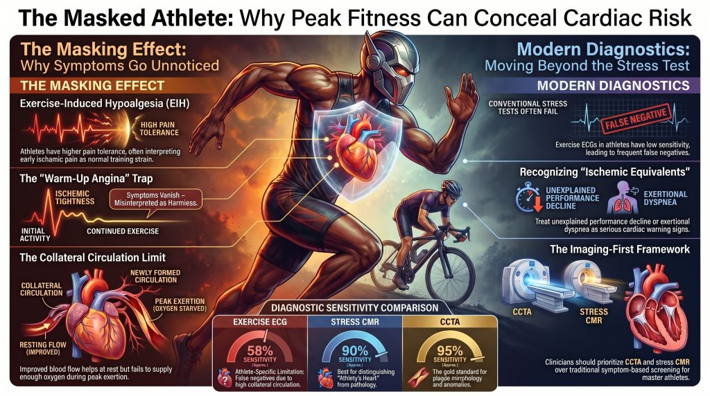
トリーンド・アスリートの心血管評価には逆説が存在する。すなわち、心臓保護をもたらす生理的適応そのもの(自律神経調節など)が、, 運動誘発性無痛覚, 、心筋効率の向上、および冠側副血行路の発達は、潜在的な病態の臨床的マーカーを同時に隠す可能性がある。本総説では、心筋虚血痛の神経生物学、運動誘発性無痛低下のメカニズム、ならびに特異な生物学的特性に関する現代のエビデンスを統合する 動脈新生 対 血管新生, 、 ウォームアップ狭心症 現象と、アスリート集団における従来の診断検査の限界です。それは冠動脈からの新たなデータを統合します 動脈 生涯にわたる持久系アスリート(MARC、Master@Heart、Cooper Center、Merghani、Papatheodorouコホート)におけるカルシウムイメージング、性別特異的なリスクパターン、生涯にわたるアポリポ蛋白B曝露のフレームワーク、ならびに高度な機能的・解剖学的イメージング(CCTA、負荷CMR、FFR/iFR、CT-FFR)の役割。その結果得られるのは、原因不明のパフォーマンス低下や労作時呼吸困難を虚血等価物として扱い、症状や亜最大負荷試験に頼るのではなくイメージングに基づく層別化を採用し、スポーツ復帰の判断を以下を通じて枠組み化する診断的枠組みである。 意思決定の共有.
1. はじめに
有酸素運動能力は、アテローム性動脈硬化に対する最も強力な防御要因の一つである。 心血管疾患, しかし、訓練されたアスリートの無視できない割合が、解剖学的に有意な冠動脈疾患を有している。 病変, 先天性冠動脈異常、または致死的な発症に至るまで臨床的に無症状のままである遺伝性心筋症1], [2]。診断上の課題は、単に疫学的なものにとどまらない。持久力トレーニングは、痛みの神経生物学、心臓感覚の自律神経調節、冠状循環の構造、そしてエネルギー効率を再構築する。 心筋 与えられたワークロードに適応する。これらの適応はそれぞれ、閾値を高めるように働く。 虚血 それはアスリートにとっては現象学的に明らかになり、臨床医にとっては心電図学的に明らかになる。.
本レビューは、仮面の運動選手(masked athlete)に対する統合的なメカニズムおよび診断の枠組みを構築するものである。側副血流能に関する過大評価された主張、運動誘発性無痛覚の内的オピオイドのみへの過度な単純化された帰属、および症状に基づく事前の参加スクリーニングへの時代遅れの依存など、先行文献におけるいくつかの繰り返し見られる誤解を修正し、運動量と(中略)の関係を再定義した近年の画像診断のエビデンスを統合するものである。, プラーク負荷, 、および成果。.
2. 心筋虚血性疼痛の病態生理
2.1 生化学的メディエーターと化学受容
冠血流の低下から数秒以内に、心筋代謝は嫌気性解糖へとシフトし、アデノシン、乳酸、水素イオン(H⁺)、ブラジキニン、プロスタグランジンの間質への蓄積を引き起こし、さらに 活性酸素種. ATPの加水分解により遊離されるアデノシンは、心臓交感神経求性線維末端のアデノシンA₁受容体に結合し、現在では狭心痛の主要な痛覚誘発性トリガーであると認識されている3], [4], [5キニノゲンのタンパク質分解によって生成されるブラジキニンは、ブラジキニンB2受容体(B2R)を介して同じ求心性神経を興奮させ、アデノシンと相乗的に作用して発火閾値を低下させる6乳酸およびH⁺の蓄積による局所的なアシドーシスは、心臓求心性神経上の酸感受性イオンチャネル(ASIC3)およびTRPV1チャネルをさらに感作させ、一方、シクロオキシゲナーゼ経路を介して合成されるプロスタグランジンは、化学感受性応答を延長・増幅させる。7].
2.2 神経経路と関連痛
交感神経系の心臓知覚神経はT1〜T5の神経節に細胞体を持ち、脊髄視床路を介して視床および体性感覚野へ投射する。これら脊髄レベルにおける心臓と体性の入力の収束が、狭心症の痛みが胸部、肩、および内側の腕へと放散する典型的な現象の原因となっている [7迷走神経求心線維は延髄の孤束核でシナプスを形成し、上部頸髄の脊髄視床丘路ニューロンを興奮させ、顎、頸部、上腹部の狭心痛に関与する。7客観的に証明された虚血と主観的な狭心痛との関係は非線形であり、複数の神経学的および生化学的入力が相互作用するため、疼痛知覚は個人間で、さらには同一人における連続した虚血発作の間でも大きく変動し得る。8].
2.3 サイレント虚血
無症候性心筋虚血とは、STセグメントの変動、血流低下、局所壁運動異常、または心筋マーカーの上昇によって示される、心筋虚血の客観的な証明である。 バイオマーカー —知覚されない場合において 狭心症 または心臓狭心症等価症状。既往歴のある患者の外来心電図コホートにおいて 冠動脈疾患, 、50%から75%までの虚血性エピソードのうち、臨床的に無症候性のものが、そして 無症候性心筋虚血 有症状発作とは独立した予後的重要性を持つ9], [10]。メカニズムには、化学受容閾値の変化、自律神経障害(特に 糖尿病)、皮質処理の違い、そして――ここで特に関連するのは――運動誘発性無痛覚である。.
3. アスリート集団における痛みの知覚と調節
3.1 運動誘発性無痛覚のメカニズム
運動誘発性無痛覚(EIH)とは、一過性の運動の後に痛みの感受性が急性に低下することを指す。トレーニングを受けたアスリートにおいて、この一時的な効果は慢性的に上昇した痛覚閾値へと定着するようである。初期の説明では、EIHはおもにβ-エンドルフィンの放出に起因するとされていたが、現在の証拠は、内因性オピオイドや、 エンドカンナビノイドシステム, 下行性抑制系, 、自律神経調節、および認知的再評価 [11], [12], [13ナロキソンによるオピオイド遮断は運動誘発性鎮痛を部分的にしか逆転させず、マウスを用いた研究から、いわゆるランナーズ・ハイはオピオイド受容体単独ではなくカンナビノイド受容体シグナル伝達に依存していることが示されている14].
機能的ニューロイメージングにより、持久系アスリートは、痛覚刺激時に視床および一次体性感覚野において減弱した活性化を示し、同時に、条件付け痛覚変調を担う下行性調節ネットワークにおける機能的結合が強化されていることが示されている15]. 統合メタアナリシスデータによると、アスリートはより高い痛みの忍容性を示すが、痛みの閾値の差はより小さく、かつ不均一である [16], [17], [18].
3.2 自律神経系、内受容感覚、および認知的適応
慢性的な持久力トレーニングは、交感神経の流出が減少した、副交感神経優位の安静時状態を確立する。アスリートは通常、内受容感覚の精度の向上を示し、それは特に心拍検出において最も顕著であるが、同時に内受容感覚の重要性に対するトップダウン制御の能力もより高く発揮し、遠心性(※求心性の誤記の可能性を考慮し文脈に合わせる場合は求心性)信号を脅威としてではなく再解釈することを可能にしている19]. エリートスポーツ心理学の中心である認知的再評価は、苦痛の感覚的シグナルが持続する場合でも、その情動的側面を抑制し、労作性不快感を脅威状態ではなく挑戦状態へと変容させる。20虚血状態において、アスリートは初期の狭心痛を通常のトレーニングによる不快感や筋肉疲労と勘違いしやすく、それが危険な診断の遅れにつながる可能性がある。.
3.3 スポーツ、性別、心理における多様性
痛みの調節機能は競技種目の特性によって異なります。持久系アスリートは、筋力系やチームスポーツのアスリートに比べ、虚血性および寒冷刺激に対する耐性が一貫して高いことを示しています [21]. EIHの性差に関する結果は一貫性が低く、男性においてより強い効果を報告する研究もあるが、文献は不均一であり、ホルモン状態、期待感、刺激のモダリティによって交絡されている [12]. 疼痛に対する特性恐怖は、アスリートとしてのステータスに関係なく、虚血性症状がどのように解釈されるかについての強力な個人レベルの予測因子であり続けている。.
4. 冠側副血行路
4.1 細動脈新生 vs 血管新生
冠状動脈側副血行路の形成には、生物学的および機能的に異なる2つの血管プロセスが寄与している。血管新生は、主に組織の低酸素状態に駆動され、HIF-1αを介した血管内皮増殖因子(VEGF)の転写を伴って、既存の微小血管から新しい毛細血管が発芽するプロセスである[22]。血管新生が改善する一方で、 毛細血管密度 組織レベルでの酸素拡散に関して、毛細血管には大量の動脈血流を処理するために必要な筋層中膜がなく、閉塞した心外膜側を代償することができない 狭窄.
細動脈・細動脈吻合部が成熟した太い動脈へと構造的にリモデリングされること22], [23]。優勢な刺激は流体せん断応力である:心外膜の狭窄が発症すると、圧力勾配が小さな休眠状態の側副血行路を通る血流を駆動し、その結果生じる内皮せん断が単球の動員、平滑筋の増殖、および 細胞外マトリックス リモデリング。このプロセスは、血管透過性の亢進、基底膜および細胞外マトリックス(ECM)の消化、平滑筋中膜の再構築、不要な血管のプルーニング(刈り込み)という4つの標準的な段階を経て進行する。23], [24]. 側副細動脈は内腔径が最大約20倍まで拡大可能であり、ポアズイユの法則に従ってコンダクタンスが非線形に大きく増加する。.
4.2 刺激としての持久力トレーニング
持続的な運動は、心拍出量と冠血流を繰り返し上昇させ、側副血管に作用するずり応力を高めるため、強力な動脈新生刺激となる。ズビンデン(Zbinden)らは、侵襲的な手法を用いて 側副血行路血流指数 バルーン留置中の測定 閉塞, 、3ヶ月間の体系的な有酸素運動トレーニングプログラムにより、正常な患者において、CFIが絶対値で約39%、相対値で60%以上増加したことが示された。 冠動脈 [25]. 続いてEXCITE試験により、高強度運動トレーニングがCFIを慢性閉塞血管と同等のレベルまで増加させることが確認された [26].
4.3 担保補償の機能的限界
側副血行路が最も発達している心臓であっても、成熟した側副血行路が供給する正常な順行性血流は、通常25~40%にとどまる [27], [28]。これは、重篤な多枝疾患を有するアスリートが無症状のまま安静時および中等度の運動を続けながらも、ピーク時には突然の虚血性代償不全に陥るという、臨床における一般的な観察結果を説明するものである。 心拍数. 動脈新生もまた自己制限的である:側副血行路が ルーメン 側副血行路が拡張すると、経側副血行路圧勾配が低下し、さらなる発育のための閾値を下回ってずり応力が低下するため、プロセスは停止する。側副血行路は保護的であるが、治癒的ではない。.
ウォーラップ狭心症、虚血性プレコンディショニング、および心筋効率
「ウォームアップ狭心症」とは、最初の運動に続いて2回目の運動を行った際に、虚血症状が軽減または消失するという臨床所見を指す。報告されている有病率は、定義やプロトコルによって異なるが、安定狭心症患者のおよそ50%から80%の範囲である [29], [303つのメカニズムが関与している:側副伝導路の追加的動員、, 虚血プレコンディショニング ミトコンドリアK-ATPチャネルおよびアデノシン放出を介した31], そして一連の外的仕事量に対する心筋の仕事量を軽減する心室-動脈連関の改善をもたらす。アスリートにとって、ウォーミングアップ狭心症には逆説的な効果があり、ランニングの最初の1キロメートル中に生じる胸部圧迫感の初期の痛みが運動の継続とともに消失し、その症状が良性であったという誤った信念を強めてしまうことがある。.
訓練されたアスリートはまた、はるかに高い 一回拍出量 そして、座りがちな個人よりも効率的な末梢酸素抽出能力を有しており、それにより、彼らはより低い値で高い外部仕事量を達成することが可能になる 二重積, 心筋酸素需要の主要な決定因子である、32]. RPPは亜最大域全体にわたって抑制されたまま維持されるため、アスリートの虚血性閾値には最大努力に非常に近づくまでアプローチされない可能性があり、無症状でのパフォーマンスと代償不全との間の警告間隔が縮小する。.
6. 運動適応によって隠蔽されるリスク表現型
マスターズアスリートにおける冠動脈疾患
35歳以上の競技者において、アテローム性動脈硬化性冠動脈疾患は、運動関連の 心臓突然死 [33]. Katzelらは、無症状のマスターズアスリートを対象とした最大運動コホート研究において、16%に無症候性虚血を同定した。これは、アスリートが優れた有酸素能力を有し、従来の検査では異常が認められなかったにもかかわらず、同年齢層の運動習慣のない男性と同等の有病率であった。 危険因子 [34]. アポE4キャリアのステータスはサイレント虚血表現型と関連していたが、コホートは小さく、この所見には再現性が必要である。現代の マスターズアスリート Screening Study (MASS) は、5年間の構造化された経過観察において、無症状の退役軍人競技者の少なからず割合に、臨床的に介入可能な心血管系の病理を確認した35].
6.2 冠動脈の異常起始
冠動脈起始異常(AAOCA)は稀であるが、若いアスリートにおいては不釣り合いなほど致死率が高い。悪性の特徴には、以下の間を走る大動脈間走行が含まれる。 大動脈 および肺動脈、鋭角な分岐、壁内近位部、およびスリット状の開口部 [36], [37]. 激しい運動時には、大血管の拡張によって異常動脈が肺動脈幹との間で圧迫され、さらに高速の血流によってスリット状の開口部が虚脱することで、これまで無症状であったアスリートに突然の高度な虚血を引き起こす可能性がある。安静時心電図および従来の運動負荷試験は概して正常である。; 冠動脈CTアンギオグラフィー is the diagnostic gold standard [38].
6.3 Hypertrophic Cardiomyopathy and the Athlete’s Heart
Hypertrophic cardiomyopathy (HCM) has historically been identified as a leading cause of sudden cardiac death in young US athletes [39]. More recent post-mortem registries — notably the United Kingdom regional registry and the NCAA decade review — show a broader distribution of etiologies in which sudden arrhythmic death syndrome (SADS, with morphologically normal hearts) and idiopathic left-ventricular hypertrophy or fibrosis rival or exceed HCM [40], [41]. The diagnostic challenge in living athletes is differentiating pathological HCM from the physiological athlete’s heart. Discriminating features favoring HCM include asymmetric septal hypertrophy disproportionate to chamber dimensions, impaired diastolic indices, 家族の歴史 of premature SCD or HCM, and patchy late gadolinium enhancement (LGE) on cardiac magnetic resonance, which independently predicts arrhythmic risk [42]. Regression of wall thickness with detraining strongly favors physiological adaptation [43].
7. Coronary Artery Calcium in Lifelong Endurance Athletes
An apparent paradox emerged over the past two decades: lifelong endurance athletes, despite favorable conventional risk factors, exhibit higher coronary artery calcium (CAC) scores than less active controls [44], [45], [46], [47]. The MARC study showed that lifelong exercise volume was associated with greater CAC and a higher prevalence of any 歯垢, but the plaque morphology was overwhelmingly calcified rather than mixed or non-calcified — a stable phenotype [44]. The longitudinal MARC-2試験 extended this: over six years of follow-up, exercise intensity (vigorous activity at ≥9 METs), rather than total volume, predicted progression of 石灰化プラーク without an increase in vulnerable 非石灰化プラーク [45]. The Master@Heart研究 compared lifelong, late-onset, and non-athletic men and likewise found greater CAC and total plaque in lifelong athletes, while the odds of vulnerable, mixed, or non-calcified plaque were not increased and were numerically lower [47].
その Cooper Center Longitudinal Study demonstrated that high physical-activity individuals with CAC < 100 had substantially lower all-cause and cardiovascular mortality than less active counterparts at the same CAC level, while the high-CAC stratum showed no significant mortality penalty for high activity [46]. Recent female-cohort data (Papatheodorou et al.) show no exercise-volume association with CAC in master female endurance athletes, indicating that the masked-athlete CAC framework cannot be extrapolated uncritically across sexes [48].
These findings reframe the masked-athlete problem. Calcified plaque in the lifelong athlete is best interpreted as the radiographic signature of stable, healed 動脈硬化 rather than active disease. CAC remains a powerful prognostic instrument when interpreted alongside plaque morphology on CCTA, lipid exposure history, and family history. It does not invalidate concern about exercise-related events; rather, it directs attention away from total CAC score and toward non-calcified and mixed plaque burden, lipid-rich morphology, and high-risk features such as low-attenuation plaque, positive remodeling, 、および napkin-ring sign.
8. Sex-Specific Considerations
The masked-athlete literature has been dominated by male cohorts, and conclusions transferred uncritically to female athletes risk substantial misclassification. The Papatheodorou cohort of master female endurance athletes found no association between lifelong exercise dose and CAC, contrasting sharply with the male MARC and Master@Heart findings [48]. Female athletes also more frequently present with atypical ischemic symptoms — exertional dyspnea, fatigue, jaw or epigastric discomfort — and are disproportionately represented among patients with ischemia and no obstructive coronary arteries (INOCA) and 心筋梗塞 with non-obstructive coronary arteries (MINOCA), in which microvascular dysfunction and vasospasm are mechanistically central [49], [50]. Sports-cardiology screening pathways should therefore (i) avoid extrapolating male CAC norms, (ii) maintain a low threshold for invasive or non-invasive functional microvascular testing in symptomatic female athletes with normal epicardial coronaries, and (iii) treat exertional dyspnea as a primary, rather than ancillary, ischemic equivalent.
9. Lifetime ApoB and Lipoprotein(a) Exposure: “Fit But Not Immune”
The Master@Heart and MARC observations are coherent with the apolipoprotein-B (アポリポ蛋白B) exposure-time framework: atherosclerotic risk is a function of cumulative ApoB-particle exposure across the life course, and exercise reduces but does not eliminate this exposure [51], [52]. Lifelong high-intensity training cannot reverse decades of LDL particle penetration into the arterial wall in genetically susceptible individuals, and elevated リポ蛋白(a) — present in approximately one in five individuals — is unmodifiable by exercise [53]. The major outcome trials of aggressive ApoB lowering (FOURIER, ODYSSEY OUTCOMES) demonstrate that even profound reduction in 動脈硬化惹起性粒子 in already-diseased patients reduces but does not abolish events [54], [55].
Practical implications for the masked athlete: (i) ApoB and Lp(a) should be measured at least once in any master athlete being risk-stratified, particularly in those with CAC > 0 or family history of premature ASCVD; (ii) the absence of conventional risk factors does not exclude clinically significant atherosclerosis when CCTA reveals plaque; (iii) the management goal is not to disqualify the athlete but to extend the asymptomatic margin by lowering residual ApoB-driven exposure to the lowest achievable level.
10. Diagnostic Testing in Athletes: Sensitivities, Specificities, and Pitfalls
10.1 The Limits of Exercise Stress Testing
Conventional exercise ECG stress testing performs poorly in the athletic population. Athletes commonly exhibit resting repolarization abnormalities, voltage-criteria left-ventricular hypertrophy, and high resting 迷走神経トーン, all of which generate false-positive ST-segment changes — particularly in women. Conversely, the very collateral and efficiency adaptations described above generate false-negative results. The Knuuti メタ分析, which has become the field-defining benchmark, reported pooled sensitivities for obstructive coronary disease of approximately 0.58 for exercise ECG, 0.85 for stress echocardiography, 0.73 for SPECT, 0.90 for stress CMR, and 0.95 for PET, with corresponding specificities ranging from 0.49 to 0.85 [56]. Submaximal protocols that terminate at 85% of age-predicted maximum heart rate systematically miss athletes whose true ischemic threshold lies near 100% of an age-exceeding maximum.
10.2 Anatomic and Advanced Imaging
Coronary CT angiography (CCTA) is the most sensitive widely available test for obstructive epicardial disease and the gold standard for diagnosing AAOCA [38], [56]. The SCOT-HEART trial demonstrated that CCTA-guided management reduces five-year coronary death and non-fatal myocardial infarction compared with standard care [57]. Cardiac magnetic resonance is the most accurate modality for distinguishing pathological hypertrophy from physiological athlete’s heart, quantifying fibrosis, and identifying phenotype-negative gene carriers [42], [43].
Table 1. Pooled diagnostic performance of common modalities for obstructive coronary disease, with athlete-specific considerations. Values are approximate and rounded from Knuuti et al. [56].
| Modality | Pooled Sensitivity | Pooled Specificity | Athlete-Specific Caveat |
| Exercise ECG | ≈ 0.58 | ≈ 0.62 | False positives from voltage LVH; false negatives from collaterals; submaximal endpoints |
| Stress Echo | ≈ 0.85 | ≈ 0.82 | Operator-dependent; suboptimal windows in muscular athletes |
| SPECT | ≈ 0.73 | ≈ 0.83 | Balanced ischemia in 3-vessel disease may falsely normalize |
| Stress CMR | ≈ 0.90 | ≈ 0.85 | Best test for HCM vs athlete’s heart and fibrosis quantification |
| 冠動脈CTアンギオグラフィー | ≈ 0.95 | ≈ 0.79 | Gold standard for AAOCA and plaque morphology |
| PET / CT-FFR | ≈ 0.95 / ≈ 0.86 | ≈ 0.83 / ≈ 0.79 | PET adds CFR; CT-FFR resolves intermediate stenoses non-invasively |
11. Functional Lesion Assessment: FFR, iFR, and CT-FFR
When CCTA reveals an intermediate stenosis in an athlete with unreliable functional testing, physiologic confirmation is required before intervention. Fractional flow reserve (FFR) and instantaneous wave-free ratio (iFR) provide pressure-derived ischemic indices; the FAME, FAME-2, and DEFINE-FLAIR/iFR-SWEDEHEART trials established that physiology-guided 血行再建術 improves outcomes and reduces unnecessary stenting compared with angiography alone [58], [59], [60]. Coronary CT–derived fractional flow reserve (CT-FFR) provides comparable diagnostic accuracy without the need for invasive instrumentation; the NXT trial reported sensitivity of 0.86 and specificity of 0.79 for ischemia-causing stenoses [61]. These functional adjuncts are particularly valuable in the athlete population, where the discrepancy between anatomic disease and functional ischemia is amplified by collateral conductance and adaptive cardiac efficiency.
12. Genetics, Family History, and CMR in Phenotype-Negative Carriers
Family history of HCM, premature SCD, or other inherited 心筋症 mandates genetic counseling and consideration of cascade testing per the 2024 AHA/ACC HCM guideline [62]. Pathogenic sarcomere variants confer lifetime disease risk that can manifest after years of athletic adaptation. Cardiac MRI is critical in phenotype-negative gene carriers because subtle myocardial crypts, focal fibrosis, or perfusion abnormalities can precede frank hypertrophy. The SHaRe registry quantified the lifetime burden of disease across genotypes and confirmed that gene-positive/phenotype-negative individuals carry a non-zero lifetime event rate [63]. In the masked-athlete framework, family history transforms a normal echocardiogram from reassuring to insufficient: CMR with LGE imaging becomes the appropriate next step.
13. Detection and Prevention Strategies
13.1 Atypical Warning Signs
Because classic crushing retrosternal pain is uncommon in trained athletes, clinicians must learn to recognize the ischemic equivalents that do present:
- Exertional dyspnea disproportionate to exercise intensity, particularly when of new or accelerating onset.
- Unexplained, sustained decline in athletic performance, training pace, or peak power.
- Atypical discomfort during high-intensity intervals — mild epigastric, jaw, or interscapular pressure that resolves with cessation.
- Exertional pre-syncope or syncope, which must always be treated as a potentially malignant marker of AAOCA, HCM, or arrhythmic substrate until proven otherwise.
13.2 A Tiered Screening Framework
The 2014 AHA 14-element history and physical remains a reference point, but the 2025 AHA/ACC scientific statement on competitive sports participation now emphasizes individualized risk assessment with imaging-based stratification rather than reliance on symptom screening alone [64]. The 2020 ESC Sports Cardiology Guidelines provide a complementary tiered framework [65]:
Tier 1 — Personal and family history with attention to premature ASCVD (< 50 years), familial SCD, and any prior unexplained syncope.
Tier 2 — Resting 12-lead ECG interpreted with the International Criteria for athlete ECG interpretation [66].
Tier 3 — Maximal symptom-limited exercise testing for masters athletes with ≥1 cardiovascular risk factor or any atypical exertional symptom; the test must be driven to genuine physiological maximum, not 85% of an age-predicted target.
Tier 4 — Anatomic and tissue-characterizing imaging (CCTA, stress CMR) in athletes who remain at elevated pre-test probability after Tiers 1–3 or whose Tier 3 results are equivocal. CAC scoring is appropriate in masters athletes ≥ 40 years with any conventional risk factor or atypical symptoms.
14. Return-to-Play After Cardiac Events
The 2025 AHA/ACC scientific statement codifies a contemporary return-to-play framework that has moved decisively away from blanket disqualification toward individualized, evidence-informed shared decision-making [64]. For master athletes following revascularization for stable obstructive disease, a structured cardiac rehabilitation program, optimization of ApoB-lowering pharmacotherapy, repeat functional and anatomic imaging, and gradual return to high-intensity training are appropriate when left-ventricular function is preserved and ischemia is absent on stress testing. Saberi and colleagues demonstrated in a randomized trial that moderate-intensity exercise modestly improves peak oxygen uptake in HCM patients without provoking arrhythmia, supporting cautious participation in many cases that previously triggered disqualification [67]. Athletes with implantable cardioverter-defibrillators have been followed in prospective registries with reassuring safety data for participation in many sports [68].
15. Shared Decision-Making in Sports Cardiology
The clinical mindset has shifted from clearing or disqualifying the athlete to risk stratification and shared decision-making [69]. Competitive athletes systematically display different risk tolerance from the general population, and the role of the スポーツ循環器専門医 is to provide a transparent, evidence-based estimate of event risk rather than to act unilaterally as a gatekeeper. The 2025 AHA/ACC framework explicitly endorses participation under close surveillance for a range of conditions previously deemed disqualifying, including selected coronary anomalies after surgical correction, post-revascularization stable coronary disease with preserved ejection fraction, and many cardiomyopathy phenotypes.
16. Limitations and Open Questions
Several caveats temper the framework presented here. First, the human pain modulation literature is heterogeneous, and effect sizes for exercise-induced hypoalgesia vary widely by stimulus type, exercise intensity, training history, and individual psychological traits. Second, the CAC findings in lifelong athletes are derived from predominantly male cohorts and do not generalize to women. Third, the often-cited Cooper Center hazard ratio of 0.52 for cardiovascular mortality applies to the high-activity stratum with CAC < 100; the high-CAC stratum showed a non-significant point estimate near unity, and this nuance must not be lost when counseling individual athletes [46]. Fourth, evolving registries continue to revise the relative weight of HCM, SADS, and idiopathic LVH/fibrosis as causes of athletic SCD, and country-specific data must be applied with care. Fifth, the optimal cadence and triggers for advanced imaging in asymptomatic master athletes remain unsettled and are an active area of guideline development.
17. Conclusions
The athlete’s heart is an extraordinary expression of physiological adaptation, but it is not invulnerable. The synergy of exercise-induced hypoalgesia, autonomic and cognitive conditioning, coronary collateral growth, and superior cardiac efficiency systematically elevates the threshold at which ischemia becomes phenomenologically obvious, narrowing the diagnostic margin between asymptomatic performance and catastrophic decompensation. Modern sports cardiology must therefore (i) treat unexplained performance decline and exertional dyspnea as ischemic equivalents, (ii) replace reliance on submaximal stress testing and symptom screening with imaging-based risk stratification, (iii) integrate plaque morphology, ApoB, and Lp(a) into the lifetime-exposure calculus, (iv) apply the masked-athlete framework with sex-specific calibration, and (v) frame return-to-sport decisions through transparent shared decision-making. The athlete’s heart is a testament to human adaptation; identifying its limits requires diagnostic vigilance proportionate to the dedication of the athlete being evaluated.
参考文献
- Maron BJ, Doerer JJ, Haas TS, Tierney DM, Mueller FO. Sudden deaths in young competitive athletes: analysis of 1866 deaths in the United States, 1980-2006. Circulation. 2009;119(8):1085-1092. doi:10.1161/CIRCULATIONAHA.108.804617
- Marijon E, Tafflet M, Celermajer DS, et al. Sports-related sudden death in the general population. Circulation. 2011;124(6):672-681. doi:10.1161/CIRCULATIONAHA.110.008979
- Crea F, Pupita G, Galassi AR, et al. Role of adenosine in pathogenesis of anginal pain. Circulation. 1990;81(1):164-172. doi:10.1161/01.cir.81.1.164
- Pan HL, Chen SR. Sensing tissue ischemia: another new function for capsaicin receptors?. Circulation. 2004;110(13):1826-1831. doi:10.1161/01.CIR.0000142618.20278.7A
- Sylvén C. Mechanisms of pain in angina pectoris–a critical review of the adenosine hypothesis. Cardiovasc Drugs Ther. 1993;7(5):745-759. doi:10.1007/BF00878926
- Tjen-A-Looi SC, Pan HL, Longhurst JC. Endogenous bradykinin activates ischaemically sensitive cardiac visceral afferents through kinin B2 receptors in cats. J Physiol. 1998;510 ( Pt 2)(Pt 2):633-641. doi:10.1111/j.1469-7793.1998.633bk.x
- Foreman RD. Mechanisms of cardiac pain. Annu Rev Physiol. 1999;61:143-167. doi:10.1146/annurev.physiol.61.1.143
- Sylvén C. Angina pectoris. Clinical characteristics, neurophysiological and molecular mechanisms. Pain. 1989;36(2):145-167. doi:10.1016/0304-3959(89)90019-5
- Cohn PF, Fox KM, Daly C. Silent myocardial ischemia. Circulation. 2003;108(10):1263-1277. doi:10.1161/01.CIR.0000088001.59265.EE
- Deedwania PC, Carbajal EV. Silent ischemia during daily life is an independent predictor of mortality in stable angina. Circulation. 1990;81(3):748-756. doi:10.1161/01.cir.81.3.748
- Koltyn KF. Analgesia following exercise: a review. Sports Med. 2000;29(2):85-98. doi:10.2165/00007256-200029020-00002
- Koltyn KF, Brellenthin AG, Cook DB, Sehgal N, Hillard C. Mechanisms of exercise-induced hypoalgesia. J Pain. 2014;15(12):1294-1304. doi:10.1016/j.jpain.2014.09.006
- Crombie KM, Brellenthin AG, Hillard CJ, Koltyn KF. Endocannabinoid and Opioid System Interactions in Exercise-Induced Hypoalgesia. Pain Med. 2018;19(1):118-123. doi:10.1093/pm/pnx058
- Fuss J, Steinle J, Bindila L, et al. A runner’s high depends on cannabinoid receptors in mice. Proc Natl Acad Sci U S A. 2015;112(42):13105-13108. doi:10.1073/pnas.1514996112
- Geisler M, Ritter A, Herbsleb M, Bär KJ, Weiss T. Neural mechanisms of pain processing differ between endurance athletes and nonathletes: A functional connectivity magnetic resonance imaging study. Hum Brain Mapp. 2021;42(18):5927-5942. doi:10.1002/hbm.25659
- Tesarz J, Schuster AK, Hartmann M, Gerhardt A, Eich W. Pain perception in athletes compared to normally active controls: a systematic review with meta-analysis. Pain. 2012;153(6):1253-1262. doi:10.1016/j.pain.2012.03.005
- Geva N, Defrin R. Enhanced pain modulation among triathletes: a possible explanation for their exceptional capabilities. Pain. 2013;154(11):2317-2323. doi:10.1016/j.pain.2013.06.031
- Pettersen SD, Aslaksen PM, Pettersen SA. Pain Processing in Elite and High-Level Athletes Compared to Non-athletes. Front Psychol. 2020;11:1908. Published 2020 Jul 28. doi:10.3389/fpsyg.2020.01908
- Boecker H, Sprenger T, Spilker ME, et al. The runner’s high: opioidergic mechanisms in the human brain. Cereb Cortex. 2008;18(11):2523-2531. doi:10.1093/cercor/bhn013
- Meijen C, Turner M, Jones MV, Sheffield D, McCarthy P. A Theory of Challenge and Threat States in Athletes: A Revised Conceptualization. Front Psychol. 2020;11:126. Published 2020 Feb 6. doi:10.3389/fpsyg.2020.00126
- Assa T, Geva N, Zarkh Y, Defrin R. The type of sport matters: Pain perception of endurance athletes versus strength athletes. Eur J Pain. 2019;23(4):686-696. doi:10.1002/ejp.1335
- Heil M, Eitenmüller I, Schmitz-Rixen T, Schaper W. Arteriogenesis versus angiogenesis: similarities and differences. J Cell Mol Med. 2006;10(1):45-55. doi:10.1111/j.1582-4934.2006.tb00290.x
- Schaper W. Collateral circulation: past and present. Basic Res Cardiol. 2009;104(1):5-21. doi:10.1007/s00395-008-0760-x
- Schaper W, Scholz D. Factors regulating arteriogenesis. Arterioscler Thromb Vasc Biol. 2003;23(7):1143-1151. doi:10.1161/01.ATV.0000069625.11230.96
- Zbinden R, Zbinden S, Meier P, et al. Coronary collateral flow in response to endurance exercise training. Eur J Cardiovasc Prev Rehabil. 2007;14(2):250-257. doi:10.1097/HJR.0b013e3280565dee
- Möbius-Winkler S, Uhlemann M, Adams V, et al. Coronary Collateral Growth Induced by Physical Exercise: Results of the Impact of Intensive Exercise Training on Coronary Collateral Circulation in Patients With Stable Coronary Artery Disease (EXCITE) Trial. Circulation. 2016;133(15):1438-1448. doi:10.1161/CIRCULATIONAHA.115.016442
- Seiler C, Stoller M, Pitt B, Meier P. The human coronary collateral circulation: development and clinical importance. Eur Heart J. 2013;34(34):2674-2682. doi:10.1093/eurheartj/eht195
- Meier P, Hemingway H, Lansky AJ, Knapp G, Pitt B, Seiler C. The impact of the coronary collateral circulation on mortality: a meta-analysis. Eur Heart J. 2012;33(5):614-621. doi:10.1093/eurheartj/ehr308
- Stewart RA, Simmonds MB, Williams MJ. Time course of “warm-up” in stable angina. Am J Cardiol. 1995;76(1):70-73. doi:10.1016/s0002-9149(99)80804-2
- Williams DO, Bass TA, Gewirtz H, Most AS. Adaptation to the stress of tachycardia in patients with coronary artery disease: insight into the mechanism of the warm-up phenomenon. Circulation. 1985;71(4):687-692. doi:10.1161/01.cir.71.4.687
- Yellon DM, Downey JM. Preconditioning the myocardium: from cellular physiology to clinical cardiology. Physiol Rev. 2003;83(4):1113-1151. doi:10.1152/physrev.00009.2003
- Gobel FL, Norstrom LA, Nelson RR, Jorgensen CR, Wang Y. The rate-pressure product as an index of myocardial oxygen consumption during exercise in patients with angina pectoris. Circulation. 1978;57(3):549-556. doi:10.1161/01.cir.57.3.549
- Maron BJ, Pelliccia A. The heart of trained athletes: cardiac remodeling and the risks of sports, including sudden death. Circulation. 2006;114(15):1633-1644. doi:10.1161/CIRCULATIONAHA.106.613562
- Katzel LI, Fleg JL, Busby-Whitehead MJ, et al. Exercise-induced silent myocardial ischemia in master athletes. Am J Cardiol. 1998;81(3):261-265. doi:10.1016/s0002-9149(97)00898-9
- Morrison BN, Isserow S, Taunton J, et al. Masters athlete screening study (MASS): incidence of cardiovascular disease and major adverse cardiac events and efficacy of screening over five years. Eur J Prev Cardiol. 2023;30(9):887-899. doi:10.1093/eurjpc/zwad090
- Cheezum MK, Liberthson RR, Shah NR, et al. Anomalous Aortic Origin of a Coronary Artery From the Inappropriate Sinus of Valsalva. J Am Coll Cardiol. 2017;69(12):1592-1608. doi:10.1016/j.jacc.2017.01.031
- Angelini P, Velasco JA, Flamm S. Coronary anomalies: incidence, pathophysiology, and clinical relevance. Circulation. 2002;105(20):2449-2454. doi:10.1161/01.cir.0000016175.49835.57
- Brothers JA, Frommelt MA, Jaquiss RDB, Myerburg RJ, Fraser CD Jr, Tweddell JS. Expert consensus guidelines: Anomalous aortic origin of a coronary artery. J Thorac Cardiovasc Surg. 2017;153(6):1440-1457. doi:10.1016/j.jtcvs.2016.06.066
- Maron BJ, Udelson JE, Bonow RO, et al. Eligibility and Disqualification Recommendations for Competitive Athletes With Cardiovascular Abnormalities: Task Force 3: Hypertrophic Cardiomyopathy, Arrhythmogenic Right Ventricular Cardiomyopathy and Other Cardiomyopathies, and Myocarditis: A Scientific Statement From the American Heart Association and American College of Cardiology. Circulation. 2015;132(22):e273-e280. doi:10.1161/CIR.0000000000000239
- Finocchiaro G, Papadakis M, Robertus JL, et al. Etiology of Sudden Death in Sports: Insights From a United Kingdom Regional Registry. J Am Coll Cardiol. 2016;67(18):2108-2115. doi:10.1016/j.jacc.2016.02.062
- Harmon KG, Asif IM, Maleszewski JJ, et al. Incidence, Cause, and Comparative Frequency of Sudden Cardiac Death in National Collegiate Athletic Association Athletes: A Decade in Review. Circulation. 2015;132(1):10-19. doi:10.1161/CIRCULATIONAHA.115.015431
- Chan RH, Maron BJ, Olivotto I, et al. Prognostic value of quantitative contrast-enhanced cardiovascular magnetic resonance for the evaluation of sudden death risk in patients with hypertrophic cardiomyopathy. Circulation. 2014;130(6):484-495. doi:10.1161/CIRCULATIONAHA.113.007094
- Pelliccia A, Maron BJ, De Luca R, Di Paolo FM, Spataro A, Culasso F. Remodeling of left ventricular hypertrophy in elite athletes after long-term deconditioning. Circulation. 2002;105(8):944-949. doi:10.1161/hc0802.104534
- Aengevaeren VL, Mosterd A, Braber TL, et al. Relationship Between Lifelong Exercise Volume and Coronary Atherosclerosis in Athletes. Circulation. 2017;136(2):138-148. doi:10.1161/CIRCULATIONAHA.117.027834
- Aengevaeren VL, Mosterd A, Bakker EA, et al. Exercise Volume Versus Intensity and the Progression of Coronary Atherosclerosis in Middle-Aged and Older Athletes: Findings From the MARC-2 Study. Circulation. 2023;147(13):993-1003. doi:10.1161/CIRCULATIONAHA.122.061173
- DeFina LF, Radford NB, Barlow CE, et al. Association of All-Cause and Cardiovascular Mortality With High Levels of Physical Activity and Concurrent Coronary Artery Calcification. JAMA Cardiol. 2019;4(2):174-181. doi:10.1001/jamacardio.2018.4628
- De Bosscher R, Dausin C, Claus P, et al. Lifelong endurance exercise and its relation with coronary atherosclerosis. Eur Heart J. 2023;44(26):2388-2399. doi:10.1093/eurheartj/ehad152
- Papatheodorou E, Aengevaeren VL, Eijsvogels TMH, et al. Prevalence of Coronary Atherosclerosis in Female Masters Endurance Athletes. Circulation. 2024;150(18):1478-1480. doi:10.1161/CIRCULATIONAHA.124.069484
- Bairey Merz CN, Pepine CJ, Walsh MN, Fleg JL. Ischemia and No Obstructive Coronary Artery Disease (INOCA): Developing Evidence-Based Therapies and Research Agenda for the Next Decade. Circulation. 2017;135(11):1075-1092. doi:10.1161/CIRCULATIONAHA.116.024534
- Tamis-Holland JE, Jneid H, Reynolds HR, et al. Contemporary Diagnosis and Management of Patients With Myocardial Infarction in the Absence of Obstructive Coronary Artery Disease: A Scientific Statement From the American Heart Association. Circulation. 2019;139(18):e891-e908. doi:10.1161/CIR.0000000000000670
- Sniderman AD, Thanassoulis G, Glavinovic T, et al. Apolipoprotein B Particles and Cardiovascular Disease: A Narrative Review. JAMA Cardiol. 2019;4(12):1287-1295. doi:10.1001/jamacardio.2019.3780
- Ference BA, Ginsberg HN, Graham I, et al. Low-density lipoproteins cause atherosclerotic cardiovascular disease. 1. Evidence from genetic, epidemiologic, and clinical studies. A consensus statement from the European Atherosclerosis Society Consensus Panel. Eur Heart J. 2017;38(32):2459-2472. doi:10.1093/eurheartj/ehx144
- Tsimikas S, Fazio S, Ferdinand KC, et al. NHLBI Working Group Recommendations to Reduce Lipoprotein(a)-Mediated Risk of Cardiovascular Disease and Aortic Stenosis. J Am Coll Cardiol. 2018;71(2):177-192. doi:10.1016/j.jacc.2017.11.014
- Sabatine MS, Giugliano RP, Keech AC, et al. Evolocumab and Clinical Outcomes in Patients with Cardiovascular Disease. N Engl J Med. 2017;376(18):1713-1722. doi:10.1056/NEJMoa1615664
- Schwartz GG, Steg PG, Szarek M, et al. Alirocumab and Cardiovascular Outcomes after Acute Coronary Syndrome. N Engl J Med. 2018;379(22):2097-2107. doi:10.1056/NEJMoa1801174
- Knuuti J, Ballo H, Juarez-Orozco LE, et al. The performance of non-invasive tests to rule-in and rule-out significant coronary artery stenosis in patients with stable angina: a meta-analysis focused on post-test disease probability. Eur Heart J. 2018;39(35):3322-3330. doi:10.1093/eurheartj/ehy267
- SCOT-HEART Investigators, Newby DE, Adamson PD, et al. Coronary CT Angiography and 5-Year Risk of Myocardial Infarction. N Engl J Med. 2018;379(10):924-933. doi:10.1056/NEJMoa1805971
- Tonino PA, De Bruyne B, Pijls NH, et al. Fractional flow reserve versus angiography for guiding percutaneous coronary intervention. N Engl J Med. 2009;360(3):213-224. doi:10.1056/NEJMoa0807611
- De Bruyne B, Pijls NH, Kalesan B, et al. Fractional flow reserve-guided PCI versus medical therapy in stable coronary disease. N Engl J Med. 2012;367(11):991-1001. doi:10.1056/NEJMoa1205361
- Davies JE, Sen S, Dehbi HM, et al. Use of the Instantaneous Wave-free Ratio or Fractional Flow Reserve in PCI. N Engl J Med. 2017;376(19):1824-1834. doi:10.1056/NEJMoa1700445
- Nørgaard BL, Leipsic J, Gaur S, et al. Diagnostic performance of noninvasive fractional flow reserve derived from coronary computed tomography angiography in suspected coronary artery disease: the NXT trial (Analysis of Coronary Blood Flow Using CT Angiography: Next Steps). J Am Coll Cardiol. 2014;63(12):1145-1155. doi:10.1016/j.jacc.2013.11.043
- Ommen SR, Ho CY, Asif IM, et al. 2024 AHA/ACC/AMSSM/HRS/PACES/SCMR Guideline for the Management of Hypertrophic Cardiomyopathy: A Report of the American Heart Association/American College of Cardiology Joint Committee on Clinical Practice Guidelines. Circulation. 2024;149(23):e1239-e1311. doi:10.1161/CIR.0000000000001250
- Ho CY, Day SM, Ashley EA, et al. Genotype and Lifetime Burden of Disease in Hypertrophic Cardiomyopathy: Insights from the Sarcomeric Human Cardiomyopathy Registry (SHaRe). Circulation. 2018;138(14):1387-1398. doi:10.1161/CIRCULATIONAHA.117.033200
- Kim JH, Baggish AL, Levine BD, et al. Clinical Considerations for Competitive Sports Participation for Athletes With Cardiovascular Abnormalities: A Scientific Statement From the American Heart Association and American College of Cardiology. Circulation. 2025;151(11):e716-e761. doi:10.1161/CIR.0000000000001297
- Pelliccia A, Sharma S, Gati S, et al. 2020 ESC Guidelines on sports cardiology and exercise in patients with cardiovascular disease. Eur Heart J. 2021;42(1):17-96. doi:10.1093/eurheartj/ehaa605
- Sharma S, Drezner JA, Baggish A, et al. International Recommendations for Electrocardiographic Interpretation in Athletes. J Am Coll Cardiol. 2017;69(8):1057-1075. doi:10.1016/j.jacc.2017.01.015
- Saberi S, Wheeler M, Bragg-Gresham J, et al. Effect of Moderate-Intensity Exercise Training on Peak Oxygen Consumption in Patients With Hypertrophic Cardiomyopathy: A Randomized Clinical Trial. JAMA. 2017;317(13):1349-1357. doi:10.1001/jama.2017.2503
- Lampert R, Olshansky B, Heidbuchel H, et al. Safety of Sports for Athletes With Implantable Cardioverter-Defibrillators: Long-Term Results of a Prospective Multinational Registry. Circulation. 2017;135(23):2310-2312. doi:10.1161/CIRCULATIONAHA.117.027828
- Baggish AL, Ackerman MJ, Putukian M, Lampert R. Shared Decision Making for Athletes with Cardiovascular Disease: Practical Considerations. Curr Sports Med Rep. 2019;18(3):76-81. doi:10.1249/JSR.0000000000000575

