
Percepción del dolor, circulación colateral y riesgo diagnóstico en atletas con insuficiencia coronaria potencial
Una síntesis mecanicista y clínica para la cardiología deportiva
Resumen
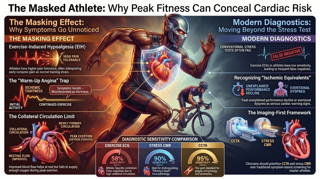
La evaluación cardiovascular de los atletas entrenados presenta una paradoja: las mismas adaptaciones fisiológicas que confieren protección cardiovascular —la modulación autonómica, hipoalgesia inducida por el ejercicio, la eficiencia cardíaca mejorada y el crecimiento de colaterales coronarias, pueden oscurecer simultáneamente los marcadores clínicos de la patología subyacente. Esta revisión sintetiza la evidencia contemporánea sobre la neurobiología del dolor isquémico miocárdico, los mecanismos de la hipoalgesia inducida por el ejercicio y la biología distinta de arteriogénesis versus angiogénesis, el angina de calentamiento fenómeno, y las limitaciones de las pruebas de diagnóstico convencionales en la población atlética. Integra datos emergentes sobre las arterias coronarias arteria imágenes de calcio en atletas de resistencia de por vida (cohortes MARC, Master@Heart, Cooper Center, Merghani, Papatheodorou), patrones de riesgo específicos según el sexo, el marco de exposición de por vida a la apolipoproteína-B y el papel de las imágenes funcionales y anatómicas avanzadas (Angio-TC coronaria, RMC de estrés, FFR/iFR, FFR por TC). El resultado es un marco diagnóstico que trata la disminución inexplicable del rendimiento y la disnea de esfuerzo como equivalentes isquémicos, emplea la estratificación basada en imágenes en lugar de depender de los síntomas o de las pruebas de esfuerzo submáximas, y enmarca las decisiones de regreso al deporte a través de toma de decisiones compartida.
1. Introducción
Aerobic fitness is among the most powerful protective factors against atherosclerotic enfermedad cardiovascular, yet a non-trivial fraction of trained athletes harbor anatomically significant coronary lesiones, congenital coronary anomalies, or inherited cardiomyopathies that remain clinically silent until catastrophic presentation [1], [2]. The diagnostic challenge is not merely epidemiological. Endurance training reshapes the neurobiology of pain, the autonomic regulation of cardiac sensation, the architecture of the coronary circulation, and the energetic efficiency with which the miocardio meets a given workload. Each of these adaptations acts to elevate the threshold at which isquemia becomes phenomenologically obvious to the athlete and electrocardiographically obvious to the clinician.
This review develops a unified mechanistic and diagnostic framework for the masked athlete. It corrects several recurring misconceptions in the prior literature — including overstated claims about collateral flow capacity, oversimplified attribution of exercise-induced hypoalgesia to endogenous opioids alone, and outdated reliance on symptom-based pre-participation screening — and integrates recent imaging evidence that has reframed the relationship between exercise volume, carga de placa, and outcome.
2. Pathophysiology of Myocardial Ischemic Pain
2.1 Biochemical Mediators and Chemosensation
Within seconds of reduced coronary perfusion, myocardial metabolism shifts toward anaerobic glycolysis, generating interstitial accumulation of adenosine, lactate, hydrogen ions (H⁺), bradykinin, prostaglandins, and especies reactivas del oxígeno. Adenosine, released from the hydrolysis of ATP, binds to adenosine A₁ receptors on cardiac sympathetic afferent endings and is now recognized as a primary nociceptive trigger of anginal pain [3], [4], [5]. Bradykinin, generated through proteolysis of kininogens, excites the same afferents through bradykinin B₂ receptors (B2R) and synergizes with adenosine to lower the firing threshold [6]. Local acidosis from lactate and H⁺ accumulation further sensitizes acid-sensing ion channels (ASIC3) and TRPV1 channels on cardiac afferents, while prostaglandins synthesized via the cyclo-oxygenase pathway prolong and amplify the chemosensitive response [7].
2.2 Neural Pathways and Referred Pain
Sympathetic cardiac afferents have cell bodies in the T1–T5 dorsal root ganglia and project via the spinothalamic tract to the thalamus and somatosensory cortex; convergence of cardiac and somatic input at these spinal levels accounts for the classic referral of anginal pain to the chest, shoulder, and inner arm [7]. Vagal afferents synapse in the nucleus tractus solitarius of the medulla; ascending projections excite upper-cervical spinothalamic neurons and contribute to anginal sensations in the jaw, neck, and epigastrium [7]. The relationship between objectively documented ischemia and subjective anginal pain is non-linear: multiple neural and biochemical inputs interact such that pain perception can vary substantially between individuals and even between successive ischemic episodes in the same individual [8].
2.3 Silent Ischemia
Silent myocardial ischemia is the objective demonstration of myocardial ischemia — by ST-segment shift, perfusion defect, regional wall-motion abnormality, or rise in cardiac biomarcadores — in the absence of perceived angina or anginal equivalents. In ambulatory ECG cohorts of patients with established enfermedad de las arterias coronarias, between 50% and 75% of ischemic episodes are clinically silent, and isquemia silenciosa carries prognostic weight independent of symptomatic episodes [9], [10]. The mechanisms include altered chemosensitive thresholds, autonomic neuropathy (notably in diabetes), cortical processing differences, and — particularly relevant here — exercise-induced hypoalgesia.
3. Pain Perception and Modulation in the Athletic Population
3.1 Mechanisms of Exercise-Induced Hypoalgesia
Exercise-induced hypoalgesia (EIH) refers to the acute reduction in pain sensitivity following a bout of exercise. In trained athletes, this transient effect appears to consolidate into a chronically elevated pain threshold. Earlier accounts attributed EIH primarily to β-endorphin release; current evidence indicates a more distributed system involving endogenous opioids, the sistema endocannabinoide, vías inhibitorias descendentes, autonomic modulation, and cognitive reappraisal [11], [12], [13]. Naloxone-mediated opioid blockade only partially reverses exercise analgesia, and murine work demonstrates that the so-called runner’s high depends on cannabinoid receptor signaling rather than opioid receptors alone [14].
Functional neuroimaging shows that endurance athletes display attenuated activation in the thalamus and primary somatosensory cortex during noxious stimulation, together with strengthened functional connectivity in descending modulatory networks responsible for conditioned pain modulation [15]. Pooled meta-analytic data confirm that athletes exhibit higher pain tolerance, although pain threshold differences are smaller and heterogeneous [16], [17], [18].
3.2 Autonomic, Interoceptive, and Cognitive Adaptation
Chronic endurance training establishes a parasympathetic-dominant resting state with reduced sympathetic outflow. Athletes typically show enhanced interoceptive accuracy — most demonstrably in heartbeat detection — but they also exhibit greater capacity for top-down regulation of interoceptive salience, allowing afferent signals to be reframed rather than alarming [19]. Cognitive reappraisal, central to elite sport psychology, recasts exertional discomfort as a challenge state rather than a threat state, suppressing the affective dimension of pain even when sensory signals persist [20]. In the ischemic context, an athlete may interpret early anginal pressure as ordinary training discomfort or muscular fatigue, leading to dangerous diagnostic delay.
3.3 Heterogeneity Across Sport, Sex, and Psychology
Pain modulation differs by sport modality. Endurance athletes consistently show higher tolerance for ischemic and cold stimuli than strength or team-sport athletes [21]. Sex differences in EIH are less consistent; some studies report stronger effects in men, but the literature is heterogeneous and confounded by hormonal status, expectancy, and stimulus modality [12]. Trait fear of pain remains a strong individual-level predictor of how ischemic symptoms are interpreted, irrespective of athletic status.
4. Coronary Collateral Circulation
4.1 Arteriogenesis Versus Angiogenesis
Two biologically and functionally distinct vascular processes contribute to the coronary collateral network. Angiogenesis is the sprouting of new capillaries from existing microvessels, driven primarily by tissue hypoxia through HIF-1α–mediated transcription of vascular endothelial growth factor (VEGF) [22]. While angiogenesis improves densidad capilar and oxygen diffusion at the tissue level, capillaries lack the muscular media required to handle bulk arterial flow and cannot compensate for an obstructive epicardial estenosis.
Arteriogenesis is the structural remodeling of pre-existing arterio-arteriolar anastomoses into mature conducting arteries [22], [23]. The dominant stimulus is fluid shear stress: when an epicardial stenosis develops, a pressure gradient drives flow through small dormant collateral channels, and the resulting endothelial shear activates monocyte recruitment, smooth-muscle proliferation, and matriz extracelular remodeling. The process proceeds in four canonical stages: increased vascular permeability, digestion of the basal lamina and ECM, reconstruction of the smooth muscle media, and pruning of redundant vessels [23], [24]. Collateral arterioles can enlarge up to roughly 20-fold in luminal diameter, producing large, non-linear increases in conductance per Poiseuille’s relation.
4.2 Endurance Training as a Stimulus
Endurance exercise is a potent arteriogenic stimulus because it repeatedly elevates cardiac output and coronary flow, raising the shear forces acting on collateral vessels. Zbinden and colleagues demonstrated, using invasive collateral flow index (CFI) measurements during balloon oclusión, that a structured three-month aerobic training program increased CFI by approximately 39% in absolute terms and over 60% in relative terms in patients with normal arterias coronarias [25]. The EXCITE trial subsequently confirmed that high-intensity exercise training increased CFI to levels comparable with chronically occluded vessels [26].
4.3 Functional Limits of Collateral Compensation
Even in the most extensively collateralized hearts, mature collaterals typically deliver only 25–40% of normal antegrade flow [27], [28]. This explains the common clinical observation that an athlete with critical multi-vessel disease may remain entirely asymptomatic during rest and moderate exertion yet suffer abrupt ischemic decompensation at peak frecuencias cardíacas. Arteriogenesis is also self-limiting: as the collateral lumen expands, the trans-collateral pressure gradient falls, shear stress drops below the threshold for further growth, and the process halts. Collaterals are protective but not curative.
5. Warm-Up Angina, Ischemic Preconditioning, and Cardiac Efficiency
Warm-up angina describes the clinical observation that ischemic symptoms during a second exertion are reduced or abolished if preceded by an initial bout of exercise. Reported prevalence varies between roughly 50% and 80% of patients with stable angina, depending on definition and protocol [29], [30]. Three mechanisms are operative: recruitment of additional collateral conductance, ischemic preconditioning mediated by mitochondrial K-ATP channels and adenosine release [31], and improved ventricular-vascular coupling that lowers the myocardial workload for a given external power output. For the athlete, warm-up angina has the perverse effect that an initial twinge of chest tightness during the first kilometer of a run may dissipate as exercise continues, reinforcing the false belief that the symptom was benign.
Trained athletes also possess substantially higher volúmenes sistólicos and more efficient peripheral oxygen extraction than sedentary individuals, allowing them to achieve high external work at a lower rate-pressure product (RPP), the principal determinant of myocardial oxygen demand [32]. Because RPP remains compressed across the submaximal range, the athlete’s ischemic threshold may not be approached until very near maximal exertion — collapsing the warning interval between asymptomatic performance and decompensation.
6. Risk Phenotypes Masked by Athletic Adaptation
6.1 Coronary Artery Disease in Master Athletes
In athletes older than 35 years, atherosclerotic coronary artery disease is the leading cause of exercise-related muerte súbita cardíaca [33]. Katzel and colleagues, in a maximal-exercise cohort of asymptomatic master athletes, identified silent ischemia in 16% — a prevalence comparable to age-matched untrained men despite the athletes’ superior aerobic capacity and unremarkable conventional factores de riesgo [34]. ApoE4 carrier status was associated with the silent-ischemia phenotype, although the cohort was small and the finding requires replication. The contemporary Atleta máster Screening Study (MASS) confirmed clinically actionable cardiovascular pathology in a non-trivial fraction of asymptomatic veteran competitors over five years of structured surveillance [35].
6.2 Anomalous Origin of a Coronary Artery
Anomalous aortic origin of a coronary artery (AAOCA) is rare but disproportionately lethal in young athletes. Malignant features include an interarterial course between the aorta and pulmonary artery, an acute angle of takeoff, an intramural proximal segment, and a slit-like ostium [36], [37]. During heavy exertion, expansion of the great vessels can compress the anomalous artery against the pulmonary trunk, and high-velocity flow may collapse the slit-like orifice — producing sudden, profound ischemia in a previously asymptomatic athlete. Resting ECG and conventional stress testing are typically normal; angiotomografía coronaria is the diagnostic gold standard [38].
6.3 Hypertrophic Cardiomyopathy and the Athlete’s Heart
Miocardiopatía hipertrófica (HCM) has historically been identified as a leading cause of sudden cardiac death in young US athletes [39]. More recent post-mortem registries — notably the United Kingdom regional registry and the NCAA decade review — show a broader distribution of etiologies in which sudden arrhythmic death syndrome (SADS, with morphologically normal hearts) and idiopathic left-ventricular hypertrophy or fibrosis rival or exceed HCM [40], [41]. The diagnostic challenge in living athletes is differentiating pathological HCM from the physiological athlete’s heart. Discriminating features favoring HCM include asymmetric septal hypertrophy disproportionate to chamber dimensions, impaired diastolic indices, historial familiar of premature SCD or HCM, and patchy realce tardío con gadolinio (LGE) on cardiac magnetic resonance, which independently predicts arrhythmic risk [42]. Regression of wall thickness with detraining strongly favors physiological adaptation [43].
7. Coronary Artery Calcium in Lifelong Endurance Athletes
An apparent paradox emerged over the past two decades: lifelong endurance athletes, despite favorable conventional risk factors, exhibit higher calcio en las arterias coronarias (CAC) scores than less active controls [44], [45], [46], [47]. The MARC study showed that lifelong exercise volume was associated with greater CAC and a higher prevalence of any placa, but the plaque morphology was overwhelmingly calcified rather than mixed or non-calcified — a stable phenotype [44]. The longitudinal Estudio MARC-2 extended this: over six years of follow-up, exercise intensity (vigorous activity at ≥9 METs), rather than total volume, predicted progression of placa calcificada without an increase in vulnerable placa no calcificada [45]. El Estudio Master@Heart compared lifelong, late-onset, and non-athletic men and likewise found greater CAC and total plaque in lifelong athletes, while the odds of vulnerable, mixed, or non-calcified plaque were not increased and were numerically lower [47].
El Estudio Longitudinal del Centro Cooper demonstrated that high physical-activity individuals with CAC < 100 had substantially lower all-cause and cardiovascular mortality than less active counterparts at the same CAC level, while the high-CAC stratum showed no significant mortality penalty for high activity [46]. Recent female-cohort data (Papatheodorou et al.) show no exercise-volume association with CAC in master female endurance athletes, indicating that the masked-athlete CAC framework cannot be extrapolated uncritically across sexes [48].
These findings reframe the masked-athlete problem. Calcified plaque in the lifelong athlete is best interpreted as the radiographic signature of stable, healed ateroesclerosis rather than active disease. CAC remains a powerful prognostic instrument when interpreted alongside plaque morphology on CCTA, lipid exposure history, and family history. It does not invalidate concern about exercise-related events; rather, it directs attention away from total CAC score and toward non-calcified and mixed plaque burden, lipid-rich morphology, and high-risk features such as placa de baja atenuación, remodelación positiva, y signo del anillo de servilleta.
8. Sex-Specific Considerations
The masked-athlete literature has been dominated by male cohorts, and conclusions transferred uncritically to female athletes risk substantial misclassification. The Papatheodorou cohort of master female endurance athletes found no association between lifelong exercise dose and CAC, contrasting sharply with the male MARC and Master@Heart findings [48]. Female athletes also more frequently present with atypical ischemic symptoms — exertional dyspnea, fatigue, jaw or epigastric discomfort — and are disproportionately represented among patients with ischemia and no obstructive coronary arteries (INOCA) and infarto de miocardio with non-obstructive coronary arteries (MINOCA), in which disfunción microvascular and vasospasm are mechanistically central [49], [50]. Sports-cardiology screening pathways should therefore (i) avoid extrapolating male CAC norms, (ii) maintain a low threshold for invasive or non-invasive functional microvascular testing in symptomatic female athletes with normal epicardial coronaries, and (iii) treat exertional dyspnea as a primary, rather than ancillary, ischemic equivalent.
9. Lifetime ApoB and Lipoprotein(a) Exposure: “Fit But Not Immune”
The Master@Heart and MARC observations are coherent with the apolipoprotein-B (ApoB) exposure-time framework: atherosclerotic risk is a function of cumulative ApoB-particle exposure across the life course, and exercise reduces but does not eliminate this exposure [51], [52]. Lifelong high-intensity training cannot reverse decades of LDL particle penetration into the arterial wall in genetically susceptible individuals, and elevated lipoproteína(a) — present in approximately one in five individuals — is unmodifiable by exercise [53]. The major outcome trials of aggressive ApoB lowering (Fourier, Odisea OUTCOMES) demonstrate that even profound reduction in partículas aterogénicas in already-diseased patients reduces but does not abolish events [54], [55].
Practical implications for the masked athlete: (i) ApoB and Lp(a) should be measured at least once in any master athlete being risk-stratified, particularly in those with CAC > 0 or family history of premature ASCVD; (ii) the absence of conventional risk factors does not exclude clinically significant atherosclerosis when CCTA reveals plaque; (iii) the management goal is not to disqualify the athlete but to extend the asymptomatic margin by lowering residual ApoB-driven exposure to the lowest achievable level.
10. Diagnostic Testing in Athletes: Sensitivities, Specificities, and Pitfalls
10.1 The Limits of Exercise Stress Testing
Conventional exercise ECG stress testing performs poorly in the athletic population. Athletes commonly exhibit resting repolarization abnormalities, voltage-criteria left-ventricular hypertrophy, and high resting tono vagal, all of which generate false-positive ST-segment changes — particularly in women. Conversely, the very collateral and efficiency adaptations described above generate false-negative results. The Knuuti metaanálisis, which has become the field-defining benchmark, reported pooled sensitivities for obstructive coronary disease of approximately 0.58 for exercise ECG, 0.85 for stress echocardiography, 0.73 for SPECT, 0.90 for stress CMR, and 0.95 for PET, with corresponding specificities ranging from 0.49 to 0.85 [56]. Submaximal protocols that terminate at 85% of age-predicted maximum heart rate systematically miss athletes whose true ischemic threshold lies near 100% of an age-exceeding maximum.
10.2 Anatomic and Advanced Imaging
Coronary CT angiography (CCTA) is the most sensitive widely available test for obstructive epicardial disease and the gold standard for diagnosing AAOCA [38], [56]. El Estudio SCOT-HEART demonstrated that CCTA-guided management reduces five-year coronary death and non-fatal myocardial infarction compared with standard care [57]. Cardiac magnetic resonance is the most accurate modality for distinguishing pathological hypertrophy from physiological athlete’s heart, quantifying fibrosis, and identifying phenotype-negative gene carriers [42], [43].
Table 1. Pooled diagnostic performance of common modalities for obstructive coronary disease, with athlete-specific considerations. Values are approximate and rounded from Knuuti et al. [56].
| Modality | Pooled Sensitivity | Pooled Specificity | Athlete-Specific Caveat |
| Exercise ECG | ≈ 0.58 | ≈ 0.62 | False positives from voltage LVH; false negatives from collaterals; submaximal endpoints |
| Stress Echo | ≈ 0.85 | ≈ 0.82 | Operator-dependent; suboptimal windows in muscular athletes |
| SPECT | ≈ 0.73 | ≈ 0.83 | Balanced ischemia in 3-vessel disease may falsely normalize |
| Stress CMR | ≈ 0.90 | ≈ 0.85 | Best test for HCM vs athlete’s heart and fibrosis quantification |
| CCTA | ≈ 0.95 | ≈ 0.79 | Gold standard for AAOCA and plaque morphology |
| PET / CT-FFR | ≈ 0.95 / ≈ 0.86 | ≈ 0.83 / ≈ 0.79 | PET adds CFR; CT-FFR resolves intermediate stenoses non-invasively |
11. Functional Lesion Assessment: FFR, iFR, and CT-FFR
When CCTA reveals an intermediate stenosis in an athlete with unreliable functional testing, physiologic confirmation is required before intervention. Fractional flow reserve (FFR) and instantaneous wave-free ratio (iFR) provide pressure-derived ischemic indices; the FAME, FAME-2, and DEFINE-FLAIR/iFR-SWEDEHEART trials established that physiology-guided revascularización improves outcomes and reduces unnecessary stenting compared with angiography alone [58], [59], [60]. Coronary CT–derived fractional flow reserve (CT-FFR) provides comparable diagnostic accuracy without the need for invasive instrumentation; the NXT trial reported sensitivity of 0.86 and specificity of 0.79 for ischemia-causing stenoses [61]. These functional adjuncts are particularly valuable in the athlete population, where the discrepancy between anatomic disease and functional ischemia is amplified by collateral conductance and adaptive cardiac efficiency.
12. Genetics, Family History, and CMR in Phenotype-Negative Carriers
Family history of HCM, premature SCD, or other inherited miocardiopatía mandates genetic counseling and consideration of cascade testing per the 2024 AHA/ACC HCM guideline [62]. Pathogenic sarcomere variants confer lifetime disease risk that can manifest after years of athletic adaptation. Cardiac MRI is critical in phenotype-negative gene carriers because subtle myocardial crypts, focal fibrosis, or perfusion abnormalities can precede frank hypertrophy. The SHaRe registry quantified the lifetime burden of disease across genotypes and confirmed that gene-positive/phenotype-negative individuals carry a non-zero lifetime event rate [63]. In the masked-athlete framework, family history transforms a normal echocardiogram from reassuring to insufficient: CMR with LGE imaging becomes the appropriate next step.
13. Detection and Prevention Strategies
13.1 Atypical Warning Signs
Because classic crushing retrosternal pain is uncommon in trained athletes, clinicians must learn to recognize the ischemic equivalents that do present:
- Exertional dyspnea disproportionate to exercise intensity, particularly when of new or accelerating onset.
- Unexplained, sustained decline in athletic performance, training pace, or peak power.
- Atypical discomfort during high-intensity intervals — mild epigastric, jaw, or interscapular pressure that resolves with cessation.
- Exertional pre-syncope or syncope, which must always be treated as a potentially malignant marker of AAOCA, HCM, or arrhythmic substrate until proven otherwise.
13.2 A Tiered Screening Framework
The 2014 AHA 14-element history and physical remains a reference point, but the 2025 AHA/ACC scientific statement on competitive sports participation now emphasizes individualized risk assessment with imaging-based stratification rather than reliance on symptom screening alone [64]. El 2020 ESC Sports Cardiology Guidelines provide a complementary tiered framework [65]:
Tier 1 — Personal and family history with attention to premature ASCVD (< 50 years), familial SCD, and any prior unexplained syncope.
Tier 2 — Resting ECG de 12 derivaciones interpreted with the International Criteria for athlete ECG interpretation [66].
Tier 3 — Maximal symptom-limited exercise testing for deportistas veteranos with ≥1 cardiovascular risk factor or any atypical exertional symptom; the test must be driven to genuine physiological maximum, not 85% of an age-predicted target.
Tier 4 — Anatomic and tissue-characterizing imaging (CCTA, stress CMR) in athletes who remain at elevated probabilidad preprueba after Tiers 1–3 or whose Tier 3 results are equivocal. CAC scoring is appropriate in masters athletes ≥ 40 years with any conventional risk factor or atypical symptoms.
14. Return-to-Play After Cardiac Events
The 2025 AHA/ACC scientific statement codifies a contemporary return-to-play framework that has moved decisively away from blanket disqualification toward individualized, evidence-informed shared decision-making [64]. For master athletes following revascularization for stable obstructive disease, a structured cardiac rehabilitation program, optimization of ApoB-lowering pharmacotherapy, repeat functional and anatomic imaging, and gradual return to high-intensity training are appropriate when left-ventricular function is preserved and ischemia is absent on stress testing. Saberi and colleagues demonstrated in a randomized trial that moderate-intensity exercise modestly improves peak oxygen uptake in HCM patients without provoking arrhythmia, supporting cautious participation in many cases that previously triggered disqualification [67]. Athletes with implantable cardioverter-defibrillators have been followed in prospective registries with reassuring safety data for participation in many sports [68].
15. Shared Decision-Making in Sports Cardiology
The clinical mindset has shifted from clearing or disqualifying the athlete to risk stratification and shared decision-making [69]. Competitive athletes systematically display different risk tolerance from the general population, and the role of the cardiólogo del deporte is to provide a transparent, evidence-based estimate of event risk rather than to act unilaterally as a gatekeeper. The 2025 AHA/ACC framework explicitly endorses participation under close surveillance for a range of conditions previously deemed disqualifying, including selected coronary anomalies after surgical correction, post-revascularization stable coronary disease with preserved fracción de eyección, and many cardiomyopathy phenotypes.
16. Limitations and Open Questions
Several caveats temper the framework presented here. First, the human pain modulation literature is heterogeneous, and effect sizes for exercise-induced hypoalgesia vary widely by stimulus type, exercise intensity, training history, and individual psychological traits. Second, the CAC findings in lifelong athletes are derived from predominantly male cohorts and do not generalize to women. Third, the often-cited Cooper Center cociente de riesgos instantáneos of 0.52 for cardiovascular mortality applies to the high-activity stratum with CAC < 100; the high-CAC stratum showed a non-significant point estimate near unity, and this nuance must not be lost when counseling individual athletes [46]. Fourth, evolving registries continue to revise the relative weight of HCM, SADS, and idiopathic LVH/fibrosis as causes of athletic SCD, and country-specific data must be applied with care. Fifth, the optimal cadence and triggers for advanced imaging in asymptomatic master athletes remain unsettled and are an active area of guideline development.
17. Conclusions
The athlete’s heart is an extraordinary expression of physiological adaptation, but it is not invulnerable. The synergy of exercise-induced hypoalgesia, autonomic and cognitive conditioning, coronary collateral growth, and superior cardiac efficiency systematically elevates the threshold at which ischemia becomes phenomenologically obvious, narrowing the diagnostic margin between asymptomatic performance and catastrophic decompensation. Modern sports cardiology must therefore (i) treat unexplained performance decline and exertional dyspnea as ischemic equivalents, (ii) replace reliance on submaximal stress testing and symptom screening with imaging-based risk stratification, (iii) integrate plaque morphology, ApoB, and Lp(a) into the lifetime-exposure calculus, (iv) apply the masked-athlete framework with sex-specific calibration, and (v) frame return-to-sport decisions through transparent shared decision-making. The athlete’s heart is a testament to human adaptation; identifying its limits requires diagnostic vigilance proportionate to the dedication of the athlete being evaluated.
Referencias
- Maron BJ, Doerer JJ, Haas TS, Tierney DM, Mueller FO. Sudden deaths in young competitive athletes: analysis of 1866 deaths in the United States, 1980-2006. Circulation. 2009;119(8):1085-1092. doi:10.1161/CIRCULATIONAHA.108.804617
- Marijon E, Tafflet M, Celermajer DS, et al. Sports-related sudden death in the general population. Circulation. 2011;124(6):672-681. doi:10.1161/CIRCULATIONAHA.110.008979
- Crea F, Pupita G, Galassi AR, et al. Role of adenosine in pathogenesis of anginal pain. Circulation. 1990;81(1):164-172. doi:10.1161/01.cir.81.1.164
- Pan HL, Chen SR. Sensing tissue ischemia: another new function for capsaicin receptors?. Circulation. 2004;110(13):1826-1831. doi:10.1161/01.CIR.0000142618.20278.7A
- Sylvén C. Mechanisms of pain in angina pectoris–a critical review of the adenosine hypothesis. Cardiovasc Drugs Ther. 1993;7(5):745-759. doi:10.1007/BF00878926
- Tjen-A-Looi SC, Pan HL, Longhurst JC. Endogenous bradykinin activates ischaemically sensitive cardiac visceral afferents through kinin B2 receptors in cats. J Physiol. 1998;510 ( Pt 2)(Pt 2):633-641. doi:10.1111/j.1469-7793.1998.633bk.x
- Foreman RD. Mechanisms of cardiac pain. Annu Rev Physiol. 1999;61:143-167. doi:10.1146/annurev.physiol.61.1.143
- Sylvén C. Angina pectoris. Clinical characteristics, neurophysiological and molecular mechanisms. Pain. 1989;36(2):145-167. doi:10.1016/0304-3959(89)90019-5
- Cohn PF, Fox KM, Daly C. Silent myocardial ischemia. Circulation. 2003;108(10):1263-1277. doi:10.1161/01.CIR.0000088001.59265.EE
- Deedwania PC, Carbajal EV. Silent ischemia during daily life is an independent predictor of mortality in stable angina. Circulation. 1990;81(3):748-756. doi:10.1161/01.cir.81.3.748
- Koltyn KF. Analgesia following exercise: a review. Sports Med. 2000;29(2):85-98. doi:10.2165/00007256-200029020-00002
- Koltyn KF, Brellenthin AG, Cook DB, Sehgal N, Hillard C. Mechanisms of exercise-induced hypoalgesia. J Pain. 2014;15(12):1294-1304. doi:10.1016/j.jpain.2014.09.006
- Crombie KM, Brellenthin AG, Hillard CJ, Koltyn KF. Endocannabinoid and Opioid System Interactions in Exercise-Induced Hypoalgesia. Pain Med. 2018;19(1):118-123. doi:10.1093/pm/pnx058
- Fuss J, Steinle J, Bindila L, et al. A runner’s high depends on cannabinoid receptors in mice. Proc Natl Acad Sci U S A. 2015;112(42):13105-13108. doi:10.1073/pnas.1514996112
- Geisler M, Ritter A, Herbsleb M, Bär KJ, Weiss T. Neural mechanisms of pain processing differ between endurance athletes and nonathletes: A functional connectivity magnetic resonance imaging study. Hum Brain Mapp. 2021;42(18):5927-5942. doi:10.1002/hbm.25659
- Tesarz J, Schuster AK, Hartmann M, Gerhardt A, Eich W. Pain perception in athletes compared to normally active controls: a systematic review with meta-analysis. Pain. 2012;153(6):1253-1262. doi:10.1016/j.pain.2012.03.005
- Geva N, Defrin R. Enhanced pain modulation among triathletes: a possible explanation for their exceptional capabilities. Pain. 2013;154(11):2317-2323. doi:10.1016/j.pain.2013.06.031
- Pettersen SD, Aslaksen PM, Pettersen SA. Pain Processing in Elite and High-Level Athletes Compared to Non-athletes. Front Psychol. 2020;11:1908. Published 2020 Jul 28. doi:10.3389/fpsyg.2020.01908
- Boecker H, Sprenger T, Spilker ME, et al. The runner’s high: opioidergic mechanisms in the human brain. Cereb Cortex. 2008;18(11):2523-2531. doi:10.1093/cercor/bhn013
- Meijen C, Turner M, Jones MV, Sheffield D, McCarthy P. A Theory of Challenge and Threat States in Athletes: A Revised Conceptualization. Front Psychol. 2020;11:126. Published 2020 Feb 6. doi:10.3389/fpsyg.2020.00126
- Assa T, Geva N, Zarkh Y, Defrin R. The type of sport matters: Pain perception of endurance athletes versus strength athletes. Eur J Pain. 2019;23(4):686-696. doi:10.1002/ejp.1335
- Heil M, Eitenmüller I, Schmitz-Rixen T, Schaper W. Arteriogenesis versus angiogenesis: similarities and differences. J Cell Mol Med. 2006;10(1):45-55. doi:10.1111/j.1582-4934.2006.tb00290.x
- Schaper W. Collateral circulation: past and present. Basic Res Cardiol. 2009;104(1):5-21. doi:10.1007/s00395-008-0760-x
- Schaper W, Scholz D. Factors regulating arteriogenesis. Arterioscler Thromb Vasc Biol. 2003;23(7):1143-1151. doi:10.1161/01.ATV.0000069625.11230.96
- Zbinden R, Zbinden S, Meier P, et al. Coronary collateral flow in response to endurance exercise training. Eur J Cardiovasc Prev Rehabil. 2007;14(2):250-257. doi:10.1097/HJR.0b013e3280565dee
- Möbius-Winkler S, Uhlemann M, Adams V, et al. Coronary Collateral Growth Induced by Physical Exercise: Results of the Impact of Intensive Exercise Training on Coronary Collateral Circulation in Patients With Stable Coronary Artery Disease (EXCITE) Trial. Circulation. 2016;133(15):1438-1448. doi:10.1161/CIRCULATIONAHA.115.016442
- Seiler C, Stoller M, Pitt B, Meier P. The human coronary collateral circulation: development and clinical importance. Eur Heart J. 2013;34(34):2674-2682. doi:10.1093/eurheartj/eht195
- Meier P, Hemingway H, Lansky AJ, Knapp G, Pitt B, Seiler C. The impact of the coronary collateral circulation on mortality: a meta-analysis. Eur Heart J. 2012;33(5):614-621. doi:10.1093/eurheartj/ehr308
- Stewart RA, Simmonds MB, Williams MJ. Time course of “warm-up” in stable angina. Am J Cardiol. 1995;76(1):70-73. doi:10.1016/s0002-9149(99)80804-2
- Williams DO, Bass TA, Gewirtz H, Most AS. Adaptation to the stress of tachycardia in patients with coronary artery disease: insight into the mechanism of the warm-up phenomenon. Circulation. 1985;71(4):687-692. doi:10.1161/01.cir.71.4.687
- Yellon DM, Downey JM. Preconditioning the myocardium: from cellular physiology to clinical cardiology. Physiol Rev. 2003;83(4):1113-1151. doi:10.1152/physrev.00009.2003
- Gobel FL, Norstrom LA, Nelson RR, Jorgensen CR, Wang Y. The rate-pressure product as an index of myocardial oxygen consumption during exercise in patients with angina pectoris. Circulation. 1978;57(3):549-556. doi:10.1161/01.cir.57.3.549
- Maron BJ, Pelliccia A. The heart of trained athletes: cardiac remodeling and the risks of sports, including sudden death. Circulation. 2006;114(15):1633-1644. doi:10.1161/CIRCULATIONAHA.106.613562
- Katzel LI, Fleg JL, Busby-Whitehead MJ, et al. Exercise-induced silent myocardial ischemia in master athletes. Am J Cardiol. 1998;81(3):261-265. doi:10.1016/s0002-9149(97)00898-9
- Morrison BN, Isserow S, Taunton J, et al. Masters athlete screening study (MASS): incidence of cardiovascular disease and major adverse cardiac events and efficacy of screening over five years. Eur J Prev Cardiol. 2023;30(9):887-899. doi:10.1093/eurjpc/zwad090
- Cheezum MK, Liberthson RR, Shah NR, et al. Anomalous Aortic Origin of a Coronary Artery From the Inappropriate Sinus of Valsalva. J Am Coll Cardiol. 2017;69(12):1592-1608. doi:10.1016/j.jacc.2017.01.031
- Angelini P, Velasco JA, Flamm S. Coronary anomalies: incidence, pathophysiology, and clinical relevance. Circulation. 2002;105(20):2449-2454. doi:10.1161/01.cir.0000016175.49835.57
- Brothers JA, Frommelt MA, Jaquiss RDB, Myerburg RJ, Fraser CD Jr, Tweddell JS. Expert consensus guidelines: Anomalous aortic origin of a coronary artery. J Thorac Cardiovasc Surg. 2017;153(6):1440-1457. doi:10.1016/j.jtcvs.2016.06.066
- Maron BJ, Udelson JE, Bonow RO, et al. Eligibility and Disqualification Recommendations for Competitive Athletes With Cardiovascular Abnormalities: Task Force 3: Hypertrophic Cardiomyopathy, Arrhythmogenic Right Ventricular Cardiomyopathy and Other Cardiomyopathies, and Myocarditis: A Scientific Statement From the American Heart Association and American College of Cardiology. Circulation. 2015;132(22):e273-e280. doi:10.1161/CIR.0000000000000239
- Finocchiaro G, Papadakis M, Robertus JL, et al. Etiology of Sudden Death in Sports: Insights From a United Kingdom Regional Registry. J Am Coll Cardiol. 2016;67(18):2108-2115. doi:10.1016/j.jacc.2016.02.062
- Harmon KG, Asif IM, Maleszewski JJ, et al. Incidence, Cause, and Comparative Frequency of Sudden Cardiac Death in National Collegiate Athletic Association Athletes: A Decade in Review. Circulation. 2015;132(1):10-19. doi:10.1161/CIRCULATIONAHA.115.015431
- Chan RH, Maron BJ, Olivotto I, et al. Prognostic value of quantitative contrast-enhanced cardiovascular magnetic resonance for the evaluation of sudden death risk in patients with hypertrophic cardiomyopathy. Circulation. 2014;130(6):484-495. doi:10.1161/CIRCULATIONAHA.113.007094
- Pelliccia A, Maron BJ, De Luca R, Di Paolo FM, Spataro A, Culasso F. Remodeling of left ventricular hypertrophy in elite athletes after long-term deconditioning. Circulation. 2002;105(8):944-949. doi:10.1161/hc0802.104534
- Aengevaeren VL, Mosterd A, Braber TL, et al. Relationship Between Lifelong Exercise Volume and Coronary Atherosclerosis in Athletes. Circulation. 2017;136(2):138-148. doi:10.1161/CIRCULATIONAHA.117.027834
- Aengevaeren VL, Mosterd A, Bakker EA, et al. Exercise Volume Versus Intensity and the Progression of Coronary Atherosclerosis in Middle-Aged and Older Athletes: Findings From the MARC-2 Study. Circulation. 2023;147(13):993-1003. doi:10.1161/CIRCULATIONAHA.122.061173
- DeFina LF, Radford NB, Barlow CE, et al. Association of All-Cause and Cardiovascular Mortality With High Levels of Physical Activity and Concurrent Coronary Artery Calcification. JAMA Cardiol. 2019;4(2):174-181. doi:10.1001/jamacardio.2018.4628
- De Bosscher R, Dausin C, Claus P, et al. Lifelong endurance exercise and its relation with coronary atherosclerosis. Eur Heart J. 2023;44(26):2388-2399. doi:10.1093/eurheartj/ehad152
- Papatheodorou E, Aengevaeren VL, Eijsvogels TMH, et al. Prevalence of Coronary Atherosclerosis in Female Masters Endurance Athletes. Circulation. 2024;150(18):1478-1480. doi:10.1161/CIRCULATIONAHA.124.069484
- Bairey Merz CN, Pepine CJ, Walsh MN, Fleg JL. Ischemia and No Obstructive Coronary Artery Disease (INOCA): Developing Evidence-Based Therapies and Research Agenda for the Next Decade. Circulation. 2017;135(11):1075-1092. doi:10.1161/CIRCULATIONAHA.116.024534
- Tamis-Holland JE, Jneid H, Reynolds HR, et al. Contemporary Diagnosis and Management of Patients With Myocardial Infarction in the Absence of Obstructive Coronary Artery Disease: A Scientific Statement From the American Heart Association. Circulation. 2019;139(18):e891-e908. doi:10.1161/CIR.0000000000000670
- Sniderman AD, Thanassoulis G, Glavinovic T, et al. Apolipoprotein B Particles and Cardiovascular Disease: A Narrative Review. JAMA Cardiol. 2019;4(12):1287-1295. doi:10.1001/jamacardio.2019.3780
- Ference BA, Ginsberg HN, Graham I, et al. Low-density lipoproteins cause atherosclerotic cardiovascular disease. 1. Evidence from genetic, epidemiologic, and clinical studies. A consensus statement from the European Atherosclerosis Society Consensus Panel. Eur Heart J. 2017;38(32):2459-2472. doi:10.1093/eurheartj/ehx144
- Tsimikas S, Fazio S, Ferdinand KC, et al. NHLBI Working Group Recommendations to Reduce Lipoprotein(a)-Mediated Risk of Cardiovascular Disease and Aortic Stenosis. J Am Coll Cardiol. 2018;71(2):177-192. doi:10.1016/j.jacc.2017.11.014
- Sabatine MS, Giugliano RP, Keech AC, et al. Evolocumab and Clinical Outcomes in Patients with Cardiovascular Disease. N Engl J Med. 2017;376(18):1713-1722. doi:10.1056/NEJMoa1615664
- Schwartz GG, Steg PG, Szarek M, et al. Alirocumab and Cardiovascular Outcomes after Acute Coronary Syndrome. N Engl J Med. 2018;379(22):2097-2107. doi:10.1056/NEJMoa1801174
- Knuuti J, Ballo H, Juarez-Orozco LE, et al. The performance of non-invasive tests to rule-in and rule-out significant coronary artery stenosis in patients with stable angina: a meta-analysis focused on post-test disease probability. Eur Heart J. 2018;39(35):3322-3330. doi:10.1093/eurheartj/ehy267
- SCOT-HEART Investigators, Newby DE, Adamson PD, et al. Coronary CT Angiography and 5-Year Risk of Myocardial Infarction. N Engl J Med. 2018;379(10):924-933. doi:10.1056/NEJMoa1805971
- Tonino PA, De Bruyne B, Pijls NH, et al. Fractional flow reserve versus angiography for guiding percutaneous coronary intervention. N Engl J Med. 2009;360(3):213-224. doi:10.1056/NEJMoa0807611
- De Bruyne B, Pijls NH, Kalesan B, et al. Fractional flow reserve-guided PCI versus medical therapy in stable coronary disease. N Engl J Med. 2012;367(11):991-1001. doi:10.1056/NEJMoa1205361
- Davies JE, Sen S, Dehbi HM, et al. Use of the Instantaneous Wave-free Ratio or Fractional Flow Reserve in PCI. N Engl J Med. 2017;376(19):1824-1834. doi:10.1056/NEJMoa1700445
- Nørgaard BL, Leipsic J, Gaur S, et al. Diagnostic performance of noninvasive fractional flow reserve derived from coronary computed tomography angiography in suspected coronary artery disease: the NXT trial (Analysis of Coronary Blood Flow Using CT Angiography: Next Steps). J Am Coll Cardiol. 2014;63(12):1145-1155. doi:10.1016/j.jacc.2013.11.043
- Ommen SR, Ho CY, Asif IM, et al. 2024 AHA/ACC/AMSSM/HRS/PACES/SCMR Guideline for the Management of Hypertrophic Cardiomyopathy: A Report of the American Heart Association/American College of Cardiology Joint Committee on Clinical Practice Guidelines. Circulation. 2024;149(23):e1239-e1311. doi:10.1161/CIR.0000000000001250
- Ho CY, Day SM, Ashley EA, et al. Genotype and Lifetime Burden of Disease in Hypertrophic Cardiomyopathy: Insights from the Sarcomeric Human Cardiomyopathy Registry (SHaRe). Circulation. 2018;138(14):1387-1398. doi:10.1161/CIRCULATIONAHA.117.033200
- Kim JH, Baggish AL, Levine BD, et al. Clinical Considerations for Competitive Sports Participation for Athletes With Cardiovascular Abnormalities: A Scientific Statement From the American Heart Association and American College of Cardiology. Circulation. 2025;151(11):e716-e761. doi:10.1161/CIR.0000000000001297
- Pelliccia A, Sharma S, Gati S, et al. 2020 ESC Guidelines on sports cardiology and exercise in patients with cardiovascular disease. Eur Heart J. 2021;42(1):17-96. doi:10.1093/eurheartj/ehaa605
- Sharma S, Drezner JA, Baggish A, et al. International Recommendations for Electrocardiographic Interpretation in Athletes. J Am Coll Cardiol. 2017;69(8):1057-1075. doi:10.1016/j.jacc.2017.01.015
- Saberi S, Wheeler M, Bragg-Gresham J, et al. Effect of Moderate-Intensity Exercise Training on Peak Oxygen Consumption in Patients With Hypertrophic Cardiomyopathy: A Randomized Clinical Trial. JAMA. 2017;317(13):1349-1357. doi:10.1001/jama.2017.2503
- Lampert R, Olshansky B, Heidbuchel H, et al. Safety of Sports for Athletes With Implantable Cardioverter-Defibrillators: Long-Term Results of a Prospective Multinational Registry. Circulation. 2017;135(23):2310-2312. doi:10.1161/CIRCULATIONAHA.117.027828
- Baggish AL, Ackerman MJ, Putukian M, Lampert R. Shared Decision Making for Athletes with Cardiovascular Disease: Practical Considerations. Curr Sports Med Rep. 2019;18(3):76-81. doi:10.1249/JSR.0000000000000575

