The Molecule Linking Your Gut to Your Heart: 5 Surprising Truths About TMAO
The Invisible Metabolic Relay: A Meta-Organismal Axis
Human health was once viewed through a purely genomic lens, yet emerging research into the gut-organ axis reveals that our physiology is managed by a “meta-organismal” relay. In this biochemical hand-off, dietary precursors are processed by the enteric microbial ecosystem into volatile intermediates, which are subsequently modified by host organs to produce systemic effects.At the center of this interface is trimethylamine N-oxide (TMAO). Historically known as an obscure marine osmolyte used by deep-sea fish to maintain fluid balance, TMAO has transitioned into a primary target for cardiovascular risk stratification. It is now recognized as a potent predictor of Major Adverse Cardio and Cerebrovascular Events (MACCE), often providing prognostic power that exceeds traditional lipid panels. The question for modern medicine has become: how does a single molecule, synthesized at the intersection of diet and the microbiome, dictate the trajectory of systemic disease?
Your Microbiome is a Chemical Factory (and Your Liver is Quality Control)
The synthesis of TMAO is a precise multi-step process that requires an obligatory contribution from gut bacteria. Without specific microbial enzymes, the precursors we eat would never be converted into this pro-atherogenic molecule.
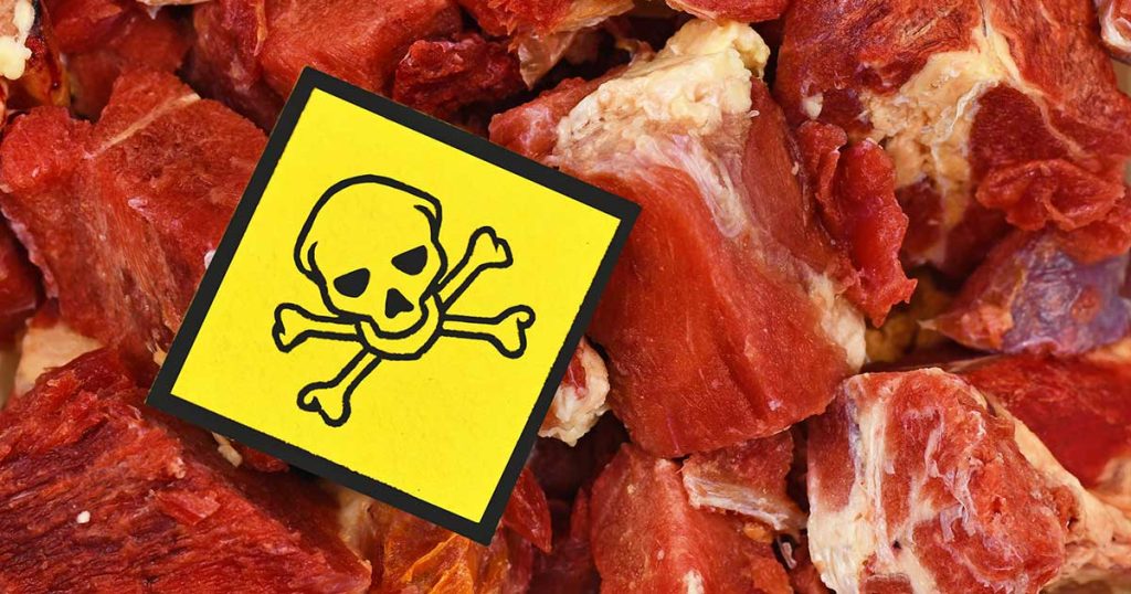
- The Microbial Gateway: When we consume choline (eggs, dairy) or L-carnitine (red meat), microbial enzymes—specifically those encoded by the cutC/D and cntA/B genes—cleave these compounds to produce trimethylamine (TMA).
- The $\gamma$ BB Nuance: Crucially, the carnitine pathway involves a significant metabolic intermediate: $\gamma$ -butyrobetaine ( $\gamma$ BB). In the proximal gut, L-carnitine is converted into $\gamma$ BB at a rate approximately 1,000-fold higher than direct TMA formation, serving as a primary reservoir for subsequent TMA production by specialized microbiota.
- Hepatic Oxidation: TMA travels via the portal vein to the liver, where it is oxidized into odorless TMAO by the flavin-containing monooxygenase 3 (FMO3) enzyme.”The production of TMAO is not a direct result of human cellular metabolism but rather a ‘meta-organismal’ process that requires an obligatory contribution from the gut microbiota.”FMO3 activity is not static; it is regulated by host factors including the farnesoid X receptor (FXR) and bile acids. When this hepatic “quality control” fails due to genetic polymorphisms, TMA accumulates, resulting in trimethylaminuria (fish odor syndrome). Under normal conditions, however, the liver efficiently converts the volatile gas into systemic TMAO, which then circulates as a bioactive metabolite.
The “Fish Paradox”—Why Seafood Isn’t the Enemy
Seafood is naturally rich in pre-formed TMAO. Consequently, a single serving of fish can spike plasma TMAO levels significantly higher than a serving of red meat. Yet, epidemiological data consistently confirms that fish consumption is cardioprotective. This apparent contradiction is resolved by three distinct factors:
- The Metabolic Route: Fish-derived TMAO is absorbed directly into the bloodstream. In contrast, meat-derived precursors must undergo microbial fermentation to produce TMA. This microbial process is often associated with other inflammatory byproducts or shifts in the microbiome (“guilt by association”) that are absent when consuming pre-formed TMAO from fish.
- Nutritional Counterbalance: Seafood provides high concentrations of omega-3 polyunsaturated fatty acids (EPA and DHA). These anti-inflammatory compounds may effectively antagonize the potential signaling harms of a transient TMAO spike.
- Transient vs. Chronic Exposure: In individuals with healthy renal function, fish-induced TMAO is cleared via glomerular filtration within 24 hours. Conversely, meat-heavy diets foster a microbiome optimized for chronic TMA production, leading to sustained, elevated baseline levels that are far more damaging than acute, diet-induced fluctuations.
TMAO is a “Rheostat” for Your Blood’s Clotting Risk
TMAO does not directly trigger coagulation; rather, it functions as a pro-thrombotic sensitizer. It acts as a biochemical rheostat , “dialing up” the reactivity of platelets to primary agonists like thrombin or collagen.The molecular mechanism involves the modulation of intracellular calcium ( $Ca^{2+}$ ) signaling. TMAO facilitates the rapid release of $Ca^{2+}$ from internal stores—specifically the dense tubular system —within the platelets. This heightened calcium flux makes platelets “twitchier” and more prone to aggregation under high-shear conditions, increasing the risk of myocardial infarction and stroke. The causal nature of this link was demonstrated by the removal of the microbial cutC gene in experimental models; eliminating the gut’s ability to produce the TMA precursor completely abolished this heightened thrombotic potential.
Beyond the Heart—The Brain and Kidney Connection
While TMAO is a staple of cardiovascular research, it is increasingly viewed as a global marker of health, with profound implications for the “Microbiota-Gut-Brain Axis” and renal longevity.
- Neurodegeneration: TMAO readily crosses the blood-brain barrier. In the central nervous system, it acts as a chemical chaperone that accelerates the aggregation of amyloid-beta and $\alpha$ -synuclein. Furthermore, it triggers pyroptosis of oligodendrocytes via the ROS-NLRP3 signaling pathway , promoting neuroinflammation and the demyelination often seen in cognitive decline.
- The Renal Vicious Cycle: TMAO is primarily cleared by the kidneys, but it also acts as a “uremic toxin.” Elevated levels promote fibrosis of renal tubulointerstitial tissues and glomerular sclerosis. This creates a destructive feedback loop: declining kidney function leads to higher TMAO retention, which in turn accelerates further renal damage.This multi-organ impact explains why multi-omics data from the UK Biobank shows that TMAO levels add significant predictive power across 17 different disease categories, reflecting systemic biological stress.
“Drugging the Microbiome” Without Killing It
Traditional approaches to microbiome modulation involved broad-spectrum antibiotics, which act as a “scorched earth” strategy. The future of TMAO management lies in “non-lethal” small-molecule inhibitors that target the enzyme (TMA lyase) rather than the bacteria themselves, avoiding the risk of antibiotic resistance.| Inhibitor Class | Key Compound | Mechanism of Action | Potency & Status || —— | —— | —— | —— || Competitive Inhibitors | DMB (found in olive oil) | Mimics choline to competitively block CutC/D | Preclinical; naturally occurring || Suicide Substrates | IMC / FMC | Irreversibly binds and deactivates the CutC/D enzyme | Nanomolar potency ( $IC_{50}$ ); high safety profile |
Compounds like Iodomethylcholine (IMC) have demonstrated the ability to lower systemic TMAO and reduce thrombotic risk without increasing bleeding time, representing a paradigm shift in precision microbiome pharmacology.
Conclusion: Toward Personalized Prevention
TMAO serves as a high-fidelity mirror reflecting the interface between our dietary inputs and our internal microbial ecosystem. It is no longer just a biomarker; it is a bioactive participant in the pathogenesis of cardiovascular, renal, and neurodegenerative diseases.As we transition toward “stratified nutrition,” an individual’s gut profile—specifically the abundance of microbial cutC genes and the activity of hepatic FMO3—will likely dictate personalized dietary interventions. By identifying “high TMA-producers” early, we can move beyond generalized advice toward targeted microbiome management. The state of your heart is, quite literally, a reflection of the invisible metabolic relay occurring in your gut. What is your internal factory producing?
Deep Dive on TMAO Research
The Meta-organismal Meta-axis of Trimethylamine N-oxide: A Comprehensive Analysis of Biochemical Pathways, Cardiometabolic Pathogenesis, and Clinical Implications
The emergence of trimethylamine N-oxide (TMAO) as a central player in cardiometabolic medicine represents a major shift in how researchers understand the interaction between diet, the gut microbiome, and human health. TMAO, a small organic compound with the molecular formula C5H11NO2, is a water-soluble amine N-oxide that has moved from being a relatively obscure osmolyte in marine biology to a widely studied candidate biomarker and potential mediator of cardiovascular risk. The synthesis of TMAO is a multi-step process that bridges the external environment (diet), the enteric microbial ecosystem (gut microbiota), and the host’s internal physiology (liver and kidneys). This report provides an exhaustive, expert-level deep dive into the TMAO axis, analyzing its biochemical architecture, the molecular mechanisms through which it may promote disease, the clinical evidence supporting its prognostic value, and the contemporary controversies regarding its status as a causal agent versus a surrogate biomarker of systemic dysbiosis and cardiometabolic risk.
The Biochemical Landscape: The Gut-Liver-Kidney Axis
The production of TMAO is not a direct result of human cellular metabolism but rather a meta-organismal process that requires an obligatory contribution from the gut microbiota. This pathway is initiated when dietary precursors are consumed and subsequently transformed into volatile intermediates that the host then modifies.
Dietary Precursors and the Microbial Gateway
The primary raw materials for TMAO synthesis are quaternary ammonium compounds found abundantly in many animal-based foods, though some plant foods also contribute through betaine and choline content. The most prominent precursors include choline (often found in the form of phosphatidylcholine or lecithin), L-carnitine, betaine, γ-butyrobetaine, and crotonobetaine.
| Dietary Precursor | Primary Food Sources | Metabolic Intermediate | Final Systemic Product |
| Choline / Phosphatidylcholine | Egg yolks, liver, dairy, soy | Trimethylamine (TMA) | Trimethylamine N-oxide (TMAO) |
| L-Carnitine | Red meat (beef, lamb), supplements | γ-Butyrobetaine (γBB) / TMA | Trimethylamine N-oxide (TMAO) |
| γ-Butyrobetaine | Pre-formed in some red meats; microbial intermediate | Trimethylamine (TMA) | Trimethylamine N-oxide (TMAO) |
| Betaine | Beets, spinach, whole grains | Trimethylamine (TMA) | Trimethylamine N-oxide (TMAO) |
| Crotonobetaine | Carnitine metabolism byproduct | Trimethylamine (TMA) | Trimethylamine N-oxide (TMAO) |
The transformation of these nutrients begins in the intestinal lumen. Dietary choline and carnitine are metabolized by specific microbial enzymes. The cleavage of the carbon-nitrogen bond in choline is catalyzed by the glycyl radical enzyme choline trimethylamine-lyase, encoded by the cutC gene, and its activating protein, encoded by cutD. This reaction releases trimethylamine (TMA) as a volatile byproduct. Similarly, L-carnitine can be metabolized through a distinct pathway involving the carnitine monooxygenase system (encoded by cntA and cntB), which also results in the production of TMA.
Recent research has added significant nuance to the carnitine pathway by identifying γ-butyrobetaine (γBB) as a major intermediary metabolite. Following the ingestion of L-carnitine, γBB is produced at a rate approximately 1,000-fold higher than the direct formation of TMA in the proximal gut. This γBB is then converted into TMA by a specialized subset of the microbiota in a secondary step. The presence of these intermediates suggests that the gut’s metabolic capacity for TMAO precursors is not a single-step reaction but a complex relay between different microbial taxa.
Hepatic Conversion and the Role of FMO3
Once TMA is produced in the gut, it is absorbed across the intestinal epithelium and transported via the portal vein to the liver. TMA is a highly volatile, malodorous compound; in humans, its rapid conversion to the non-odorous TMAO is a critical detoxification step. This oxidation is catalyzed by the flavin-containing monooxygenase (FMO) family of enzymes, specifically the FMO3 isoform, which accounts for the vast majority of TMA-to-TMAO conversion in the liver.
The activity of FMO3 is a major determinant of circulating TMAO levels. Genetic polymorphisms in the FMO3 gene can lead to reduced enzymatic activity, resulting in the accumulation of TMA and the condition known as trimethylaminuria, or fish-odor syndrome, where TMA is excreted in sweat, breath, and urine. Conversely, increased FMO3 activity – which is influenced by host factors including bile-acid signaling and FXR-related pathways – can raise systemic TMAO concentrations even in the absence of extreme precursor intake. After its formation in the liver, TMAO enters the systemic circulation and is primarily eliminated by the kidneys through glomerular filtration.
Molecular Mechanisms of Action: How TMAO May Drive Disease
The pathogenicity of TMAO has been linked to a diverse range of mechanisms that influence cellular stress, inflammatory signaling, thrombosis, and lipid homeostasis. Experimental models have moved beyond simple correlation to identify intracellular targets that TMAO may modulate at physiologic or pathophysiologic concentrations. Still, many of these mechanisms remain best established in preclinical systems rather than definitive human intervention studies.
Cholesterol Metabolism and Reverse Cholesterol Transport
One of the hallmark pro-atherogenic mechanisms attributed to TMAO is disruption of cholesterol homeostasis. Under normal conditions, the body maintains balance through reverse cholesterol transport (RCT), where excess cholesterol from peripheral macrophages is transported back to the liver for excretion in bile. TMAO has been shown in animal models to impair this process.
In murine models, dietary supplementation of TMAO leads to a roughly 35% reduction in RCT capacity. This impairment appears to occur through several parallel routes:
- Macrophage Foam Cell Formation: TMAO upregulates the expression of scavenger receptors, specifically CD36 and scavenger receptor A (SR-A), which facilitate the uptake of modified LDL into macrophages. This increases the rate at which macrophages are converted into pro-inflammatory foam cells within the arterial wall.
- Bile Acid Inhibition: TMAO suppresses the expression of key hepatic enzymes involved in bile acid synthesis, most notably CYP7A1 and CYP27A1. By reducing the conversion of cholesterol into bile acids, TMAO can restrict a major route for cholesterol elimination.
- Bile Acid Transport: Proteomic studies have suggested that TMAO downregulates the abundance of bile acid transporters, further disrupting the flux of cholesterol metabolites and promoting accumulation.
Endothelial Dysfunction and the NLRP3 Inflammasome
TMAO acts as a stimulus for vascular inflammation and endothelial dysfunction, both of which are important early steps in atherogenesis. One frequently discussed pathway in this context is activation of the NLRP3 inflammasome.
The proposed mechanism involves mitochondrial dysfunction and the accumulation of mitochondrial reactive oxygen species (mtROS). TMAO has been reported to suppress expression of the mitochondrial deacetylase SIRT3, leading to hyperacetylation and reduced activity of superoxide dismutase 2 (SOD2). This loss of antioxidant buffering may promote an oxidative burst that activates thioredoxin-interacting protein (TXNIP), which in turn triggers assembly of the NLRP3 inflammasome. Inflammasome activation then promotes cleavage of pro-caspase-1 and release of the pro-inflammatory cytokines IL-1β and IL-18.
| Signaling Pathway | Cellular Effect of TMAO | Resulting Pathology |
| NLRP3 / TXNIP | Activation of inflammasome and cytokine release (IL-1β, IL-18) | Endothelial inflammation, vascular injury |
| NF-κB | Increased expression of VCAM-1 and ICAM-1 | Enhanced leukocyte adhesion and migration |
| MAPK / ERK | Phosphorylation of IκB-related inflammatory signaling nodes | Pro-inflammatory gene transcription |
| PERK (UPR) | Activation of endoplasmic reticulum stress signaling | FoxO1 induction and metabolic dysfunction |
Beyond inflammation, TMAO may also interfere with endothelial self-repair. In cell-based studies, it impairs the proliferation and migration of human umbilical vein endothelial cells (HUVECs) and can activate protein kinase C (PKC), further stabilizing a pro-inflammatory endothelial phenotype.
Platelet Hyper-reactivity and Thrombotic Potential
TMAO is widely described as a pro-thrombotic co-metabolite. It does not function as a primary agonist like thrombin or collagen but instead appears to sensitize platelets to these stimuli. The molecular basis for this effect lies in the modulation of intracellular calcium (Ca2+) signaling. TMAO facilitates the release of Ca2+ from internal platelet stores, leading to heightened aggregation and faster thrombus formation under high-shear conditions. Importantly, removal of microbial TMA-generating capacity in experimental systems eliminates this heightened thrombotic phenotype, supporting the concept that the gut microbiome can function as a rheostat for systemic clotting risk.
Clinical Evidence: TMAO as a Global Prognostic Marker
Since the initial report linking TMAO to cardiovascular disease in 2011, multiple prospective cohort studies and meta-analyses have evaluated TMAO as an independent predictor of major adverse cardiovascular and cerebrovascular events (MACCE or MACE).
Cardiovascular Disease and All-Cause Mortality
Large-scale meta-analyses involving tens of thousands of participants have provided quantitative evidence for a link between higher plasma TMAO and adverse outcomes. One commonly cited meta-analysis of 14 studies (15,662 participants) found that high plasma TMAO levels were associated with a hazard ratio (HR) of 1.91 for all-cause mortality compared with lower levels.
| Clinical Outcome | Subjects / Cohorts | Statistical Estimate (HR/RR) | Confidence Interval (95%) |
| All-Cause Mortality | 15,662 subjects | HR: 1.91 | 1.40-2.61 |
| MACCE / MACE | 13,944 subjects | HR: 1.67 | 1.33-2.11 |
| Cardiovascular Events | 10,245 subjects | HR: 1.23 | 1.07-1.42 |
| Mortality (Long-term) | 218 subjects (malnourished) | HR: 2.01 | 1.23-3.31 |
Dose-response analyses have suggested that risk rises incrementally with higher TMAO concentrations. In one meta-analysis, every 10 µmol/L increase in plasma TMAO was associated with an approximately 7.6% higher relative risk of all-cause mortality. These associations often remain statistically significant after adjustment for age, sex, BMI, blood pressure, LDL cholesterol, and smoking status, suggesting that TMAO may capture prognostic information not fully reflected in traditional risk factors alone.
Heart Failure and Cardiac Remodeling
In patients with heart failure (HF), TMAO levels appear to function as both a marker of disease severity and a possible contributor to progression. HF patients often exhibit higher TMAO levels than healthy controls, with levels correlating with BNP and NYHA functional class. Mechanistically, TMAO has been linked to adverse remodeling through several pathways:
- Fibrosis: TMAO may augment histone methylation and promote endothelial-to-myofibroblast transformation, increasing collagen deposition in the heart.
- Energy Metabolism: TMAO has been linked to impaired myocardial energy handling, including effects on oxidative phosphorylation and the ATP/creatine phosphate ratio.
- Direct Toxicity: In some animal models, high-dose TMAO supplementation attenuates the cardioprotective benefits of exercise and worsens myocardial inflammation.
The association of heart failure risk has also been examined in diverse community-based cohorts such as the Cardiovascular Health Study (CHS) and the Multi-Ethnic Study of Atherosclerosis (MESA). In these cohorts, higher concentrations of TMAO (HR 1.15) and its precursor choline (HR 1.44) were independently associated with incident HF, with some analyses suggesting stronger associations in Black and Hispanic/Latino populations.
Chronic Kidney Disease and the Renal Conundrum
The relationship between TMAO and chronic kidney disease (CKD) is complex and bidirectional. Because TMAO is cleared by the kidneys, its concentration rises as renal function declines. In patients with end-stage renal disease (ESRD), TMAO levels can exceed 90 µmol/L, compared with roughly 3 µmol/L in healthy controls.
However, TMAO is not merely a passive marker of kidney damage. Experimental work suggests it may act as a uremic toxin that promotes renal tubulointerstitial fibrosis and glomerular sclerosis. This creates a vicious cycle in which kidney damage leads to higher TMAO, and higher TMAO may further worsen kidney injury. Successful renal transplantation produces a marked fall in plasma TMAO, reinforcing that renal clearance is a primary determinant of systemic levels in this population.
Controversies and Critical Evaluation: Causality vs. Association
The rapid rise of TMAO as a cardiovascular risk factor has been met with substantial scientific scrutiny, particularly around whether it is a causal driver of disease or a marker of a pro-atherogenic diet, impaired renal clearance, or broader microbiome imbalance.
The Fish Paradox
The best-known controversy is the fish paradox. Fish and seafood are naturally rich in pre-formed TMAO, which they use as an osmoprotectant. Consuming fish can lead to immediate and substantial increases in plasma TMAO – often larger than those observed after red-meat feeding. Yet fish intake is consistently associated with cardioprotective dietary patterns and, in many epidemiologic datasets, lower cardiovascular risk.
Several explanations have been proposed:
- Metabolic Route: Fish-derived TMAO is absorbed directly as TMAO, whereas red-meat-derived precursors often require gut microbial conversion to TMA first. The microbial production process may travel with broader dietary and microbiome features that matter independently of TMAO itself.
- Nutritional Counterbalance: Fish provides omega-3 polyunsaturated fatty acids (EPA and DHA), which may offset or outweigh any potential harm from transient TMAO elevation.
- Transient Exposure: In people with normal renal function, fish-induced TMAO elevations typically return toward baseline within about 24 hours. Chronic microbial production on meat-rich diets may produce more sustained exposure than acute postprandial spikes.
Mendelian Randomization and Genetic Insights
To address causality, investigators have used Mendelian randomization (MR), which relies on genetic variants as instrumental variables to estimate the effect of an exposure such as TMAO on outcomes such as coronary artery disease or stroke.
The MR literature is mixed. Some studies have not supported a direct causal relationship between genetically predicted higher TMAO and coronary artery disease or stroke. This has strengthened the argument that elevated TMAO may sometimes reflect reverse causality, especially when CKD, diabetes, or other chronic disorders elevate TMAO secondarily. However, other MR analyses have suggested possible causal relationships with systolic blood pressure and type 2 diabetes risk. These conflicting results imply that, if TMAO is causal, its effects may be pathway-specific, population-specific, or modified by renal function, diet, and host genetics.
Confounding by Renal Function and Diet
A major limitation in TMAO research is confounding by renal function. Because TMAO depends heavily on glomerular filtration, estimated glomerular filtration rate (eGFR) can materially influence any observed association between TMAO and cardiovascular outcomes. Some studies have found that after adjustment for renal markers, the predictive strength of TMAO is attenuated. Diet quality is another major confounder; higher TMAO often travels with Western-style dietary patterns that are independently linked to cardiovascular risk.
Dietary and Lifestyle Influences on the TMAO Pathway
Since TMAO production is fundamentally a diet-microbiome interaction, lifestyle changes remain a practical first-line strategy for managing elevated levels.
Nutritional Modulation
Dietary patterns strongly shape the metabolic capacity of the gut microbiota.
- Plant-Based and Mediterranean Diets: These patterns are generally associated with lower TMAO levels and with broader cardiometabolic benefit. Higher fiber intake supports a more diverse microbiome and may reduce the abundance or activity of TMA-producing taxa.
- Red Meat and Eggs: These are major contributors of carnitine and choline. Replacing red meat with plant proteins or, in some contexts, white meat can lower TMAO levels substantially over short timeframes.
- The Ketogenic Diet: Because ketogenic diets may rely heavily on eggs, meat, and dairy, they can raise TMAO in some individuals, a consideration that should be weighed against other metabolic effects.
Microbiome-Targeted Strategies
Beyond diet, direct modulation of the gut microbiota is an active area of investigation.
- Antibiotics: Broad-spectrum antibiotics can markedly reduce TMAO production in humans and animal models, but this is not a practical long-term strategy because of resistance, collateral microbiome injury, and rebound effects.
- Probiotics and Prebiotics: Some probiotic strains, particularly Bifidobacterium and Lactobacillus, and prebiotic fibers such as inulin have shown potential to blunt postprandial TMAO responses, though human evidence remains limited and heterogeneous.
Therapeutic Strategies: Pharmacological Inhibition
The most active pharmacologic strategy for lowering TMAO has focused on inhibiting the microbial enzymes that produce TMA, ideally with gut-restricted, non-lethal compounds.
TMA Lyase Inhibitors
The most promising therapeutic avenue is inhibition of microbial TMA lyases, especially the CutC/D system. Unlike antibiotics, these agents are designed to block enzyme activity without killing the bacteria, reducing selective pressure for resistance.
- 3,3-Dimethyl-1-butanol (DMB): A structural analogue of choline present in small amounts in some foods. DMB acts as a competitive inhibitor of microbial TMA lyases. In animal models, DMB reduces plasma TMAO, inhibits foam-cell formation, and attenuates atherosclerosis.
- Halomethylcholines (IMC and FMC): Iodomethylcholine (IMC) and fluoromethylcholine (FMC) are second-generation, mechanism-based inhibitors that are substantially more potent than DMB in preclinical work. Studies suggest that IMC can lower TMAO for sustained periods, reshape host cholesterol metabolism, and reduce thrombotic potential without clearly increasing bleeding time in animal models.
| Inhibitor Class | Key Compound | Mechanism of Action | Clinical / Research Status |
| Competitive Inhibitor | DMB | Choline analogue; blocks CutC/D | Preclinical |
| Suicide Substrate | IMC / FMC | Irreversibly inhibits CutC/D | Preclinical |
| Hepatic Inhibitor | FMO3 inhibitors | Blocks host TMA oxidation | Limited by toxicity / trimethylaminuria risk |
| Indirect Modulator | Canagliflozin | May alter gut-host metabolic signaling | Clinically approved, but not as a TMAO-specific therapy |
Host Enzyme Inhibition
Inhibiting hepatic FMO3 is another theoretical way to lower TMAO, but the approach is difficult. FMO3 participates in the metabolism of many xenobiotics, and strong inhibition risks off-target toxicity as well as trimethylaminuria, which would likely be poorly tolerated in routine clinical practice.
Emerging Frontiers: Neurodegeneration and Multi-omics
The scope of TMAO research is expanding beyond cardiometabolic disease into neurodegeneration and systems-biology approaches to precision medicine.
TMAO and the Microbiota-Gut-Brain Axis
Emerging studies suggest that TMAO may participate in the pathogenesis of neurodegenerative disease, including Alzheimer’s disease and Parkinson’s disease. TMAO has been detected in cerebrospinal fluid, and experimental work suggests it can influence blood-brain barrier biology and neuroinflammatory signaling.
In the brain, proposed mechanisms include:
- Protein Aggregation: TMAO can function as a chemical chaperone. In disease-relevant contexts, it has been reported to influence aggregation of α-synuclein and amyloid-β.
- Neuroinflammation: TMAO may activate astrocytes and microglia, increasing release of inflammatory mediators such as TNF-α and IL-6.
- Demyelination: In hypertensive animal models, TMAO has been linked to oligodendrocyte pyroptosis through ROS-NLRP3 signaling, promoting white-matter injury.
Multi-omics and Precision Medicine
The integration of metabolomics, proteomics, metagenomics, and clinical phenotyping is producing a more complete view of the TMAO axis. Large biobank analyses using machine learning suggest that adding multi-omics features may improve disease prediction beyond traditional clinical markers alone. Precision nutrition is a particularly important future application. By characterizing an individual’s gut microbial TMA-producing capacity – for example, cutC abundance – along with host genetic features such as FMO3 variants, clinicians may eventually tailor dietary recommendations more precisely.
Critical Evaluation and Future Directions
Despite more than a decade of intensive study, several gaps must be closed before TMAO measurement or targeted reduction becomes standard clinical practice.
Research Gaps and Needs
- Human Clinical Trials: TMA-lyase inhibition has shown strong promise in animals, but randomized, placebo-controlled human trials are still lacking.
- Causality in Diverse Populations: Mixed Mendelian-randomization findings suggest that TMAO’s role may vary by ancestry, renal function, metabolic status, and baseline diet.
- Standardization of Assays: Broader clinical use would require standardized assays and agreed-upon reference or risk ranges.
- Long-term Effects of Microbiome Modulation: Chronic manipulation of microbial metabolism could have unintended downstream effects that remain poorly characterized.
Conclusion: Should TMAO Be a Clinical Target?
The current evidence supports TMAO as a highly informative marker of the diet-microbiome-host interface and a plausible mechanistic contributor to cardiometabolic disease in at least some settings. Its consistent association with mortality and cardiovascular events, together with biologically plausible mechanisms in preclinical studies, makes it an attractive candidate for risk stratification and future therapeutic targeting.
At the same time, the debate over absolute causality remains unsettled. Renal function, dietary pattern, host genetics, and microbiome composition all complicate interpretation. For now, the most defensible clinical position is that TMAO is a useful research and prognostic biomarker with emerging mechanistic relevance, but not yet a universally accepted stand-alone treatment target. In higher-risk patients, elevated TMAO may still identify an opportunity for stronger dietary counseling, tighter renal and cardiometabolic surveillance, and, eventually, precision microbiome-directed interventions.
Key Takeaways for Clinical Practice
- Synthesis: TMAO is a meta-organismal metabolite produced from dietary choline and carnitine through coordinated actions of the gut microbiota and hepatic FMO3.
- Prognostic Value: Higher plasma TMAO is associated with increased risk of MACE and all-cause mortality; in one meta-analysis, each 10 µmol/L increase was associated with roughly 7.6% higher mortality risk.
- Mechanisms: Proposed disease mechanisms include impaired reverse cholesterol transport, inflammasome activation, endothelial dysfunction, and platelet sensitization.
- Confounding: Interpretation of TMAO should always account for renal function and diet, because kidney clearance is a dominant determinant of circulating levels.
- Intervention: Current management centers on dietary pattern – especially Mediterranean-style or more plant-forward eating – while microbial enzyme inhibitors remain experimental.
- Broad Impact: TMAO is also being studied in neuroinflammation, cognitive decline, and neurodegenerative disease, though those links remain less mature than the cardiovascular literature.
Peer-Reviewed Citation Remap
- Tang WH, Hazen SL. The contributory role of gut microbiota in cardiovascular disease. J Clin Invest. 2014;124(10):4204-4211. doi:10.1172/JCI72331
- Wang Z, Klipfell E, Bennett BJ, et al. Gut flora metabolism of phosphatidylcholine promotes cardiovascular disease. Nature. 2011;472(7341):57-63. doi:10.1038/nature09922
- Chen ML, Zhu XH, Ran L, Lang HD, Yi L, Mi MT. Trimethylamine-N-Oxide Induces Vascular Inflammation by Activating the NLRP3 Inflammasome Through the SIRT3-SOD2-mtROS Signaling Pathway. J Am Heart Assoc. 2017;6(9):e006347. Published 2017 Sep 4. doi:10.1161/JAHA.117.006347
- Tang WHW, Lemaitre RN, Jensen PN, et al. Trimethylamine N-Oxide and Related Gut Microbe-Derived Metabolites and Incident Heart Failure Development in Community-Based Populations. Circ Heart Fail. 2024;17(8):e011569. doi:10.1161/CIRCHEARTFAILURE.124.011569
- Roberts AB, Gu X, Buffa JA, et al. Development of a gut microbe-targeted nonlethal therapeutic to inhibit thrombosis potential. Nat Med. 2018;24(9):1407-1417. doi:10.1038/s41591-018-0128-1
- Koeth RA, Levison BS, Culley MK, et al. γ-Butyrobetaine is a proatherogenic intermediate in gut microbial metabolism of L-carnitine to TMAO. Cell Metab. 2014;20(5):799-812. doi:10.1016/j.cmet.2014.10.006
- Cho CE, Caudill MA. Trimethylamine-N-Oxide: Friend, Foe, or Simply Caught in the Cross-Fire?. Trends Endocrinol Metab. 2017;28(2):121-130. doi:10.1016/j.tem.2016.10.005
- Wang Z, Tang WHW, O’Connell T, et al. Circulating trimethylamine N-oxide levels following fish or seafood consumption. Eur J Nutr. 2022;61(5):2357-2364. doi:10.1007/s00394-022-02803-4
- Schiattarella GG, Sannino A, Toscano E, et al. Gut microbe-generated metabolite trimethylamine-N-oxide as cardiovascular risk biomarker: a systematic review and dose-response meta-analysis. Eur Heart J. 2017;38(39):2948-2956. doi:10.1093/eurheartj/ehx342
- Li X, Geng J, Zhao J, et al. Trimethylamine N-Oxide Exacerbates Cardiac Fibrosis via Activating the NLRP3 Inflammasome. Front Physiol. 2019;10:866. Published 2019 Jul 9. doi:10.3389/fphys.2019.00866
- Hu X, Ren H, Cao Y. The association between trimethylamine N-oxide levels and ischemic stroke occurrence: a meta-analysis and Mendelian randomization study. BMC Neurol. 2023;23(1):413. Published 2023 Nov 21. doi:10.1186/s12883-023-03458-2
- Koeth RA, Levison BS, Culley MK, et al. γ-Butyrobetaine is a proatherogenic intermediate in gut microbial metabolism of L-carnitine to TMAO. Cell Metab. 2014;20(5):799-812. doi:10.1016/j.cmet.2014.10.006
- Pathak P, Helsley RN, Brown AL, et al. Small molecule inhibition of gut microbial choline trimethylamine lyase activity alters host cholesterol and bile acid metabolism. Am J Physiol Heart Circ Physiol. 2020;318(6):H1474-H1486. doi:10.1152/ajpheart.00584.2019
- Li XS, Obeid S, Klingenberg R, et al. Gut microbiota-dependent trimethylamine N-oxide in acute coronary syndromes: a prognostic marker for incident cardiovascular events beyond traditional risk factors. Eur Heart J. 2017;38(11):814-824. doi:10.1093/eurheartj/ehw582
- Stubbs JR, House JA, Ocque AJ, et al. Serum Trimethylamine-N-Oxide is Elevated in CKD and Correlates with Coronary Atherosclerosis Burden. J Am Soc Nephrol. 2016;27(1):305-313. doi:10.1681/ASN.2014111063
- Tang WH, Hazen SL. Microbiome, trimethylamine N-oxide, and cardiometabolic disease. Transl Res. 2017;179:108-115. doi:10.1016/j.trsl.2016.07.007
- Heianza Y, Ma W, DiDonato JA, et al. Long-Term Changes in Gut Microbial Metabolite Trimethylamine N-Oxide and Coronary Heart Disease Risk. J Am Coll Cardiol. 2020;75(7):763-772. doi:10.1016/j.jacc.2019.11.060
- Wang Z, Roberts AB, Buffa JA, et al. Non-lethal Inhibition of Gut Microbial Trimethylamine Production for the Treatment of Atherosclerosis. Cell. 2015;163(7):1585-1595. doi:10.1016/j.cell.2015.11.055
- Vogt NM, Romano KA, Darst BF, et al. The gut microbiota-derived metabolite trimethylamine N-oxide is elevated in Alzheimer’s disease. Alzheimers Res Ther. 2018;10(1):124. Published 2018 Dec 22. doi:10.1186/s13195-018-0451-2
- Ji X, Tian L, Niu S, Yao S, Qu C. Trimethylamine N-oxide promotes demyelination in spontaneous hypertension rats through enhancing pyroptosis of oligodendrocytes. Front Aging Neurosci. 2022;14:963876. Published 2022 Aug 22. doi:10.3389/fnagi.2022.963876
- Obeid R, Mohr L, White BA, et al. Circulating trimethylamine N-oxide and cardiovascular, cerebral, and renal diseases including mortality: Umbrella review of published systematic reviews and meta-analyses. Nutr Metab Cardiovasc Dis. 2025;35(8):103908. doi:10.1016/j.numecd.2025.103908
- Zeevi D, Korem T, Zmora N, et al. Personalized Nutrition by Prediction of Glycemic Responses. Cell. 2015;163(5):1079-1094. doi:10.1016/j.cell.2015.11.001