
هل تُعتبر درجات كالسيوم القلب العالية سيئة للرياضيين الذين يمارسون رياضات التحمل؟
مفارقة الرياضي: النموذج السريري للكالسيوم في الشريان التاجي: تقييم المخاطر لدى الأفراد الذين لا أعراض لديهم
شهد مشهد طب القلب الوقائي تحولاً عميقاً مع ظهور التصوير دون السريري، وتحديداً قياس كمية كالسيوم الشريان التاجي. لعقود، اعتمد تقييم المخاطر القلبية الوعائية بشكل شبه حصري على نماذج احتمالية مستمدة من بيانات رصدية واسعة النطاق. وفي حين أن أدوات مثل معدل خطر فرامينغهام و معادلات الأتراب المجمعة أرست أساساً لـ الوقاية الأولية, ، تظل قياسات غير مباشرة للمخاطر، حيث تقدر احتمالية الإصابة بالمرض بناءً على المؤشرات البديلة مثل كوليسترول المستويات و ضغط الدم. يمثل دمج تقييم نقاط الكالسيوم التاجي في الممارسة السريرية تحولاً نحو نهج قائم على المرض، حيث يتيح التصور المباشر عبء تصلب الشرايين تصنيفاً فردياً للغاية للمخاطر. في عامة السكان، يعد وجود ومدى الكالسيوم التاجي مؤشراً قوياً و متنبئ مستقل المستقبل احشاء عضلة القلب ومعدلات الوفيات القلبية الوعائية.¹ ومع ذلك، فقد حددت مجموعة متزايدة من الأدلة ظاهرة غير متوقعة لدى رياضيي التحمل المتقدمين في السن: هؤلاء الأفراد، الذين غالباً ما يمتلكون تفوقاً اللياقة القلبية التنفسية وikفض معدل الوفيات الإجمالي، وغالباً ما يُظهرون درجات أعلى لتكلس الشرايين التاجية والمزيد من الشرايين التاجية لوحة من نظرائهم الخمول²، يستكشف هذا التقرير الأسس التقنية لتسجيل نقاط الكالسيوم التاجي (CAC)، وأدائه التنبئي بين عامة السكان، والآليات البيولوجية المقترحة لتفسير مفارقة الرياضيين، وفائدته المقارنة مقارنة بالوسائل الحديثة الأخرى التنبؤ بالمخاطر أدوات.
الأسس التقنية لحساب نقاط الكالسيوم في الشريان التاجي
تاجري شريان الكالسيوم (CAC) هو مؤشر عالي التخصص لشرايين القلب تصلب الشرايين وبديل عملي للشريان التاجي العام عبء اللويحات.¹,³ تطوير لويحة متكلسة في الشرايين التاجية عملية بيولوجية نشطة ومنظمة وليست مجرد تراكم سلبي للمعادن. وعادة ما تعكس مراحل أكثر تقدمًا من سلسلة تصلب الشرايين وترتبط ارتباطًا وثيقًا بعبء اللويحات الإجمالي في الشرايين التاجية النخاعية القلبيّة.¹،³ لأنه تکلس يعكس جزءاً فقط من تصلب الشرايين الكلي، فإن وجود الكالسيوم القابل للاكتشاف يشير عموماً إلى عبء كامن أكبر يمكن أن يشمل كلاً من الترسبات المتكلسة وغير المتكلسة عصيدي.¹,³
ميكانيكا التصوير المقطعي المحوسب وطريقة أغاتستون
الطريقة القياسية لتقييم الكالسيوم التاجي هي من خلال الفحص بدون صبغة،, تخطيط القلب الكهربائي مُتزامَنٌ مع تخطيطِ القلبِ الكهربائيّ التصوير المقطعي المحوسب (الطبقات المحورية المقطعية) للصدر. لضمان الحصول على صور عالية الجودة وتقليل التشويش الحركي الناتج عن دورة القلب، تُلتقط الصور عادةً أثناء حبس التنفس لمرة واحدة باستخدام تخطيط القلب المترابط الاستباقي.تتيح هذه المزامنة للمسح الضوئي التقاط الصور بشكل أساسي أثناء انبساط القلب، عندما يكون القلب في أثبت حالاته.³ الحديثة التصوير المقطعي المحوسب متعدد الكشفات أنظمة حلّت محل التصوير المقطعي بالحزمة الإلكترونية السابق إلى حد كبير نظراً لتوفرها الواسع وارتفاع دقة مكانية.³
يتم تحقيق قياس كمية الكالسيوم التاجي عادةً من خلال مقياس أغاتستون, ، وهي طريقة شبه كمية وُصفت في الأصل عام 1990. ولتُصنّف على أنها متكلسة إصابة, ، يجب أن تكون كثافة البؤرة التاجية عادةً ≥130 وحدات هاونسفيلد ومساحة تعادل أو تزيد عن 3 بكسلات متجاورة (≈1 مم²) في معايير التصوير القياسية.³ يتم حساب نقاط أغاتستون لآفة واحدة عن طريق ضرب مساحة الآفة بعامل كثافة مرجح يعتمد على ذروة التوهين داخل تلك الآفة: عامل 1 لـ 130–199 وحدة هاونسفيلد، و2 لـ 200–299 وحدة هاونسفيلد، و3 لـ 300–399 وحدة هاونسفيلد، و4 لـ ≥400 وحدة هاونسفيلد.³ إجمالي نقاط أغاتستون هو المجموع التراكمي للنقاط عبر الشريان التاجي الأيسر الرئيسي، والشريان الأمامي النازل الأيسر، والشريان المنعطف، والشريان التاجي الأيمن.³ في حين توجد قياسات أخرى مثل حجم الكالسيوم ونقاط الكتلة، فإن الإرشادات السريرية الرئيسية ومعظم أدلة النتائج تواصل إعطاء الأولوية لنقاط أغاتستون لتقييم المخاطر الوقائية الروتينية.⁴،⁵
السلامة والتكلفة والنتائج عرضية الاستعمال
التعرض الإشعاعي الناتج عن تصوير الشريان التاجي بالأشعة المقطعية لتقييم الكالسيوم يكون منخفضاً بشكل عام، ولكنه يختلف باختلاف جهاز الماسح الضوئي والبروتوكول المستخدم. لقد تم نشر تقديرات لجرعة الإشعاع الناتجة عن فحص الكالسيوم والمخاطر المرتبطة بها، وغالباً ما يُشار إلى البروتوكولات الحديثة النموذجية بحوالي 1 ملي سيفرت في ظل الظروف المثلى.⁶ يتضمن مجال الرؤية أجزاءً من الرئتين، والمنصف، والجزء العلوي من البطن، لذا فإن النتائج العرضية خارج القلب ليست شائعة فحسب، بل قد تؤدي إلى إجراء فحوصات إضافية لاحقة اعتماداً على ممارسات الإبلاغ المحلية.³,⁵
الدقة التنبؤية و“قوة الصفر” بين عامة السكان
تم إباتة الفائدة السريرية لتسجيل نقاط الكالسيوم التاجي (CAC) من خلال مجموعات طولية كبيرة بما في ذلك دراسة التعددية العرقية لتصلب الشرايين, ، مما يدعم قدرة (CAC) على تحسين تقييم المخاطر بما يتجاوز العوامل التقليدية.⁷

التأثير الإنذاري للنتيجة الصفرية
تشير نتيجة دراسة التكسير التاجي (أغاتستون) البالغة 0 إلى عدم وجود لويحات متكلسة قابلة للاكتشاف، وترتبط بمعدلات أحداث منخفضة للغاية على المدى القصير إلى المتوسط لدى العديد من البالغين الذين لا يعانون من أعراض، وغالبًا ما تدعم خفض تقدير المخاطر. إعادة تصنيف في السياقات المناسبة.⁴,⁵,⁷
لماذا يمكن لنتيجة “معدل الكالسيوم التاجي = صفر” أن تفوت الكشف عن اللويحات (النسب المئوية الرئيسية)
يكشف اختبار CAC عن اللويحات المتكلسة وليس لوحة غير متكلسة. في المجموعات التي تظهر عليها الأعراض والتي تم تقييمها باستخدام تصوير الأوعية المرجلة بالشرايين التاجية بالأشعة المقطعية (CCTA), clinically meaningful plaque can exist despite CAC=0.
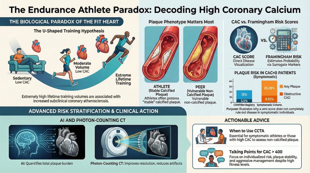
في تأكيد السجل analysis of symptomatic patients undergoing CCTA, among those with CAC=0, 13% had nonobstructive CAD, 3.5% had ≥50% تضيق, and 1.4% had ≥70% stenosis.⁸
In a large symptomatic cohort study focused on CAC=0 patients undergoing CCTA, 25.28% had any plaque and 9.32% had obstructive CAD.¹⁰
Age materially modifies the “rule-out” value of CAC=0, with younger symptomatic patients more likely to have non-calcified disease.⁹
These findings are the practical reason CAC=0 should not be interpreted as “no coronary disease,” particularly when symptoms are present.
Risk Reclassification and Ethnicity
Traditional حاسبات المخاطر provide population-based estimates and may misclassify individuals. Adding CAC can improve discrimination and reclassification, especially among patients initially categorized as borderline or intermediate risk.⁴,⁵,⁷
The Paradox of the Endurance Athlete: High CAC in Highly Fit Individuals
The relationship between exercise and heart health is not purely linear. Cardiac imaging studies of masters athletes (often defined as individuals >35 years with years of high-intensity training) have shown that some long-term endurance athletes have higher CAC prevalence and/or greater plaque burden than risk-factor–matched controls.²
Research from the Master@Heart Cohort
في Master@Heart study, lifelong endurance athletes demonstrated higher coronary atherosclerosis burden compared with controls, emphasizing that increased fitness does not uniformly equate to lower measured plaque burden on imaging.² This has contributed to “U-shaped” or “reverse J-shaped” hypotheses proposing that while moderate exercise is protective, very high lifetime training volumes may be associated with increased subclinical coronary atherosclerosis in some individuals.²

Plaque Phenotype, Fitness, and Event Rates
A key paradox is that despite higher CAC in some endurance athletes, many studies observe favorable outcomes influenced by high cardiorespiratory fitness and overall risk-factor profiles.⁴,¹¹ Still, a high CAC score in an athlete should be interpreted as evidence of underlying atherosclerosis requiring clinical attention and individualized risk discussion—not dismissed as “benign athletic calcification.”²,¹¹
Comparative Analysis: CAC vs. Other Risk Prediction Tools
CAC complements traditional risk scores by providing a direct, disease-based measure. Guidelines support CAC particularly when treatment decisions (e.g., statin initiation) are uncertain in borderline/intermediate-risk patients.⁴,⁵ CCTA directly visualizes both calcified and non-calcified plaque and is therefore better suited to symptomatic evaluation and plaque characterization, but is more complex and typically requires iodinated contrast.³,⁹
Future Directions: Photon-Counting CT and AI-Enabled Plaque Quantification
Photon-counting CT (PCCT) is an emerging CT detector technology that can improve spatial resolution and reduce تحلّق الضوء, potentially improving calcium quantification and plaque characterization in select settings.¹²,¹³
In parallel, AI-enabled quantitative plaque analysis on CCTA has been evaluated in peer-reviewed studies, including REVEALPLAQUE, supporting automated plaque quantification as a step toward more granular anatomic risk assessment beyond a single calcium sum.¹⁴
Conclusion
الـ نقاط كالسيوم الشريان التاجي is an important tool for cardiovascular risk stratification. By moving beyond statistical probability and visualizing disease burden, CAC enables more individualized primary prevention and المشاركة في صنع القرار.⁴,⁵,⁷ Its “power of zero” can identify individuals at very low near-term risk in the appropriate (asymptomatic) context; however, CAC=0 does not exclude non-calcified plaque, and symptomatic patients can still harbor meaningful plaque and even obstructive disease on CCTA.⁸–¹⁰
In masters endurance athletes, CAC highlights a complex reality: some lifelong athletes demonstrate greater plaque burden and higher CAC, but outcomes are influenced by fitness, risk-factor profiles, and plaque characteristics that vary by cohort.²,¹¹ The practical takeaway remains that CAC is best interpreted in clinical context—useful as a risk modifier and decision aid, not as a stand-alone verdict.⁴,⁵
Corrected References
- Parikh P, Shah N, Ahmed H, Schoenhagen P, Fares M. Coronary artery calcium scoring: Its practicality and clinical utility in primary care. Cleve Clin J Med. 2018;85(9):707-716. doi:10.3949/ccjm.85a.17097
- De Bosscher R, Dausin C, Claus P, et al. Lifelong endurance exercise and its relation with coronary atherosclerosis. Eur Heart J. 2023;44(26):2388-2399. doi:10.1093/eurheartj/ehad152
- Vatsa N, Faaborg-Andersen C, Dong T, Blaha MJ, Shaw LJ, Quintana RA. Coronary Atherosclerotic Plaque Burden Assessment by Computed Tomography and Its Clinical Implications. Circ Cardiovasc Imaging. 2024;17(8):e016443. doi:10.1161/CIRCIMAGING.123.016443
- Grundy SM, Stone NJ, Bailey AL, et al. 2018 AHA/ACC/AACVPR/AAPA/ABC/ACPM/ADA/AGS/APhA/ASPC/NLA/PCNA Guideline on the Management of Blood Cholesterol: A Report of the American College of Cardiology/American Heart Association Task Force on Clinical Practice Guidelines. Circulation. 2019;139(25):e1082-e1143. doi:10.1161/CIR.0000000000000625
- Foraker R, Sperling L, Bratzke L, et al. Opportunistic Detection of Coronary Artery Calcium on Noncardiac Chest Computed Tomography: An Emerging Tool for Cardiovascular Disease Prevention: A Scientific Statement From the American Heart Association. Circulation. 2025;152(19):e391-e401. doi:10.1161/CIR.0000000000001382
- Kim KP, Einstein AJ, Berrington de González A. Coronary artery calcification screening: estimated radiation dose and cancer risk. Arch Intern Med. 2009;169(13):1188-1194. doi:10.1001/archinternmed.2009.162
- Bild DE, Bluemke DA, Burke GL, et al. Multi-Ethnic Study of Atherosclerosis: objectives and design. Am J Epidemiol. 2002;156(9):871-881. doi:10.1093/aje/kwf113
- Villines TC, Hulten EA, Shaw LJ, et al. Prevalence and severity of coronary artery disease and adverse events among symptomatic patients with coronary artery calcification scores of zero undergoing coronary computed tomography angiography: results from the CONFIRM (Coronary CT Angiography Evaluation for Clinical Outcomes: An International Multicenter) registry. J Am Coll Cardiol. 2011;58(24):2533-2540. doi:10.1016/j.jacc.2011.10.851
- Mortensen MB, Gaur S, Frimmer A, et al. Association of Age With the Diagnostic Value of Coronary Artery Calcium Score for Ruling Out Coronary Stenosis in Symptomatic Patients. JAMA Cardiol. 2022;7(1):36-44. doi:10.1001/jamacardio.2021.4406
- Yu YT, Hou ZH, Lu B, et al. Prevalence of coronary artery disease in symptomatic patients with zero coronary artery calcium score in different age population. Int J Cardiovasc Imaging. 2021;37(2):723-729. doi:10.1007/s10554-020-02028-8
- DeFina LF, Radford NB, Barlow CE, et al. Association of All-Cause and Cardiovascular Mortality With High Levels of Physical Activity and Concurrent Coronary Artery Calcification. JAMA Cardiol. 2019;4(2):174-181. doi:10.1001/jamacardio.2018.4628
- Symons R, Sandfort V, Mallek M, Ulzheimer S, Pourmorteza A. Coronary artery calcium scoring with photon-counting CT: first in vivo human experience. Int J Cardiovasc Imaging. 2019;35(4):733-739. doi:10.1007/s10554-018-1499-6
- Flohr T, Schmidt B, Ulzheimer S, Alkadhi H. Cardiac imaging with photon counting CT. Br J Radiol. 2023;96(1152):20230407. doi:10.1259/bjr.20230407
- Ramasamy A, Sokooti H, Zhang X, et al. Novel near-infrared spectroscopy-intravascular ultrasound-based deep-learning methodology for accurate coronary computed tomography plaque quantification and characterization. Eur Heart J Open. 2023;3(5):oead090. Published 2023 Oct 30. doi:10.1093/ehjopen/oead090

