
Progresión fisiopatológica e impacto funcional sistémico de la aterosclerosis subclínica
Un análisis del curso de la vida desde la juventud hasta la mitad de la vida
Resumen
Aterosclerótico enfermedad cardiovascular (ASCVD) sigue siendo la principal causa de mortalidad en todo el mundo, y sus manifestaciones clínicas representan la culminación en etapa tardía de una trayectoria biológica que comienza décadas antes —a menudo en la vida fetal y sin duda en la segunda década—. Esta revisión integra la biología vascular estructural, la hemodinámica, la causalidad lipídica, inflamación, fenotipos específicos de sexo, neurodegeneración y reversibilidad del estilo de vida en un marco unificado para comprender ateroesclerosis subclínica como una enfermedad activa, progresiva y modificable. Examinamos la heterogeneidad regional en la arquitectura de la pared arterial y el papel de vasa vasorum, la divergencia de las grandes arterias ateroesclerosis de la enfermedad de pequeños vasos cerebrales, los determinantes hemodinámicos de placa la localización y la evidencia longitudinal de Día de pago, Bogalusa, CARDIA, MESA, y la serie fetal FELIC que anclan el inicio de la enfermedad en la vida temprana. Sintetizamos evidencia de aleatorización mendeliana, genética de poblaciones y ensayos de intervención que establecen apolipoproteína que contiene b lipoproteínas como el impulsor causal necesario de aterogénesis, y situamos a la Lp(a), la inflamación, la hematopoyesis clonal, disfunción endotelial, rarefacción microvascular y rigidez arterial dentro de este marco. Concluimos con las imágenes y biomarcador herramientas que permiten la detección durante la ventana preclínica prolongada, los fenotipos específicos de sexo que históricamente han sido poco reconocidos y la evidencia de los ensayos, más prominentemente la Ensayo sobre el estilo de vida y el corazón, la serie de casos de Esselstyn, STARS y el documentado por IVUS estatinas y ensayos de regresión con inhibidores de PCSK9 (REVERSIÓN, ASTEROIDE, Saturno, GLAGOV, PACMAN-AMI, Huygens) — lo que respalda la conclusión de que la aterosclerosis subclínica es, cuando se aborda de forma temprana y agresiva, una enfermedad reversible.
Palabras clave
Aterosclerosis subclínica; apolipoproteína B; lipoproteína(a); disfunción endotelial; vasa vasorum; enfermedad de pequeños vasos cerebrales; coronario arteria calcio; velocidad de onda de pulso; regresión de la placa; alimento integral dieta basada en plantas.
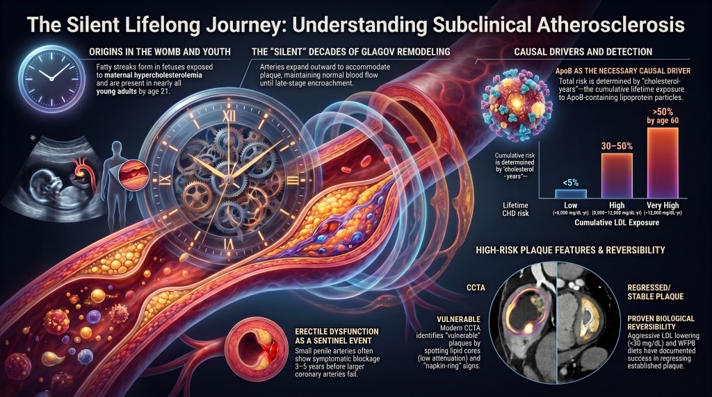
1. Introducción
La aparición de la ECVA clínica es la culminación de una trayectoria biológica silenciosa y de múltiples décadas que a menudo comienza en la segunda década de la vida —y en el caso de los fetos expuestos a una madre hipercolesterolemia, incluso antes del nacimiento. La aterosclerosis subclínica se refiere a la presencia de alteraciones estructurales en la pared arterial —incluyendo engrosamiento intimal, la retención de lipoproteínas, la acumulación de células espumosas y la formación temprana de placas, que preceden a las manifestaciones sintomáticas evidentes como angina, infarto de miocardio, infarto isquémico, claudicación periférica o demencia vascular. Esta progresión silenciosa está gobernada por una interacción estrechamente acoplada entre fuerzas hemodinámicas, variaciones estructurales en la pared arterial, el flujo de lipoproteínas, la activación inmunitaria y el estrés metabólico.
Aunque tradicionalmente se ha enmarcado como una enfermedad de la senescencia, los estudios de cohortes longitudinales y los datos de autopsias han cambiado fundamentalmente el enfoque hacia los adultos jóvenes y los adolescentes, revelando que sutiles déficits funcionales cotidianos —que van desde una velocidad de procesamiento cognitivo disminuida hasta una reserva renal y eréctil alterada— surgen mucho antes de que se alcancen los umbrales clínicos tradicionales para el diagnóstico. La formulación más rigurosa de este concepto a lo largo de la vida es la de exposición acumulativa o “años de colesterol” modelo, en el cual la integral de la concentración de partículas de apoB plasmática a lo largo del tiempo predice tanto el momento como la gravedad de los eventos clínicos [7], [9].
Esta revisión ofrece una síntesis exhaustiva de la fisiopatología, la distribución anatómica y las consecuencias funcionales de la enfermedad vascular subclínica en todo el árbol arterial humano, prestando especial atención a (i) la biología divergente de la aterosclerosis de grandes arterias y la enfermedad de pequeños vasos cerebrales; (ii) la centralidad causal de las lipoproteínas que contienen apoB y la contribución de la Lp(a), la inflamación y la hematopoyesis clonal; (iii) los fenotipos específicos de cada sexo; (iv) las modalidades de detección emergentes; y (v) la evidencia basada en ensayos clínicos de que la enfermedad subclínica es reversible cuando se aborda de forma temprana y agresiva.
2. Las lipoproteínas que contienen apolipoproteína B como el factor causal necesario
2.1 La síntesis de aleatorización mendeliana
Aleatorización mendeliana (MR) proporciona una de las formas más rigurosas de inferencia causal disponibles fuera ensayos controlados aleatorizados. Al explotar la asignación aleatoria de alelos en la concepción, los estudios RM estiman el efecto de por vida de una exposición determinada genéticamente sin la confusión de causalidad inversa, covariables de estilo de vida o error de medición. Ference y sus colegas analizaron 50 polimorfismos en nueve genes relacionados con las LDL en 312 321 participantes y establecieron que cada disminución genética de 1 mmol/L (≈38.7 mg/dL) en el c-LDL confiere un riesgo relativo reducción de aproximadamente el 54.5 por ciento para enfermedad coronaria — un efecto aproximadamente tres veces mayor por unidad LDL que el de las estatinas iniciadas en la mediana edad1].
El relación dosis-respuesta es loglineal sin un umbral detectable, lo que respalda un modelo de exposición acumulativa. Un segundo estudio de RM que utiliza polimorfismos en PCSK9, HMGCR, y NPC1L1 demostró que la reducción del riesgo guarda una estrecha correlación con la magnitud y la duración de la reducción del c-LDL, lo que respalda la conclusión de que la exposición acumulada a lo largo de la vida a las lipoproteínas que contienen apoB es un determinante central de la aterogénesis, mientras que la vía molecular específica a través de la cual se reduce el c-LDL es menos importante para la reducción del riesgo de ECVA [3]. Una Declaración de Consenso de la Sociedad Europea de Ateroesclerosis de 2017 analizó más de 200 estudios genéticos, epidemiológicos prospectivos y de intervención aleatorizados, y concluyó que el LDL cumple con el Criterios de Bradford Hill para la causalidad en la ECVAS4], y la actualización de la EAS de 2020 extendió esta conclusión a las lipoproteínas que contienen apoB en general [19].
2.2 La ApoB como la partícula unificadora
Cada partícula de lipoproteína aterogénica importante — VLDL, IDL, LDL, Lp(a) y quilomicrón remanentes — transportan una sola molécula de apolipoproteína B: apoB-100 para partículas de origen hepático y apoB-48 para restos intestinales. ApoB por lo tanto cuenta partículas aterogénicas, mientras que el C-LDL mide una carga cuya proporción respecto al transportador varía con el estado metabólico. En el UK Biobank análisis de 389,529 participantes, la apoB fue el predictor dominante de infarto de miocardio; el c-LDL y el c-no-HDL perdieron significancia estadística tras el ajuste por apoB5]. En estados de discordancia — donde el c-LDL es normal pero el número de partículas es alto, como en síndrome metabólico y resistencia a la insulina — la apoB devela un riesgo aterogénico sustancial que una estrategia basada únicamente en el c-LDL pasaría por alto6].
2.3 Exposición acumulada y el concepto de “colesterol-años”
El modelo de exposición acumulativa trata el riesgo de ASCVD como una función de la integral de la concentración plasmática de c-LDL a lo largo del tiempo, análogo a “paquetes-año”para el tabaco. Varios análisis respaldan una relación de dosis-tiempo entre la exposición al C-LDL y los eventos de ECVA. El “umbral” para la enfermedad clínicamente aparente en hombres se ha descrito en algunos análisis como aproximadamente 5,000 mg/dL·años —por ejemplo, LDL de 125 mg/dL × 40 años, lo que corresponde a una edad de ~50 años en las poblaciones occidentales modernas—, pero el valor exacto de LDL-año en el cual surge la enfermedad clínica depende del modelo y debe presentarse como ilustrativo en lugar de como un límite biológico universal [7], [9].
El “LDL años placa”el marco reformula la prevención cuantitativamente en torno a la carga acumulativa. Por ejemplo, un LDL de por vida de 70 mg/dL × 80 años produce ≈5,600 mg/dL·años; un LDL de 130 mg/dL × 50 años produce ≈6,500 mg/dL·años; y la heterocigota no tratada hipercolesterolemia familiar los pacientes con LDL de 250 mg/dL superan los umbrales ilustrativos comunes a principios de los treinta años. Estos cálculos son útiles para comunicar la biología de dosis y tiempo y para explicar por qué la HF heterocigota no tratada conlleva un alto riesgo de cardiopatía coronaria durante toda la vida, pero no deben presentarse como umbrales de riesgo individual validados7], [8], [9].
| Categoría | Exposición acumulativa a las LDL | Trayectoria aproximada | Riesgo ilustrativo de cardiopatía coronaria a lo largo de la vida |
| Bajo | <5,000 mg/dL·año | LDL 70 × 70 años | <5% |
| Intermedio | 5,000–8,000 mg/dL·año | LDL 100 × 60 años | 10–20% |
| Alto | 8,000–12,000 mg/dL·año | LDL 130 × 65 años | 30–50% |
| Muy alto | >12,000 mg/dL·año | LDL 190 × 65 años (HeFH) | >50% a los 60 años |
Tabla 1. Categorías ilustrativas de exposición acumulada al colesterol LDL y riesgo aproximado de cardiopatía coronaria a lo largo de la vida. Adaptado de Ference et al. y Domanski et al.7], [8], [9]. Estas categorías son ayudas de comunicación ilustrativas, no umbrales de riesgo individual validados por guías clínicas.
2.4 Tsimane y Hadza: El experimento natural de un LDL bajo durante toda la vida
Kaplan y sus colegas examinaron las puntuaciones de CAC en 705 Tsimane recolectores-horticultores de la Amazonía boliviana de entre 40 y 94 años. El C-LDL medio fue de 91 mg/dL, y el 85 por ciento de los adultos de 40 años o más tenían un CAC de cero, en comparación con aproximadamente el 50 por ciento de los adultos estadounidenses de edad similar en MESA. El sesenta y cinco por ciento de los mayores de 75 años tenían un CAC de cero —el nivel más bajo de aterosclerosis coronaria jamás medido en ninguna población— y solo el 8 por ciento tenía un CAC ≥100, frente a más del 50 por ciento en poblaciones estadounidenses de la misma edad [10].
Este experimento natural respalda la conclusión de que mantener un C-LDL relativamente bajo a lo largo de la vida, combinado con una alta actividad física y un mínimo fumar, se asocia con una prevalencia muy baja de enfermedad coronaria calcificación. No debe plantearse como una prueba de que el c-LDL ≤90 mg/dL elimina prácticamente la aterosclerosis coronaria, ya que el fenotipo de los tsimanes refleja múltiples exposiciones a lo largo de la vida y no el c-LDL de forma aislada. Se ha descrito a los hadza de Tanzania como una población de cazadores-recolectores físicamente activa con características cardiometabólicas favorables en los estudios disponibles, pero no se han realizado imágenes directas de CAC en este grupo; por lo tanto, las conclusiones sobre la ausencia de enfermedad coronaria clínica en los hadza son inferenciales en lugar de directas [11].
2.5 Niveles bajos de LDL genéticos de por vida: PCSK9 y ANGPTL3
Cohen y sus colegas identificaron mutaciones naturales de pérdida de función en PCSK9 (R46L, Y142X, C679X) que reducen el LDL entre un 15 y un 40 por ciento a lo largo de la vida. Los portadores de variantes seleccionadas mostraron reducciones importantes en los eventos de cardiopatía coronaria, y las estimaciones más llamativas —incluidas reducciones cercanas al 88 por ciento para algunas variantes— reflejan tamaños del efecto que varían según la variante y la ascendencia12]. Tales tamaños del efecto superan con creces lo que cualquier intervención farmacológica iniciada en la mitad de la vida puede lograr. De manera similar, las variantes homocigóticas de pérdida de función en ANGPTL3 producir hipobetalipoproteinemia con LDL muy baja y triglicéridos; las series epidemiológicas describen un riesgo de ASCVD notablemente reducido y favorable perfiles lipídicos en dichos portadores, aunque las tasas de eventos no se han cuantificado en cohortes aleatorizadas13].
Estos experimentos genéticos establecen una condición de contorno útil: cuando la carga de partículas de apoB durante toda la vida es genéticamente baja, la aterosclerosis aparece marcadamente suprimida incluso sin intervención farmacológica. El mimetismo farmacológico de estos fenotipos —a través de anticuerpos monoclonales contra PCSK9, agentes de ARNsi e inhibidores de ANGPTL3— ha demostrado una reducción aditiva de eventos además de la terapia con estatinas. Los análisis de Fourier y la extensión abierta FOURIER respalda tasas de eventos progresivamente más bajas entre los pacientes que alcanzan niveles muy bajos de LDL, incluidos LDL <20 mg/dL, sin una señal de seguridad excesiva durante el seguimiento disponible18].
2.6 Por qué un LDL muy bajo y sostenido reduce notablemente la aterogénesis
Endotelial transcitosis de LDL en el íntima depende de la concentración, y la retención mediada por proteoglicanos —el paradigma de respuesta a la retención de Tabas, Williams y Borén— se vuelve menos probable a medida que disminuye el flujo de partículas de apoB83]. Los recién nacidos tienen un C-LDL de ≈30 mg/dL, y la ateroesclerosis lesiones no se detectan típicamente en esta etapa; esto debe interpretarse como evidencia de que un C-LDL de por vida muy bajo es incompatible con las etapas iniciales de la aterogénesis a escala poblacional, en lugar de como una afirmación de que la aterogénesis sea biológicamente imposible a cualquier valor único de C-LDL. Las cohortes con C-LDL de por vida <70 mg/dL (pérdida de función de PCSK9, pérdida de función de ANGPTL3, HF tratada) demuestran reducciones notables en las tasas de eventos, con regresión de la placa documentada por IVUS con un C-LDL durante el tratamiento <60 mg/dL en los ensayos REVERSAL, ASTEROID, SATURN y GLAGOV [14], [15], [16], [17].
FOURIER-OLE extendido evolocumab seguimiento durante una mediana de aproximadamente 5 años e informó una reducción continua de eventos con un c-LDL durante el tratamiento <20 mg/dL sin señales de seguridad adicionales durante el seguimiento disponible18]. Una demostración formal de dosis-respuesta sin que se haya establecido firmemente una meseta a estos niveles muy bajos de LDL, los datos disponibles son consistentes con un beneficio continuo y ninguna toxicidad compensatoria hasta el nivel de LDL más bajo alcanzado hasta la fecha en las poblaciones de los ensayos.
2.7 Reconocimiento y refutación de la postura de los escépticos de las LDL
Un pequeño grupo de autores —de forma más destacada Ravnskov, Diamond y Kendrick— han argumentado que el LDL no es causal, citando estudios observacionales en poblaciones de edad avanzada en los que el LDL parece no ser predictivo (el llamado “paradoja lipídica”). Las refutaciones estándar están bien establecidas. Primero, la causalidad inversa predomina en la vejez: la enfermedad crónica, la malabsorción y la fragilidad reducen el c-LDL, sesgando la asociación entre el c-LDL y la mortalidad en los análisis transversales. Segundo, sesgo de supervivencia selecciona a individuos genéticamente protegidos entre aquellos que alcanzan la edad de 80 años con LDL alto. Tercero, la atenuación del riesgo relativo con la edad no implica la atenuación de riesgo absoluto; la tasa de eventos absolutos aumenta dramáticamente con la edad. Cuarto, la evidencia genética y farmacológica es mutuamente corroborativa en aproximadamente veinte líneas independientes de investigación, satisfaciendo los criterios de triangulación para la causalidad [4], [6], [19La postura escéptica se basa casi por completo en datos observacionales mientras ignora la evidencia convergente de la genética y de los ensayos aleatorizados.
3. Determinantes estructurales y el papel de los vasa vasorum en la aterogénesis
3.1 Profundidad crítica y la unidad lamelar
En las arterias sistémicas de gran calibre, las demandas metabólicas de la pared gruesa superan la capacidad de difusión simple de oxígeno desde el lumen. La “profundidad crítica” se define como el límite fisiológico de la difusión de oxígeno y nutrientes a partir de la sangre luminal, establecido por Geiringer en aproximadamente 0.5 mm (≈500 μm), o más o menos 29–30 unidades lamelares de la pared medial [78], [79]. Cada unidad laminar consta de una lámina elástica y su capa asociada de células de músculo liso y matriz extracelular, de aproximadamente 15 μm de espesor. Los segmentos de pared más gruesos que este umbral requieren una red microvascular intrínseca —los vasa vasorum— para mantener la viabilidad de la capa media externa y adventicia.
Los vasa vasorum se clasifican en vasa vasorum interna, que se originan directamente de la luz arterial, y vasa vasorum externa, que provienen de ramas distantes y penetran en la adventicia. Las investigaciones de Geiringer establecieron que los vasa vasorum son abundantes en la adventicia y el tercio externo de la media, mientras que los ≈0.5 mm internos (≈30 unidades laminares) de la pared permanecen avasculares y se nutren únicamente mediante difusión luminal.79]. Cuando la pared se engrosa debido a la placa ateroesclerótica o la hiperplasia hipertensiva, la distancia de difusión aumenta, generando un entorno hipóxico en las capas más profundas. La hipoxia desencadena una señalización angiogénica dependiente de HIF-1α, lo que lleva a la proliferación de vasa vasorum que pueden penetrar la lámina elástica interna y entrar en la placa misma, donde sirven tanto como conductos para células inflamatorias como fuentes frágiles de hemorragia intraplaca.
3.2 Nutrición vascular extracraneal vs. intracraneal
La vasculatura intracraneal presenta un perfil arquitectónico único en comparación con la circulación sistémica. Las arterias intracraneales sanas se caracterizan por una túnica media más delgada, una adventicia menos abundante y una relativa escasez de fibras elásticas fibras; muchos carecen de una lámina elástica externa bien definida. Una característica distintiva principal es que los vasos intracraneales están bañados en líquido cefalorraquídeo rico en nutrientes, lo que proporciona soporte metabólico a través de la difusión externa y compensa parcialmente la relativa ausencia de vasa vasorum en las primeras etapas de la vida.
Los estudios modernos de autopsia e imagen han refinado la perspectiva histórica de que los vasa vasorum están ausentes en el cerebro. En una serie de autopsias de 50 casos, se identificaron vasa vasorum en el 72 por ciento de los pacientes, localizados principalmente en la túnica adventicia. Su distribución es marcadamente no uniforme: los vasa vasorum se encuentran con mayor frecuencia en los segmentos proximales —las arterias vertebrales, la arteria basilar y la porción intracraneal de la interna arteria carótida — que en los segmentos distales, como las arterias cerebral media o cerebral anterior. Aunque la aterosclerosis puede ocurrir en ausencia de vasa vasorum, su desarrollo está fuertemente asociado con la progresión de la enfermedad intracraneal: en la arteria vertebral intracraneal, la presencia de vasa vasorum adventiciales se correlaciona con una mayor carga de placa, una calcificación intraplaca más densa y una estenosis luminal más severa estenosis.
| Tipo de embarcación | Espesor de pared | Vasa vasorum (etapa temprana) | Fuente nutricional |
| Aorta / sistémico grande | >1.0 mm (frecuentemente 1.5–2.0 mm) | Abundante en la adventicia y la media externa | Difusión luminal + VV |
| carótida extracraneal | ≈0,6–1,0 mm | Presente en la adventicia | Difusión luminal + VV |
| ACI / AV intracraneal | ≈0.2–0.3 mm | Escasos; principalmente segmentos proximales | Difusión luminal + LCR |
| ACM / ACA distal | <0.2 mm; carece de LME | Ausente o raro | Difusión luminal + LCR |
| Arteriolas penetrantes | de 40 a 200 μm de diámetro | Ausente | difusión luminal + líquido intersticial |
Tabla 2. Arquitectura comparativa del grosor de la pared arterial, la densidad de los vasa vasorum y la fuente nutricional en las circulaciones sistémica y cerebral. EEL, lámina elástica externa; ICA, arteria carótida interna; VA, arteria vertebral; MCA, arteria cerebral media; ACA, arteria cerebral anterior; VV, vasa vasorum; CSF, líquido cefalorraquídeo.
4. Fuerzas hemodinámicas y localización de la placa
4.1 Esfuerzo cortante laminar frente a perturbado
La naturaleza focal de la aterosclerosis está dictada por la interacción entre el flujo sanguíneo y la geometría arterial. El endotelio sirve como mecanosensor, traduciendo el estrés físico en señalización biológica a través de los cilios primarios, las integrinas y el medio mediado por el glucocálix mecanotransducción, y canales iónicos (especialmente Piezo1 y TRPV4). En los segmentos arteriales rectos, el flujo es laminar, lo que genera un nivel alto y unidireccional esfuerzo cortante en la pared (típicamente de 1 a 7 Pa, o de 10 a 70 dyn/cm²). Este entorno mantiene un fenotipo endotelial aterorresistente caracterizado por la activación sostenida de óxido nítrico sintasa endotelial (eNOS), fosforilación de los factores de transcripción KLF2 y KLF4, y supresión de la señalización de NF-κB, con el resultado de que óxido nítrico la producción es alta, adhesión leucocitaria se suprime y la permeabilidad íntima permanece baja85].
Por el contrario, en los puntos de bifurcación, ramificación y áreas de alta curvatura, el flujo se vuelve “perturbado”. Estas zonas ateropronas experimentan una tensión de corte en la pared promediada en el tiempo baja (a menudo <0.4 Pa) y un alto índice de corte oscilatorio, con la dirección de la fuerza de fricción invirtiéndose durante el ciclo cardíaco. En las regiones de flujo perturbado, el endotelio sufre un cambio fenotípico: las uniones estrechas se aflojan, lo que permite una mayor transcitosis de LDL hacia la íntima; la expresión de VCAM-1, ICAM-1, la E-selectina y la MCP-1 capturan monocitos y linfocitos T circulantes; especies reactivas del oxígeno la generación aumenta a través de la activación de la NADPH oxidasa; y el eje protector KLF2/eNOS es suprimido.
El estudio seminal de dinámica de fluidos computacional realizado por Ku y sus colegas en la bifurcación carotídea humana demostró una estrecha concordancia espacial entre las regiones de bajo cizallamiento y el engrosamiento íntimal, el estudio empírico fundacional de la teoría hemodinámica de la aterogénesis [71]. Este patrón se repite en cada punto de ramificación del árbol arterial: los segmentos proximales de la DA y la CX, el bulbo carotídeo, la bifurcación aórtica abdominal y los ostios de las arterias renales exhiben esta vulnerabilidad dependiente de la geometría.
4.2 Comportamiento celular en el microambiente de la placa
Una vez retenidas en la íntima, las LDL sufren una modificación oxidativa por la mieloperoxidasa, la lipooxigenasa y las especies reactivas del oxígeno. LDL oxidada es reconocido por los receptores scavenger (CD36, SR-A) en células residentes y recién reclutadas macrófagos, que lo internalizan y se convierten células espumosas. En las lesiones subclínicas tempranas, la muerte de las células espumosas se equilibra con efrocitosis — la eliminación de células apoptóticas por los macrófagos vecinos —, pero a medida que el microentorno se vuelve cada vez más tóxico, la eferocitosis falla, los restos apoptóticos y necróticos se acumulan, y un núcleo necrótico formas. Las células de músculo liso vascular (VSMC) cambian simultáneamente de un fenotipo contráctil a uno sintético, migrando de la túnica media a la íntima, donde secretan una matriz rica en colágeno cápsula fibrosa que inicialmente estabiliza la lesión. El equilibrio entre la reparación que engrosa la capa fibrosa y la inflamación que expande el núcleo determina si una placa permanece estable o progresa hacia la vulnerabilidad82], [83], [84].
5. Disfunción endotelial como la lesión detectable más temprana
La disfunción endotelial precede a cualquier lesión estructural detectable por grosor íntima-media carotídeo (GIMC), la puntuación de calcio en las arterias coronarias o la angiografía. Es funcional, dinámica y parcialmente reversible, y por lo tanto representa la ventana práctica más temprana para la intervención primordial.
Dilatación mediada por flujo
Braquial dilatación mediada por flujo (DMF), medidos por ultrasonido después de un manguito en el antebrazo de 5 minutos oclusión, cuantifica la vasodilatación dependiente del endotelio (en gran medida mediada por óxido nítrico). Una FMD más baja se asocia con un mayor riesgo cardiovascular, pero la FMD depende del protocolo y ningún punto de corte universal único se aplica en todos los laboratorios; los valores por debajo de aproximadamente 5 a 7 por ciento se consideran comúnmente anormales en contextos de investigación. En el Estudio Multiétnico de Aterosclerosis (MESA), la FMD aportó información pronóstica independiente más allá del riesgo de Framingham y el IMT-C [33]. La FMD está deteriorada en sujetos con niveles de LDL incluso levemente elevados, insulina resistencia, sin tratar hipertensión, o exposición crónica a la contaminación del aire por partículas.
5.2 Índice de hiperemia reactiva
El índice de hiperemia reactiva (RHI), medido de forma no invasiva mediante pletismografía digital (EndoPAT), refleja la microvascularización función endotelial en la yema del dedo después de la hiperemia reactiva. Se ha utilizado un umbral de RHI de alrededor de 1.67 para identificar la disfunción endotelial coronaria o la aterosclerosis coronaria temprana en cohortes seleccionadas; la sensibilidad y la especificidad dependen de la población y del estándar de referencia [34]. RHI es independiente del operador, tiene buena reproducibilidad y se ha utilizado como resultado en ensayos de intervención farmacológica y en el estilo de vida.
5.3 Degradación del glucocálix
El glicocáliz endotelial es una capa superficial de tipo gel de 0,5 a 3 μm compuesta de proteoglicanosheparán sulfato, sulfato de condroitina), glicoproteínas (sindecán-1) y plasma adsorbido proteínas. Modula la transcitosis de LDL, el rodamiento de leucocitos y la mecanotransducción por esfuerzo cortante. Glicocálix degradación impulsada por LDL oxidada, hiperglucemia, el TNF-α y las especies reactivas de oxígeno, expone moléculas de adhesión y aumenta el flujo de lipoproteínas en la íntima. El adelgazamiento del glucocáliz, detectable mediante imágenes sublinguales de campo oscuro lateral y por niveles elevados de syndecan-1 y hialuronano en plasma, ocurre antes de la alteración medible de la dilatación mediada por flujo (DMF) y se encuentra entre las primeras anomalías detectables en la enfermedad subclínica [35], [36].
5.4 Disfunción microvascular que precede a la enfermedad macrovascular
Reserva de flujo coronario (CFR) por debajo de 2.0 por tomografía por emisión de positrones, por debajo de 2.5 mediante Doppler transtorácico, o por debajo de 2.0 mediante resonancia magnética cardíaca es un predictor independiente de eventos cardiovasculares incluso en ausencia de enfermedad epicárdica obstructiva. La disfunción endotelial microvascular puede ocurrir con coronarias normales angiografías, proporcionando el sustrato para el síndrome de isquemia con no obstructiva arterias coronarias (INOCA), discutida en la Sección 8 [37].
6. Mapeo anatómico de la enfermedad vascular subclínica
La aterosclerosis es una enfermedad sistémica pero no uniforme, que favorece sitios anatómicos específicos caracterizados por una geometría compleja y un flujo alterado. El mapeo preciso en todos los lechos vasculares revela el patrón predecible y dependiente de la geometría de la localización de la placa.
6.1 Lechos cerebrovasculares y cervicales
En el cuello, la bifurcación carotídea y la arteria carótida interna proximal son los sitios principales para el desarrollo temprano de placas, debido a la separación del flujo, la recirculación y la baja fricción oscilatoria en el bulbo carotídeo. Dentro del cráneo, la enfermedad se observa con mayor frecuencia en la ACI intracraneal (sifón carotídeo) y en los segmentos proximales de las ramas principales del polígono de Willis. Las series de autopsias indican que la aterosclerosis intracraneal se retrasa con respecto a la enfermedad extracraneal por aproximadamente 15 a 20 años; las lesiones estables son más comunes en la ACI, mientras que las lesiones más dinámicas y progresivas se encuentran en la ACM, la ACA y las arterias cerebrales posteriores.
Es importante destacar que, enfermedad aterosclerótica intracraneal representa aproximadamente el 9 por ciento de golpes en poblaciones blancas, pero del 30 al 50 por ciento de los accidentes cerebrovasculares en poblaciones de Asia oriental, negras e hispanas [73], [74]. Esta disparidad racial persiste tras el ajuste por las variables tradicionales factores de riesgo, lo que sugiere contribuciones genéticas y estructurales subyacentes. El ensayo SAMMPRIS estableció que el manejo médico es superior a la colocación de un stent para la enfermedad intracraneal aterosclerótica sintomática con una estenosis del ≥70 por ciento [73].
6.2 Vasculatura coronaria y aorta
La aterosclerosis coronaria se inicia típicamente en los segmentos proximales de las arterias epicárdicas, y la descendente anterior (DA) proximal muestra la mayor prevalencia de placas —particularmente dentro de los primeros 40 mm— debido a su ángulo de salida agudo y a la alta alteración del flujo generada en las bifurcaciones de las ramas diagonales y septales. Las bifurcaciones de la DA con las diagonales, la circunfleja (Cx) con las marginales obtusas y la coronaria derecha (CD) con la arteria descendente posterior muestran todas una predilección en las paredes laterales de baja fricción (esfuerzo cortante bajo) de las ramas secundarias.
El estudio de los Determinantes Patobiológicos de la Aterosclerosis en los Jóvenes (PDAY) confirmó que estrías grasas y se presentan lesiones elevadas en las coronarias de individuos de tan solo 15 a 34 años. En la aorta, existe un claro gradiente de susceptibilidad: la aorta abdominal está más gravemente afectada que la aorta torácica, con la mayor concentración de placas en la aorta abdominal distal y en la bifurcación aórtica.
6.3 Arterias renales y mesentéricas
La estenosis de la arteria renal aterosclerótica (EARA) representa aproximadamente el 90 por ciento de los casos de estenosis de la arteria renal y afecta principalmente los ostios de la arteria renal y los 2 cm proximales de la arteria renal principal. Por el contrario, la displasia fibromuscular, que representa el ≈10 por ciento restante, suele afectar los segmentos medio y distal o las ramas intrarrenales. La enfermedad de la arteria mesentérica —que afecta el tronco celíaco y las arterias mesentéricas superior e inferior— también ocurre con mayor frecuencia en los orígenes aórticos, donde la turbulencia del flujo es mayor.
6.4 Arterias de las extremidades inferiores y de la pelvis
La enfermedad arterial periférica sigue una progresión predecible desde las arterias elásticas hasta las musculares. La aterosclerosis suele aparecer primero en las arterias elásticas suprainguinales (aorta e ilíacas) antes de progresar a las arterias musculares infrainguinales (femoral, poplítea y tibial). La enfermedad vascular pélvica involucra frecuentemente a la arteria pudenda interna (API), en la que se ha documentado una estenosis u oclusión significativa en aproximadamente el 54 por ciento de los hombres examinados por enfermedad de las arterias coronarias —una prevalencia extraordinaria que refleja el pequeño calibre y la exposición compartida a factores de riesgo de estos vasos. Las ramas distales de la arteria pudenda interna, incluidas las arterias cavernosas, son singularmente susceptibles debido a su pequeño diámetro (0.5-1 mm), como se analiza en la Sección 9.
7. Diferenciación entre aterosclerosis de grandes arterias y enfermedad de pequeños vasos cerebrales
La distinción entre la aterosclerosis clásica y enfermedad de los pequeños vasos cerebrales (EPVC) es fundamental para comprender los mecanismos divergentes de lesión vascular en todo el cuerpo humano.
7.1 Por qué las arteriolas penetrantes no desarrollan aterosclerosis clásica
Las arteriolas penetrantes (de 40 a 200 μm de diámetro) que irrigan el cerebro profundo —los ganglios basales, el tálamo, la cápsula interna y la sustancia blanca periventricular— no presentan la forma excéntrica, placas ricas en lípidos típico de la aterosclerosis de grandes vasos. Tres factores estructurales y biológicos explican esta divergencia:
Primero, simplicidad estructural: estos vasos carecen de la estructura lamelar multicapa y de las láminas elásticas interna y externa bien definidas que sustentan la arquitectura clásica de la placa. Segundo, barrera hematoencefálica especialización: el células endoteliales de estos vasos son componentes especializados de la unidad neurovascular, con uniones estrechas formadas por claudina-5, ocludina y ZO-1, respaldadas por pericitos, pies terminales de astrocitos y un entorno metabólico único que difiere fundamentalmente de los vasos sistémicos. Tercero, la ausencia de vasa vasorum: estos vasos dependen enteramente de la difusión luminal y externa y no poseen la red microvascular intrínseca que puede ser cooptada para la nutrición de la placa en arterias más grandes.
7.2 El espectro de la patología de los pequeños vasos
En lugar de la aterosclerosis clásica, las arteriolas penetrantes desarrollan un conjunto distinto de patologías denominadas colectivamente enfermedad de los pequeños vasos cerebrales. La lipohialinosis, caracterizada originalmente por C. Miller Fisher como “desorganización segmentaria de la pared arteriolar”, implica la acumulación de agregados céreos y vidriosos de lípidos y proteínas dentro de la pared del vaso, necrosis fibrinoide de las células de músculo liso de la capa media y estrechamiento luminal. Es impulsada principalmente por la hipertensión crónica y es el sustrato principal de los infartos lacunares en las arteriolas penetrantes lenticuloestriadas, talamoperforantes y pontinas.
La arteriolosclerosis hiperplásica implica un engrosamiento concéntrico en “piel de cebolla” de la pared debido a la proliferación de músculo liso y la duplicación de la membrana basal, y es más característica de la hipertensión maligna o acelerada. El microateroma se refiere a lesiones oclusivas en vasos penetrantes más grandes (200-800 μm); estos comparten algunas características con la aterosclerosis (células espumosas, retención de lípidos), pero típicamente ocurren en el origen proximal del vaso perforante y reflejan la propagación de la enfermedad de las arterias grandes hacia las ramas.
Angiopatía amiloide cerebral (AAC) implica el depósito progresivo de β-amiloide (Aβ) —predominantemente Aβ40 y en menor medida Aβ42— en la túnica media y adventicia de las arteriolas corticales y leptomeníngeas (típicamente de <2 mm de calibre). La CAA es independiente de la hipertensión y es un factor contribuyente importante a las microhemorragias lobares, la siderosis superficial y la hemorragia subaracnoidea de la convexidad [23], [24]. El cSVD patológicamente avanzado muestra frecuentemente una transición desde la disfunción endotelial hasta la alteración de la BHE: la falla de la BHE permite que componentes séricos tóxicos —fibrinógeno, IgG, complemento— se extravasen hacia el cerebro, lo que desencadena inflamación perivascular, “dilatación forzada” y acumulación de tejido conjuntivo que deja a los vasos sanguíneos descendentes vulnerables a daños por alta presión.
8. Evidencia de la primera infancia y trayectorias de riesgo longitudinales
8.1 Napoli/FELIC: Estrías grasas fetales
La lesión ateroesclerótica más temprana documentada en humanos es fetal. Napoli y colaboradores, en el estudio FELIC (Destino de las Lesiones Tempranas en Niños), demostraron que las estrías grasas se forman en la íntima aórtica de los fetos, con una acumulación intimal de LDL y su oxidación antes del reclutamiento de monocitos. La formación de estrías grasas fetales se vio muy incrementada por la hipercolesterolemia materna durante el embarazo, lo que sugiere que el entorno lipídico materno programa la vulnerabilidad vascular de la descendencia décadas antes de la enfermedad clínica [52].
Estas observaciones transformaron la concepción de la aterosclerosis, pasando de ser una enfermedad de la mediana edad a una enfermedad de curso vital iniciada en el útero, en la que el entorno prenatal establece la trayectoria de la acumulación intimal posterior de LDL.
8.2 PDAY y el Estudio del Corazón de Bogalusa
El Estudio PDAY realizó autopsias estandarizadas a más de 3000 personas de entre 15 y 34 años que murieron por traumatismos, catalogando la prevalencia y la extensión de las estrías grasas y las lesiones elevadas en las arterias coronarias y la aorta. Las estrías grasas estuvieron presentes en prácticamente todas las aortas y en las coronarias de la mayoría al final de los veintiocho años; las lesiones elevadas aparecieron en aproximadamente el 20 por ciento de los hombres de 30 a 34 años. El estudio desarrolló la puntuación de riesgo PDAY, que demostró que el tabaquismo, colesterol no HDL, y la hipertensión en los jóvenes son fuertes predictores de calcificación avanzada décadas más tarde.
El Estudio del corazón de Bogalusa extendieron estas observaciones a una cohorte comunitaria longitudinal, demostrando que los factores de riesgo medidos a los 5-17 años predicen la morbilidad subclínica en la edad adulta. Berenson y colaboradores, en datos de autopsias de 204 jóvenes de 2 a 39 años que murieron por traumatismos, documentaron estrías grasas aórticas en aproximadamente la mitad de los niños de 2 a 15 años, elevándose a casi el 100 por ciento a los 21 años; estrías grasas coronarias en el 8 por ciento de los niños de 2 a 15 años y en el 69 por ciento de aquellos de 26 a 39 años; y lesiones elevadas coronarias en el 3 por ciento de los de 6 a 15 años y en el 30 por ciento de los de 26 a 39 años. El número de factores de riesgo cardiovascular se correlacionó directamente con la extensión tanto de las estrías grasas como de las placas fibrosas [53].
8.3 El estudio CARDIA
El estudio de Desarrollo del Riesgo de Arteriopatía Coronaria en Adultos Jóvenes (CARDIA, por sus siglas en inglés) hizo un seguimiento a 5115 participantes de entre 18 y 30 años durante más de 35 años de seguimiento. Varios hallazgos han dado forma a la cardiología preventiva contemporánea. Primero, la exposición sostenida a niveles de c-LDL incluso modestamente elevados y presión arterial durante los años veinte y treinta contribuye al riesgo de ASCVD de manera independiente a los niveles de riesgo más adelante en la vida. Segundo, para el año 25 (edad promedio 50 años), aproximadamente el 27.7 por ciento de los participantes tenían calcio en la arteria coronaria detectable y aproximadamente el 53 por ciento tenía calcio en la aorta abdominal. Tercero, los participantes que adoptaron hábitos saludables —dieta, ejercicio, dejar de fumar, mantenimiento del peso— durante la adultez joven tuvieron significativamente menos enfermedad subclínica en la mediana edad, con reducciones absolutas en el riesgo de eventos sustancialmente mayores que las que la intervención en la mediana edad puede lograr.
8.4 Aterosclerosis pediátrica y en adolescentes: Hipercolesterolemia familiar
Hipercolesterolemia familiar heterocigota (HeFH; prevalencia ≈1:250) se presenta con C-LDL de 190–400 mg/dL desde el nacimiento, resultado de variantes patógenas en LDLR, APOB, o raramente PCSK9. Sin tratar, la HeFH conlleva un riesgo de cardiopatía coronaria de aproximadamente el 50 por ciento a la edad de 50 años en los hombres. El IMTC está significativamente elevado en los niños con HeFH a la edad de 8 a 10 años. El histórico ensayo clínico de Wiegman y sus colegas demostró que el inicio de estatinas entre los 8 y los 18 años normalizó la progresión del IMTC en comparación con sus pares [54]. El seguimiento a 20 años de esa cohorte (Luirink y colaboradores) mostró que el tratamiento temprano con estatinas redujo el riesgo de infarto de miocardio en aproximadamente un 75 por ciento en comparación con los padres no tratados, lo que constituye una de las demostraciones más contundentes en cualquier campo de la medicina de que la intervención temprana y sostenida puede alterar fundamentalmente la trayectoria de una enfermedad determinada genéticamente [55].
9. Impacto funcional de la enfermedad vascular subclínica en adultos jóvenes y de mediana edad
La aterosclerosis subclínica se describe a menudo como “silenciosa”, sin embargo, investigaciones rigurosas demuestran déficits funcionales mensurables mucho antes de que se alcancen los umbrales clínicos tradicionales.
9.1 Función cognitiva y deterioro neurovascular
En la cohorte CARDIA, los niveles más altos de CAC y calcio en la aorta abdominal a las edades de 43 a 55 años se asociaron significativamente con peores puntuaciones en las pruebas de velocidad psicomotora (Test de sustitución de símbolos de dígitos), atención sostenida y memoria verbal. Esta relación persiste tras el ajuste por edad, sexo, educación y factores de riesgo tradicionales, lo que sugiere que las lesiones calcificadas avanzadas en la mediana edad reflejan toda una vida de estrés vascular que también afecta la microvasculatura cerebral y la integridad de la sustancia blanca. Se ha encontrado que la inteligencia a los 19 años se correlaciona inversamente con el estado de la placa carotídea a los 60 años, una asociación probablemente mediada por la influencia a largo plazo de la capacidad cognitiva en el nivel socioeconómico, el acceso a la atención médica y adherencia a los estilos de vida saludables.
9.2 Capacidad aeróbica y tolerancia al ejercicio
La relación entre la actividad física y la enfermedad subclínica es bidireccional. Alta condición cardiorrespiratoria en la adultez joven protege contra el desarrollo de CAC y el aumento del IMTc de 15 a 25 años después. Por el contrario, la presencia de aterosclerosis subclínica deteriora sutilmente la tolerancia al ejercicio: la enfermedad subclínica reduce la reserva vascular —la capacidad de las arterias para dilatarse y aumentar el flujo durante el esfuerzo máximo—, lo que contribuye a una fatiga más temprana y a un V̇O₂ máximo reducido. Las personas con baja capacidad cardiorrespiratoria tienen de dos a tres veces más probabilidades de morir prematuramente por ECVA, incluso cuando se emparejan por factores de riesgo tradicionales.
9.3 Sueño, fatiga y estado de ánimo
Las duraciones extremas del sueño (8 horas) y la mala calidad subjetiva del sueño se asocian con una mayor prevalencia de CAC y una mayor velocidad de onda de pulso. La irregularidad en la duración del sueño —una variación superior a 120 minutos a lo largo de una semana— está vinculada a una prevalencia un 33 por ciento mayor de una alta carga de CAC. La desalineación circadiana impulsa la inflamación crónica de bajo grado y la activación del sistema nervioso simpático, lo que predispone a los individuos a la aterosclerosis subclínica. Aunque los vínculos causales directos con los trastornos del estado de ánimo aún están emergiendo, la combinación de fatiga de origen vascular, alteración del sueño e inflamación crónica contribuye a una reducida calidad de vida.
9.4 Disfunción eréctil como evento centinela: La hipótesis del tamaño de las arterias
La disfunción eréctil (DE) suele ser la primera manifestación clínica de la enfermedad vascular sistémica. La arteria pudenda interna (1-2 mm) y sus ramas cavernosas (0,5-1 mm) son notablemente más pequeñas que las arterias coronarias proximales (3-4 mm), la carótida interna (5-7 mm) o la arteria femoral (6-8 mm). De acuerdo con la hipótesis del tamaño de la arteria articulada por Montorsi y sus colegas, la ateroesclerosis equivalente carga de placa produce estenosis hemodinámicamente significativa primero en los vasos pequeños, de modo que la circulación cavernosa alcanza el umbral de afectación sintomática años antes de que lo haga la circulación coronaria80].
Los hombres con disfunción eréctil vascular tienen una prevalencia notablemente mayor de enfermedad coronaria subclínica; el ensayo COBRA informó que la disfunción eréctil vasculógena precedió a la aparición sintomática de la enfermedad coronaria en un promedio de aproximadamente 3 años (rango de 2 a 5 años) [80]. Esta relación temporal convierte a la DE en un marcador de alerta clínicamente procesable: todo hombre que se presente con DE vasculogénica amerita una evaluación formal del riesgo cardiovascular, que a menudo incluye la puntuación de calcio coronario (CAC), perfiles lipídicos con apoB y Lp(a), y la consideración de terapia antiplaquetaria y hipolipemiante.
9.5 Función renal y reserva renal
En adultos jóvenes sin hipertensión, la función renal (estimada filtración glomerular tasa, eGFR) se asocia de forma independiente con rigidez arterial medida mediante la velocidad de la onda de pulso braquial-tobillo (baPWV). Análisis de mediación indica que la eGFR media la relación entre la presión sistólica y presión arterial diastólica y la aterosclerosis subclínica, particularmente en los varones, lo que sugiere que la presión arterial elevada influye en la salud cardiovascular en parte a través de una reducción temprana de la reserva renal, con cambios endocrinos y oxidativos secundarios que aceleran la progresión aterosclerótica.
10. Diferencias específicas por sexo en la aterosclerosis subclínica
La enfermedad cardiovascular se ha estudiado históricamente en cohortes predominantemente masculinas, y el reconocimiento de fenotipos femeninos distintos se ha retrasado décadas con respecto a la biología correspondiente. Varios mecanismos producen diferencias específicas de sexo en la enfermedad subclínica.
10.1 Disfunción microvascular coronaria
Las mujeres se ven afectadas de manera desproporcionada por disfunción microvascular coronaria (DMC), el cual es el mecanismo dominante de isquemia en el 50-60 por ciento de las mujeres con angina y arterias coronarias no obstructivas. El estudio Women's Ischemia Syndrome Evaluation (WISE) estableció a la MDC como un importante fenotipo subclínico específico de sexo con implicaciones pronósticas equivalentes a la EAC obstructiva [38].
10.2 INOCA y MINOCA
Isquemia con arterias coronarias no obstructivas (INOCA, por sus siglas en inglés) se refiere a la isquemia sintomática en presencia de una estenosis coronaria <50 por ciento; aproximadamente el 70 por ciento de los pacientes son mujeres. El infarto de miocardio con arterias coronarias no obstructivas (MINOCA, por sus siglas en inglés) se refiere al infarto de miocardio con una estenosis <50 por ciento, representa del 5 al 15 por ciento de todos los infartos de miocardio y es de dos a tres veces más frecuente en mujeres que en hombres. Los mecanismos incluyen disfunción microvascular, erosión de la placa (en lugar de ruptura), vasoespasmo epicárdico y disección espontánea de la arteria coronaria. El diagnóstico requiere pruebas funcionales coronarias invasivas —provocación con acetilcolina, reserva de flujo inducida por adenosina e imagen intravascular—, modalidades que aún no están disponibles de manera consistente fuera de centros especializados [39].
10.3 El embarazo como una prueba de estrés vascular
Resultados adversos del embarazo — preeclampsia, hipertensión gestacional, diabetes gestacional, el parto prematuro, confieren un aumento de 2 a 4 veces en el riesgo de por vida de eventos cardiovasculares. El embarazo se reconoce ahora como un “fisiológico prueba de estrés”que expone disfunción vascular y metabólica latente; la preeclampsia en particular se asocia con un mayor CIMT, mayor VOP y función endotelial alterada años o décadas después del embarazo índice [40]. Una declaración científica de la Asociación Americana del Corazón de 2020 recomienda incorporar los antecedentes de embarazos en la evaluación rutinaria del riesgo cardiovascular en las mujeres.
10.4 Transición a la menopausia y progresión subclínica acelerada
El estudio Study of Women’s Health Across the Nation (SWAN) demostró una progresión acelerada del IMT-c y una rigidez arterial durante la perimenopausia tardía y la posmenopausia temprana, con una progresión mediana del IMT-c que se duplica aproximadamente en el año anterior al año posterior a la última menstruación. Estrógeno la abstinencia elimina sus efectos tónicos sobre el metabolismo de los lípidos, la función endotelial y el fenotipo del músculo liso vascular. Los hallazgos del estudio SWAN han impulsado el reconocimiento de que la menopausia la transición es un periodo vulnerable para prevención primaria en lugar de un punto de conexión estático posterior al evento41].
10.5 Disección espontánea de la arteria coronaria
Disección espontánea de la arteria coronaria es responsable de una proporción desproporcionada de síndromes coronarios agudos en mujeres jóvenes: más del 90 por ciento de los casos de DACS ocurren en mujeres, particularmente en el período periparto y en aquellas de 40 a 55 años. Aproximadamente la mitad de los pacientes con DACS tienen displasia fibromuscular coexistente. La DACS no es aterosclerótica, pero su identificación ha reformulado el diagnóstico diferencial del IAM en mujeres jóvenes42].
| Fenotipo | ¿Predominantemente femenino? | Mecanismo | Detección |
| INOCA | Sí (~70%) | Disfunción microvascular; vasoespasmo | Prueba de provocación con acetilcolina; RCF |
| MINOCA | Sí (2–3×) | Erosión de placa; SCAD; vasoespasmo | OCT; IVUS |
| SCAD | Sí (>90%) | hematoma intramural en la pared coronaria | Angiografía coronaria + OCT |
| Legado de la preeclampsia | Exclusivamente para mujeres | Sensibilización endotelial; riesgo de hipertensión | PA de por vida; GIMT |
| IMC acelerado por la menopausia | Sí | Privación de estrógenos | CIMT seriada; VOP |
Tabla 3. Fenotipos subclínicos y clínicos de predominio femenino de la enfermedad cardiovascular. RCF, reserva de flujo coronario; OCT, tomografía de coherencia óptica; IVUS, ecografía intravascular; SCAD, disección espontánea de las arterias coronarias; CIMT, carótida grosor íntima-media; VOP, velocidad de onda de pulso.
11. La interfaz vascular-neurodegenerativa: la aterosclerosis y la enfermedad de Alzheimer
La distinción binaria histórica entre la demencia vascular y la enfermedad de Alzheimer (EA) es cada vez más insostenible. Los factores vasculares —tanto la aterosclerosis de grandes vasos como la enfermedad de pequeños vasos— se reconocen ahora como contribuyentes centrales al desarrollo y la trayectoria de la patología de la EA, y una fracción sustancial de la EA diagnosticada clínicamente presenta una patología mixta, vascular y neurodegenerativa, en la autopsia.
11.1 Oligemia y depuración amiloide
La ateroesclerosis grave del círculo de Willis es significativamente más frecuente en cerebros con EA que en controles de la misma edad. La reducción de la perfusión cerebral —“oligemia” en lugar de isquemia franca— facilita la acumulación de β-amiloide (Aβ) al aumentar su producción y alterar su aclaramiento a través de las vías perivasculares y glinfáticas. El propio Aβ es vasoactivo, produce la constricción de las arterias cerebrales y agrava aún más el estado oligémico en un ciclo autorregenerativo de deterioro neurovascular.
11.2 Barrera hematoencefálica y daño hipocampal
Trabajos recientes han documentado que la aterosclerosis sistémica está asociada con la patología amiloide y tau mediada por la disfunción de la BHE en el hipocampo [23El daño vascular produce apoptosis endotelial y de células de músculo liso, lo que exacerba la angiopatía amiloide cerebral y promueve la acumulación perivascular de tau. Los macrófagos dentro de las placas ateroscleróticas pueden procesar la proteína precursora amiloide derivada de plaquetas en Aβ40 y Aβ42, lo que proporciona un vínculo biológico directo entre la aterosclerosis sistémica impulsada por ApoB y la enfermedad neurodegenerativa.
12. Inflamación en la aterogénesis subclínica
12.1 hs-PCR y riesgo inflamatorio residual
Proteína C reactiva de alta sensibilidad (PCR-as) integra la señalización de la interleucina-6 en la fase inicial y ha sido validada como una predictor independiente de eventos vasculares en Júpiter y múltiples juicios posteriores [28]. En pacientes tratados con estatinas, “riesgo inflamatorio residual” —definido como una hs-CRP ≥2 mg/L a pesar de un nivel de LDL <70 mg/dL— sigue siendo un potente predictor de eventos recurrentes; en muchas cohortes de prevención secundaria, el riesgo inflamatorio residual ahora supera el riesgo residual colesterol riesgo en magnitud29].
12.2 El eje IL-1β / IL-6: CANTOS
El Canakinumab Antiinflamatorio Trombosis Estudio de resultados (CANTOS) proporcionó evidencia aleatoria de que tratar la inflamación, sin que ello implique un efecto reductor del colesterol LDL, puede reducir los eventos cardiovasculares. El canakinumab —un anticuerpo monoclonal contra la IL-1β— redujo el evento primario eventos cardiovasculares adversos mayores (MACE) en aproximadamente un 15 por ciento con la dosis de 150 mg sin reducir el C-LDL, con el mayor beneficio en aquellos con la mayor reducción en IL-6 [30]. El estudio respalda la conclusión de que la señalización inflamatoria contribuye a los eventos recurrentes a través de mecanismos que no se explican por completo con la sola reducción del colesterol LDL, e identifica el inflamasoma NLRP3 → El eje IL-1β → IL-6 → CRP como objetivo terapéutico clínicamente viable. Actualmente, la aterosclerosis se entiende mejor como una enfermedad impulsada por procesos paralelos y parcialmente independientes —la retención de partículas de apoB y la activación del sistema inmunitario innato—, los cuales pueden abordarse para lograr una reducción óptima de los eventos.
12.3 Hematopoyesis clonal de potencial indeterminado
La hematopoyesis clonal de potencial indeterminado (CHIP) se refiere a la presencia de mutaciones somáticas adquiridas en células madre hematopoyéticas —más comúnmente en DNMT3A, TET2, ASXL1 y JAK2— sin una neoplasia hematológica evidente. La CHIP es detectable en menos del 1 por ciento de los adultos jóvenes, pero en aproximadamente el 10 por ciento de los individuos mayores de 70 años. Jaiswal y sus colaboradores demostraron que los portadores de CHIP tienen un riesgo aproximadamente 2 veces mayor de cardiopatía coronaria y un IAM más temprano (por 5 a 10 años) [31Mecanísticamente, las células mieloides mutantes secretan un exceso de IL-1β e IL-6, lo que acelera la inflamación vascular; de manera consistente con esto, varios estudios han informado una mayor carga o progresión de la aterosclerosis subclínica en portadores de CHIP, aunque los tamaños del efecto varían según la mutación, la cohorte y el criterio de valoración por imagen. El CHIP representa un eje emergente importante de riesgo cardiovascular que no se detecta mediante la medición de factores de riesgo tradicionales.
12.4 Relación neutrófilos-linfocitos
La relación neutrófilos-linfocitos (NLR), que se puede calcular fácilmente a partir de cualquier hemograma completo, es un marcador económico de la inflamación sistémica. Las revisiones y los metaanálisis asocian un NLR más alto con un mayor riesgo cardiovascular y una aterosclerosis subclínica acelerada, pero los valores de corte varían según la población y el contexto clínico; el umbral de NLR de >3,0, que se cita con frecuencia, se presenta mejor como un valor de corte en el contexto de la investigación que como un umbral diagnóstico respaldado por las guías clínicas [32].
13. Lipoproteína(a) como un factor de riesgo causal independiente
13.1 Evidencia genética y mendeliana
La lipoproteína (a) [Lp(a)] está determinada genéticamente, en su mayor parte, por la variación en el locus LPA, ubicado en el cromosoma 6q26-q27. La Estudio de Población General de Copenhague demostraron una relación dosis-respuesta gradual entre la variación en el número de copias del kringle IV-2 del LPA y el riesgo de infarto de miocardio; los estudios de aleatorización mendeliana han confirmado la causalidad con magnitudes de efecto por unidad mayores que las del colesterol LDL [20], [21], [27].
13.2 Mecanismos de patogenicidad
La Lp(a) está formada por una partícula similar a la LDL (una molécula de apoB-100, colesterol y fosfolípidos) unida covalentemente mediante un único enlace disulfuro entre Cys4326 de la apoB y Cys4057 de la apolipoproteína (a) [apo(a)]. La apo(a) es estructuralmente homóloga a plasminógeno, que cuenta con 10 repeticiones del dominio kringle IV y un único dominio kringle V, pero carece de actividad proteolítica. Convergen múltiples mecanismos de patogenicidad:
En primer lugar, la Lp(a) es el principal transportador de fosfolípidos oxidados en las lipoproteínas apoB; aproximadamente el 85 por ciento de los OxPL plasmáticos asociados a la apoB se encuentran unidos a la Lp(a). Los OxPL activan las células endoteliales, los monocitos y plaquetas [22]. En segundo lugar, el sitio de unión a la lisina kringle IV-10 de la apo(a) compite con el plasminógeno por la fibrina, lo que afecta fibrinolisis y haciendo que la Lp(a) sea protrombótica [23]. En tercer lugar, la Lp(a) induce la expresión de IL-6, IL-8 y MCP-1 en las células vasculares, lo que contribuye al fenotipo proinflamatorio. En cuarto lugar, la Lp(a) se asocia con la calcificación válvula aórtica estenosis, con datos de aleatorización mendeliana que establecen una relación causal [24].
13.3 Prevalencia e implicaciones clínicas
Aproximadamente el 20 por ciento de la población mundial presenta niveles de Lp(a) superiores a 50 mg/dL (más de 125 nmol/L), umbral a partir del cual el riesgo cardiovascular aumenta considerablemente; aproximadamente 1 de cada 5 personas con infarto de miocardio prematuro presenta niveles elevados de Lp(a) [25], [26]. La distribución varía según el origen étnico: es más alta en los africanos occidentales, intermedia en los europeos y más baja en los asiáticos orientales.
Los niveles elevados de Lp(a) deben considerarse un factor de riesgo causal independiente que puede coexistir con la ausencia, la presencia leve o la presencia avanzada de calcio en las arterias coronarias. En el estudio MESA y en el Estudio del Corazón de Dallas, tanto los niveles elevados de Lp(a) como el CAC se asociaron de manera independiente con el riesgo de ASCVD, y los participantes que presentaban tanto niveles elevados de Lp(a) como un CAC ≥100 tuvieron el mayor riesgo observado [87]. La medición única de Lp(a) cuenta ahora con el respaldo de las directrices actuales de la EAS/ESC y de la actualización específica de 2024 de la Asociación Nacional de Lípidos sobre el Lp(a) en la práctica clínica [25], [86]. La evaluación de la enfermedad subclínica debe incluir la consideración de los fenotipos relacionados con la Lp(a) cuando el CAC, la carga de placa, la calcificación de la válvula aórtica o los eventos se presenten de manera desproporcionada con respecto a los factores de riesgo tradicionales. Terapias emergentes: oligonucleótidos antisentido (pelar carne) y los agentes de siRNA dirigidos al ARNm del LPA —pueden reducir los niveles de Lp(a) entre un 80 y un 98 por ciento y actualmente se encuentran en ensayos de fase 3 sobre resultados cardiovasculares.
14. Contribuciones microvasculares y rarefacción capilar
14.1 Pérdida de densidad capilar en la hipertensión
Rarefacción capilar —una reducción en el número de capilares perfundidos por unidad de volumen de tejido— es un rasgo característico de la hipertensión esencial y precede al desarrollo de una elevación fija de la presión arterial. Antonios y sus colegas demostraron una reducción de aproximadamente el 14 por ciento en la parte dorsal de los dedos densidad capilar en personas hipertensas frente a personas normotensas [56]. La rarefacción aumenta la resistencia vascular periférica y contribuye a la disfunción orgánica en los riñones, la retina y el corazón.
14.2 Disfunción microvascular coronaria
La disfunción microvascular coronaria (DMC) afecta a aproximadamente la mitad de las mujeres con dolor de pecho y a una cuarta parte de los hombres. El diagnóstico se basa en una reserva de flujo coronario 25 medido de manera invasiva. El pronóstico a largo plazo es preocupante: incluso en ausencia de enfermedad epicárdica obstructiva, la CMD duplica el riesgo de eventos cardiovasculares mayores (MACE). El estudio ISCHEMIA El subestudio y los estudios de continuación WISE-CVD han establecido que la CMD es una entidad clínicamente importante [37], [38].
14.3 La microvasculatura retiniana como ventana
El estrechamiento de las arteriolas retinianas —cuantificado mediante una menor relación entre arteriolas y vénulas en la oftalmoscopia o mediante angiografía por tomografía de coherencia óptica— predice eventos cardiovasculares independientemente de los factores de riesgo tradicionales. La retina es el único lecho vascular que se puede visualizar directamente in vivo y proporciona una evaluación no invasiva de la salud microvascular sistémica [57].
15. Mecanismos de la progresión asintomática: remodelación de Glagov y placa vulnerable
15.1 La remodelación externa y el fenómeno de Glagov
Glagov y sus colegas demostraron en 1987 que las arterias coronarias humanas pasan por un proceso de remodelación en dos etapas como respuesta a la acumulación de placa. En la fase compensatoria, mientras el área de la placa se mantiene por debajo de aproximadamente el 40 por ciento del área delimitada por la lámina elástica interna, el área total del vaso aumenta de tal manera que el área de la luz se mantiene aproximadamente constante o incluso aumenta ligeramente [77]. Esto remodelación exterior permite que una carga considerable de placa coexista sin que se restrinja el flujo sanguíneo en reposo —y, por lo tanto, sin síntomas ni anomalías en la luminografía estándar—. Solo en la fase de invasión, cuando el área de la placa supera aproximadamente el 40 por ciento del área de la capa interna-lúmen (IEL), el vaso se vuelve incapaz de expandirse más y la luz comienza a estrecharse rápidamente. Esta mecánica biológica explica por qué la angiografía coronaria subestima sistemáticamente la carga de placa y por qué una sola angiografía coronaria negativa no descarta la presencia de una enfermedad subclínica clínicamente significativa.
15.2 Reserva de flujo y colateralización
El sistema cardiovascular cuenta con una reserva funcional considerable. En los lechos coronario y renal, el flujo sanguíneo en reposo suele mantenerse hasta que la estenosis supera el 60–70 por ciento del diámetro luminal; en los lechos del músculo esquelético, las reservas de flujo son aún mayores. La lenta progresión de la enfermedad subclínica a menudo permite el desarrollo de circulación colateral — vías vasculares alternativas que eluden las lesiones obstructivas — lo que enmascara aún más la enfermedad primaria y contribuye a su curso asintomático.
15.3 El concepto de placa vulnerable
No todas las placas son igual de peligrosas. La clasificación de Virmani–Stary definió la “fibroateroma de capa fina” (TCFA) como una placa con una capa fibrosa de menos de 65 μm que recubre una gran núcleo necrótico rico en lípidos (más del 10 por ciento de volumen de placa), con infiltración de macrófagos en la capa superficial, hemorragia intraplaca y remodelación positiva (hacia afuera) [81], [82]. El estudio PROSPECT utilizó ultrasonido intravascular de tres vasos con histología virtual para caracterizar a 697 pacientes con síndromes coronarios agudos y demostró que lesiones culpables Los eventos posteriores se dieron principalmente en pacientes con TCFA con una carga de placa ≥70 por ciento y un área luminal mínima ≤4,0 mm² [72].
| Característica | Umbral | cociente de riesgos instantáneos para MACE |
| Espesor de la capa fibrosa | <65 μm (TCFA) | ≈5–7× |
| Volumen del núcleo necrótico | >10% de volumen de placa | ≈2–3× |
| Carga de placa | Sección transversal ≥70% | ≈5× (PROSPECT) |
| Área luminal mínima (IVUS) | ≤4,0 mm² | ≈3× |
| Remodelación positiva índice | ≥1,10 | ≈2,5× |
| Placa de baja atenuación (CCTA) | <30 HU | ≈2,5–3× |
| Letrero en forma de servilletero (CCTA) | Cualitativo | ≈5× |
Tabla 4. Características de la placa vulnerable y razones de riesgo aproximadas para eventos cardiovasculares adversos graves. Rangos sintetizados a partir de datos de los estudios PROSPECT, ICONIC, SCOT-HEART y CRISP-CT [63], [64], [65], [67], [72].
16. Velocidad de la onda de pulso, rigidez arterial y calcificación vascular
16.1 Dos fenotipos distintos de calcificación
La calcificación vascular no es un fenómeno homogéneo. Coexisten dos fenotipos distintos con diferentes mecanismos, pronósticos e implicaciones terapéuticas. La calcificación intimal se produce dentro de las placas ateroscleróticas ricas en lípidos y es el sustrato que se cuantifica mediante el método de Agatston puntuación de calcio en las arterias coronarias; está determinada por la apoB y permite predecir eventos. La calcificación medial, descrita clásicamente por Mönckeberg, se produce dentro de la túnica media independientemente de los lípidos y está determinada por la transdiferenciación osteogénica de las células del músculo liso vascular; es más prominente en enfermedad renal crónica, tipo 2 diabetes, y el envejecimiento [58].
16.2 Fragmentación de la elastina
Aórtico elastina tiene una vida media de aproximadamente 70 años y es prácticamente no renovable en el sistema vascular adulto. El estrés mecánico cíclico, la actividad de las MMP-2 y MMP-9, y las enzimas elastolíticas como las catepsinas S y K provocan la fragmentación de la elastina, lo que aumenta directamente la velocidad de la onda de pulso. La pérdida de la elasticidad de la elastina desplaza la carga mecánica hacia el colágeno —una fibra entre 100 y 1000 veces más rígida que la elastina—, lo que produce el endurecimiento progresivo de la aorta característico de envejecimiento vascular.
16.3 Cristalización del fosfato de calcio
En la ERC, la hiperfosfatemia impulsa la apoptosis de las células musculares lisas vasculares (VSMC) y la liberación de vesículas de la matriz, lo que da lugar a la formación de cristales de hidroxiapatita. El pirofosfato, la fetuina-A y la proteína Gla de la matriz son inhibidores fisiológicos cuyos niveles disminuyen con la edad y en la ERC. La klotho —una proteína transmembrana y circulante expresada en el riñón que actúa como correceptor del FGF23— disminuye con la edad, y su deficiencia acelera directamente la calcificación vascular. El FGF23 en sí mismo se asocia de manera independiente con hipertrofia ventricular izquierda, la calcificación vascular y la mortalidad cardiovascular [58], [59].
16.4 Valores de corte de la velocidad de la onda del pulso
La velocidad de la onda de pulso, la medida no invasiva de referencia para evaluar la rigidez arterial, cuenta con umbrales pronósticos bien establecidos. La velocidad de la onda de pulso carótida-femoral (PWV carótida-femoral, cfPWV) es el estándar de referencia; el documento de consenso de la Sociedad Europea de Hipertensión y la Sociedad Europea de Cardiología estableció >10 m/s como el umbral para un riesgo cardiovascular elevado, tras la corrección por la trayectoria real de la onda de pulso [60]. La velocidad de propagación del pulso (PWV) del brazo al tobillo (baPWV), utilizada principalmente en poblaciones del este de Asia, tiene un umbral comúnmente aplicado de >14 m/s para indicar un riesgo elevado y de >18 m/s para indicar un endurecimiento arterial grave [61].
16.5 Índice de aumento y CAVI
El índice de aumento (AIx@75), derivado del análisis de la onda de pulso, refleja la reflexión de la onda desde la periferia y se eleva en casos de rigidez arterial; los umbrales superiores al 25 por ciento se asocian con un riesgo elevado [60]. El índice vascular cardio-tobillo (CAVI) es una medida de la rigidez arterial independiente de la presión arterial; los valores superiores a 9 indican un riesgo elevado [62].
17. Métodos de detección de la enfermedad vascular subclínica
17.1 Índice de calcio coronario
Tomografía computarizada del corazón sin contraste con cálculo del Puntaje de Agatston sigue siendo la prueba única más ampliamente validada para detectar la enfermedad coronaria subclínica. Un CAC de cero en adultos asintomáticos implica un riesgo muy bajo de MACE a 10 años, y el CAC se incorpora como herramienta de evaluación de riesgo en las guías de colesterol de la AHA/ACC de 2018 y en las de la ESC/EAS de 2019 dislipidemia directrices. La dosis de radiación es de aproximadamente 1 mSv, comparable a la exposición al fondo natural durante varios meses.
17.2 Angiografía coronaria por tomografía computarizada y características de las placas de alto riesgo
Angiografía por tomografía computarizada coronaria (CCTA) proporciona información sobre la morfología completa del vaso y permite identificar características de las placas de alto riesgo que no se detectan únicamente con la escala de Agatston. Estas características incluyen placas de baja atenuación (<30 HU dentro de la placa, lo que indica un núcleo necrótico rico en lípidos; HR para MACE ≈2,5–3,0); remodelación positiva (índice de remodelación ≥1,10); calcificación irregular; y el signo del «anillo de servilleta» (baja atenuación central rodeada por un borde de mayor atenuación; HR ≈5) [63], [64].
El análisis SCOT-HEART, de cinco años de duración, demostró que el tratamiento guiado por angiografía computarizada coronaria (CCTA) redujo los infartos de miocardio mortales y no mortales en un 41 por ciento en comparación con la atención estándar en pacientes sintomáticos [65]. Los registros PROMISE y CONFIRM2 han aportado una mayor validación pronóstica.
17.3 Índice de atenuación de la grasa pericoronaria
El índice de atenuación de la grasa pericoronaria (FAI), introducido por Antonopoulos y sus colegas, cuantifica la atenuación tomográfica del tejido adiposo pericoronario dentro de una zona radial definida. Los segmentos coronarios inflamados desplazan la atenuación adiposa local hacia valores de HU menos negativos (menos lípidos, más agua), lo que refleja la señalización inflamatoria paracrina entre la placa coronaria y la grasa circundante [66]. El estudio CRISP-CT demostró que un FAI perivascular elevado predice la mortalidad cardíaca independientemente de la carga de placa, observándose la señal más fuerte en los casos de FAI elevado alrededor de la arteria coronaria derecha proximal [67]. Las métricas derivadas del FAI ahora se incorporan a plataformas comerciales de posprocesamiento; EE. UU. se ha informado de la autorización de la FDA para algunos productos de esta familia (por ejemplo, CaRi-Plaque), mientras que CaRi-Heart/FAI-Score cuenta con autorización regulatoria en determinados mercados fuera de EE. UU. y su estatus regulatorio sigue en evolución en Estados Unidos, por lo que las declaraciones específicas del etiquetado deben verificarse por producto y fecha.
17.4 CCTA cuantitativa con IA
La angiografía coronaria cuantitativa por tomografía computarizada (AI-QCT) mejorada con inteligencia artificial y aprendizaje profundo —como lo demuestran las plataformas de análisis de placa Cleerly y HeartFlow— automatiza la segmentación del volumen total de la placa; los componentes calcificados, no calcificados y de baja atenuación; los índices de remodelación; y la atenuación de la grasa pericoronaria. Un estudio multicéntrico internacional de 2022 realizado por Lin y sus colegas validó un flujo de trabajo de CCTA basado en el aprendizaje profundo comparándolo con lectores expertos y demostró una reproducibilidad y un desempeño pronóstico favorables para la placa, la estenosis y predicción de riesgos [88]. Los métodos de AI-QCT se utilizan cada vez más en el ámbito clínico, pero las afirmaciones específicas de cada plataforma deben estar respaldadas por datos de validación propios de dicha plataforma.
17.5 Resonancia magnética de las paredes de los vasos sanguíneos
La resonancia magnética de alta resolución de 3 teslas de la pared vascular con secuencias de sangre negra (T1 con preparación DANTE, SPACE, MERGE) permite obtener imágenes directas de la pared arterial, lo que permite diferenciar la aterosclerosis intracraneal (realce excéntrico y periférico) de la vasculitis (realce concéntrico) y la disección (hematoma intramural). La resonancia magnética de la pared vascular detecta la placa antes de que se produzca un estrechamiento luminal y resulta particularmente valiosa para evaluar la enfermedad aterosclerótica intracraneal [69].
17.6 Tomografía de coherencia óptica
La tomografía de coherencia óptica (OCT) intravascular proporciona imágenes transversales de las placas coronarias con una resolución de 10 μm, lo que permite medir directamente el grosor de la capa fibrosa e identificar microcalcificaciones, la erosión de la placa y el trombo intracoronario. La OCT es la modalidad de imagen de elección para caracterizar la morfología de la placa en los casos de MINOCA y SCAD.
17.7 Imágenes microvasculares por ultrasonido
La ecografía con contraste y la microscopía de localización por ultrasonido de superresolución (ULM) muestran imágenes de los vasa vasorum neovascularización dentro de las placas carotídeas y detectar la rarefacción microvascular con una resolución a escala micrométrica, lo que proporciona una lectura funcional de la inflamación de la placa y la microcirculación tisular [70].
17.8 Grosor íntima-media carotídea, índice tobillo-brazo y Doppler renal
El grosor íntima-media carotídeo >0,9 mm (y un IMT focal >1,5 mm que define una placa) en una ecografía en modo B de alta resolución predice eventos cardiovasculares y se utiliza ampliamente en la investigación pediátrica y sobre la hipercolesterolemia familiar (HF). Un índice tobillo-brazo 180–200 cm/s en el ostium renal y una relación renal-aórtica >3,5 es la prueba de detección de elección para la estenosis de la arteria renal.
| Lecho vascular | Modalidad preferida | Parámetro clave / umbral |
| Coronario | Tomografía computarizada sin contraste (Agatston) | Un índice CAC >0 indica una enfermedad subclínica |
| Coronario | CCTA | LAP, NRS, PR; FAI; volúmenes de placa AI-QCT |
| Carótida | Ecografía | CIMT >0,9 mm; placa definida como un IMT focal >1,5 mm |
| Extremidad inferior | Índice tobillo-brazo | Un ABI <0,9 indica una EAP significativa |
| Cerebro (grande) | Resonancia magnética de la pared vascular | Engrosamiento excéntrico de la pared; realce con medio de contraste |
| Sistémico | Velocidad de la onda de pulso | cfPWV >10 m/s; baPWV >14 m/s |
| Renal | Ecografía dúplex / ARM | PSV >180–200 cm/s; índice renoaórtico >3,5 |
| Endotelial | FMD braquial; EndoPAT RHI | FMD <7%; RHI <1,67 |
Tabla 5. Modalidades de imagen y fisiológicas preferidas para la detección de enfermedad vascular subclínica en todo el árbol arterial. CAC, calcio coronario; CCTA, angiografía coronaria por tomografía computada; LAP, placa de baja atenuación; NRS, signo del anillo de servilleta; PR, remodelación positiva; FAI, índice de atenuación de la grasa pericoronaria; AI-QCT, tomografía computarizada coronaria cuantitativa con AI; CIMT, grosor íntima-media carotídeo; PWV, velocidad de la onda de pulso; FMD, dilatación mediada por flujo; RHI, índice de hiperemia reactiva.
18. Factores genéticos y riesgo poligénico
18.1 El locus 9p21
El locus 9p21.3 del cromosoma fue identificado mediante un estudio de asociación de todo el genoma en 2007 como la primera región de susceptibilidad a la cardiopatía coronaria (CC) que se ha replicado de manera sólida. Cada alelo de riesgo confiere un aumento de aproximadamente el 25 por ciento en el riesgo de CC, independientemente de todos los factores de riesgo tradicionales conocidos. El locus contiene el ARN no codificante largo ANRIL y se encuentra adyacente a los genes supresores de tumores CDKN2A/CDKN2B; el mecanismo propuesto implica una desregulación de la proliferación y la senescencia del músculo liso vascular [75].
18.2 Variantes de LDLR, APOB y PCSK9
La hipercolesterolemia familiar es causada por variantes patógenas en el gen LDLR (más de 2,000 mutaciones descritas), en el gen APOB (R3500Q, la variante canónica de la apoB defectuosa familiar) o, en raras ocasiones, por variantes de ganancia de función en el gen PCSK9. La prevalencia de la HF heterocigótica es de aproximadamente 1 en 250 en la mayoría de las poblaciones; la HF homocigótica es de aproximadamente 1 en 300 000. Las variantes con pérdida de función en el gen PCSK9 (R46L, Y142X, C679X) reducen el LDL a lo largo de toda la vida y confieren reducciones del 30 al 88 por ciento en la enfermedad coronaria [12].
18.3 Puntuaciones de riesgo poligénico
Khera y sus colegas construyeron un puntuación de riesgo poligénico que incluye 6.6 millones de variantes y demostró que el 8 por ciento de la población con mayor riesgo tiene un riesgo tres veces mayor de padecer cardiopatía coronaria —un efecto equivalente en magnitud al de la hipercolesterolemia familiar monogénica—. Puntuaciones poligénicas aportan información pronóstica adicional más allá de los factores de riesgo tradicionales y las variantes mendelianas, y se están incorporando cada vez más a modelos de estratificación de riesgo de múltiples niveles [76].
19. Reversibilidad de los hábitos de vida y regresión de la placa: la evidencia de los ensayos clínicos
Una pregunta fundamental para el médico en la práctica clínica es si la aterosclerosis subclínica puede revertirse una vez que se ha establecido. La evidencia disponible, basada en imágenes y procedente de estudios aleatorizados y series de casos, respalda la conclusión de que, con una reducción suficiente de la exposición a las partículas de apoB y de la inflamación, es posible lograr una regresión tanto funcional como estructural de la placa.
19.1 El estudio Lifestyle Heart Trial (Ornish)
El estudio Lifestyle Heart Trial asignó al azar a 48 pacientes con enfermedad coronaria (EC) documentada mediante angiografía a una intervención integral sobre el estilo de vida o a la atención habitual. La intervención combinó una dieta basada en alimentos vegetales integrales (WFPB) (≈10 por ciento de las calorías provenientes de grasas), ejercicio aeróbico, manejo del estrés mediante el yoga y la meditación, apoyo grupal y programas para dejar de fumar. Angiografía coronaria cuantitativa al cabo de 1 año se observó un porcentaje promedio de estenosis de diámetro disminuyó del 40,0 por ciento al 37,8 por ciento en el grupo experimental, frente a un aumento del 42,7 por ciento al 46,1 por ciento en el grupo de control (p < 0,001 entre grupos) [43]. Al cabo de 5 años de seguimiento, el grupo experimental mostró una regresión continua hasta el 37,3 por ciento, frente a una progresión del grupo de control hasta el 51,9 por ciento, con aproximadamente 2,5 veces menos eventos cardíacos en el grupo de intervención [44]. El estudio Lifestyle Heart Trial es una de las intervenciones aleatorias sobre el estilo de vida más conocidas que ha demostrado regresión angiográfica de aterosclerosis coronaria.
19.2 La serie de casos de Esselstyn
Esselstyn y sus colegas realizaron un seguimiento prospectivo de 198 pacientes consecutivos, seleccionados por ellos mismos, con enfermedad coronaria (EC) confirmada que adoptaron una dieta basada en alimentos de origen vegetal (WFPB) sin aceite. De ellos, 177 (89,4 por ciento) se mantuvieron fieles a la dieta durante un seguimiento promedio de 3,7 años. Los pacientes que cumplieron con la dieta presentaron una tasa de recurrencia de eventos del 0,6 por ciento, mientras que los que no la cumplieron presentaron una tasa de eventos del 62 por ciento, y varios pacientes mostraron regresión coronaria documentada mediante angiografía. Las principales limitaciones son bien conocidas: la ausencia de un grupo de control paralelo, la autoselección de los participantes, los efectos de selección relacionados con la adherencia y la dependencia de la verificación de los resultados basada en los expedientes médicos. Los hallazgos se presentan mejor como datos de apoyo y generadores de hipótesis, en consonancia con la evidencia de los ensayos intensivos sobre el estilo de vida mencionados anteriormente, en lugar de como prueba de que la intervención basada en una dieta de alimentos de origen vegetal (WFPB) sea excepcionalmente eficaz [45].
19.3 STARS y el ensayo de Heidelberg
El Estudio de Regresión de la Aterosclerosis de St. Thomas (STARS) asignó al azar a 90 hombres con enfermedad coronaria (EC) a recibir atención habitual, una dieta hipolipemiante o una dieta combinada con colestiramina. La angiografía cuantitativa realizada a los 39 meses mostró regresión en el 38 por ciento del grupo de dieta, en el 33 por ciento del grupo de dieta más medicamento y en solo el 4 por ciento de los pacientes que recibieron atención habitual, con progresión en el 15 por ciento, el 12 por ciento y el 46 por ciento, respectivamente [46]. El estudio de Heidelberg (Schuler y colaboradores) demostró que, mediante una intervención multifactorial que combinaba una dieta baja en grasas y ejercicio estructurado, se reducía la progresión angiográfica y se observaba una regresión moderada al cabo de un año [47].
19.4 Ensayos de regresión con estatinas e inhibidores de la PCSK9 documentados mediante IVUS
Desde el estudio REVERSAL en 2004, la ecografía intravascular ha permitido cuantificar con alta resolución los cambios en volumen percentual de ateroma (PAV) y volumen total de ateroma (VTA) en respuesta al tratamiento hipolipemiante. El conjunto de datos acumulados establece una clara relación dosis-respuesta: a medida que desciende progresivamente el nivel de LDL durante el tratamiento, se produce una regresión de la placa cada vez mayor.
| Prueba | Año | Terapia | Se logró el nivel de LDL | Resultado |
| Estilo de vida y corazón | 1990, 1998 | Dieta basada en alimentos de origen vegetal (WFPB) + estilo de vida multimodal | ≈95 mg/dL | Estenosis −7,91 TP9T (relativa); aproximadamente 2,5 veces menos eventos |
| Serie Esselstyn | 1999, 2014 | Dieta WFPB sin aceite | Variable; reducción intensiva de los niveles de LDL | 0,61 TP9T de eventos recurrentes en los participantes que permanecieron en el estudio durante un promedio de 3,7 años; sin control, autoselección |
| ESTRELLAS | 1992 | Dieta ± colestiramina | ≈125 mg/dL | Regresión 38% frente a atención habitual 4% |
| REVERSIÓN | 2004 | Atorvastatina 80 mg | 79 mg/dL | Estable en PAV (Δ −0,4%) |
| ASTEROIDE | 2006 | Rosuvastatina 40 mg | 60,8 mg/dL | PAV −0,98%, TAV −6,8% |
| Saturno | 2011 | Rosuva 40 frente a Atorva 80 | 62 / 70 mg/dL | PAV −1,22% / −0,99% |
| GLAGOV | 2016 | Evolocumab + estatina | 36,6 mg/dL | PAV −0,95% frente a +0,05% |
| PACMAN-AMI | 2022 | Alirocumab + estatina | 23,5 mg/dL | PAV −2.13%; aumento del grosor de la capa fibrosa |
| Huygens | 2022 | Evolocumab + estatina | 28,1 mg/dL | Espesor de la capa fibrosa: +42,7 μm |
Tabla 6. Ensayos de regresión de la placa verificada mediante imágenes, organizados según la intensidad de la intervención y los niveles de LDL-C durante el tratamiento. PAV, porcentaje ateroma volumen; TAV, volumen total de ateroma; WFPB, dieta basada en alimentos vegetales integrales [14], [15], [16], [17], [43], [44], [45], [46], [48], [49].
El estudio REVERSAL demostró que la progresión se detuvo prácticamente por completo con niveles de LDL ≈79 mg/dL mediante un tratamiento con estatinas en dosis altas [14]. El estudio ASTEROID fue el primero en demostrar una regresión clara —la PAV se redujo en un 0,98 por ciento y la TAV en un 6,8 por ciento— con un nivel de LDL durante el tratamiento de 60,8 mg/dL y una dosis de 40 mg de rosuvastatina [15]. El estudio SATURN amplió la comparación para incluir 80 mg de atorvastatina [16]. El estudio GLAGOV asignó al azar a 968 pacientes que recibían un tratamiento optimizado con estatinas a recibir evolocumab o placebo y se observó una regresión del PAV del 0.95 por ciento en el grupo de evolocumab, con un nivel de LDL durante el tratamiento de 36.6 mg/dL [17]. El estudio PACMAN-AMI, en pacientes que habían sufrido un infarto agudo de miocardio, demostró una reducción de la PAV del 2.13 por ciento y una estabilización simultánea de la morfología de la placa —aumento del grosor de la capa fibrosa, disminución de la reserva lipídica— con un nivel de LDL durante el tratamiento de 23.5 mg/dL con alirocumab además de estatina de alta intensidad [48]. HUYGENS, al utilizar la OCT para medir directamente el grosor de la capa fibrosa, demostró un aumento de 42,7 μm con un nivel de LDL en tratamiento de 28,1 mg/dL, lo que provocó que muchas placas pasaran de ser del tipo TCFA a ser de capa gruesa fibroateroma clasificación [49].
El mensaje general que se desprende de estos ensayos es que se ha observado repetidamente una regresión y estabilización de la placa con una reducción intensiva del colesterol LDL, especialmente cuando el nivel de colesterol LDL alcanzado se encuentra muy por debajo de los 70 mg/dL; la magnitud de la regresión varía según la carga de placa inicial, la modalidad de imagen, la relación apoB/colesterol LDL alcanzada y el tratamiento de fondo.
19.5 La fase subclínica como ventana óptima para la intervención
Una vez que... placa vulnerable una vez que se ha formado —con su núcleo necrótico, su delgada capa fibrosa y su remodelación positiva—, incluso una terapia agresiva puede reducir y estabilizar el riesgo, pero no elimina todos los eventos residuales, incluso cuando se alcanza un nivel muy bajo de LDL-C [18]. Por el contrario, prevención primordial — Evitar el primer episodio de transcitosis y retención al mantener bajos los niveles de apoB a lo largo de toda la vida — se aproxima más al gran beneficio a lo largo de la vida que se observa en los casos de niveles genéticamente más bajos de LDL. La implicación práctica es directa: comenzar temprano, reducir los niveles lo suficiente y mantener el control de los factores de riesgo de manera indefinida.
19.6 La paradoja del CAC
Se ha observado que el tratamiento con estatinas aumenta el volumen de calcio coronario, aun cuando reduce las tasas de eventos. Esto no indica necesariamente una progresión perjudicial: las estatinas pueden aumentar la densidad de calcio de la placa (un marcador asociado con la cicatrización y la estabilización) al tiempo que reducen el componente vulnerable y rico en lípidos de la placa. Dado que la puntuación de Agatston pondera la densidad del calcio (asignando puntuaciones más altas al calcio más denso), una lesión estabilizada puede dar lugar a una puntuación de Agatston más alta, incluso cuando el riesgo de eventos disminuye. La puntuación de densidad y volumen del CAC, desarrollada por Criqui y sus colegas, aborda esta paradoja al demostrar que, para una puntuación total de Agatston dada, una mayor densidad del CAC predice menos eventos [50], [51]. La implicación clínica es que las evaluaciones seriadas de la puntuación del CAC durante el tratamiento con estatinas deben interpretarse tomando como referencia la densidad y la morfología, y no solo el índice de Agatston.
20. Síntesis e implicaciones para la práctica
La aterosclerosis subclínica no es un estado pasivo del envejecimiento, sino un síndrome biológico activo y progresivo con consecuencias medibles en la edad adulta temprana —y, cada vez más, en la infancia e incluso en el útero—. El conjunto de pruebas analizadas aquí respalda varias conclusiones integradas.
En primer lugar, la aterogénesis está determinada causalmente por las lipoproteínas que contienen apoB. La convergencia de la evidencia procedente de la aleatorización mendeliana, los estudios genéticos familiares y los ensayos aleatorios, en aproximadamente veinte líneas de investigación independientes, establece la causalidad de la apoB con un rigor sin igual en la mayoría de las áreas de la medicina. El riesgo es proporcional a la integral de la exposición a las partículas de apoB a lo largo del tiempo. La consecuencia es que la prevención primordial —mantener niveles bajos de apoB a lo largo de toda la vida— produce la mayor reducción posible de eventos.
En segundo lugar, las vías paralelas no lipídicas amplifican o modulan la aterogénesis impulsada por la apoB. La Lp(a), la inflamación (NLRP3 → IL-1β → IL-6 → CRP), la hematopoyesis clonal, la hipertensión, la desregulación glucémica y el endurecimiento arterial aportan, cada uno de ellos, componentes de riesgo con mecanismos distintos. Una prevención integral aborda todos estos ejes, no solo el del LDL.
En tercer lugar, la divergencia entre la aterosclerosis de las arterias grandes y la enfermedad de los vasos cerebrales pequeños refleja un umbral estructural y hemodinámico fundamental. La complejidad de la pared vascular y la presencia de vasa vasorum permiten la formación de placas con núcleo lipídico; en cambio, la arquitectura más simple de las arteriolas penetrantes favorece la lipohialinosis, el microateroma y la angiopatía amiloide. Cada patología tiene su propio perfil de factores de riesgo y su enfoque terapéutico óptimo.
En cuarto lugar, el “silencio clínico” de la enfermedad subclínica se mantiene gracias a la remodelación adaptativa —la Fenómeno de Glagov — y por la reserva fisiológica en la capacidad de flujo. Este silencio se ve interrumpido periódicamente por sutiles deterioros funcionales en la velocidad del procesamiento cognitivo, la regularidad del sueño, la tolerancia al ejercicio y la función eréctil. Estas señales funcionales, cuando se reconocen adecuadamente, ofrecen una ventana de oportunidad para la intervención clínica años antes de que se presente el primer episodio manifiesto.
En quinto lugar, los fenotipos específicos de cada sexo —INOCA, MINOCA, SCAD, efectos residuales de la preeclampsia y progresión del grosor íntima-media carotídeo (CIMT) acelerada por la menopausia— han sido históricamente subdiagnosticados y requieren protocolos clínicos específicos.
En sexto lugar, la evidencia clínica —desde el estudio Lifestyle Heart Trial hasta los estudios modernos de regresión con IVUS con inhibidores de la PCSK9 — respalda la conclusión de que la aterosclerosis subclínica es, cuando se trata de manera temprana y agresiva, parcialmente reversible. Es posible estabilizar la placa, engrosar las capas fibrosas y reducir las tasas de eventos. Tanto los patrones alimentarios basados en alimentos integrales de origen vegetal como la reducción farmacológica del LDL producen regresión en sus respectivos contextos de ensayo; la intervención integral en el estilo de vida complementa, en lugar de reemplazar, la terapia farmacológica adecuada.
La perspectiva del ciclo de vida que ofrecen los estudios Napoli/FELIC, Bogalusa, PDAY, CARDIA, MESA y el experimento natural de los tsimane transmite un mensaje práctico: la integridad vascular en la vejez se basa en la exposición a los factores de riesgo durante la juventud. La detección temprana mediante la puntuación de CAC, la velocidad de la onda de pulso, la resonancia magnética de la pared vascular, el FAI y las nuevas técnicas de imagen potenciadas por IA, combinada con un manejo agresivo de los factores de riesgo que aborde la apoB, Lp(a), la inflamación, la presión arterial, la glucemia, el sueño, la condición física y la exposición al tabaco, ofrece la posibilidad realista de reducir la morbilidad cardiovascular hasta el último momento de la vida. El objetivo de la cardiología preventiva ya no es posponer el primer evento por una década; es la eliminación de la enfermedad aterosclerótica clínica como causa habitual de sufrimiento humano.
Referencias
- Ference BA, Yoo W, Alesh I, et al. Effect of long-term exposure to lower low-density lipoprotein cholesterol beginning early in life on the risk of coronary heart disease: a Mendelian randomization analysis. J Am Coll Cardiol. 2012;60(25):2631-2639. doi:10.1016/j.jacc.2012.09.017
- Ference BA, Kastelein JJP, Catapano AL. Lipids and Lipoproteins in 2020. JAMA. 2020;324(6):595-596. doi:10.1001/jama.2020.5685
- Ference BA, Robinson JG, Brook RD, et al. Variation in PCSK9 and HMGCR and Risk of Cardiovascular Disease and Diabetes. N Engl J Med. 2016;375(22):2144-2153. doi:10.1056/NEJMoa1604304
- Ference BA, Ginsberg HN, Graham I, et al. Low-density lipoproteins cause atherosclerotic cardiovascular disease. 1. Evidence from genetic, epidemiologic, and clinical studies. A consensus statement from the European Atherosclerosis Society Consensus Panel. Eur Heart J. 2017;38(32):2459-2472. doi:10.1093/eurheartj/ehx144
- Marston NA, Giugliano RP, Melloni GEM, et al. Association of Apolipoprotein B-Containing Lipoproteins and Risk of Myocardial Infarction in Individuals With and Without Atherosclerosis: Distinguishing Between Particle Concentration, Type, and Content. JAMA Cardiol. 2022;7(3):250-256. doi:10.1001/jamacardio.2021.5083
- Sniderman AD, Thanassoulis G, Glavinovic T, et al. Apolipoprotein B Particles and Cardiovascular Disease: A Narrative Review. JAMA Cardiol. 2019;4(12):1287-1295. doi:10.1001/jamacardio.2019.3780
- Ference BA, Bhatt DL, Catapano AL, et al. Association of Genetic Variants Related to Combined Exposure to Lower Low-Density Lipoproteins and Lower Systolic Blood Pressure With Lifetime Risk of Cardiovascular Disease. JAMA. 2019;322(14):1381-1391. doi:10.1001/jama.2019.14120
- Vogel RA. The new cholesterol guidelines: finally more light than heat. J Am Coll Cardiol. 2014;64(9):920-921. doi:10.1016/j.jacc.2014.06.1168
- Domanski MJ, Tian X, Wu CO, et al. Time Course of LDL Cholesterol Exposure and Cardiovascular Disease Event Risk. J Am Coll Cardiol. 2020;76(13):1507-1516. doi:10.1016/j.jacc.2020.07.059
- Kaplan H, Thompson RC, Trumble BC, et al. Coronary atherosclerosis in indigenous South American Tsimane: a cross-sectional cohort study. Lancet. 2017;389(10080):1730-1739. doi:10.1016/S0140-6736(17)30752-3
- Pontzer H, Wood BM, Raichlen DA. Hunter-gatherers as models in public health. Obes Rev. 2018;19 Suppl 1:24-35. doi:10.1111/obr.12785
- Cohen JC, Boerwinkle E, Mosley TH Jr, Hobbs HH. Sequence variations in PCSK9, low LDL, and protection against coronary heart disease. N Engl J Med. 2006;354(12):1264-1272. doi:10.1056/NEJMoa054013
- Stitziel NO, Khera AV, Wang X, et al. ANGPTL3 Deficiency and Protection Against Coronary Artery Disease. J Am Coll Cardiol. 2017;69(16):2054-2063. doi:10.1016/j.jacc.2017.02.030
- Nissen SE, Tuzcu EM, Schoenhagen P, et al. Effect of intensive compared with moderate lipid-lowering therapy on progression of coronary atherosclerosis: a randomized controlled trial. JAMA. 2004;291(9):1071-1080. doi:10.1001/jama.291.9.1071
- Nissen SE, Nicholls SJ, Sipahi I, et al. Effect of very high-intensity statin therapy on regression of coronary atherosclerosis: the ASTEROID trial. JAMA. 2006;295(13):1556-1565. doi:10.1001/jama.295.13.jpc60002
- Nicholls SJ, Ballantyne CM, Barter PJ, et al. Effect of two intensive statin regimens on progression of coronary disease. N Engl J Med. 2011;365(22):2078-2087. doi:10.1056/NEJMoa1110874
- Nicholls SJ, Puri R, Anderson T, et al. Effect of Evolocumab on Progression of Coronary Disease in Statin-Treated Patients: The GLAGOV Randomized Clinical Trial. JAMA. 2016;316(22):2373-2384. doi:10.1001/jama.2016.16951
- Gaba P, O’Donoghue ML, Park JG, et al. Association Between Achieved Low-Density Lipoprotein Cholesterol Levels and Long-Term Cardiovascular and Safety Outcomes: An Analysis of FOURIER-OLE. Circulation. 2023;147(16):1192-1203. doi:10.1161/CIRCULATIONAHA.122.063399
- Borén J, Chapman MJ, Krauss RM, et al. Low-density lipoproteins cause atherosclerotic cardiovascular disease: pathophysiological, genetic, and therapeutic insights: a consensus statement from the European Atherosclerosis Society Consensus Panel. Eur Heart J. 2020;41(24):2313-2330. doi:10.1093/eurheartj/ehz962
- Kamstrup PR, Tybjaerg-Hansen A, Steffensen R, Nordestgaard BG. Genetically elevated lipoprotein(a) and increased risk of myocardial infarction. JAMA. 2009;301(22):2331-2339. doi:10.1001/jama.2009.801
- Clarke R, Peden JF, Hopewell JC, et al. Genetic variants associated with Lp(a) lipoprotein level and coronary disease. N Engl J Med. 2009;361(26):2518-2528. doi:10.1056/NEJMoa0902604
- Tsimikas S. A Test in Context: Lipoprotein(a): Diagnosis, Prognosis, Controversies, and Emerging Therapies. J Am Coll Cardiol. 2017;69(6):692-711. doi:10.1016/j.jacc.2016.11.042
- Koschinsky ML, Marcovina SM. Structure-function relationships in apolipoprotein(a): insights into lipoprotein(a) assembly and pathogenicity. Curr Opin Lipidol. 2004;15(2):167-174. doi:10.1097/00041433-200404000-00009
- Thanassoulis G, Campbell CY, Owens DS, et al. Genetic associations with valvular calcification and aortic stenosis. N Engl J Med. 2013;368(6):503-512. doi:10.1056/NEJMoa1109034
- Kronenberg F, Mora S, Stroes ESG, et al. Lipoprotein(a) in atherosclerotic cardiovascular disease and aortic stenosis: a European Atherosclerosis Society consensus statement. Eur Heart J. 2022;43(39):3925-3946. doi:10.1093/eurheartj/ehac361
- Nordestgaard BG, Chapman MJ, Ray K, et al. Lipoprotein(a) as a cardiovascular risk factor: current status. Eur Heart J. 2010;31(23):2844-2853. doi:10.1093/eurheartj/ehq386
- Kamstrup PR, Benn M, Tybjaerg-Hansen A, Nordestgaard BG. Extreme lipoprotein(a) levels and risk of myocardial infarction in the general population: the Copenhagen City Heart Study. Circulation. 2008;117(2):176-184. doi:10.1161/CIRCULATIONAHA.107.715698
- Ridker PM, Danielson E, Fonseca FA, et al. Rosuvastatin to prevent vascular events in men and women with elevated C-reactive protein. N Engl J Med. 2008;359(21):2195-2207. doi:10.1056/NEJMoa0807646
- Ridker PM. How Common Is Residual Inflammatory Risk?. Circ Res. 2017;120(4):617-619. doi:10.1161/CIRCRESAHA.116.310527
- Ridker PM, Everett BM, Thuren T, et al. Antiinflammatory Therapy with Canakinumab for Atherosclerotic Disease. N Engl J Med. 2017;377(12):1119-1131. doi:10.1056/NEJMoa1707914
- Jaiswal S, Natarajan P, Silver AJ, et al. Clonal Hematopoiesis and Risk of Atherosclerotic Cardiovascular Disease. N Engl J Med. 2017;377(2):111-121. doi:10.1056/NEJMoa1701719
- Bhat T, Teli S, Rijal J, et al. Neutrophil to lymphocyte ratio and cardiovascular diseases: a review. Expert Rev Cardiovasc Ther. 2013;11(1):55-59. doi:10.1586/erc.12.159
- Yeboah J, Folsom AR, Burke GL, et al. Predictive value of brachial flow-mediated dilation for incident cardiovascular events in a population-based study: the multi-ethnic study of atherosclerosis. Circulation. 2009;120(6):502-509. doi:10.1161/CIRCULATIONAHA.109.864801
- Bonetti PO, Pumper GM, Higano ST, Holmes DR Jr, Kuvin JT, Lerman A. Noninvasive identification of patients with early coronary atherosclerosis by assessment of digital reactive hyperemia. J Am Coll Cardiol. 2004;44(11):2137-2141. doi:10.1016/j.jacc.2004.08.062
- van den Berg BM, Spaan JA, Rolf TM, Vink H. Atherogenic region and diet diminish glycocalyx dimension and increase intima-to-media ratios at murine carotid artery bifurcation. Am J Physiol Heart Circ Physiol. 2006;290(2):H915-H920. doi:10.1152/ajpheart.00051.2005
- Becker BF, Chappell D, Bruegger D, Annecke T, Jacob M. Therapeutic strategies targeting the endothelial glycocalyx: acute deficits, but great potential. Cardiovasc Res. 2010;87(2):300-310. doi:10.1093/cvr/cvq137
- Taqueti VR, Di Carli MF. Coronary Microvascular Disease Pathogenic Mechanisms and Therapeutic Options: JACC State-of-the-Art Review. J Am Coll Cardiol. 2018;72(21):2625-2641. doi:10.1016/j.jacc.2018.09.042
- Shaw LJ, Bairey Merz CN, Pepine CJ, et al. Insights from the NHLBI-Sponsored Women’s Ischemia Syndrome Evaluation (WISE) Study: Part I: gender differences in traditional and novel risk factors, symptom evaluation, and gender-optimized diagnostic strategies. J Am Coll Cardiol. 2006;47(3 Suppl):S4-S20. doi:10.1016/j.jacc.2005.01.072
- Tamis-Holland JE, Jneid H, Reynolds HR, et al. Contemporary Diagnosis and Management of Patients With Myocardial Infarction in the Absence of Obstructive Coronary Artery Disease: A Scientific Statement From the American Heart Association. Circulation. 2019;139(18):e891-e908. doi:10.1161/CIR.0000000000000670
- Bellamy L, Casas JP, Hingorani AD, Williams DJ. Pre-eclampsia and risk of cardiovascular disease and cancer in later life: systematic review and meta-analysis. BMJ. 2007;335(7627):974. doi:10.1136/bmj.39335.385301.BE
- El Khoudary SR, Aggarwal B, Beckie TM, et al. Menopause Transition and Cardiovascular Disease Risk: Implications for Timing of Early Prevention: A Scientific Statement From the American Heart Association. Circulation. 2020;142(25):e506-e532. doi:10.1161/CIR.0000000000000912
- Hayes SN, Kim ESH, Saw J, et al. Spontaneous Coronary Artery Dissection: Current State of the Science: A Scientific Statement From the American Heart Association. Circulation. 2018;137(19):e523-e557. doi:10.1161/CIR.0000000000000564
- Ornish D, Brown SE, Scherwitz LW, et al. Can lifestyle changes reverse coronary heart disease? The Lifestyle Heart Trial. Lancet. 1990;336(8708):129-133. doi:10.1016/0140-6736(90)91656-u
- Ornish D, Scherwitz LW, Billings JH, et al. Intensive lifestyle changes for reversal of coronary heart disease. JAMA. 1998;280(23):2001-2007. doi:10.1001/jama.280.23.2001
- Esselstyn CB Jr, Gendy G, Doyle J, Golubic M, Roizen MF. A way to reverse CAD?. J Fam Pract. 2014;63(7):356-364b.
- Watts GF, Lewis B, Brunt JN, et al. Effects on coronary artery disease of lipid-lowering diet, or diet plus cholestyramine, in the St Thomas’ Atherosclerosis Regression Study (STARS). Lancet. 1992;339(8793):563-569. doi:10.1016/0140-6736(92)90863-x
- Schuler G, Hambrecht R, Schlierf G, et al. Regular physical exercise and low-fat diet. Effects on progression of coronary artery disease. Circulation. 1992;86(1):1-11. doi:10.1161/01.cir.86.1.1
- Räber L, Ueki Y, Otsuka T, et al. Effect of Alirocumab Added to High-Intensity Statin Therapy on Coronary Atherosclerosis in Patients With Acute Myocardial Infarction: The PACMAN-AMI Randomized Clinical Trial. JAMA. 2022;327(18):1771-1781. doi:10.1001/jama.2022.5218
- Nicholls SJ, Kataoka Y, Nissen SE, et al. Effect of Evolocumab on Coronary Plaque Phenotype and Burden in Statin-Treated Patients Following Myocardial Infarction. JACC Cardiovasc Imaging. 2022;15(7):1308-1321. doi:10.1016/j.jcmg.2022.03.002
- Criqui MH, Denenberg JO, Ix JH, et al. Calcium density of coronary artery plaque and risk of incident cardiovascular events. JAMA. 2014;311(3):271-278. doi:10.1001/jama.2013.282535
- Henein M, Granåsen G, Wiklund U, et al. High dose and long-term statin therapy accelerate coronary artery calcification. Int J Cardiol. 2015;184:581-586. doi:10.1016/j.ijcard.2015.02.072
- Napoli C, D’Armiento FP, Mancini FP, et al. Fatty streak formation occurs in human fetal aortas and is greatly enhanced by maternal hypercholesterolemia. Intimal accumulation of low density lipoprotein and its oxidation precede monocyte recruitment into early atherosclerotic lesions. J Clin Invest. 1997;100(11):2680-2690. doi:10.1172/JCI119813
- Berenson GS, Srinivasan SR, Bao W, Newman WP 3rd, Tracy RE, Wattigney WA. Association between multiple cardiovascular risk factors and atherosclerosis in children and young adults. The Bogalusa Heart Study. N Engl J Med. 1998;338(23):1650-1656. doi:10.1056/NEJM199806043382302
- Wiegman A, Hutten BA, de Groot E, et al. Efficacy and safety of statin therapy in children with familial hypercholesterolemia: a randomized controlled trial. JAMA. 2004;292(3):331-337. doi:10.1001/jama.292.3.331
- Luirink IK, Wiegman A, Kusters DM, et al. 20-Year Follow-up of Statins in Children with Familial Hypercholesterolemia. N Engl J Med. 2019;381(16):1547-1556. doi:10.1056/NEJMoa1816454
- Antonios TF, Singer DR, Markandu ND, Mortimer PS, MacGregor GA. Rarefaction of skin capillaries in borderline essential hypertension suggests an early structural abnormality. Hypertension. 1999;34(4 Pt 1):655-658. doi:10.1161/01.hyp.34.4.655
- Wong TY, Klein R, Sharrett AR, et al. Retinal arteriolar narrowing and risk of coronary heart disease in men and women. The Atherosclerosis Risk in Communities Study. JAMA. 2002;287(9):1153-1159. doi:10.1001/jama.287.9.1153
- Demer LL, Tintut Y. Vascular calcification: pathobiology of a multifaceted disease. Circulation. 2008;117(22):2938-2948. doi:10.1161/CIRCULATIONAHA.107.743161
- Gutiérrez OM, Mannstadt M, Isakova T, et al. Fibroblast growth factor 23 and mortality among patients undergoing hemodialysis. N Engl J Med. 2008;359(6):584-592. doi:10.1056/NEJMoa0706130
- Van Bortel LM, Laurent S, Boutouyrie P, et al. Expert consensus document on the measurement of aortic stiffness in daily practice using carotid-femoral pulse wave velocity. J Hypertens. 2012;30(3):445-448. doi:10.1097/HJH.0b013e32834fa8b0
- Munakata M. Brachial-Ankle Pulse Wave Velocity: Background, Method, and Clinical Evidence. Pulse (Basel). 2016;3(3-4):195-204. doi:10.1159/000443740
- Shirai K, Utino J, Otsuka K, Takata M. A novel blood pressure-independent arterial wall stiffness parameter; cardio-ankle vascular index (CAVI). J Atheroscler Thromb. 2006;13(2):101-107. doi:10.5551/jat.13.101
- Motoyama S, Ito H, Sarai M, et al. Plaque Characterization by Coronary Computed Tomography Angiography and the Likelihood of Acute Coronary Events in Mid-Term Follow-Up. J Am Coll Cardiol. 2015;66(4):337-346. doi:10.1016/j.jacc.2015.05.069
- Maurovich-Horvat P, Ferencik M, Voros S, Merkely B, Hoffmann U. Comprehensive plaque assessment by coronary CT angiography. Nat Rev Cardiol. 2014;11(7):390-402. doi:10.1038/nrcardio.2014.60
- SCOT-HEART Investigators, Newby DE, Adamson PD, et al. Coronary CT Angiography and 5-Year Risk of Myocardial Infarction. N Engl J Med. 2018;379(10):924-933. doi:10.1056/NEJMoa1805971
- Antonopoulos AS, Sanna F, Sabharwal N, et al. Detecting human coronary inflammation by imaging perivascular fat. Sci Transl Med. 2017;9(398):eaal2658. doi:10.1126/scitranslmed.aal2658
- Oikonomou EK, Marwan M, Desai MY, et al. Non-invasive detection of coronary inflammation using computed tomography and prediction of residual cardiovascular risk (the CRISP CT study): a post-hoc analysis of prospective outcome data. Lancet. 2018;392(10151):929-939. doi:10.1016/S0140-6736(18)31114-0
- Min JK, Leipsic J, Pencina MJ, et al. Diagnostic accuracy of fractional flow reserve from anatomic CT angiography. JAMA. 2012;308(12):1237-1245. doi:10.1001/2012.jama.11274
- Kang N, Qiao Y, Wasserman BA. Essentials for Interpreting Intracranial Vessel Wall MRI Results: State of the Art. Radiology. 2021;300(3):492-505. doi:10.1148/radiol.2021204096
- Christensen-Jeffries K, Couture O, Dayton PA, et al. Super-resolution Ultrasound Imaging. Ultrasound Med Biol. 2020;46(4):865-891. doi:10.1016/j.ultrasmedbio.2019.11.013
- Ku DN, Giddens DP, Zarins CK, Glagov S. Pulsatile flow and atherosclerosis in the human carotid bifurcation. Positive correlation between plaque location and low oscillating shear stress. Arteriosclerosis. 1985;5(3):293-302. doi:10.1161/01.atv.5.3.293
- Stone GW, Maehara A, Lansky AJ, et al. A prospective natural-history study of coronary atherosclerosis. N Engl J Med. 2011;364(3):226-235. doi:10.1056/NEJMoa1002358
- Chimowitz MI, Lynn MJ, Derdeyn CP, et al. Stenting versus aggressive medical therapy for intracranial arterial stenosis. N Engl J Med. 2011;365(11):993-1003. doi:10.1056/NEJMoa1105335
- Wang Y, Meng R, Liu G, et al. Intracranial atherosclerotic disease. Neurobiol Dis. 2019;124:118-132. doi:10.1016/j.nbd.2018.11.008
- Helgadottir A, Thorleifsson G, Manolescu A, et al. A common variant on chromosome 9p21 affects the risk of myocardial infarction. Science. 2007;316(5830):1491-1493. doi:10.1126/science.1142842
- Khera AV, Chaffin M, Aragam KG, et al. Genome-wide polygenic scores for common diseases identify individuals with risk equivalent to monogenic mutations. Nat Genet. 2018;50(9):1219-1224. doi:10.1038/s41588-018-0183-z
- Glagov S, Weisenberg E, Zarins CK, Stankunavicius R, Kolettis GJ. Compensatory enlargement of human atherosclerotic coronary arteries. N Engl J Med. 1987;316(22):1371-1375. doi:10.1056/NEJM198705283162204
- Wolinsky H, Glagov S. A lamellar unit of aortic medial structure and function in mammals. Circ Res. 1967;20(1):99-111. doi:10.1161/01.res.20.1.99
- Okita Y. An intimal cylinder in the descending aorta. J Thorac Cardiovasc Surg. 2015;150(3):e35-e36. doi:10.1016/j.jtcvs.2015.06.043
- Montorsi P, Ravagnani PM, Galli S, et al. Association between erectile dysfunction and coronary artery disease. Role of coronary clinical presentation and extent of coronary vessels involvement: the COBRA trial. Eur Heart J. 2006;27(22):2632-2639. doi:10.1093/eurheartj/ehl142
- Fuster V, Moreno PR, Fayad ZA, Corti R, Badimon JJ. Atherothrombosis and high-risk plaque: part I: evolving concepts. J Am Coll Cardiol. 2005;46(6):937-954. doi:10.1016/j.jacc.2005.03.074
- Virmani R, Burke AP, Farb A, Kolodgie FD. Pathology of the vulnerable plaque. J Am Coll Cardiol. 2006;47(8 Suppl):C13-C18. doi:10.1016/j.jacc.2005.10.065
- Tabas I, Williams KJ, Borén J. Subendothelial lipoprotein retention as the initiating process in atherosclerosis: update and therapeutic implications. Circulation. 2007;116(16):1832-1844. doi:10.1161/CIRCULATIONAHA.106.676890
- Hansson GK. Inflammation, atherosclerosis, and coronary artery disease. N Engl J Med. 2005;352(16):1685-1695. doi:10.1056/NEJMra043430
- Gimbrone MA Jr, García-Cardeña G. Endothelial Cell Dysfunction and the Pathobiology of Atherosclerosis. Circ Res. 2016;118(4):620-636. doi:10.1161/CIRCRESAHA.115.306301
- Koschinsky ML, Bajaj A, Boffa MB, et al. A focused update to the 2019 NLA scientific statement on use of lipoprotein(a) in clinical practice. J Clin Lipidol. 2024;18(3):e308-e319. doi:10.1016/j.jacl.2024.03.001
- Mehta A, Vasquez N, Ayers CR, et al. Independent Association of Lipoprotein(a) and Coronary Artery Calcification With Atherosclerotic Cardiovascular Risk. J Am Coll Cardiol. 2022;79(8):757-768. doi:10.1016/j.jacc.2021.11.058
- Lin A, Manral N, McElhinney P, et al. Deep learning-enabled coronary CT angiography for plaque and stenosis quantification and cardiac risk prediction: an international multicentre study. Lancet Digit Health. 2022;4(4):e256-e265. doi:10.1016/S2589-7500(22)00022-X

