
De klinische beoordeling van kransslagaderkalk (CAC) heeft de afgelopen drie decennia een paradigmaverschuiving ondergaan, waarbij het zich heeft ontwikkeld van een onderzoekshulpmiddel tot een centraal instrument in de cardiovasculaire risicostratificatie. Centraal in deze transformatie staat het concept van de kalkscore van de kransslagaders van nul — algemeen bekend als de “kracht van nul” — dat is ontpopt als een van de krachtigste negatieve risicomarkers in de preventieve cardiologie1]. In adequat geselecteerde populaties biedt een CAC-score van nul aanzienlijke prognostische geruststelling op korte en middellange termijn. De interpretatie van CAC = klinische contexten is echter niet uniform. Restrisico wijkt aanzienlijk af tussen asymptomatische personen die ondergaan primaire preventie screening en symptomatische patiënten die zich presenteren met pijn op de borst.
De aanwezigheid van coronaire kalk vertegenwoordigt een biologische respons op chronische lipideninfiltratie en ontsteking binnen de slagader intima [2]. Verkalking weerspiegelt geavanceerde atherosclerotische remodellering met betrekking tot osteogene differentiatie en extracellulaire matrix veranderingen. Calcificatie die echter zichtbaar is op een CT-scan zonder contrast legt echter alleen macroscopische mineralisatie vast en kwantificeert niet niet-verkalkte plaque componenten, waaronder lipiderijke necrotische kernen en vezig weefsel [3]. De afwezigheid van calcium staat dus niet gelijk aan de afwezigheid van aderverkalking.
Biologische grondslagen van kransslagaderverkalking
CAC-scorebepaling wordt uitgevoerd met behulp van contrastvrije, ECG-gesynchroniseerde CT Beeldvorming. De Agatston-score vermenigvuldigt tandplak oppervlakte volgens een dichtheidswegingsfactor afgeleid van piekdemping ≥ 130 Hounsfield-eenheden (HE) [2]. Laesies onder deze drempel worden niet als verkalkt beschouwd.
Bij contrastversterkte coronaire CT-angiografie (CCTA), plaque-karakterisering onderscheidt necrotische kern, fibrovetachtige, fibreuze en verkalkte componenten op basis van attenuatiebereiken [3Belangrijk is dat ruptuurgevoelige plaques vaak grote necrotische kernen bevatten met dunne vezelige kappen en minimaal macrocalcificatie [4].
Er bestaat een cruciaal mechanistisch verschil tussen macrocalcificatie en microverkalking. Dichte macrocalcificaties die zichtbaar zijn op een CT-scan weerspiegelen vaak plaquestabilisatie en genezing. Daarentegen kunnen microscopisch kleine verkalkingen – die kleiner zijn dan de resolutie van een CT-scan – mechanische spanning binnen de vezelkap veroorzaken en de kans op scheuren vergroten [5]. Daarom sluit CAC = 0 macroscopische verkalking uit, maar sluit het geen microverkalkingsactiviteit uit of kwetsbare plaque biologie.
De Asymptomatische Bevolking: De Kracht van Nul
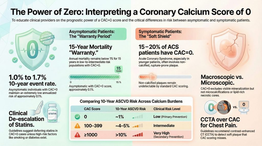
In de Multi-Ethnic Study of Atherosclerosis (MESA), ongeveer 50% asymptomatische volwassenen van middelbare leeftijd vertoonden bij aanvang een CAC-waarde van 0 [6]. Uit langdurige follow-up bleek dat de 10-jaars incidentie van ASCVD-voorvallen bij deze personen ongeveer 1–1,7% bedroeg, wat overeenkomt met een risico op jaarbasis van bijna 0,1% [6,7]. Deze eventpercentages zijn aanzienlijk lager dan voorspeld door traditionele risicomodellen bij veel personen met een gemiddeld risico.
Zelfs minimale verkalking (CAC 1–10) zorgt voor een ongeveer drievoudige toename in kransslagaderziekte risico vergeleken met CAC = 06]. Dit betekent dat CAC = 0 eerder een biologisch onderscheiden fenotype met een laag risico vertegenwoordigt dan simpelweg het laagste punt op een continuüm.
Getekend risico over CAC-categorieën
Het risico neemt stapsgewijs toe naarmate de calciumlast toeneemt7,8]:
- CAC = 0: ~1% 10-jaarsrisico
- CAC 1–99: ~1,91 TP9T 10-jaarsrisico
- CAC 100–399: ~4–5% 10-jaarsrisico
- CAC ≥400: ~7–91 TP9T 10-jaarsrisico
- CAC ≥1000: >10%, gericht op doelgroepen voor secundaire preventie
Individuen met een CAC ≥1000 vertonen cardiovasculaire mortaliteitscijfers die vergelijkbaar zijn met stabiele secundaire-preventiecohorten, waaronder cohorten vergelijkbaar met FOURIER-studie deelnemers8]. Deze waarneming ondersteunt agressieve cholesterolverlagende strategieën in extreme CAC-categorieën.
NNT-modellering en therapeutische toewijzing
CAC-scoring verfijnt statine allocatie-efficiëntie. Onder personen met een gemiddeld risico:
- CAC = 0: 5 jaar Benodigd aantal te behandelen patiënten (NNT) overschrijdt vaak 100
• CAC >100: 5-jaars NNT varieert vaak tussen 12–25 [1,9]
Het meeste absolute statinevoordeel komt ten goede aan patiënten met een meetbare gecalcificeerde last. 2018 ACC/AHA cholesterol guidelines recommend that statin therapy may be deferred in asymptomatic individuals with CAC = 0 unless diabetes, roken, or strong familiegeschiedenis is present [9].
Soft Plaque and “Warranty” Periods
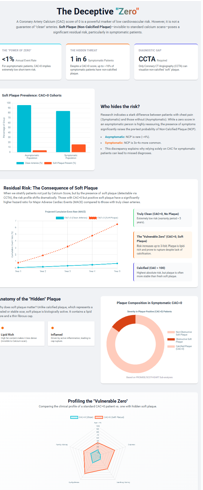
The Miami Heart Study demonstrated that approximately 16% of asymptomatic individuals with CAC = 0 have non-calcified plaque on CCTA [10]. However, obstructive stenosis (≥50%) was present in only 0.8%, and high-risk plaque features were uncommon. Thus, while soft plaque is detectable, it rarely reaches clinical significance in asymptomatic zero-CAC individuals.
A prospective study of 9,715 asymptomatic individuals demonstrated that CAC = 0 confers a 15-year mortality “warranty period” in low-to-intermediate risk populations [11]. Annual mortality remained below 1% during this interval. However, this warranty is shortened in diabetics and heavy smokers due to accelerated plaque progression [12].
Sex-Specific Nuance and Ethnic Variability
Women generally exhibit lower absolute CAC prevalence compared with men but may harbor a greater proportion of non-calcified plaque [13]. Consequently, while CAC = 0 remains prognostically powerful in women, symptomatic female patients may require more cautious interpretation. Women presenting with chest pain frequently demonstrate non-obstructive CAD or microvascular dysfunction, conditions not reliably captured by CAC scoring alone.
MESA demonstrated significant ethnic heterogeneity in CAC prevalence [6]. White individuals exhibit the highest CAC prevalence, followed by Hispanic, Black, and Chinese American populations. Despite differences in prevalence, event rates at comparable CAC levels are similar across ethnic groups, reinforcing that CAC = 0 confers prognostic reassurance across diverse populations.
The Symptomatic Population: Residual Risk
In patients presenting with stable chest pain, CAC = 0 does not exclude coronary disease. Approximately 7–16% demonstrate plaque on CCTA, and 1–2% have obstructive CAD ≥50% [14]. In de PROMISE trial, 16.5% of symptomatic zero-CAC patients had non-obstructive CAD, and 1.5% had obstructive disease [15].
Acute Coronary Syndrome and Zero CAC
Pooled analyses demonstrate that 15–20% of patients presenting with acuut coronair syndroom (ACS)—particularly younger individuals—may have CAC = 0 at presentation [16]. These findings reflect the predominance of non-calcified, rupture-prone plaque. Accordingly, the 2021 AHA/ACC Chest Pain Guideline recommends CCTA rather than CAC scoring when evaluating possible acute coronary syndrome [17].
Detailed Plaque Morphology: ICONIC Insights
De ICONIC trial quantified plaque components in symptomatic zero-CAC patients who subsequently developed ACS [4]:
- Fibrous plaque: 29.4 mm³ (ACS) vs 5.5 mm³ (controls)
• Fibrofatty plaque: 27.3 mm³ vs 1.3 mm³
• Necrotic-core plaque: 2.8 mm³ vs 0.0 mm³
These quantitative differences confirm that substantial vulnerable plaque burden may exist in the absence of detectable calcification.
Bayesian Interpretation and Risk Modifiers
Diagnostic performance depends on pre-test probability [18]. In asymptomatic individuals with low baseline risk, CAC = 0 reduces post-test probability to near-negligible levels. In symptomatic patients with higher pre-test probability, residual risk remains clinically meaningful despite a zero score.
Additional risk modifiers must be considered. Parental premature hart- en vaatziekten independently increases risk, even among individuals with CAC = 0 [19]. Furthermore, an elevated Atherogenic Index of Plasma (AIP) correlates with calcification burden and major adverse cardiovascular events independent of traditional risicofactoren [20].
Conclusion
The coronary slagader calciumscore of zero is among the most powerful negative risk markers in cardiovascular medicine. In asymptomatic individuals, it confers a prolonged mortality warranty and supports selective de-escalation of pharmacotherapy. In symptomatic patients, however, CAC = 0 represents a “soft shield”—associated with lower risk than positive scores but insufficient to exclude obstructive or vulnerable plaque. Future risk stratification must integrate calcified and non-calcified plaque assessment, clinical phenotype, and individualized risk modifiers rather than relying solely on binary calcium absence.
Referenties
- Hussain A, Ballantyne CM, Nambi V. Zero Coronary Artery Calcium Score: Desirable, but Enough?. Circulation. 2020;142(10):917-919. doi:10.1161/CIRCULATIONAHA.119.045026
- Czaja-Ziółkowska MZ, Wasilewski J, Gąsior M, Głowacki J. An update on the coronary calcium score: a review for clinicians. Postepy Kardiol Interwencyjnej. 2022;18(3):201-205. doi:10.5114/aic.2022.121035
- Vatsa N, Faaborg-Andersen C, Dong T, Blaha MJ, Shaw LJ, Quintana RA. Coronary Atherosclerotic Plaque Burden Assessment by Computed Tomography and Its Clinical Implications. Circ Cardiovasc Imaging. 2024;17(8):e016443. doi:10.1161/CIRCIMAGING.123.016443
- Jonas RA, Nurmohamed NS, Crabtree TR, et al. CTA-Derived Plaque Characteristics and Risk of Acute Coronary Syndrome in Patients With Coronary Artery Calcium Score of Zero: Insights From the ICONIC Trial. AJR Am J Roentgenol. 2025;225(1):e2431476. doi:10.2214/AJR.24.31476
- Hutcheson JD, Goettsch C, Bertazzo S, et al. Genesis and growth of extracellular-vesicle-derived microcalcification in atherosclerotic plaques. Nat Mater. 2016;15(3):335-343. doi:10.1038/nmat4519
- Detrano R, Guerci AD, Carr JJ, et al. Coronary calcium as a predictor of coronary events in four racial or ethnic groups. N Engl J Med. 2008;358(13):1336-1345. doi:10.1056/NEJMoa072100
- Budoff MJ, Shaw LJ, Liu ST, et al. Long-term prognosis associated with coronary calcification: observations from a registry of 25,253 patients. J Am Coll Cardiol. 2007;49(18):1860-1870. doi:10.1016/j.jacc.2006.10.079
- Dzaye O, Razavi AC, Michos ED, et al. Coronary artery calcium scores indicating secondary prevention level risk: Findings from the CAC consortium and FOURIER trial. Atherosclerosis. 2022;347:70-76. doi:10.1016/j.atherosclerosis.2022.02.006
- Grundy SM, Stone NJ, Bailey AL, et al. 2018 AHA/ACC/AACVPR/AAPA/ABC/ACPM/ADA/AGS/APhA/ASPC/NLA/PCNA Guideline on the Management of Blood Cholesterol: A Report of the American College of Cardiology/American Heart Association Task Force on Clinical Practice Guidelines. Circulation. 2019;139(25):e1082-e1143. doi:10.1161/CIR.0000000000000625
- Nasir K, Cainzos-Achirica M, Valero-Elizondo J, et al. Coronary Atherosclerosis in an Asymptomatic U.S. Population: Miami Heart Study at Baptist Health South Florida. JACC Cardiovasc Imaging. 2022;15(9):1604-1618. doi:10.1016/j.jcmg.2022.03.010
- Valenti V, Ó Hartaigh B, Heo R, et al. A 15-Year Warranty Period for Asymptomatic Individuals Without Coronary Artery Calcium: A Prospective Follow-Up of 9,715 Individuals. JACC Cardiovasc Imaging. 2015;8(8):900-909. doi:10.1016/j.jcmg.2015.01.025
- McClelland RL, Jorgensen NW, Budoff M, et al. 10-Year Coronary Heart Disease Risk Prediction Using Coronary Artery Calcium and Traditional Risk Factors: Derivation in the MESA (Multi-Ethnic Study of Atherosclerosis) With Validation in the HNR (Heinz Nixdorf Recall) Study and the DHS (Dallas Heart Study). J Am Coll Cardiol. 2015;66(15):1643-1653. doi:10.1016/j.jacc.2015.08.035
- Shaw LJ, Raggi P, Callister TQ, Berman DS. Prognostic value of coronary artery calcium screening in asymptomatic smokers and non-smokers. Eur Heart J. 2006;27(8):968-975. doi:10.1093/eurheartj/ehi750
- Wang X, Le EPV, Rajani NK, et al. A zero coronary artery calcium score in patients with stable chest pain is associated with a good prognosis, despite risk of non-calcified plaques. Open Heart. 2019;6(1):e000945. Published 2019 Apr 11. doi:10.1136/openhrt-2018-000945
- Foldyna B, Mayrhofer T, Lu MT, et al. Prognostic value of CT-derived coronary artery disease characteristics varies by ASCVD risk: insights from the PROMISE trial. Eur Radiol. 2023;33(7):4657-4667. doi:10.1007/s00330-023-09430-5
- Gottlieb I, Miller JM, Arbab-Zadeh A, et al. The absence of coronary calcification does not exclude obstructive coronary artery disease or the need for revascularization in patients referred for conventional coronary angiography. J Am Coll Cardiol. 2010;55(7):627-634. doi:10.1016/j.jacc.2009.07.072
- Gulati M, Levy PD, Mukherjee D, et al. 2021 AHA/ACC/ASE/CHEST/SAEM/SCCT/SCMR Guideline for the Evaluation and Diagnosis of Chest Pain: A Report of the American College of Cardiology/American Heart Association Joint Committee on Clinical Practice Guidelines. Circulation. 2021;144(22):e368-e454. doi:10.1161/CIR.0000000000001029
- Greenland P, Bonow RO, Brundage BH, et al. ACCF/AHA 2007 clinical expert consensus document on coronary artery calcium scoring by computed tomography in global cardiovascular risk assessment and in evaluation of patients with chest pain: a report of the American College of Cardiology Foundation Clinical Expert Consensus Task Force (ACCF/AHA Writing Committee to Update the 2000 Expert Consensus Document on Electron Beam Computed Tomography). Circulation. 2007;115(3):402-426. doi:10.1161/CIRCULATIONAHA..107.181425
- Lloyd-Jones DM, Nam BH, D’Agostino RB Sr, et al. Parental cardiovascular disease as a risk factor for cardiovascular disease in middle-aged adults: a prospective study of parents and offspring. JAMA. 2004;291(18):2204-2211. doi:10.1001/jama.291.18.2204
- Yao H, Feng G, Liu Y, Chen Y, Shao C, Wang Z. Coronary artery calcification burden, atherogenic index of plasma, and risk of adverse cardiovascular events in the general population: evidence from a mediation analysis. Lipids Health Dis. 2024;23(1):258. Published 2024 Aug 20. doi:10.1186/s12944-024-02255-1

