
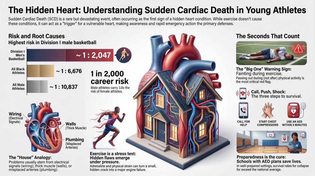
年轻人心脏性猝死
流行病学, ,疾病机制、诊断与预防
1. 范围与定义
心脏性猝死 传统上定义为:在有目击者的情况下,于症状发作后一小时内发生的意料之外的心源性猝死;或者在无目击者的情况下,在死者生前最后一次被看到健康存活后的24小时内发生的心源性猝死。. 心脏骤停 描述了恢复循环时的同一事件;心脏骤停与死亡的区别主要取决于反应的速度和质量,这就是为什么最好将这两者视为具有不同结果的单一临床实体,而不是两个独立的现象1].
本篇综述涉及大约35岁以下的人群。这一年龄分界线并非随意划定。在此年龄之上,冠状动脉 动脉粥样硬化 在病因学图景中逐渐占据主导地位,且临床问题也与成人趋于一致 缺血性心脏病. 在此之下,其病因主要为遗传性、先天性或获得性非动脉粥样硬化性疾病,相应的诊断和预防策略也有所不同。.
被研究最多的群体是竞技运动员,原因有三:他们有系统的名册记录,他们接受会产生数据的医学评估,且他们的死亡会被报道。这造成了显著的查明不对称性。从绝对数量来看,非运动员年轻人的突然死亡更为常见,但其特征却远未得到充分阐明;以下内容大多源自运动员队列,并以无法量化的精确度适用于普通年轻人群。.
2. 流行病学
2.1 发病率与测量问题
历史发病率估计值因方法学局限性而受到削弱,这些局限性使其系统性地偏低:依赖媒体报道和自愿登记系统、使用基于参与人数估计而非确切队列的分母推导出的不精确分母、宽泛且异质的年龄范围,以及未能区分劳力性事件与非劳力性事件 [2,3].
一项针对美国全国大学体育协会(NCAA)运动员、为期20年的多数据库监测研究弥补了大部分此类局限性。该研究涵盖了2002年至2022年期间共9,106,516个运动员年,采用四种独立病例确定策略,在总共1,102例死亡病例中识别出143例经裁定的心源性猝死(SCD)病例[4,5]. 没有任何单一来源报告的病例数超过82%——这一发现有助于解释为何此前的单一来源估计值较低。.
由此产生的总体发生率为 每 63,682 名运动员-年 1 例 (95% CI 1:54,065–1:75,010) [4].
相比之下,丹麦对运动员猝死(SCD)报告实行强制规定,其竞技运动员在运动期间或运动后一小时内发生事件的年发病率约为每10万中有1.2例。由于病例定义、运动归因和人群的不同,跨国家数据集的直接比较不可靠,且不同系列之间的明显差异往往反映的是确定方法,而非真正的风险差异。.
2.2 按性别、种族和运动项目进行危险分层
风险并非平均分布。NCAA的监测数据显示出明显且一致的梯度。.
表 1. 2002–2022 年 NCAA 运动员按人口统计学和运动分层的心源性猝死发生率 [4]
| 地层 | 发生率(每名运动员-年) | 95% CI | 4年职业生涯风险 | 比例与总体 |
| 一级男子篮球赛,白人 | 1:5,848 | 1:2,498–1:13,691 | ~1:1,462 | 10.9 |
| 一级男子篮球赛,黑人 | 1:7,696 | 宽阔的,重叠的 | ~1:1,924 | 8.3 |
| NCAA第一级别男子篮球,整体 | 1:8,188 | — | ~1:2,047 | 7.8 |
| 篮球,所有组别 | 1:19,164 | — | ~1:4,791 | 3.3 |
| 美式橄榄球,所有级别 | 1:31,743 | — | ~1:7,936 | 2.0 |
| 所有黑人运动员 | 1:26,704 | 1:20,417–1:34,925 | ~1:6,676 | 2.4 |
| 所有男运动员 | 1:43,348 | 1:36,228–1:51,867 | ~1:10,837 | 1.5 |
| 所有白人运动员 | 1:74,581 | 1:60,247–1:92,326 | ~1:18,645 | 0.85 |
| 所有女运动员 | 1:164,504 | 1:110,552–1:244,787 | ~1:41,126 | 0.39 |
| 总体队列 | 1:63,682 | 1:54,065–1:75,010 | ~1:15,921 | 1.00(参考) |
职业风险是源出版物中使用的简单的四倍年化近似值 [4].
性别. 男性运动员的发病率约为女性运动员的 3.8 倍(在二手资料中通常四舍五入为四倍)[4]。该解释尚未完全确立,但可能是多因素导致的:相对于身体大小而言更大的左心室质量和室壁厚度、自主神经反应和复极的差异、某些疾病的高患病率 致心律失常性基质, ,以及运动暴露强度和类型的差异6]。值得注意的是,通道病的性别差异较小——尤其是 长QT综合征, ,其中女性性别是 风险因素 对于青春期后的事件——比结构性心肌病更甚。.
种族. 黑人运动员的发病率约为白人运动员的2.8倍 [4]。这一发现需要谨慎解读。该文献中的种族是一个社会变量,作为一组未测量因素的不完善代理变量:这些因素包括不同运动项目和位置的参与度差异、体型、, 高血压 患病率、镰状细胞性状、社会经济医疗可及性、健康的结构性决定因素,以及可能与血统相关的心脏表型。当代指南明确将此背景下的种族视为一种社会政治构建,并呼吁在未来的研究中纳入健康的社会决定因素1]. 实际意义在于,“黑人种族”识别出风险升高,却没有解释原因,因此它是临床算法的糟糕基础。.
体育. 在所有研究过的运动项目中,篮球的风险最高,且这种风险具有独立性:在对性别和种族进行多变量调整后,参与篮球运动仍与风险升高相关(比值比 2.75,95% 置信区间 1.73–4.34)[4] 美式足球由于其庞大的阵容人数,贡献了绝对数量最多的病例。足球同样被认为是高发病率的项目之一。这些运动的共同特征——高动态、急停急起、重复的最大强度发力以及自主神经的急剧转换——很可能是机制性的而非偶然的,尽管这一假说尚未得到直接验证 [7,8].
一个经常被重复的错误值得纠正。在第一级别男子篮球中,白人球员的点估计值(1:5,848)超过了黑人球员(1:7,696)[4]。次要来源通常会颠倒这一点。这两个估计值都基于事件发生数量极少且范围广泛、相互重叠的情况 置信区间, ,并且篮球内部的排序不应被视为已经在任何一个方向上确定。.
2.3 时间趋势
在研究期间,NCAA运动员的SCD发病率每五年下降约29%(五年发病率比为0.71, 95% 置信区间 0.61–0.82),而同一人群中的非心血管死亡率则保持不变(发病率比 0.98,95% 置信区间 0.94–1.04)[4].
心血管死亡率下降的特异性具有提示意义,但该研究并非旨在确定其机制,其作者也未将这一趋势归因于任何单一干预措施。可能的促成因素包括在资源充足的项目中更广泛地使用心电图、改进的应急行动计划以及 自动体外除颤器 部署、对劳力性预警症状的更好识别以及病例确查的长期变化。厘清这些因素仍然是一个尚未解决的问题,不应将这一趋势引为任何特定预防策略有效性的证据。.
3. 病因:疾病谱
3.1 病因分布
在有充分信息以便进行分析的 143 起 NCAA 案件(共 118 起)中 裁决 [4]:
- 尸检阴性不明原因猝死: 19.5%
- 特发性左心室肥厚 或者可能 心肌病: 16.9%
- 肥厚型心肌病: 12.7%
- 提示:先天性冠状动脉 动脉 异常, 致心律失常性心肌病, 心肌炎, 主动脉夹层, ,以及其他原因
此分布与历史情况有实质性不同,在历史情况中,肥厚型心肌病被描述为单一的主要病因[2,3]。这一转变反映的是法医严谨性的提高,而不是疾病生物学本身的变化:标准化的专家仲裁将许多较少被识别的病例重新归类为特发性或原因不明。 系统评价 仅凭临界室壁厚度就归因于肥厚型心肌病。.
在更广泛的文献中,尸检未能确定结构性病因的病例比例约为10%至42%,这种差异主要取决于检查的彻底程度、病理学家的专业水平以及是否遵循了针对心脏的特定检查流程。.
对预防工作的启示是直接的且未得到充分认识的: 在这些数据中,经裁定占比最大的单一类别是未发现结构性异常。. 因此,仅关注结构性疾病的策略留下了相当大一部分未能解释或未被发现的事件。这一比例并非固定不变——它随法医鉴定方法、送检病理学家的专业知识以及是否 分子尸检 ——被执行,但非结构性和尸检阴性原因的重要性在各个系列中是一致的。.
3.2 为什么体力劳累会诱发事件
运动性猝死最好通过“基质-触发因素-调节因素”的框架来理解。易损的基质——解剖学、结构性或离子通道异常——是必要的,但往往是不够的。运动提供了将潜在易损性转化为 心室颤动 [7,8].
儿茶酚胺风暴. 运动会导致显著的交感神经激活,循环中的儿茶酚胺数倍上升。β-肾上腺素能刺激使整个……的不应期异质性缩短 心肌, ,增加自律性,并增强肌浆网的钙负荷。在钙处理异常的情况下——最典型的是儿茶酚胺敏感性多形性室性心动过速——这直接具有致心律失常作用 [8].
需求 缺血. 在冠状动脉解剖异常、心肌桥或严重的左心室流出道梗阻的情况下,运动诱发的心肌耗氧量增加无法得到供氧的匹配。缺血反过来会导致局部传导减慢和复极离散,这正是折返的基质。.
机械和血流动力学应力. 室壁应力增加、心腔扩张以及针对梗阻的强力收缩会急性牵拉心肌。机械电反馈——即由牵拉激活的离子通道改变膜电位——提供了一种将机械负荷与电不稳定性联系起来的合理解释机制,这可能与致心律失常性心肌病有关,在该疾病中,运动似乎既会加速病情进展,又会诱发发作。.
电解质和酸碱平衡失调。. 运动导致工作肌肉释放钾离子、细胞内酸中毒、容量耗竭,而在持久耐力运动中还会导致低钠血症。每种因素都会改变传导和复极化过程 [9].
自主过渡. 在运动停止时交感神经张力的突然撤除和迷走神经活性的激增会产生一段电不均一期。相当一部分与用力相关的事件发生在运动刚结束时而不是运动过程中,而篮球、足球和橄榄球的停顿交替结构可能会反复重现这些转变[8].
底物进展. 在某些情况下,运动不仅是诱因,更是疾病的驱动因素。由桥斑蛋白-2(plakophilin-2)变异引起的致心律失常性心肌病最能说明这一点,耐力运动量与更早的表型表达、更高的心律失常负荷以及结构进展相关[1,10].
3.3 心肌病
肥厚型心肌病. 由无法解释的定义所界定 左心室肥厚, 通常具有遗传性肌节基础,尽管约有一半的患者没有可识别的致病变异。表型、临床病程和心律失常风险差异很大。猝死的机制包括由心肌细胞排列紊乱和间质纤维化引起的心室性心律失常、来自 微血管功能障碍, ,以及由动态流出道梗阻导致的血流动力学崩溃。风险分层结合了最大室壁厚度,, 家族史 猝死、原因不明的晕厥、非持续性室性心动过速、左心房大小、流出道压差、心尖动脉瘤以及……的范围 钆迟增强 在心脏磁共振上。携带 致病性变异 没有肥厚性表型的心律失常风险低1].
致心律失常性心肌病. 其特征为心室功能障碍——右心室、左心室或双心室——伴有与心腔扩张或收缩功能受损程度不相称的心室心律失常负荷。大多数已确定的变异影响桥粒 蛋白质, ,其中致arrhythmogenic right ventricular cardiomyopathy基因(注:上下文未明时按通常医学语境,此处省略了主语)中以桥粒斑菲素蛋白-2(plakophilin-2)最为常见,尽管约有一半的病例基因型为阴性。基因型显著改变了风险:由桥粒斑菲素蛋白-2介导的疾病表现出明显的运动相关加速进展,且参与耐力运动会增加心律失常风险,而对于非桥粒斑菲素蛋白-2介导及基因型阴性的疾病,是否存在类似风险尚无定论。10区分早期的致心律失常性心肌病与耐力训练运动员的右心室扩张是运动心脏病学中最困难的问题之一1].
扩张型心肌病. 左心室或双心室扩张伴收缩功能障碍,约60%基因型呈阴性。心律失常风险随以下情况的降低而增加: 射血分数, ,症状和瘢痕负担,在特定的基因亚型(A/C型核纤层蛋白、桥粒斑蛋白和伴肌动蛋白-C)中高得不成比例,无论射血分数如何,这些亚型都需要更密切的监测。初步证据表明,在A/C型核纤层蛋白相关疾病中,更高的累积终生运动暴露与较低的射血分数相关,这提出了运动既会诱发心律失常又会促进病情进展的可能性[1].
左心室高小梁化. 肌小梁显著伴深陷的肌小梁间隐窝,既往称为左心室心肌致密化不全。它不再被认为是一种独立的心肌病。在无共存的肥厚性或扩张性表型、室性心律失常或症状的情况下,尚未证明会发生不良事件,因此无症状者的孤立性过度肌小梁化最好被视为一种形态学变异 [1].
3.4 通道病
遗传性心律失常综合征可在心脏结构正常的情况下导致猝死,是尸检阴性猝死的一个重要的可识别原因。.
长 QT 综合征. 室性复极延迟,易诱发尖端扭转性室性心动过速。基因型可预测诱因:LQT1 (KCNQ1) 事件通常由体力活动诱发,游泳是一个特征性诱因;LQT2 (KCNH2) 事件与听觉惊吓、情绪和产后时期相关;LQT3 (SCN5A) 事件主要在静息或睡眠时发生。诊断依据校正的 QT 间期、症状史、家族史和基因检测。有两个临床上重要的注意事项:运动员的 QTc 阈值与一般人群不同,且 QT 间期可能会间歇性恢复正常,因此单次正常的静息 QTc 不能排除诊断。隐匿性长 QT 综合征——基因型阳性但 QTc 持续正常——见于相当一部分变异基因携带者 [1].
儿茶酚胺敏感性多形性室性心动过速. 最常见的原因是ryanodine受体(RYR2)变异,在肾上腺素能应激下产生异常的舒张期钙释放。它是 通道病 最具体地与体力 exertion 相关,通常在运动期间的可重复心率阈值下产生双向或多形性室性心动过速。静息心电图通常是正常的,这使得该病无法通过静息心电图筛查发现。对于临床怀疑 CPVT 的患者,运动试验具有很高的诊断率,通常会在一致的心率阈值下重现心律失常,并且对于有劳力性晕厥且静息心电图正常的患者来说,这是合适的检查方法。1].
布鲁加达综合征. 右胸导联呈穹隆型 ST 段抬高为特征,少数病例与 SCN5A 变异相关。发作主要发生于静息状态、睡眠中或发热时,而非运动期间,尽管伴随运动的发热是一个已知的诱因 [1].
预激综合征图形. 附加房室旁路。发生猝死的情况为 心房颤动 沿着具有短不应期的传导径路快速传导,并退化为心室颤动。与此处讨论的大多数病因不同,这是一种易于检出且可明确治疗的疾病,静息心电图具有诊断价值 [1].
3.5 先天性冠状动脉畸形
冠状动脉异位起源是导致运动中猝死的主要原因之一,也是常规筛查中最不易被检测出的原因之一。.
风险最高变异是左冠状动脉异常起源于右窦,且在动脉间走行,位于 主动脉 以及肺动脉。建议的高危特征包括主动脉壁内壁段、狭缝状近端开口、锐角起步以及较长的壁内长度。其机制被认为涉及运动时随着大血管扩张而产生的动态压迫和开口畸变,从而产生间歇性缺血,在发作间期可能不留任何固定的异常 [1].
右冠状动脉异常起源伴动脉间走行更为常见且风险较低;许多受累个体无症状。其他变异——室间隔内、主动脉后、肺动脉前走行——通常是良性的。.
临床上面临的挑战是,受累患者在发生首发事件之前通常无症状,静息心电图通常正常,且即使在高危解剖结构的情况下,运动负荷试验也可能无法诱发出缺血。明确诊断需要进行解剖学成像,最常采用的是冠状动脉计算机断层扫描血管造影或心脏磁共振检查 [1].
心肌桥, 冠状动脉肌桥是指一段冠状动脉穿行于心肌之中,这是一种常见且通常为偶发的解剖变异。只有当肌桥较深或较长且引起可证实的局部缺血时,它才具有临床意义。1].
3.6 心肌炎和获得性疾病
心肌炎通过急性心律失常导致猝死 炎症, ,水肿以及随后的纤维化。其重要性部分在于它可能是暂时性的:在心肌炎性活动期进行剧烈运动具有致心律失常性,但这种风险会随着炎症的消退而大幅降低。这正是标准建议的核心所在,即在症状和炎症的客观证据消失之前避免劳累。新兴的心脏磁共振数据表明,经过筛选的运动员或许可以比早期指南假设的3至6个月间隔期更早安全复出,尽管证据基础仍然有限,且复出应以炎症的客观消退为依据,而不仅仅取决于过去的时间 [1,11].
主动脉病变. 马凡综合征, 洛伊斯-迪茨综合征, ,血管型埃勒斯-当洛斯综合征和二叶式主动脉瓣相关的主动脉病变易诱发急性主动脉夹层。这是年轻人猝死的一种罕见原因,但其独特之处在于可以通过体格检查和影像学检查发现,并且其风险与主动脉尺寸相关,从而允许采取基于阈值的管理方法。显著的主动脉扩大在年轻运动员中很罕见——无论体型如何,男性直径大于 42 毫米、女性直径大于 40 毫米都是不寻常的——因此发现此类情况需要评估潜在的主动脉病变,而不是将其归因于训练。1].
心律失常性二尖瓣脱垂. 大多数二尖瓣脱垂是良性的,但有一个亚群——特征性地表现为双叶脱垂伴二尖瓣环分离、下外侧壁延迟钆增强以及复杂性室性期前收缩——与猝死相关。乳头肌牵拉及随之而来的局部纤维化提供了一个合理的致心律失常基质 [1].
心脏震荡. 在复极易损期受到胸前钝性撞击导致结构正常的的心脏发生心室颤动。这是一种机械现象而非疾病现象,不受任何筛查策略的影响,通过即时除颤可以存活[1].
镰状细胞性状. 与劳力性虚脱和死亡相关,特别是在炎热环境下进行高强度体能训练期间,其机制涉及劳力性 横纹肌溶解症 而是代谢紊乱而非原发性心律失常。此处将其收录是因为它表现为运动中的突然晕厥,与鉴别诊断和预防方案相关 [1].
3.7 遗传架构:外显率、修饰基因以及基因型的局限性
上述大多数疾病都是遗传性的,但在这一领域,遗传的表现远不像“遗传性心脏病”这个词所暗示的那样具有决定性,而基因型与结果之间的差距正是许多临床难点所在。.
外显不全和变异度表达. 携带致病变异并不一定会可靠地导致疾病,即便致病,在拥有相同变异的家族内部,其严重程度也存在很大差异。通过临床确诊家族(因有人发病而被识别)得出的外显率估计值,在应用于偶然发现或通过人群筛查发现的变异时,会大大夸大风险。来自未筛选队列的估计值则持续偏低。这带来了直接的后果:对于肥厚型心肌病基因型阳性但表型阴性的个体而言,其心律失常风险较低,受到的对待与已表现出表型的患者有很大不同 [1].
年龄依赖性表达. 肥厚型、致心律失常型和扩张型心肌病的表型通常在青春期和成年早期显现,而不是自出生时即存在。14岁时的正常评估并不能排除24岁时患病的可能。这正是基因型阳性亲属需要进行连续评估而非单次筛查即告清除的原因,这也解释了为什么随访时间足够长的筛查队列最终会记录到曾通过筛查且结果正常者的死亡病例。.
修饰基因与多基因背景。. 相同的变异在不同的遗传背景下会产生不同的表型。具有单独微小效应的常见变异通过多基因评分进行聚合,似乎会改变心肌病的透显率和严重程度——这可能解释了单基因模型无法解释的部分家族内变异性。这项工作比冠心病领域的同类文献处于更早的阶段,多基因评分在此情况下尚不具备临床可操作性,但其发展方向是一个模型:其中罕见的致病变异设定了易感性,而常见变异、环境以及训练负荷决定了疾病是否以及何时出现。.
基因型阴性疾病. 大约一半的肥厚型和心律失常型心肌病病例,以及约60%的扩张型心肌病病例,均未发现可确定的致病变异。患者基因检测结果呈阴性并不排除遗传性疾病,也不应因此省略对亲属的临床筛查。.
临床意义未明变异. 这些是在缺乏明确表型时广泛基因 panel 检测最常见的结果,并且它们经常被误解为中等风险的结果。其实不然:在有待进一步明确之前,它们是无信息价值的 重新分类, ,随着证据的积累,这种情况可能双向发生。在没有表型或家族史的个体中进行检测主要会产生这类结果,这是反对将基因检测作为主要筛查手段的主要论据 [1].
3.8 尸检阴性不明原因猝 sudden unexplained death 和分子尸检
当包含毒理学和组织学检查在内的全面尸检仍未查明死因时,该死因被归类为尸检阴性不明原因猝死,在某些研究系列中也称为心脏性猝死综合征。这一分类是暂时的,而非最终定论。.
尸检基因检测在相当一部分病例中发现了具有临床干预价值的致病性或可能致病性变异。 将现行的ACMG分类标准应用于302例经专家裁定的病例,在13%名死者中发现了可采取临床干预措施的变异,主要涉及儿茶酚胺能多形性室性心动过速和长QT综合征,其中RYR2基因与这些疾病的关系最为密切 [12]. 如果进行尸检,则在相当大比例的家系中可确立具有临床意义的诊断。 遗传学 并结合了对幸存亲属的临床评估。.
各系列报告的检出率差异显著——从低于4%到约30%不等——且在很大程度上取决于所采用的变异分类框架。严格遵循ACMG标准的系列报告的检出率较低;而采用较宽松阈值的系列报告的检出率较高,这主要是因为它们将意义未明的变异计入诊断性变异。 因此,除非分类标准一致,否则跨系列比较并不可靠 [12].
有三点需要说明。首先,真正原因不明的死亡比例低于尸检结果阴性率所显示的水平。其次,分子尸检需要妥善保存标本,这取决于法医的实践,而往往并未实施。第三,也是临床上最重要的一点,对死者的确诊能够对在世的亲属进行级联筛查——这使得死后评估成为一项针对整个家庭的预防策略,而不仅仅是确定死因。.
4. 运动员心脏:适应与疾病
持续训练产生的的心脏重构,在表型上与上述疾病有所重叠。区分适应性变化与病理性改变是该领域的核心诊断难题,而误判则会带来双向的代价。.
4.1 结构适应
Endurance training produces predominantly 离心性重塑 — chamber enlargement with proportionate wall thickening — driven by sustained volume loading. Strength training produces relatively more concentric change. Most athletes exhibit a mixed pattern reflecting the actual demands of their sport.
Adaptation is modified by sport, sex, body size, ethnicity, and training duration. Left ventricular wall thickness in the 13–15 mm range in men, and right ventricular dilation in endurance athletes, fall into diagnostic gray zones overlapping hypertrophic and arrhythmogenic cardiomyopathy respectively. Left ventricular end-diastolic dimensions of 60 mm or more occur in a meaningful minority of trained athletes without valvular or myocardial disease [1,11].
Features favoring physiological adaptation include symmetric enlargement of all four chambers, normal or supranormal diastolic function, absence of late gadolinium enhancement, normal functional capacity, appropriate 血压 response to exercise, and regression with detraining. Features favoring pathology include asymmetric hypertrophy, impaired diastolic function, late gadolinium enhancement, a family history of cardiomyopathy or premature sudden death, and marked ECG abnormality out of proportion to the structural findings [1].
4.2 Electrical adaptation
Training produces a reproducible set of ECG changes: sinus bradycardia and sinus arrhythmia from increased 迷走神经张力, ectopic atrial and junctional rhythms, first-degree and Mobitz type I atrioventricular block, incomplete right bundle branch block, isolated voltage criteria for ventricular hypertrophy, and early repolarization. These are physiological, require no evaluation in an asymptomatic person without concerning family history, and their misclassification as pathological was the principal historical barrier to ECG-based screening [13].
Remodeling and repolarization vary by ethnicity, not only between Black and White athletes. Athletes of African and Afro-Caribbean descent show greater left ventricular wall thickness for a given body size and a distinctive repolarization pattern — J-point elevation with convex ST-segment elevation followed by T-wave inversion confined to leads V1–V4 — which is benign. Applying criteria derived from White European cohorts without this accommodation more than doubles the false-positive rate in Black athletes [14]. Distinct patterns have also been described in athletes of Middle Eastern, South Asian, and East Asian descent, and normative data for these groups remain comparatively sparse — a gap that propagates directly into screening performance wherever population-specific reference values do not exist.
Age matters similarly: T-wave inversion in leads V1–V3 in athletes aged 16 or younger represents a persistent juvenile pattern rather than disease [13].
5. Clinical Presentation and Warning Symptoms
The defining clinical feature of these conditions is that most affected individuals are asymptomatic until the index event. Where symptoms do occur, they are frequently attributed to deconditioning, dehydration, anxiety, or normal exertional limitation — by the individual, by coaching staff, and often by clinicians.
Symptoms warranting evaluation before further participation:
Exertional syncope or near-syncope. The single most important warning symptom. Syncope during exertion, as distinct from immediately after cessation, should be presumed cardiac until proven otherwise. Post-exertional syncope is more often neurally mediated but does not exclude cardiac causes.
Exertional chest pain. Particularly if reproducible at a consistent workload, suggesting demand ischemia from anomalous coronary anatomy or outflow obstruction.
Exertional dyspnea disproportionate to conditioning, especially where it represents a change from established baseline capacity.
Palpitations with exertion, particularly abrupt in onset and offset, or associated with lightheadedness.
Unexplained decline in performance not attributable to training load, illness, or injury.
The family history is as informative as the personal history: premature sudden death before age 50 in a 一级亲属, known inherited cardiomyopathy or arrhythmia syndrome, unexplained drowning, single-vehicle accidents without explanation, or unexplained seizure disorder — the last because long QT syndrome and CPVT events are frequently misdiagnosed as epilepsy [1,15].
6. Diagnostic Evaluation
6.1 Electrocardiography and the evolution of interpretation criteria
The resting 12导联心电图 detects the electrical signatures of cardiomyopathy and channelopathy, and is the single most informative low-cost test in this population. Its historical limitation was not sensitivity but specificity: applying general-population criteria to trained athletes generated false-positive rates that made systematic use impractical.
Interpretation criteria have been refined substantially over fifteen years.
Table 2. Evolution of athlete ECG interpretation criteria
| Criteria set | 年 | False positives, White athletes | False positives, Black athletes | Principal change |
| ESC recommendations [16] | 2010 | 16.2% | 40.4% | Training-related versus unrelated dichotomy; no ethnicity-specific criteria; most T-wave inversion classified abnormal |
| Seattle Criteria [17] | 2013 | 7.1% | 18.4% | Black athlete repolarization pattern recognized as physiological; QTc thresholds raised to ≥470 ms (male) and ≥480 ms (female) |
| Refined Criteria [14] | 2014 | 5.3% | 11.5% | Isolated axis deviation and isolated atrial enlargement reclassified as non-triggering |
| International Criteria [13] | 2017 | ~1.3–3.0% | ~4.2–6.8% | Formal normal/borderline/abnormal categories; two or more borderline findings required to trigger evaluation |
The 2017 International Criteria define exactly five borderline findings: left axis deviation, right axis deviation, 左心房增大, right atrial enlargement, and complete right bundle branch block [13]. Any single one of these in isolation does not warrant evaluation in an asymptomatic athlete without concerning family history; two or more do. Isolated voltage criteria for left or right ventricular hypertrophy are classified as normal, not borderline — a point frequently misstated.
Two caveats matter. First, the reported false-positive rates derive from expert or specialist-supervised interpretation; rates under non-specialist reading are consistently higher, and the magnitude of that gap has not been established at scale. Second, gains in specificity have not been free: in masters and pediatric cohorts, the International Criteria have missed diagnoses that earlier criteria would have flagged, including dilated cardiomyopathy presenting with isolated left axis deviation.
6.2 Diagnostic versus screening sensitivity
An important distinction is frequently elided. Diagnostic sensitivity is the probability that a person with established, fully expressed disease has an abnormal ECG. Screening sensitivity is the probability that an asymptomatic adolescent with early, incomplete, or concealed expression is correctly identified in a mass-screening setting.
The second is systematically lower than the first. Adolescent phenotype is often immature — both hypertrophic and arrhythmogenic cardiomyopathy express progressively — dynamic conditions are intermittently normal at rest, and field acquisition and interpretation conditions are inferior to those of a referral laboratory. Sensitivity figures quoted from referral cohorts of patients with known disease, commonly cited near 98% for hypertrophic cardiomyopathy, should not be presented as screening performance [18].
The most instructive evidence comes from long-term follow-up of a screened cohort. Among 11,168 adolescent English Football Association players screened with questionnaire, examination, ECG, and echocardiography, conditions associated with sudden death were identified in 0.38%, with ECG abnormal in 86% of those individuals versus 7% for history and 5% for physical examination [19]. Over a mean 10.6 years of follow-up, however, eight athletes died of cardiac causes — six of whom had screened normal at age 16, at a mean of 6.8 years after screening, most from cardiomyopathies not detectable at the time of testing.
A normal screen is not durable clearance. This is a property of progressive disease, not a failure of the test.
6.3 Secondary evaluation
An abnormal primary screen or concerning symptom initiates further testing, selected by the suspected condition rather than applied as a fixed panel.
Transthoracic echocardiography assesses wall thickness, chamber dimensions, systolic and diastolic function, valvular structure, aortic dimensions, and in many cases coronary origins.
Cardiac magnetic resonance provides superior tissue characterization. Late gadolinium enhancement identifies fibrosis and scar; T2-weighted imaging and parametric mapping identify edema and active inflammation. It is often decisive in distinguishing physiological hypertrophy from cardiomyopathy, in evaluating the right ventricle for arrhythmogenic cardiomyopathy, and in diagnosing myocarditis. Late gadolinium enhancement confined to right ventricular insertion points is a recognized finding without established adverse prognostic significance.
Exercise testing should be sport-specific and designed to provoke symptoms at the intensity actually encountered in competition, rather than terminated at arbitrary heart-rate targets or performed pharmacologically. It is essential in suspected CPVT, in evaluating ventricular ectopy, and in assessing ischemia in anomalous coronary anatomy.
Ambulatory rhythm monitoring characterizes arrhythmia burden and its relationship to activity; extended monitoring is often required given the intermittency of clinically relevant events.
Coronary imaging by CT angiography or magnetic resonance is required to define coronary origin and course when anomalous anatomy is suspected.
Genetic testing confirms diagnosis where the phenotype is established, informs risk stratification in specific conditions — plakophilin-2 in arrhythmogenic cardiomyopathy, lamin A/C in dilated cardiomyopathy, genotype in long QT syndrome — and enables cascade screening of relatives. It performs poorly as a primary diagnostic test in the absence of phenotype or family history, where it principally generates variants of uncertain significance.
Notably, cardiac imaging, exercise testing, and ambulatory monitoring have insufficient evidence to support their use as primary screening tools in asymptomatic individuals [1]. Their role is in secondary evaluation.
6.4 Emerging technologies
Artificial intelligence applied to the ECG. Deep learning models trained on large ECG corpora can identify conditions that lack a 特征性的 signature to the human eye. Convolutional neural networks have been developed to detect hypertrophic cardiomyopathy from the 12-lead ECG alone, with performance maintained on external validation across diverse international cohorts and in pediatric and adolescent populations [20]. Comparable models identify electrocardiographically concealed long QT syndrome — genotype-positive individuals with a normal measured QTc — which conventional interval measurement cannot detect by definition [21].
The potential relevance to this field is obvious: the principal limitation of ECG screening is not cost but interpretive accuracy, and an algorithm that improves specificity without sacrificing sensitivity would change the calculus directly. Several important caveats apply. Models trained predominantly on clinical populations may perform differently in trained athletes, whose baseline ECGs differ systematically from those of the general population, and athlete-specific validation remains limited. Performance in the demographic groups with the highest false-positive rates under conventional criteria has not been separately established. And an algorithm that flags disease without an accessible pathway to secondary evaluation reproduces the equity problem described in Section 7.5 rather than solving it. AI-ECG is best understood at present as a promising adjunct under active validation rather than an established screening tool.
Wearable and consumer devices. Smartwatches, adhesive patch monitors, and consumer single-lead and multi-lead ECG devices are now widely used by athletes, and increasingly generate cardiac data that reaches clinicians unsolicited. Their genuine strengths are duration and opportunism: a patch monitor worn for two weeks or a smartwatch worn continuously may capture a symptomatic paroxysmal arrhythmia that a resting ECG and a 24-hour Holter both miss, and this is a real diagnostic contribution in a person with intermittent palpitations.
Their limitations are equally clear. Single-lead recordings cannot assess axis, chamber enlargement, repolarization across the precordium, or most of the criteria on which athlete ECG interpretation depends. Automated rhythm classification is optimized for atrial fibrillation detection in older populations and performs poorly for the arrhythmias relevant here. Signal quality during exercise — precisely when it would be most valuable — is frequently inadequate. And the false-positive burden generated by consumer devices in young, healthy, highly motivated users is substantial and largely unquantified.
Wearables complement rather than replace structured evaluation. A consumer device recording during a symptomatic episode is valuable evidence; a normal consumer recording is not clearance, and should not be treated as one by the athlete or the clinician.
7. Screening: What It Achieves and What It Does Not
Screening is one component of prevention. This section assesses it on its own terms rather than treating it as the organizing question of the field.
7.1 The rationale and its limits
Preparticipation cardiovascular screening aims to identify people with unrecognized disease in time for management that reduces risk. Its effectiveness depends on a chain of conditions: the disease must be detectable before the event, the test must detect it at acceptable cost and false-positive burden, effective management must exist, and affected individuals must have access to it.
That chain has weak links. Resting ECG has very limited sensitivity for anomalous coronary origin, catecholaminergic polymorphic ventricular tachycardia, early or concealed arrhythmogenic cardiomyopathy, aortopathy, and significant valve disease, and by definition contributes nothing in cases that prove autopsy-negative — the largest single category. Screening addresses a subset of the problem.
7.2 Comparative performance of screening components
Meta-analytic estimates in athletic populations give ECG a sensitivity approaching 90–94% for conditions detectable by ECG, with specificity near 93%, against approximately 20% sensitivity for history and 9% for physical examination [18]. Evidence drawn specifically from NCAA populations has since been synthesized separately [22]. The qualifier matters: this figure describes performance against cardiomyopathies and channelopathies with electrical signatures, not against the full spectrum of causes, and should not be read as 94% sensitivity for sudden death risk overall. Contemporary guidance cites a comparable range, placing history and physical sensitivity for silent cardiac conditions at 10–20% and noting that adding ECG raises the sensitivity of the preparticipation evaluation to approximately 94% [1].
The argument for ECG is therefore not that it performs well in absolute terms; it is that history and physical examination alone detect only a minority of relevant conditions. This is not an argument against the history and physical examination, which identifies symptomatic individuals, elicits family history, and detects Marfan stigmata, pathological murmurs, diminished femoral pulses, and hypertension — none of which an ECG reliably captures.
7.3 Guideline positions
European guidance has long recommended ECG-inclusive screening [11,16]. North American guidance historically endorsed a standardized 14-point history and physical examination without recommending universal ECG [15].
That divide has narrowed substantially. The 2025 American Heart Association / American College of Cardiology scientific statement holds that inclusion of a resting 12-lead ECG is reasonable, because it improves detection of underlying cardiac conditions in asymptomatic athletes relative to history and physical examination alone. The endorsement carries three conditions: clinicians must be adequately trained in contemporary athlete-specific interpretation criteria; programs must ensure access to secondary evaluation, including the financial and logistical resources for systematic downstream assessment; and because no approach provides absolute protection, an 应急行动计划 must be in place wherever people train and compete [1].
The unresolved questions are therefore not whether ECG may be used, but whether it should be universally implemented, how programs should be resourced to use it safely, and whether incremental benefit justifies downstream cost and harm.
7.4 Health economics
Published cost-effectiveness estimates for adding ECG to 体检前筛查 span a wide range, driven more by assumption choice than by data.
Table 3. Published economic evaluations of ECG-inclusive screening
| Analysis | Comparison | 结果 |
| Wheeler et al. (2010) [23] | ECG + H&P vs. H&P alone | $42,900 per life-year saved (95% CI $21,200–$71,300) |
| Wheeler et al. (2010) [23] | ECG + H&P vs. no screening | $76,100 per life-year saved ($62,400–$130,000) |
| Schoenbaum et al. (2012) [24] | H&P then ECG vs. H&P alone | $68,800 per QALY |
| Schoenbaum et al. (2012) [24] | ECG alone vs. H&P alone | $37,700 per QALY |
| Halkin et al. (2012) [25] | National program, US extrapolation | $10.6–14.4 million per life saved |
Two of the three principal analyses return figures within commonly cited US willingness-to-pay thresholds. The third, returning figures orders of magnitude higher, does so partly by loading recurring annual history and physical costs into the ECG arm — an accounting choice contested in subsequent literature [25].
Every one of these analyses is governed by a single assumption: the 相对危险度 reduction conferred by detection and subsequent management. That parameter is not well established, and as discussed in Section 8 the evidence underlying it has shifted.
7.5 Equity
Screening programs can widen the inequities they are intended to reduce. The mechanism is the secondary evaluation, not the primary screen.
An abnormal screening result initiates a diagnostic cascade — echocardiography, cardiac magnetic resonance, ambulatory monitoring, genetic evaluation, subspecialty consultation — that in unassisted settings the family must fund. Uninsured and underinsured individuals face two consequences: they may exit the pathway without completing evaluation, continuing to participate with uncharacterized risk; and their absence from outcome data systematically biases estimates of disease prevalence, specificity, and cost-effectiveness in a favorable direction.
This is compounded by the ethnic disparity in false-positive rates. Even under contemporary criteria, false-positive findings remain more frequent among Black athletes [14], meaning that the population most likely to be referred for costly secondary evaluation overlaps with the population least likely to be able to afford it. Contemporary guidance states directly that screening programs without appropriate downstream resources have the potential to harm athletes from underrepresented racial and ethnic groups [1].
A screening program without a funded pathway to secondary evaluation is not a neutral intervention. It is a mechanism for identifying risk in people who cannot then act on it.
7.6 Psychological consequences
Screening programs are evaluated almost exclusively on detection and cost. The psychological consequences are real, are borne disproportionately by people who turn out not to have disease, and are rarely measured.
The false-positive interval. Between an abnormal screening result and its resolution, an athlete is a person who has been told their heart may be dangerously abnormal. That interval is frequently weeks and sometimes months, determined by appointment availability, insurance authorization, and imaging capacity rather than by clinical urgency. Documented consequences include anxiety, intrusive thoughts about dying during exertion, sleep disruption, and withdrawal from training during a period in which no restriction has actually been imposed. Where the athlete’s identity is substantially organized around sport — as it is for many at collegiate and elite level — the threat is to selfhood as much as to health.
Resolution is incomplete. Reassurance after a negative workup does not reliably return people to baseline. Residual health anxiety, continued symptom vigilance, and reduced training intensity have been described persisting after formal clearance, a pattern familiar from other screening contexts. Because false positives outnumber true positives by a large factor at any plausible operating point, this is not a marginal harm affecting a handful of people; it is the modal experience of an abnormal screen.
True positives carry their own burden. A diagnosis in a young person may bring restriction or modification of participation, loss of athletic identity, disrupted scholarship or professional prospects, defibrillator carriage in adolescence, and the knowledge of inherited risk extending to siblings and future children. Depression and anxiety are recognized sequelae of disqualification, and the transition out of competitive sport is itself a period of elevated psychological risk independent of the cardiac diagnosis.
Implications. Three follow directly. Time from abnormal screen to definitive resolution is a clinically meaningful quality metric, not merely an operational one, and shortening it is a genuine intervention. Communication of an abnormal result should convey the base rate — that most abnormal screens resolve without disease — rather than deferring all interpretation to the specialist. And psychological support should be planned into screening programs rather than improvised, particularly for athletes who are restricted or who transition out of competition.
8. Management and Sports Participation
8.1 The shift from disqualification to shared decision-making
Historical practice restricted individuals with identified 心血管疾病 from competitive sport more or less categorically. That paradigm has been substantially abandoned, on both ethical and empirical grounds [1,11].
The empirical case rests on outcome data:
- In a prospective multinational 观察性研究 of individuals with hypertrophic cardiomyopathy — 1,534 patients plus 126 genotype-positive, phenotype-negative individuals across 42 centers — those engaging in vigorous exercise, including competitive athletes, did not experience higher rates of death, resuscitated arrest, appropriate defibrillator shock, or arrhythmic syncope than moderate exercisers or sedentary participants [26].
- A prospective multinational registry of athletes with implantable cardioverter-defibrillators found no deaths, resuscitated arrests, or arrhythmia-related injuries during sport over long-term follow-up [27].
- Contemporary cohorts of elite athletes with genetic heart disease who returned to competition under expert supervision report low rates of breakthrough events [28].
The ethical case is that the historical model rested on the premise that athletes cannot make informed decisions about their own risk — a position that is neither supported by evidence nor consistent with the standards applied elsewhere in medicine.
Shared decision-making does not imply equal risk across conditions. It is a process for incorporating an individual’s values into a decision under uncertainty, not a conclusion that all diagnoses carry comparable danger or that all participation requests should be accommodated. The risk attached to genotype-positive, phenotype-negative status differs by orders of magnitude from that attached to plakophilin-2-mediated arrhythmogenic cardiomyopathy in an endurance athlete, and the framework is designed to make that difference explicit rather than to dissolve it.
The 2025 AHA/ACC statement is explicit that it does not issue disqualification recommendations, but rather clinical considerations to inform shared decision-making. Under this framework a uniform approach of restriction should not be applied to individuals with cardiomyopathy; participation is instead determined through a process incorporating accurate diagnosis, condition-specific risk stratification, guideline-directed treatment, disclosure of known and unknown risks, and the individual’s own values and risk tolerance. For those under 18, parents or guardians participate directly [1].
8.2 Where risk remains prohibitive
Shared decision-making does not mean that all participation is endorsed. Situations where risk is understood to outweigh benefit include arrhythmogenic cardiomyopathy caused by plakophilin-2 variants, particularly with endurance sport; active myocarditis or pericarditis; unrepaired anomalous origin of the left coronary artery with an interarterial course; severe symptomatic 主动脉瓣狭窄; heritable thoracic aortic disease with aortic dilation; and prior aortic dissection. Participation is also generally deferred during diagnostic evaluation and until guideline-directed therapy is optimized [1].
8.3 Condition-specific management
Management is disease-specific and is the mechanism by which detection produces benefit: beta-blockade in long QT syndrome and CPVT, with flecainide and sympathetic denervation in selected CPVT cases; defibrillator implantation where risk stratification indicates, though never solely to enable sport participation; 导管消融术 for accessory pathways and selected ventricular arrhythmias; surgical reimplantation or unroofing for high-risk coronary anomalies; septal reduction for obstructive hypertrophic cardiomyopathy; aortic surgery at guideline thresholds; and exercise prescription modification where disease progression is exercise-associated [1].
Two points deserve emphasis. First, individuals who discontinue competitive sport should be counseled on the established health benefits of continued recreational physical activity — a transition, not a cessation. Second, longitudinal surveillance is required regardless of the participation decision, because phenotype evolves and the original decision may require revisiting.
8.4 Return to play and longitudinal surveillance
A participation decision is a point on a trajectory rather than a conclusion, and the surveillance that follows is what makes continued participation defensible.
Serial imaging at intervals determined by condition and rate of change. Genotype-positive, phenotype-negative individuals in conditions where exercise may precipitate phenotypic conversion — plakophilin-2-mediated arrhythmogenic cardiomyopathy most clearly — warrant close longitudinal surveillance, with imaging intervals individualized to phenotype, exercise exposure, and evidence of disease progression, while continuing to compete [1]. Aortic dimensions in aortopathy are followed on a schedule set by absolute diameter and rate of growth, with side-by-side comparison of images rather than reliance on prior reports, since inter-study measurement variability can exceed true annual change.
Repeat exercise testing, sport-specific and to the intensity actually encountered in competition, to confirm continued absence of provoked arrhythmia or ischemia and to verify the efficacy of pharmacological suppression where it has been prescribed.
Ambulatory rhythm monitoring to track arrhythmia burden over time, and device interrogation where a defibrillator is present, including appropriate and inappropriate therapy history.
Structured re-evaluation of the decision itself. The shared decision-making conversation should be revisited periodically rather than treated as settled — because the evidence base is changing, because the individual’s own risk tolerance may change, and because phenotype progression may move a person from one risk category to another without symptoms.
Post-intervention return — after surgical coronary reimplantation, aortic repair, ablation, or device implantation — follows condition-specific intervals governed by healing, demonstrated absence of ischemia or inducible arrhythmia, and normal ventricular function, rather than by elapsed time alone.
9. Secondary Prevention: Emergency Response
Because no screening strategy detects all disease, and because some causes are undetectable in principle, survival in a substantial proportion of events is determined entirely by what happens in the first minutes.
9.1 Determinants of survival
Survival depends on rapid recognition, immediate high-quality chest compressions, and early defibrillation, and additionally on rhythm at collapse, underlying substrate, arrest location, emergency medical services interval, airway management, post-arrest care, and neurological injury.
Recognition is the most common failure point in athletic settings, and the one most amenable to training. Bystanders are primed to interpret the collapse of a young athlete as anything other than 心脏骤停. Agonal breathing is mistaken for breathing; seizure-like activity, common in the first seconds of arrest, is mistaken for a primary neurological event. A venue with excellent defibrillator coverage can still lose several minutes to a delayed recognition decision — which is why time-to-first-compression should be measured and drilled separately from time-to-shock, since the two fail independently.
9.2 Emergency action plans
An adequate plan comprises a written, venue-specific document reviewed at least annually with local emergency medical services and physically accessible at each site; a designated coordinator responsible for oversight; defibrillator placement supporting a collapse-to-shock interval of three minutes or less from any point of athletic activity; documented CPR and defibrillator training among athletic trainers, coaching staff, and strength staff; rehearsal drills with time-to-first-compression and time-to-shock recorded; and a coordinated transport plan to a designated receiving facility [1].
Where such plans have been prospectively studied in high school settings, survival to hospital discharge has substantially exceeded rates observed for out-of-hospital cardiac arrest generally [29] — a difference attributable to witnessed collapse, trained responders, and immediate defibrillator availability rather than to any characteristic of the individuals.
A final observation on policy. Sudden cardiac arrest and death will continue to occur irrespective of screening strategy and participation decisions, and their occurrence should not be interpreted as evidence that a shared decision-making approach has failed. Sound policy is not well made in the immediate aftermath of an individual event.
10. Family Evaluation
A diagnosis of inherited heart disease is a diagnosis about a family. Where a proband is identified — living or deceased — first-degree relatives warrant clinical evaluation, and cascade genetic testing where a pathogenic variant has been identified [1].
This applies with particular force after an unexplained death. Postmortem specimen retention adequate for genetic analysis is not universal practice among medical examiners, and its absence forecloses the family’s diagnostic pathway permanently. Where molecular autopsy is combined with clinical evaluation of surviving relatives, a clinically relevant diagnosis is established in a substantially higher proportion of families than by either approach alone [12].
Cascade evaluation identifies relatives who carry risk before it manifests — the only circumstance in this field where prevention operates on a clearly identified population rather than an unselected one, and correspondingly the setting where the yield of evaluation is highest.
11. Knowledge Gaps
Several questions central to this field remain unresolved, and are unlikely to be settled by the study designs typically proposed.
Whether screening reduces mortality cannot be established by randomized trial. At an incidence near 1 per 63,682 athlete-years [4], demonstrating a 50% relative reduction with conventional power would require on the order of ten million athlete-years of observation. The Italian regional experience and the contradictory Israeli national experience are both observational [30,31], both confounded by secular trend and ascertainment change, and both are why the question remains open after four decades. This is a structural feature of studying a rare outcome, not a deficiency of effort.
The real-world false-positive rate is unknown. Published rates derive from expert interpretation. The rate achieved by clinicians who would actually staff population screening has not been established at scale, and it is the parameter that determines whether ECG-inclusive screening is feasible outside academic centers.
The magnitude of benefit from detection is uncertain and has probably narrowed. Legacy economic models assume that detection leads to restriction and that restriction prevents death. The second link has weakened considerably. Benefit now plausibly derives more from disease-specific treatment, family cascade screening, and targeted emergency preparedness than from removal from sport.
The true cost and completion rate of the diagnostic cascade are not established, because existing data derive from settings in which financially constrained individuals exit the pathway before completion.
The mechanism of the observed decline in incidence is unknown, and attributing it to screening is not supported by the data.
Sudden death in non-athletic young people is comparatively uncharacterized. The population is larger, the ascertainment poorer, and the preventive infrastructure absent.
12. Summary
Sudden cardiac death in the young is rare, concentrated, and heterogeneous in cause. Risk varies several-fold by sex, race, and sport, with Division I male basketball players the highest-risk studied group at roughly 1 in 2,000 over a four-year career. The etiologic picture is no longer dominated by hypertrophic cardiomyopathy: autopsy-negative sudden unexplained death is the most common single finding, and a meaningful fraction of these cases prove on molecular autopsy to be inherited arrhythmia syndromes.
Exercise acts as a trigger through catecholaminergic surge, demand ischemia, mechanical stress, electrolyte shift, and abrupt autonomic transition — and in at least one condition, plakophilin-2-mediated arrhythmogenic cardiomyopathy, as a driver of disease progression rather than merely a trigger.
Distinguishing physiological cardiac adaptation from disease is the central diagnostic challenge, and criteria for doing so on the ECG have improved markedly, reducing false-positive rates from roughly 40% to under 7% in Black athletes across fifteen years of refinement. Contemporary guidance on both sides of the Atlantic now regards ECG-inclusive screening as reasonable, conditioned on trained interpretation, assured access to secondary evaluation, and emergency preparedness.
Management has moved from categorical disqualification to shared decision-making, supported by outcome data showing lower risk from continued participation than was historically assumed. Detection produces benefit principally through disease-specific treatment, family cascade screening, and targeted preparedness rather than through removal from sport.
No screening strategy prevents all events. Emergency action planning, rapid recognition, immediate compressions, and early defibrillation remain the last and most reliable line of prevention, and in well-prepared settings save a majority of those who arrest.
参考文献
- Kim JH, Baggish AL, Levine BD, et al. Clinical Considerations for Competitive Sports Participation for Athletes With Cardiovascular Abnormalities: A Scientific Statement From the American Heart Association and American College of Cardiology. J Am Coll Cardiol. 2025;85(10):1059-1108. doi:10.1016/j.jacc.2024.12.025
- Maron BJ, Doerer JJ, Haas TS, Tierney DM, Mueller FO. Sudden deaths in young competitive athletes: analysis of 1866 deaths in the United States, 1980-2006. Circulation. 2009;119(8):1085-1092. doi:10.1161/CIRCULATIONAHA.108.804617
- Harmon KG, Asif IM, Maleszewski JJ, et al. Incidence, Cause, and Comparative Frequency of Sudden Cardiac Death in National Collegiate Athletic Association Athletes: A Decade in Review. Circulation. 2015;132(1):10-19. doi:10.1161/CIRCULATIONAHA.115.015431
- Petek BJ, Churchill TW, Moulson N, et al. Sudden Cardiac Death in National Collegiate Athletic Association Athletes: A 20-Year Study. Circulation. 2024;149(2):80-90. doi:10.1161/CIRCULATIONAHA.123.065908
- Moulson N, Petek BJ, Ackerman MJ, et al. Rationale and Design of the ORCCA (Outcomes Registry for Cardiac Conditions in Athletes) Study. J Am Heart Assoc. 2023;12(11):e029052. doi:10.1161/JAHA.122.029052
- Pelliccia A, Maron BJ, Culasso F, Spataro A, Caselli G. Athlete’s heart in women. Echocardiographic characterization of highly trained elite female athletes. JAMA. 1996;276(3):211-215. doi:10.1001/jama.276.3.211
- Albert CM, Mittleman MA, Chae CU, Lee IM, Hennekens CH, Manson JE. Triggering of sudden death from cardiac causes by vigorous exertion. N Engl J Med. 2000;343(19):1355-1361. doi:10.1056/NEJM200011093431902
- Franklin BA, Thompson PD, Al-Zaiti SS, et al. Exercise-Related Acute Cardiovascular Events and Potential Deleterious Adaptations Following Long-Term Exercise Training: Placing the Risks Into Perspective—An Update: A Scientific Statement From the American Heart Association. Circulation. 2020;141(13):e705-e736. doi:10.1161/CIR.0000000000000749
- Sejersted OM, Sjøgaard G. Dynamics and consequences of potassium shifts in skeletal muscle and heart during exercise. Physiol Rev. 2000;80(4):1411-1481. doi:10.1152/physrev.2000.80.4.1411
- James CA, Bhonsale A, Tichnell C, et al. Exercise increases age-related penetrance and arrhythmic risk in arrhythmogenic right ventricular dysplasia/cardiomyopathy-associated desmosomal mutation carriers. J Am Coll Cardiol. 2013;62(14):1290-1297. doi:10.1016/j.jacc.2013.06.033
- Sharma S, Pelliccia A, Gati S. The ‘Ten Commandments’ for the 2020 ESC Guidelines on Sports Cardiology and Exercise in Patients with Cardiovascular Disease. Eur Heart J. 2021;42(1):6-7. doi:10.1093/eurheartj/ehaa735
- Lahrouchi N, Raju H, Lodder EM, et al. Utility of Post-Mortem Genetic Testing in Cases of Sudden Arrhythmic Death Syndrome. J Am Coll Cardiol. 2017;69(17):2134-2145. doi:10.1016/j.jacc.2017.02.046
- Drezner JA, Sharma S, Baggish A, et al. International criteria for electrocardiographic interpretation in athletes: Consensus statement. Br J Sports Med. 2017;51(9):704-731. doi:10.1136/bjsports-2016-097331
- Sheikh N, Papadakis M, Ghani S, et al. Comparison of electrocardiographic criteria for the detection of cardiac abnormalities in elite black and white athletes. Circulation. 2014;129(16):1637-1649. doi:10.1161/CIRCULATIONAHA.113.006179
- Maron BJ, Levine BD, Washington RL, et al. Eligibility and Disqualification Recommendations for Competitive Athletes With Cardiovascular Abnormalities: Task Force 2: Preparticipation Screening for Cardiovascular Disease in Competitive Athletes: A Scientific Statement From the American Heart Association and American College of Cardiology. Circulation. 2015;132(22):e267-e272. doi:10.1161/CIR.0000000000000238
- Corrado D, Pelliccia A, Heidbuchel H, et al. Recommendations for interpretation of 12-lead electrocardiogram in the athlete. Eur Heart J. 2010;31(2):243-259. doi:10.1093/eurheartj/ehp473
- Drezner JA, Ackerman MJ, Anderson J, et al. Electrocardiographic interpretation in athletes: the ‘Seattle criteria’. Br J Sports Med. 2013;47(3):122-124. doi:10.1136/bjsports-2012-092067
- Harmon KG, Zigman M, Drezner JA. The effectiveness of screening history, physical exam, and ECG to detect potentially lethal cardiac disorders in athletes: a systematic review/meta-analysis. J Electrocardiol. 2015;48(3):329-338. doi:10.1016/j.jelectrocard.2015.02.001
- Malhotra A, Dhutia H, Finocchiaro G, et al. Outcomes of Cardiac Screening in Adolescent Soccer Players. N Engl J Med. 2018;379(6):524-534. doi:10.1056/NEJMoa1714719
- Siontis KC, Wieczorek MA, Maanja M, et al. Hypertrophic cardiomyopathy detection with artificial intelligence electrocardiography in international cohorts: an external validation study. Eur Heart J Digit Health. 2024;5(4):416-426. Published 2024 Apr 15. doi:10.1093/ehjdh/ztae029
- Bos JM, Attia ZI, Albert DE, Noseworthy PA, Friedman PA, Ackerman MJ. Use of Artificial Intelligence and Deep Neural Networks in Evaluation of Patients With Electrocardiographically Concealed Long QT Syndrome From the Surface 12-Lead Electrocardiogram. JAMA Cardiol. 2021;6(5):532-538. doi:10.1001/jamacardio.2020.7422
- Conway JJ, Bennett D, Asif IM, Teramoto M, Toresdahl BG. Pre-participation cardiovascular screening among NCAA athletes: a systematic review and meta-analysis of 27 891 athletes. Br J Sports Med. 2026;60(5):379-387. Published 2026 Mar 12. doi:10.1136/bjsports-2025-110791
- Wheeler MT, Heidenreich PA, Froelicher VF, Hlatky MA, Ashley EA. Cost-effectiveness of preparticipation screening for prevention of sudden cardiac death in young athletes. Ann Intern Med. 2010;152(5):276-286. doi:10.7326/0003-4819-152-5-201003020-00005
- Schoenbaum M, Denchev P, Vitiello B, Kaltman JR. Economic evaluation of strategies to reduce sudden cardiac death in young athletes. Pediatrics. 2012;130(2):e380-e389. doi:10.1542/peds.2011-3241
- Halkin A, Steinvil A, Rosso R, Adler A, Rozovski U, Viskin S. Preventing sudden death of athletes with electrocardiographic screening: what is the absolute benefit and how much will it cost?. J Am Coll Cardiol. 2012;60(22):2271-2276. doi:10.1016/j.jacc.2012.09.003
- Lampert R, Ackerman MJ, Marino BS, et al. Vigorous Exercise in Patients With Hypertrophic Cardiomyopathy. JAMA Cardiol. 2023;8(6):595-605. doi:10.1001/jamacardio.2023.1042
- Lampert R, Olshansky B, Heidbuchel H, et al. Safety of Sports for Athletes With Implantable Cardioverter-Defibrillators: Long-Term Results of a Prospective Multinational Registry. Circulation. 2017;135(23):2310-2312. doi:10.1161/CIRCULATIONAHA.117.027828
- Martinez KA, Bos JM, Baggish AL, et al. Return-to-Play for Elite Athletes With Genetic Heart Diseases Predisposing to Sudden Cardiac Death. J Am Coll Cardiol. 2023;82(8):661-670. doi:10.1016/j.jacc.2023.05.059
- Drezner JA, Toresdahl BG, Rao AL, Huszti E, Harmon KG. Outcomes from sudden cardiac arrest in US high schools: a 2-year prospective study from the National Registry for AED Use in Sports. Br J Sports Med. 2013;47(18):1179-1183. doi:10.1136/bjsports-2013-092786
- Corrado D, Basso C, Pavei A, Michieli P, Schiavon M, Thiene G. Trends in sudden cardiovascular death in young competitive athletes after implementation of a preparticipation screening program. JAMA. 2006;296(13):1593-1601. doi:10.1001/jama.296.13.1593
- Steinvil A, Chundadze T, Zeltser D, et al. Mandatory electrocardiographic screening of athletes to reduce their risk for sudden death proven fact or wishful thinking?. J Am Coll Cardiol. 2011;57(11):1291-1296. doi:10.1016/j.jacc.2010.10.037

