
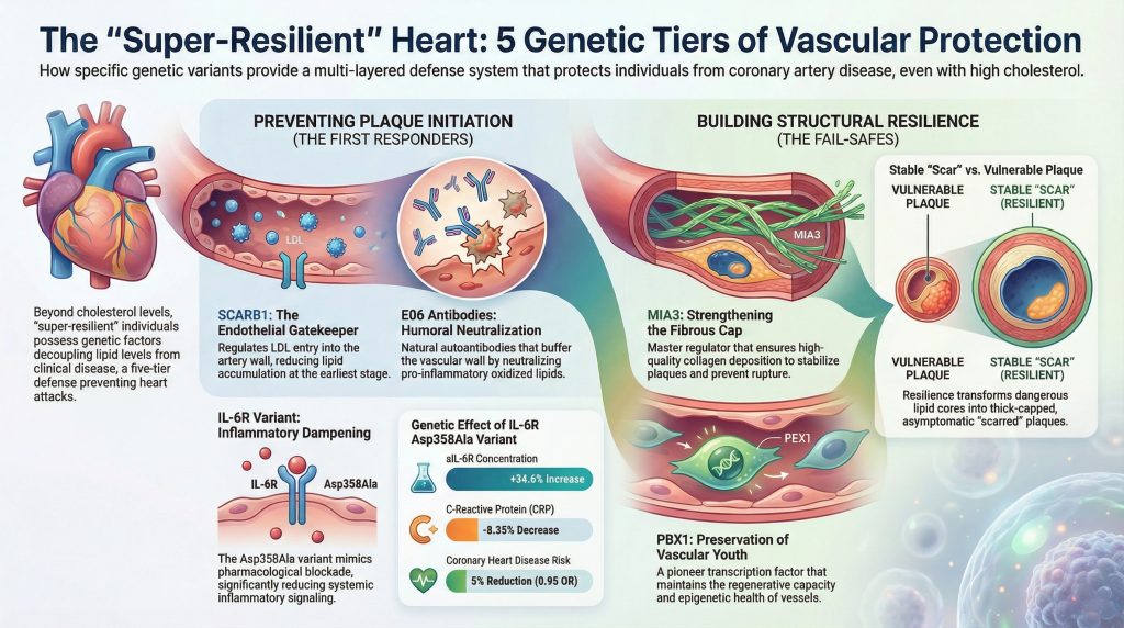
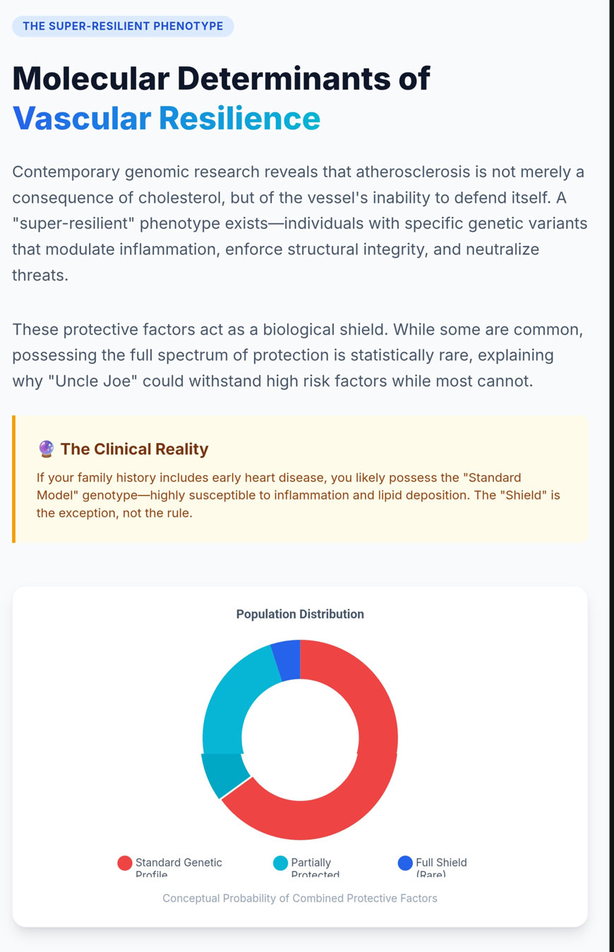
की ऐतिहासिक अवधारणा धमनी काठिन्य परिचलन के बीच एक रैखिक संबंध पर लगातार ध्यान केंद्रित किया गया है कोलेस्ट्रॉल स्तरों और ऑब्सट्रक्टिव के विकास कोरोनरी धमनी रोग. हालांकि, समकालीन जीनोमिक अनुसंधान, विशेष रूप से बड़े पैमाने पर जीनोम-व्यापी एसोसिएशन अध्ययन और मेंडेलियन रैंडमाइजेशन विश्लेषणों से पता चलता है कि एक काफी अधिक जटिल परिदृश्य सामने आया है [1यह अब स्पष्ट हो गया है कि कुछ व्यक्तियों में एक “अत्यंत-प्रतिरोधी” संवहनी फेनोटाइप होता है, जो उच्च स्तर की प्रणालीगत को सहन करने की क्षमता की विशेषता है। जोखिम कारक रुप्चर-प्रोन एथेरोस्क्लेरोटिक विकसित किए बिना घाव [1]. यह लचीलापन आनुवंशिक कारकों की एक श्रृंखला द्वारा संचालित होता है जो कई स्तरों पर संवाहिनी प्रतिक्रिया को नियंत्रित करते हैं: प्रणालीगत और स्थानीय सूजन संबंधी संकेतन, वाहिका दीवार की संरचनात्मक अखंडता, और इसके पार एंडोथेलियल परिवहन लाइपोप्रोटीन, और प्रो-ऑक्सीडेटिव खतरों का सहज प्रतिरक्षा न्यूनीकरण [1]. इस समझ का केंद्र इंटरलुकॉन-6 रिसेप्टर वेरिएंट p.Asp358Ala द्वारा निभाई जाने वाली भूमिकाएँ हैं [1–3], परिवहन प्रोटीन MIA3/TANGO1 [6,7], कचरा रिसेप्टर SCARB1 [8–10], अग्रणी प्रतिलेखन कारक PBX1 [11–13], और ऑक्सीकृत फॉस्फोलिपिड्स के खिलाफ प्राकृतिक ऑटोएंटीबॉडी की सुरक्षात्मक गतिविधि, जैसे कि E06 [14–16].
आईएल-6 रिसेप्टर मार्ग का इन्फ्लेमेटरी मॉड्यूलेशन
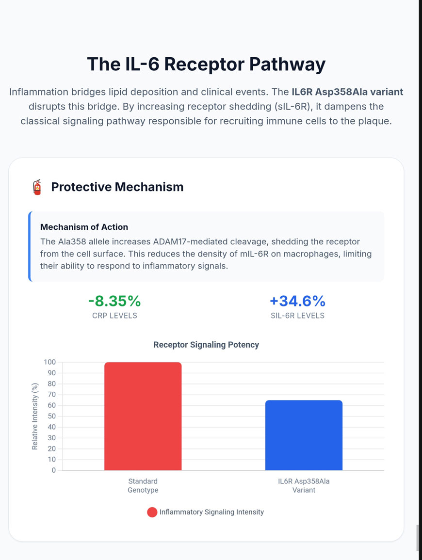
सूजन का एक मौलिक चालक है एथरोएजेनेसिस, जो लिपिड जमा होने और इसके जैसी नैदानिक घटनाओं के बीच पुल के रूप में कार्य करता है मायोकार्डियल इंफार्क्शन [1]. इस प्रक्रिया में शामिल साइटोकाइन्स में, इंटरलुकिन-6 (IL-6) प्रतिरक्षा प्रतिक्रिया के एक बहुआयामी (प्लियोट्रॉपिक) विनियामक के रूप में केंद्रीय स्थान रखता है, जिसे मुख्य रूप से संश्लेषित किया जाता है मैक्रोफेज, टी कोशिकाओं और एडिपोसाइट्स [2]. का सिग्नलिंग आर्किटेक्चर आईएल-६ इसे क्लासिकल सिग्नलिंग—जो कि झिल्ली-बद्ध IL-6 रिसेप्टर (mIL-6R) के माध्यम से होती है—और ट्रांस-सिग्नलिंग, जो रिसेप्टर (sIL-6R) के घुलनशील रूप द्वारा मध्यस्थता करती है, में विभाजित किया गया है [2जबकि क्लासिक सिग्नलिंग mIL-6R व्यक्त करने वाली कोशिकाओं तक सीमित है (जैसे कि हेपैटोसाइट्स और कुछ प्रतिरक्षा सबसेट), ट्रांस-सिग्नलिंग IL-6 को सिग्नल-ट्रांसड्यूसिंग सबयूनिट gp130 व्यक्त करने वाली किसी भी कोशिका को सक्रिय करने की अनुमति देती है, जिससे साइटोकाइन की प्रो-इंफ्लेमेटरी पहुंच संवहनी (vascular) तक व्यापक हो जाती है। एंडोथेलियम और अन्य महत्वपूर्ण ऊतक [2].
Asp358Ala वेरिएंट और शेडिंग तंत्र
IL6R जीन में rs2228145 एकल न्यूक्लियोटाइड बहुरूपता (SNP) की पहचान, जिसके परिणामस्वरूप p.Asp358Ala प्रतिस्थापन होता है, ने एक अनूठा आनुवंशिक दृष्टिकोण प्रदान किया है जिसके माध्यम से इसके कारण भूमिका का निरीक्षण किया जा सके। कोरोनरी हृदय रोग [1]. यह गैर-पर्यायवाची वेरिएंट जीन की स्थिति 1073 पर एडेनिन से साइटोसिन में संक्रमण को शामिल करता है, जो एक्सॉन 9 के भीतर स्थित है जो रिसेप्टर के बाह्य कोशिकीय डोमेन को एनकोड करता है [2]. यह प्रतिस्थापन मेटालोप्रेसिडेस ADAM17 द्वारा पहचाने जाने वाले विदलन स्थल (वेलिन 356) के अत्यधिक निकट होता है, जिसे mIL-6R को sIL-6R में बदलने के लिए जिम्मेदार “शेडडेस” के रूप में भी जाना जाता है [2].
एलानिन (C) एलिल की उपस्थिति रिसेप्टर की प्रोटियोलाइटिक विभाजन के प्रति संवेदनशीलता को महत्वपूर्ण रूप से बढ़ाती है, जिससे रिसेप्टर वितरण में गहरा बदलाव आता है। Ala358 वेरिएंट के वाहक मोनोसाइट्स और CD4+ टी कोशिकाओं पर mIL-6R की सतही अभिव्यक्ति में उल्लेखनीय कमी दिखाते हैं—प्रति एलिल 28% तक की कमी—जबकि साथ ही परिसंचारी sIL-6R के स्तर में भी पर्याप्त वृद्धि होती है, जो आमतौर पर माइनर एलिल की प्रति प्रतिलिपि पर 34.6% की वृद्धि होती है [2].
मेंडेलियन रैंडमाइजेशन और कार्मल प्रोटेक्शन सांख्यिकी
मेंडेलियन के लिए एक आनुवंशिक उपकरण के रूप में Asp358Ala वेरिएंट का उपयोग करना यादृच्छिकीकरण मानव आबादी में आईएल-6 रिसेप्टर अवरोध के दीर्घकालिक प्रभावों के आकलन की अनुमति देता है1]. साक्ष्य लगातार यह प्रदर्शित करते हैं कि इस तंत्र के माध्यम से IL-6 सिगनलिंग का शमन कार्डियोवैस्कुलर और सूजन संबंधी बीमारियों के एक स्पेक्ट्रम के खिलाफ सुरक्षात्मक प्रभाव प्रदान करता है [1–3IL-6R मोनोक्लोनल एंटीबॉडी के यादृच्छिक परीक्षणों में देखे गए प्रभावों से आनुवंशिक निष्कर्षों की तुलना करके टोसिलिज़ुमैब, शोधकर्ताओं ने पुष्टि की है कि यह आनुवंशिक वेरिएंट प्रभावी रूप से औषधीय अवरोध की नकल करता है [1].
तालिका 1. फेनोटाइप या बायोमार्कर संघटन
| प्रकटिक या जैवचिह्न | प्रभाव का आकार (प्रति Ala358 एलिल) | 95% विश्वास अंतराल | पी-मान |
| आईएल-6आर प्लाज्मा सांद्रता | +34.6% (सापेक्ष वृद्धि) | [33.2%, 36.1%] | < 1 × 10⁻²⁰ |
| सी-रिएक्टिव प्रोटीन (सीआरपी) | -8.35% (सापेक्ष कमी) | [-9.38%, -7.31%] | < 1 × 10⁻¹⁵ |
| फाइब्रिनोजेन सांद्रता | -0.85% (सापेक्ष कमी) | [-1.10%, -0.60%] | 1.2 × 10⁻¹⁰ |
| कोरोनरी हृदय रोग (सीएचडी) | 0.95 (अनुपात संभावना) | [0.93, 0.97] | 1.53 × 10⁻⁵ |
| परिधीय धमनी रोग (पीएडी) | 0.91 (अनुपात संभावना) | [0.88, 0.94] | 6.2 × 10⁻⁹ |
अनुसंधान संघों द्वारा 133,449 व्यक्तियों तक का विश्लेषण करके संश्लेषित डेटा [1].
इन निष्कर्षों की सांख्यिकीय महत्वता यह दर्शाती है कि IL-6R संकेतन केवल रोग का एक मार्कर नहीं बल्कि एक कारणगत सहभागी है। CAD के जोखिम में कमी तब भी होती है जब sIL-6R से बंधे परिसंचारी IL-6 अणुओं की अर्ध-आयु में संभावित वृद्धि होती है; शुद्ध जैविक प्रभाव निर्माण और अस्थिरता के लिए आवश्यक सूजन प्रतिक्रिया में कमी है। पट्टिका [1]. यह आनुवंशिक “मंदन” रोकता है धमनी आमतौर पर लिपिड प्रतिधारण से जुड़ी विशाल भड़काऊ प्रतिक्रिया को उत्पन्न करने से, प्रभावी रूप से की उपस्थिति को अलग करते हुए एलडीएल नैदानिक रोग के विकास से। बांग्लादेशी आबादी में हाल के अध्ययनों से यह और अधिक पुष्ट होता है कि यह बहुआकारता टाइप 2 के जोखिम को कम करती है। मधुमेह और उच्च रक्तचाप [5].
चिकित्सीय निहितार्थ और व्यापक सुरक्षात्मक प्रोफ़ाइल
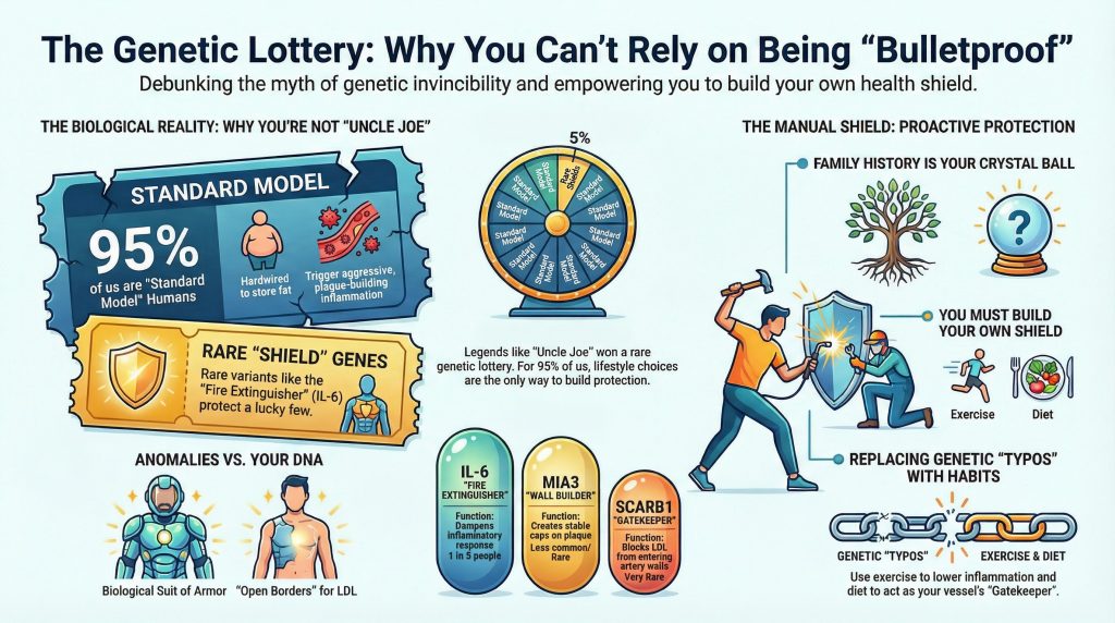
Asp358Ala वेरिएंट से प्राप्त अंतर्दृष्टियों का दवा विकास पर गहरा प्रभाव है। चूंकि यह वेरिएंट पारंपरिक जोखिम कारकों जैसे को महत्वपूर्ण रूप से प्रभावित किए बिना (जिसका प्रमाण कम CRP और फाइब्रिनोजन से मिलता है) प्रणालीगत सूजन को कम करता है। रक्तचाप, यह कम करने के लिए IL-6R अवरोध को एक सटीक उपकरण के रूप में उजागर करता है अवशिष्ट सूजन का जोखिम [1इसके अलावा, इस वेरिएंट के सुरक्षात्मक प्रभाव दीर्घकालिक से भी आगे तक फैले हुए हैं। हृदय रोग गंभीर तीव्र बीमारियों तक। मेंडेलियन रैंडमाइजेशन अध्ययनों से संकेत मिला है कि IL-6R अवरोधन सेप्सिस की घटनाओं में कमी और गंभीर COVID-19 में बेहतर परिणामों से कारणगत रूप से जुड़ा हुआ है।4].
हालाँकि, यह तंत्र पूरी तरह से समझौतों से रहित नहीं है। जबकि Asp358Ala एलिल CAD से बचाता है और रुमेटीइड आर्थराइटिस, यह कुछ संक्रमणों के प्रति संवेदनशीलता बढ़ा सकता है, जो फार्माकोलॉजिकल IL-6R एंटैगोनिस्ट्स के दुष्प्रभाव प्रोफ़ाइल के समान है [2]. यह IL-6/IL-6R अक्ष की सूक्ष्म समझ की आवश्यकता को रेखांकित करता है, जहाँ वेरिएंट सूजन संबंधी सुरक्षा और मेज़बान रक्षा के संतुलन में एक “प्राकृतिक प्रयोग” के रूप में कार्य करता है।.
MIA3 और रेशेदार टोपी की संरचनात्मक अखंडता
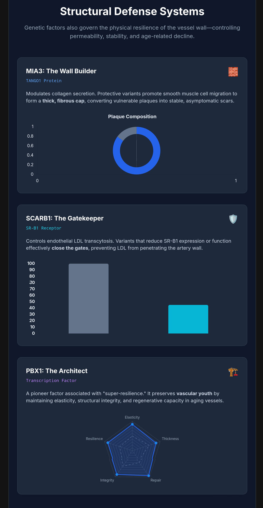
जबकि सूजन संबंधी समायोजन प्लाक के आरंभ को रोकता है, एक बार बन जाने पर घाव की स्थिरता मुख्यतः रक्तवाहिनी की दीवार की संरचनात्मक विशेषताओं द्वारा नियंत्रित होती है। MIA3 जीन, जो प्रोटीन TANGO1 (Transport and Golgi Organization 1) को एन्कोड करता है, एक आधारशिला के रूप में उभरा है। संवहनी लचीलापन के स्राव में इसकी भूमिका के माध्यम से एक्स्ट्रासेलुलर मैट्रिक्स घटक [6].
TANGO1 जैव रसायन और कार्गो निर्यात
TANGO1 एक विकासक्रमिक रूप से संरक्षित प्रोटीन है जो एंडोप्लास्मिक रेटिकुलम (ER) झिल्ली में स्थित है। इसका प्राथमिक कार्य उन बड़े स्रावी कार्गोओं के निर्यात को सुगम बनाना है जो मानक में फिट होने के लिए बहुत बड़े होते हैं। कॉपी vesicles, जिनका व्यास आमतौर पर केवल 60–80 एनएम होता है [6]. बड़े अणु, जैसे प्रोकोलेजन—जो 300 nm तक की लंबाई तक पहुँच सकते हैं—के लिए एक विशेष परिवहन तंत्र की आवश्यकता होती है। TANGO1 इसे ER निकास स्थलों (ERES) पर अंगूठी-नुमा ढांचे में व्यवस्थित होकर और ER-गोल्जी मध्यवर्ती कम्पार्टमेंट (ERGIC) झिल्लियों को शामिल करके एक अस्थायी, विस्तारित स्रावी सुरंग बनाने के लिए हासिल करता है [6].
यह प्रोटीन ल्यूमिनल SH3-सदृश डोमेन का उपयोग करके विशेष रूप से ट्रिपल-हेलीकल कोलेजन मोटिफ्स और चैपरोन HSP47 से बंधता है, जिससे यह सुनिश्चित होता है कि केवल सही ढंग से मुड़े हुए प्रोटीन ही निर्यात के लिए पैकेज किए जाते हैं।6]. यह यंत्रणा कई कोलेजन वर्गों के परिवहन के लिए आवश्यक है, जिनमें अंतःकोशिकीय तंतुमय कोलेजन (I, II, और III), गैर-तंतुमय आधार झिल्ली कोलेजन (IV), और FACIT कोलेजन (IX) शामिल हैं [6].
rs67180937 वेरिएंट और वीएसएमसी फेनोटाइपिक स्थिरता
रक्तवाहिनी में MIA3 अभिव्यक्ति का आनुवंशिक नियमन यह निर्धारित करने के लिए महत्वपूर्ण है कि क्या एक प्लाक एक खतरनाक, फटने-प्रवण घाव बनता है या एक स्थिर “दाग” बनता है। जीनोम-व्यापी एसोसिएशन अध्ययनों ने क्रोमोसोम 1q41 पर एक लोकेस की पहचान की है जहाँ प्रमुख SNP rs67180937 का G एलिल कोरोनरी धमनी रोग के बढ़े हुए जोखिम से जुड़ा हुआ है [7]. यंत्रणात्मक अध्ययनों से पता चला है कि यह जोखिम एलिल संवहनी में MIA3 की कम अभिव्यक्ति से संबंधित है। चिकनी पेशी कोशिकाएं (VSMCs) और विकास कारकों के प्रति एक उल्लेखनीय रूप से कम प्रोलिफ़रेटिव प्रतिक्रिया [7].
एक लचीले संवहनी रूपात्मक प्रकार में, उच्च MIA3 अभिव्यक्ति VSMCs को एक सुरक्षात्मक, सिंथेटिक रूपात्मक प्रकार की ओर संक्रमण को बढ़ावा देती है। ये कोशिकाएँ … की ओर प्रवास करती हैं। इंटिमा और एक मोटे, को बनाने के लिए आवश्यक कोलेजन-युक्त बाह्यकोशीय मैट्रिक्स का उत्पादन करें, फाइब्रस कैप के ऊपर लिपिड कोर एक पट्टिका की7].
तालिका 2. MIA3 अभिव्यक्ति के अनुसार प्लाक की विशेषताएँ
| प्लाक की विशेषता | एमआईए3 की कम अभिव्यक्ति (जोखिम जीनोटाइप) | उच्च MIA3 अभिव्यक्ति (संरक्षणात्मक जीनोटाइप) |
| वीएसएमसी प्रसार | कम; टोपी में कम कोशिकाएँ | सुदृढ़; मजबूत कोशिकीय उपस्थिति |
| कोलेजन जमाव | क्षतिग्रस्त; पतला, नाजुक मैट्रिक्स | कुशल; मोटी, सुरक्षात्मक रेशेदार टोपी |
| नैदानिक परिणाम | फटने-प्रवण “कमजोर” धब्बा | स्थिर, लक्षणरहित “दागदार” पट्टिका |
मानव दाता VSMCs और चूहे के मॉडलों पर किए गए अध्ययनों से संश्लेषित डेटा [7].
MIA3 की प्रासंगिकता मानव कोरोनरी धमनी के घावों में देखे गए अवलोकनों से और भी पुष्ट होती है, जहाँ MIA3 प्रोटीन की प्रचुरता में थिन-कैप क्षेत्रों में महत्वपूर्ण कमी देखी जाती है। अस्थिर प्लाक स्थिर घावों के मोटे कैप्स की तुलना में [7].
SCARB1 और लिपोप्रोटीन ट्रांससाइटोसिस का द्वारपालन
एथेरोजेनेसिस में एक महत्वपूर्ण प्रारंभिक कदम लो-डेन्सिटी लिपोप्रोटीन (एलडीएल) का भौतिक प्रवेश है। उपएन्डोथीलियल स्थान. अब इसे एक अत्यधिक विनियमित पारकोशिकीय परिवहन प्रक्रिया के रूप में मान्यता प्राप्त है जिसे ट्रांससाइटोसिस, मुख्य रूप से स्कैवेन्जर रिसेप्टर क्लास बी टाइप 1 द्वारा मध्यस्थता की जाती है (SR-B1), SCARB1 जीन द्वारा एन्कोड किया गया [8].
DOCK4/Rac1 संकेत अक्ष
एंडोथीलियल SR-B1 द्वारा LDL कणों को आंतरिक रूप से ग्रहण करने और परिवहन करने की प्रक्रिया एक जटिल संकेत श्रृंखला से जुड़ी होती है। जब LDL, SR-B1 के बाह्य डोमेन से बंधता है, तो रिसेप्टर के एक विशिष्ट साइटोप्लाज्मिक डोमेन ग्वानिन न्यूक्लियोटाइड एक्सचेंज फैक्टर को आकर्षित करता है। डॉक4 (साइटोकिनेसिस समर्पक 4) [8]. यह भर्ती स्मॉल GTPase Rac1 के सक्रियण के लिए एक पूर्वापेक्षा है, जो परिवहन के लिए आवश्यक एक्टिन साइटोस्केलेटन पुनर्विन्यासों का संचालन करता है [8].
तालिका 3. एलडीएल ट्रांससाइटोसिस में आणविक घटक का कार्य
| आणविक घटक | एलडीएल ट्रांससाइटोसिस में कार्य |
| एसआर-बी1 (एससीएआरबी1) | ल्यूमिनल सतह पर एलडीएल ग्रहण के लिए प्राथमिक रिसेप्टर |
| आईक्यूएएसईएसएल मोटिफ | अनुकूलक प्रोटीनों के लिए साइटोप्लाज्मिक डॉकिंग साइट |
| डॉक4 | गुआनिन न्यूक्लियोटाइड एक्सचेंज फैक्टर; SR-B1 में भर्ती करता है |
| आरएसी1 | एक्टिन-मध्यस्थता वाली आंतरिक ग्रहण प्रक्रिया को प्रेरित करने वाला जीटीपीएज़ |
एंडोथीलियल कोशिका परिवहन अध्ययनों से संश्लेषित डेटा [8].
SR-B1 के एंडोथीलियल-विशिष्ट निष्कासन वाले चूहों पर किए गए प्रयोगात्मक साक्ष्यों ने धमनी की दीवार में एलडीएल की डिलीवरी में 60–80% की कमी दिखाई, जिससे एथेरोस्क्लेरोटिक घाव क्षेत्र में भारी कमी आई [8]. यह इंगित करता है कि SCARB1 में ऐसे वेरिएंट्स जो इस ट्रांससाइटोसिस की दर को कम करते हैं, मूल रूप से एथेरोस्क्लेरोसिस के “प्रवेश” चरण में इसके विकास को रोक सकते हैं [8].
मानव SCARB1 वेरिएंट्स और लिपोप्रोटीन विरोधाभास
जिगर में, SR-B1 चयनात्मक अवशोषण के लिए प्रमुख रिसेप्टर है। कोलेस्ट्रिल एस्टर से एचडीएल. परिणामस्वरूप, SCARB1 में दुर्लभ हानि-कार्य वेरिएंट्स परिसंचारी HDL कोलेस्ट्रॉल के अत्यधिक स्तर तक बढ़ सकते हैं, फिर भी ये व्यक्ति अक्सर बढ़े हुए कोरोनरी जोखिम का सामना करते हैं क्योंकि रिवर्स कोलेस्ट्रॉल ट्रांसपोर्ट बाधित है [9,10].
तालिका 4. SCARB1 आनुवंशिक वेरिएंट का प्रभाव
| SCARB1 आनुवंशिक वेरिएंट | पर प्रभाव लिपिड प्रोफ़ाइल | रक्तवाहिनी संबंधी जोखिम पर प्रभाव |
| rs5888 (टीटी जीनोटाइप) | उच्च एचडीएल-सी से संबंधित | अक्सर सुरक्षात्मक (विशेषकर गैर-एशियाई पुरुषों में) |
| rs144334493 (हटाव) | FOXA1 बाइंडिंग को कम करता है; SR-B1 को घटाता है। | सीएचडी के प्रति बढ़ी हुई संवेदनशीलता |
| S129L प्रतिस्थापन | संयुक्त उच्च एचडीएल-सी और उच्च एलपी(ए) | कम एलपी(ए) निकासी; उच्च जोखिम |
मेटा-विश्लेषणों और कोहोर्ट अध्ययनों से संश्लेषित डेटा [10].
लचीलापन यकृत और एंडोथीलियम दोनों में रिसेप्टर के कार्य की दक्षता से जुड़ा होता है। अनुकूल बहुआकारिकताएँ जो यकृत की रिवर्स कोलेस्ट्रॉल परिवहन को प्रभावित किए बिना एंडोथीलियल ट्रांससाइटोसिस को कम करती हैं, आदर्श “सुपर-लचीला” जीनोटाइप का प्रतिनिधित्व करती हैं।8–10].
PBX1 और रक्तवाहिनी की जवानी की रक्षा
संवहनी उम्र बढ़ने यह कई प्रकार के हृदय संबंधी रोगों का पूर्ववर्ती है। हाल के अध्ययनों ने के पास वेरिएंट्स की पहचान की है। पीबीएक्स1 (प्रि-बी-सेल ल्यूकेमिया होमोबॉक्स 1) बुढ़ापे में असाधारण रक्तवाहिनी स्वास्थ्य से जुड़े जीन के रूप में [11].
एक अग्रदूत प्रतिलेखन कारक के रूप में PBX1
PBX1 होमियोबॉक्स ट्रांसक्रिप्शन फैक्टरों के TALE (थ्री अमीनोएसिड लूप एक्सटेंशन) वर्ग से संबंधित है और एक “पायनियर फैक्टर” के रूप में कार्य करता है, जिसका अर्थ है कि यह बंद क्रोमैटिन तक पहुँच सकता है और विशिष्ट जीनों को ट्रांसक्रिप्शनल सक्रियण के लिए चिह्नित कर सकता है।12,13]. हृदय-रक्तवाहिनी तंत्र में, PBX1 वे प्रतिलेखन मार्ग समन्वयित करता है जो महाधमनी के पैटर्न निर्माण और हृदय के निकास मार्ग के विकास को नियंत्रित करते हैं [12,13]. बुढ़ापे में वयस्क रक्तवाहिनी में, एंडोथीलियल क्षति की मरम्मत और एंडोथीलियल पूर्वज कोशिका आबादी के रखरखाव के लिए PBX1 अभिव्यक्ति आवश्यक है [11–13].
एपिजेनेटिक नियमन और संवहनी लचीलापन
PBX1 वेरिएंट्स से जुड़ी लचीलापन संभवतः उनकी रक्तवाहिनी की दीवार की युवा एपिजेनेटिक अवस्था को बनाए रखने की क्षमता से उत्पन्न होती है। मरम्मत जीनों की उच्च अभिव्यक्ति बनाए रखते हुए और कोशिकीय ऑक्सीडेटिव तनाव और अपोप्टोसिस में, PBX1 संरचनात्मक क्षय को रोकता है।12,13].
प्राकृतिक एंटी-ऑक्सएलडीएल एंटीबॉडी और ह्यूमरल रक्षा
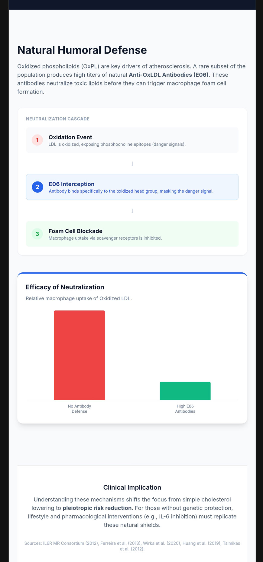
यहाँ तक कि जब लिपोप्रोटीन रक्तवाहिनी की दीवार में प्रवेश कर ऑक्सीकृत (OxLDL) हो जाते हैं, तब भी वे E06 जैसी प्राकृतिक रूप से मौजूद ऑटोएंटीबॉडीज़ द्वारा निष्क्रिय किए जा सकते हैं, इससे पहले कि वे सूजन संबंधी प्रतिक्रिया को उत्प्रेरित करें। फोम सेल चक्र15].
E06 एंटीबॉडी और फॉस्फोकोलाइन एपिटोप
E06 एक प्राकृतिक IgM ऑटोएंटीबॉडी है जो विशेष रूप से ऑक्सीडाइज़्ड फॉस्फोलिपिड्स (OxPL) के फॉस्फोकोलाइन (PC) हेडग्रुप से बंधती है।15sn-2 फैटी एसिड खंड के ऑक्सीकरण पर, पीसी हेडग्रुप एक नियोएपिटोप या “खतरे के संकेत” के रूप में उजागर हो जाता है।15].
तालिका 5. E06 द्वारा जैविक इकाई मान्यता
| जैविक इकाई | E06 द्वारा मान्यता | कार्य करने का तरीका |
| नेटिव एलडीएल | नहीं | सामान्य लिपिड चयापचय में हस्तक्षेप से बचाव करता है |
| तांबे-ऑक्सीकृत एलडीएल | हाँ | सतह पर खंडित OxPL से जुड़ता है |
| लाइपोप्रोटीन(ए) | हाँ | एपो(ए) से कॉवैलेंट रूप से बंधे ऑक्सपीएल को निष्क्रिय करता है। |
| अपॉप्टोटिक कोशिकाएँ | हाँ | क्षतिग्रस्त झिल्लियों पर उजागर पीसी को पहचानता है |
प्रतिरक्षा संबंधी परीक्षणों और संरचनात्मक अध्ययनों से संश्लेषित डेटा [15].
E06 की सुरक्षात्मक क्रिया दोहरी है: यह मैक्रोफेज स्कैवेंजर रिसेप्टर्स द्वारा इन कणों के अवशोषण को रोकता है, जिससे फोम सेल का निर्माण नहीं होता, और TLR-2 तथा TLR-4 जैसे रिसेप्टर्स के माध्यम से प्रदाह-प्रेरक संकेतन को निष्क्रिय कर देता है।15].
चिकित्सीय संभावनाएँ और नैदानिक सहसंबंध
E06-सदृश एंटीबॉडीज़ के उच्च प्राकृतिक स्तर का अस्तित्व एक मजबूत “जन्मजात प्रतिरक्षण” प्रदान करता है। इसने VB-201 जैसे चिकित्सीय उम्मीदवारों के विकास को प्रेरित किया है, जो CD14 और TLR-2 से बंधने के लिए प्रदाहजनक OxPLs के साथ प्रतिस्पर्धा करने के लिए डिज़ाइन किया गया एक लघु-अणु लेसिनोक्सॉइड है।16]. OxPL-apoB के उच्च स्तर (E06-आधारित ELISA द्वारा मापा गया) कोरोनरी, फेमोरल और करोटिड धमनी रोग14].
संदर्भ
- Interleukin-6 Receptor Mendelian Randomisation Analysis (IL6R MR) Consortium, Swerdlow DI, Holmes MV, et al. The interleukin-6 receptor as a target for prevention of coronary heart disease: a mendelian randomisation analysis. Lancet. 2012;379(9822):1214-1224. doi:10.1016/S0140-6736(12)60110-X
- Ferreira RC, Freitag DF, Cutler AJ, et al. Functional IL6R 358Ala allele impairs classical IL-6 receptor signaling and influences risk of diverse inflammatory diseases. PLoS Genet. 2013;9(4):e1003444. doi:10.1371/journal.pgen.1003444
- Levin MG, Klarin D, Georgakis MK, et al. A Missense Variant in the IL-6 Receptor and Protection From Peripheral Artery Disease. Circ Res. 2021;129(10):968-970. doi:10.1161/CIRCRESAHA.121.319589
- Hamilton FW, Thomas M, Arnold D, et al. Therapeutic potential of IL6R blockade for the treatment of sepsis and sepsis-related death: A Mendelian randomisation study. PLoS Med. 2023;20(1):e1004174. Published 2023 Jan 30. doi:10.1371/journal.pmed.1004174
- Aparicio-Siegmund S, Garbers Y, Flynn CM, et al. The IL-6-neutralizing sIL-6R-sgp130 buffer system is disturbed in patients with type 2 diabetes. Am J Physiol Endocrinol Metab. 2019;317(2):E411-E420. doi:10.1152/ajpendo.00166.2019
- Wilson DG, Phamluong K, Li L, et al. Global defects in collagen secretion in a Mia3/TANGO1 knockout mouse. J Cell Biol. 2011;193(5):935-951. doi:10.1083/jcb.201007162
- Aherrahrou R, Guo L, Nagraj VP, et al. Genetic Regulation of Atherosclerosis-Relevant Phenotypes in Human Vascular Smooth Muscle Cells. Circ Res. 2020;127(12):1552-1565. doi:10.1161/CIRCRESAHA.120.317415
- Huang L, Chambliss KL, Gao X, et al. SR-B1 drives endothelial cell LDL transcytosis via DOCK4 to promote atherosclerosis. Nature. 2019;569(7757):565-569. doi:10.1038/s41586-019-1140-4
- Hu S, Hu D, Wei H, et al. Functional Deletion/Insertion Promoter Variants in SCARB1 Associated With Increased Susceptibility to Lipid Profile Abnormalities and Coronary Heart Disease. Front Cardiovasc Med. 2022;8:800873. Published 2022 Jan 13. doi:10.3389/fcvm.2021.800873
- Ye LF, Zheng YR, Zhang QG, Yu JW, Wang LH. Meta-analysis of the association between SCARB1 polymorphism and fasting blood lipid levels. Oncotarget. 2017;8(46):81145-81153. Published 2017 Sep 14. doi:10.18632/oncotarget.20867
- Wen Y, Chen H, Wang Y, et al. Extracellular vesicle-derived TP53BP1, CD34, and PBX1 from human peripheral blood serve as potential biomarkers for the assessment and prediction of vascular aging. Hereditas. 2024;161(1):3. Published 2024 Jan 3. doi:10.1186/s41065-023-00306-8
- Crisafulli L, Brindisi M, Liturri MG, Sobacchi C, Ficara F. PBX1: a TALE of two seasons-key roles during development and in cancer. Front Cell Dev Biol. 2024;12:1372873. Published 2024 Feb 9. doi:10.3389/fcell.2024.1372873
- Chen H, Yu Z, Niu Y, Wang L, Xu K, Liu J. Research progress of PBX1 in developmental and regenerative medicine. Int J Med Sci. 2023;20(2):225-231. Published 2023 Jan 22. doi:10.7150/ijms.80262
- Taleb A, Witztum JL, Tsimikas S. Oxidized phospholipids on apoB-100-containing lipoproteins: a biomarker predicting cardiovascular disease and cardiovascular events. Biomark Med. 2011;5(5):673-694. doi:10.2217/bmm.11.60
- Hörkkö S, Bird DA, Miller E, et al. Monoclonal autoantibodies specific for oxidized phospholipids or oxidized phospholipid-protein adducts inhibit macrophage uptake of oxidized low-density lipoproteins. J Clin Invest. 1999;103(1):117-128. doi:10.1172/JCI4533
- Mendel I, Feige E, Yacov N, et al. VB-201, an oxidized phospholipid small molecule, inhibits CD14- and Toll-like receptor-2-dependent innate cell activation and constrains atherosclerosis. Clin Exp Immunol. 2014;175(1):126-137. doi:10.1111/cei.12212

