
यह पृष्ठ स्वचालित रूप से अनुवादित किया गया है। यदि कोई विसंगति है, तो अंग्रेज़ी संस्करण प्रामाणिक है।.

संशोधित: 16 जुलाई, 2026

एलडीएल “बैड” कोलेस्ट्रॉल: अपोलोपाइप्रोटीन बी, इमेजिंग रेसोल्यूशन, और 60 वर्ष की आयु में “सामान्य” कोरोनरी धमनियों का भ्रम

लेखक: पीटर मेगडल, पीएचडी

इस लेख का उपयोग कैसे करें

चिकित्सा अस्वीकरण: यह लेख केवल शिक्षा के लिए है और चिकित्सा सलाह नहीं है। व्यक्तिगत मार्गदर्शन के लिए हमेशा अपने चिकित्सक से परामर्श लें।.

आसान पाठ

“परफेक्ट” 60 वर्षीय व्यक्ति का रहस्य

कल्पना कीजिए कि जॉन नाम का एक व्यक्ति है जो अभी 60 साल का हुआ है। जॉन बहुत अच्छा महसूस करता है। वह सैर पर जाता है, हरी सब्जियां खाता है, और अभी भी अपने पोते-पोतियों के साथ तालमेल बिठा सकता है। वह हाल ही में एक बड़े चेक-अप के लिए अपने डॉक्टर के पास गया था। डॉक्टर ने उसके चार्ट देखे, उसकी रक्तचाप, और उसे एक “सामान्य” रिपोर्ट दी। उसकी जाँच में कोई बड़ी रुकावट नहीं दिखाई दी, और उसका कोलेस्ट्रॉल संख्याएँ “ठीक” लग रही थीं। जॉन कार्यालय से यह महसूस करते हुए निकला कि उसका दिल किसी किशोर जैसा है।.

हालाँकि, जॉन “द चेक-अप मिस्ट्री” में फंस सकते हैं। यह एक ऐसी स्थिति है जहाँ परीक्षण ऊपर से अच्छे दिखते हैं, लेकिन शरीर के अंदर की वास्तविकता बहुत अलग होती है।.

इसे साठ साल पहले बने एक पुराने घर की तरह समझिए। फ़ुटपाथ से, घर बहुत शानदार दिखता है। इस पर चमकीले पेंट की एक ताज़ा परत और एक बेहतरीन घास का मैदान है। लेकिन अगर आप दीवारों को फाड़कर देखें, तो आपको पुराने, जंग लगे पाइप और ऐसे तार मिल सकते हैं जो छिलने लगे हैं। घर के पास से गुजरने वाले किसी भी व्यक्ति को यह “सामान्य” लगता है, लेकिन इसकी संरचना गहराई में पुरानी हो रही है जहाँ कोई देख नहीं सकता।.

यह पोस्ट आपको यह दिखाने के लिए है कि कैसे हृदय की सामान्य जाँचें अक्सर हृदय रोग के “छिपे हुए” खतरों को नजरअंदाज कर देती हैं। हम इस बात पर चर्चा करने जा रहे हैं कि क्यों एक स्पष्ट स्वास्थ्य रिपोर्ट सिर्फ एक धोखा हो सकती है और जैसे-जैसे हमारी उम्र बढ़ती है, हमारी धमनियों के बारे में विज्ञान वास्तव में क्या कहता है।.

मुख्य बात 1: आपकी धमनियों ने किशोर अवस्था में ही बूढ़ा होना शुरू कर दिया था

अधिकांश लोग सोचते हैं कि हृदय रोग “बुजुर्गों” की समस्या है। उनका मानना है कि यदि वे 30 या 40 वर्ष के हैं, तो उनकी धमनियां “माइक्रोस्कोप के नीचे बिल्कुल साफ” हैं। लेकिन चिकित्सा अनुसंधान एक बहुत ही अलग, और कहीं अधिक सच्ची कहानी दिखाता है।.

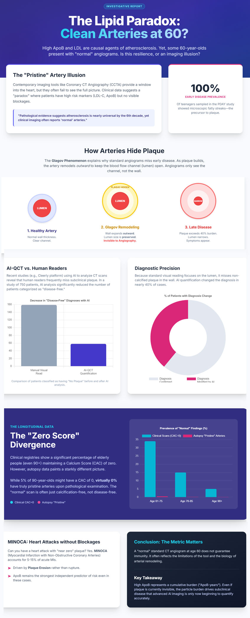
डॉक्टर मानक परीक्षण पर जो देखता है (जिसे हम क्लिनिकल रियलिटी कहते हैं) और वैज्ञानिक शरीर के ऊतकों को सीधे देखने पर जो देखता है (जिसे हम फोरेंसिक रियलिटी कहते हैं) उसके बीच एक बहुत बड़ा अंतर है। वैज्ञानिकों ने दुर्घटनाओं में मरने वाले किशोरों और युवा वयस्कों की धमनियों का अध्ययन किया है। दो प्रसिद्ध अध्ययन, “पीडीएवाई”अध्ययन और“बोगलूस हार्ट स्टडी,”, कुछ ऐसा मिला है जो हमारे स्वास्थ्य के बारे में हमारे सोचने के तरीके को बदल देना चाहिए।.

लगभग उन सभी किशोरों में हृदय रोग के शुरुआती संकेत पहले से ही मौजूद थे। वैज्ञानिक इन्हें “वसायुक्त रेखाएँ.ये दिल की नलियों की परत में जमने वाली वसा के छोटे-छोटे पीले ढेर होते हैं।.

विश्लेषण और प्रतिबिंब: यह जानकर एक झटका लगता है कि पश्चिमी दुनिया में जब तक कोई 40 वर्ष की आयु तक पहुँचता है, तब तक “परिपूर्ण” धमनियां मूल रूप से मौजूद ही नहीं होती हैं। हम यह सोचते हुए अपना जीवन बिता देते हैं कि जब भी हम दौड़ने जाते हैं या सलाद खाते हैं, तो हम एक साफ स्लेट के साथ शुरुआत कर रहे हैं। लेकिन “कचरा” हाई स्कूल के समय से ही जमा होता आ रहा है। जब तक कोई 60 वर्ष का होता है, तब तक यह विचार कि उनकी धमनियां “अक्षुण्ण” हैं, लगभग हमेशा एक ऐसी जांच द्वारा की गई भूल होती है जो उतनी शक्तिशाली नहीं होती है। जैसा कि विशेषज्ञ कहते हैं:

“जीवन के छठे दशक तक, पश्चिमी आबादी में पूरी तरह से साफ कोरोनरी धमनियां लगभग अनुपस्थित होती हैं।”

मुख्य बात 2: एलडीएल (LDL) आपके जोखिम को मापने का गलत तरीका है

जब आप रक्त परीक्षण करवाते हैं, तो आपका डॉक्टर आमतौर पर “LDL-C” देखता है। यह मापता है वज़न आपके रक्त में “खराब” कोलेस्ट्रॉल का। लेकिन विज्ञान यह पता लगा रहा है कि खतरे को मापने का वजन सबसे अच्छा तरीका नहीं है। इसके बजाय, हमें इस बात पर ध्यान देना चाहिए कि “अपोबी.”

इसे समझने के लिए, आइए एक उदाहरण का उपयोग करें सूटकेस और यात्री.

एक दरवाज़े की कल्पना करें (आपकी धमनी दीवार)। एलडीएल-सी (LDL-C) एयरपोर्ट से आने वाले सभी सामान (luggage) के कुल वजन की तरह है। लेकिन ApoB सूटकेस की वास्तविक संख्या है। इससे कोई फर्क नहीं पड़ता कि सूटकेस भारी है या हल्का; यदि बहुत अधिक सूटकेस हैं, तो वे दरवाजे पर फंस जाएंगे और ढेर लगा देंगे।.

हृदय रोग का कारण बनने वाले प्रत्येक “खराब” कण में ठीक एक ApoB होता है। प्रोटीन इस पर काम कर रहे हैं। इससे ApoB “कण भार”—वास्तव में ऐसी कितनी चीज़ें हैं जो आपके दिल की नली में फँस सकती हैं।.

समय की शक्तिएपोबी-इयर्स): यह सिर्फ इस बारे में नहीं है कि आज आपके पास कितने “सूटकेस” हैं। यह इस बारे में है कि वे दरवाज़े से अंदर आने की कोशिश कितने लंबे समय से कर रहे हैं। वैज्ञानिक इसे “ApoB-years” कहते हैं। 40 साल तक उच्च ApoB होना, 5 साल तक रहने की तुलना में कहीं अधिक खतरनाक है। यह धीरे-धीरे टपकने वाले नल की तरह है; कई सालों में, एक छोटा सा रिसाव भी तहखाने में बाढ़ ला सकता है।.

लिपोप्रोटीन मीट्रिक यह क्या मापता है यह क्यों महत्वपूर्ण है
एलडीएल-सी (वजन) कोलेस्ट्रॉल का कुल द्रव्यमान या भार।. यदि आपके पास कई छोटे कण हैं तो यह गलत हो सकता है।.
Apob (मात्रा) “खराब” कणों की वास्तविक संख्या।. जोखिम का सबसे मजबूत भविष्यवक्ता; जो आपको वास्तविक “बोझ” बताता है।”
एलपी(ए) (चिपचिपा) एक विशेष, अत्यधिक चिपचिपा कण।. एक स्वतंत्र जोखिम कारक इससे थक्का जमने की संभावना बढ़ जाती है।.

मुख्य बात 3: “ग्लागोव प्रभाव” - आपकी धमनियां कचरे को कैसे छिपाती हैं

डॉक्टर अक्सर इस जमाव को क्यों नहीं देख पाते हैं? यह “ग्लागोव प्रभाव.”

अपनी धमनी को एक वॉक-इन कोठरी की तरह समझें। जैसे ही आप ज़्यादा से ज़्यादा “कचरा” ठूंसना शुरू करते हैं (पट्टिका) को कोठरी में, आप इसे तुरंत दरवाजे को ब्लॉक नहीं करने देते हैं। इसके बजाय, आप कोठरी की पिछली दीवार को अगले कमरे में और आगे धकेल देते हैं। दरवाजे से झाँकने वाले किसी व्यक्ति के लिए, कोठरी खाली और साफ़ दिखती है। लेकिन वास्तव में, दीवारें छिपे हुए कबाड़ से बाहर की ओर उभरी हुई हैं।.

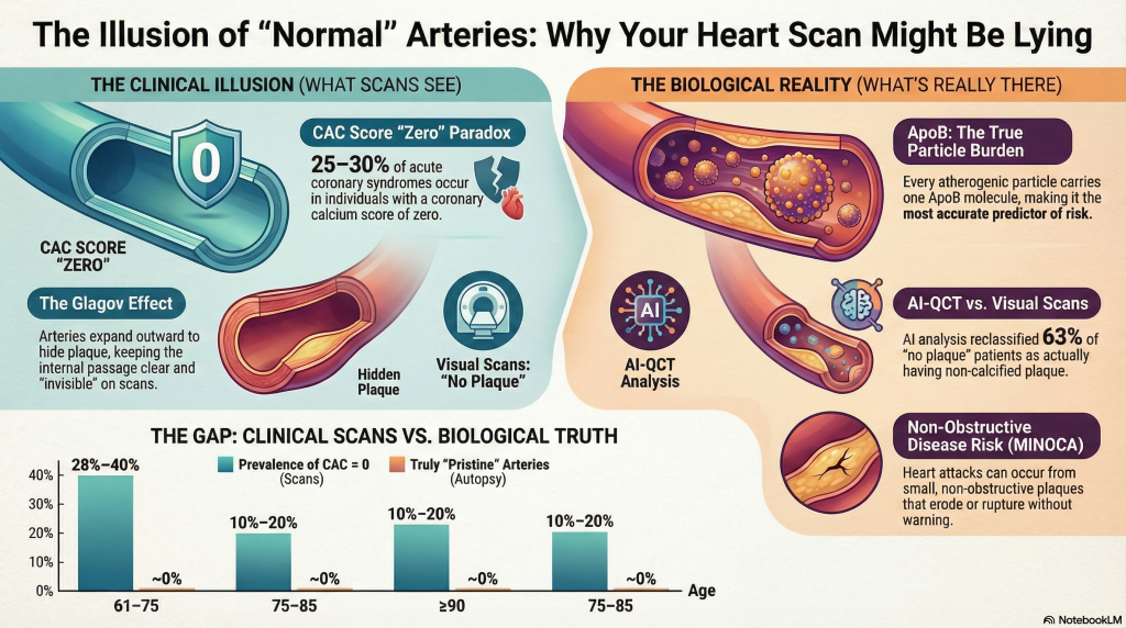
आपकी धमनियां भी ऐसा ही करती हैं। जब पट्टिका (प्लाक) जमा होती है, तो धमनी की दीवार की बाहरी परत वास्तव में बाहर की ओर फैल जाती है। यह “हॉलवे” (जहां से रक्त बहता है) को पूरी तरह खुला रखता है।.

विश्लेषण और प्रतिबिंब: यही कारण है कि मानक परीक्षण जैसे एंजियोग्राम वे हमें असफल करते हैं। वे केवल उस खुले स्थान को देखते हैं जहाँ रक्त प्रवाहित होता है। वे "उभरी हुई" दीवारों के अंदर छिपी प्लाक को नहीं देख सकते। वास्तव में, आपकी धमनियाँ 40% प्लाक से भरी हो सकती हैं इससे पहले कि रक्त प्रवाह धीमा होना भी शुरू हो। जब तक एक मानक परीक्षण में "जाम" दिखता है, तब तक यह बीमारी दशकों से बढ़ रही होती है। हम "गलियारे" को देख रहे हैं जबकि "दीवारें" कचरे से भरी हुई हैं।.

निष्कर्ष 4: क्यों “शून्य” कैल्शियम स्कोर “जेल से बाहर निकलने का मुफ्त कार्ड” नहीं है

बहुत से लोग एक परीक्षण करवाते हैं जिसे कोरोनरी धमनी कैल्शियम (सीएसी) स्कोर। यह परीक्षण “कठोर” प्लाक की तलाश करता है - वह प्लाक जो कैल्शियम में बदल गया है, जो हड्डी जैसा होता है। यदि आपका स्कोर शून्य है, तो आप सोच सकते हैं कि आपका जोखिम शून्य है।.

लेकिन “शून्य” स्कोर का मतलब यह नहीं है कि आपके पास शून्य पट्टिका (प्लाक) है। इसका मतलब सिर्फ यह है कि आपके पास नहीं है कैल्सीफाइड पट्टिका.

चिकित्सा का विरोधाभास: यह वह हिस्सा है जो कई लोगों को हैरान करता है। कैल्शियम वास्तव में “प्लाक हीलिंग” (घाव भरने) का संकेत है। यह एक निशान की तरह है जिसका उपयोग शरीर घाव को ढकने के लिए करता है। जब शरीर धमनी में कोई गड़बड़ी देखता है, तो वह उसे स्थिर करने के लिए उस पर “पत्थर जैसा आवरण” बनाने की कोशिश करता है।.

  1. हार्ड प्लाक यह पुराना, क्षत-विक्षत और कैल्सीकृत है। यह समस्या को ठीक करने का शरीर का अपना तरीका है।.
  2. सॉफ्ट प्लाक: यह युवा, “चिकना” प्लाक है। यह बहुत अधिक खतरनाक है क्योंकि यह “जैविक रूप से सक्रिय” और अस्थिर है।.

सॉफ्ट प्लाक को एक पिम्पल की तरह समझें। अगर यह “फूट” (रुप्चर हो) जाता है, तो यह अचानक खून का थक्का, जिससे एक दिल का दौरा. सख्त प्लाक वास्तव में फटने की संभावना कम होती है। डेटा दिखाता है कि 25% से 30% तक के हृदयाघात उन लोगों में होते हैं जिन्हें पहले से था कैल्शियम स्कोर शून्य का।.

विश्लेषण और प्रतिबिंब: शून्य स्कोर उच्च स्कोर से बेहतर है, लेकिन यह “समय की एक झलक” है। यह आपको दिखाता है कि शरीर में कहाँ पहले ही निशान बन चुके हैं। यह आपकी धमनियों की दीवारों में वर्तमान में जमा हो रही “नरम चिकनाई” के बारे में कुछ नहीं बताता है। उच्च ApoB वाले किसी व्यक्ति के लिए, शून्य स्कोर का आमतौर पर यही अर्थ होता है कि नुकसान अभी तक “पत्थर” में नहीं बदला है। आग अभी भी जल रही है; बस उसने स्कैन को देखने के लिए कोई राख नहीं छोड़ी है।.

निष्कर्ष 5: एआई मानव आँख से अधिक स्मार्ट है

डॉक्टर बहुत समझदार होते हैं, लेकिन वे स्क्रीन पर मानव आँख जो देख सकती है, उससे सीमित होते हैं। “नामक नई तकनीक“एआई-क्यूसीटी”(जैसे क्लियरली प्लेटफॉर्म) खेल को बदल रहा है। यह तकनीक कृत्रिम बुद्धिमत्ता (आर्टिफिशियल इंटेलिजेंस) का उपयोग करके हृदय के स्कैन को किसी इंसान की तुलना में कहीं अधिक बारीकी से देखती है।.

750 रोगियों के एक अध्ययन में, डॉक्टरों ने स्कैन देखे और 159 ऐसे लोगों की पहचान की, जिनके बारे में उन्हें लगा कि उनमें बिल्कुल भी “प्लाक” नहीं है। वे “साफ” दिख रहे थे। हालाँकि, जब एआई (AI) ने बिल्कुल उन्हीं स्कैन को देखा, तो उसने पाया कि उनमें से कई लोगों के शरीर में वास्तव में छिपा हुआ प्लाक था।.

  • मानव नेत्र 159 लोगों को बताया गया कि उनमें “कोई प्लाक नहीं” था।”
  • एआई आँख उन लोगों में से वास्तव में केवल 58 लोगों को कोई पट्टिका (प्लाक) नहीं थी।.

एआई ने पाया कि जिन 101 लोगों को डॉक्टरों ने “स्वस्थ” (संक्रमण मुक्त) समझा था, दरअसल उनमें बिना-कैल्शियम वाला प्लाक (खतरनाक “नरम” प्रकार)। एआई एक इंसान की तुलना में प्लाक को “माप” (सटीक मात्रा को मापना) और “विशेषता बता सकता है” (यह किस चीज़ से बना है यह देखना) इंसानों से बेहतर कर सकता है। यह दिखाता है कि डॉक्टरों द्वारा की जाने वाली मानक दृश्य जाँच लगभग हमेशा यह चूक जाती है कि वास्तव में वहाँ कितनी बीमारी मौजूद है।.

निष्कर्ष 6: “मिनोका” चेतावनी - बिना ब्लॉकेज के दिल के दौरे

हम अक्सर सोचते हैं कि दिल का दौरा केवल तब होता है जब कोई पाइप 100% पूरी तरह से बंद हो जाए, जैसे सिंक में जाम हो गया नालिका। लेकिन एक ऐसी स्थिति होती है जिसे "“मिनोका.ऐसा तब होता है जब किसी को दिल का दौरा (हार्ट अटैक) पड़ता है, भले ही उनकी धमनियां पूरी तरह से खुली हुई दिखती हों।.

यह सभी हृदयाघातों का लगभग 5% से 15% हिस्सा है।.

यह कैसे होता है? ऐसा इसलिए होता है क्योंकि प्लाक का एक छोटा सा, “नरम” उभार भी फट सकता है। जब ऐसा होता है, तो शरीर रक्त का थक्का बनाकर उस फटी हुई जगह को ठीक करने की कोशिश करता है। वही थक्का दिल को रोक देता है, भले ही कुछ ही पल पहले धमनी (आर्टरी) खुद “खुली” हुई दिख रही हो।.

विश्लेषण और प्रतिबिंब: यह “भ्रम” का सबसे डरावना हिस्सा है। सोमवार को आपकी रिपोर्ट “सामान्य” आ सकती है और मंगलवार को दिल का दौरा पड़ सकता है क्योंकि सॉफ्ट प्लाक का एक छोटा सा, छिपा हुआ उभार फटने का फैसला कर लेता है। उस मरीज के डर की कल्पना करें जिसे यह बताया गया हो कि उसकी “नमें साफ हैं” और एक हफ्ते बाद वह आपातकालीन कक्ष (इमरजेंसी रूम) में पहुंच जाए। यही कारण है कि अपने “पार्टिकल काउंट” (ApoB) को कम रखना सबसे अच्छा बचाव है। आपके पास जितने कम “सूटकेस” होंगे, उतनी ही कम संभावना होगी कि पहले स्थान पर इनमें से कोई छोटा लेकिन घातक “उभार” बने।.

सारांश: भ्रम के पार

हमें दिल की बीमारी के बारे में यह सोचना बंद करना होगा कि यह “जाम पाइप” की समस्या है जो केवल बूढ़े लोगों को होती है। इसके बजाय, हमें इसे “कणों के जमाव” की जीवनभर चलने वाली प्रक्रिया के रूप में देखने की जरूरत है।”

“क्लिन आर्टरी” (स्वच्छ धमनी) का भ्रम इसलिए होता है क्योंकि हमारे उपकरण अक्सर बीमारी के शुरुआती चरणों को देखने के लिए पर्याप्त रूप से शक्तिशाली नहीं होते हैं। हमारी धमनियों में चौड़े खुले “गलियारे” हो सकते हैं, लेकिन “दीवारें” दशकों पुरानी छिपी हुई पट्टिका (प्लाक) से भरी हो सकती हैं।.

आयु बनाम पट्टिका: वास्तविकता का अंतर

आयु वर्ग “क्लिनिकल स्कैन पर ”स्वच्छ” परिणाम वास्तविकता में संभवतः “प्राचीन” धमनियाँ (ऑटोप्सी)
61–75 वर्ष 28% से 40% लोग "साफ" दिखते हैं“ ~0% (Almost nobody is truly clean)
75–85 Years 10% to 20% of people look “clean” ~0% (Almost nobody is truly clean)
90+ Years ~5% of people look “clean” ~0% (Almost nobody is truly clean)

Final Thought: Next time you talk to your doctor about your heart, don’t just settle for a “normal” report. You should ask a deeper question: “Doctor, my tests look normal, but what is my ApoB count, and how do we know I don’t have hidden soft plaque?” Understanding your true “particle burden” and the hidden state of your artery walls could save your life by spotting the danger before the “illusion” disappears.

गहन अध्ययन

Apolipoprotein B, Imaging Resolution, and the Illusion of “Normal” Coronary Arteries at Age 60

The clinical assertion that individuals maintaining markedly elevated levels of low-density लाइपोप्रोटीन कोलेस्ट्रॉल (LDL-C) and एपोलिपोप्राटीन Bअपोबी) can possess anatomically “normal” कोरोनरी धमनियां at age 60 represents a diagnostic paradox. This phenomenon often hinges on the resolution of the imaging tool used. While contemporary tools like the कोरोनरी धमनी कैल्शियम (सीएसी) score and standard visual Coronary Computed Tomography Angiography (CCTA) provide a high-resolution window into the arterial wall, they exist in tension with pathological evidence which suggests that धमनी काठिन्य is a nearly universal condition of human aging, and clinical data showing that दिल के दौरे can occur even in the absence of significant anatomical blockages [1].
The Causal Framework of Lipoprotein-Driven Atherogenesis

The consensus establishes that prolonged exposure to ApoB-containing lipoproteins is the primary driver of atherosclerotic पट्टिका [2]. Each atherogenic particle—including एलडीएल, वीएलडीएल, और IDL—carries a single molecule of ApoB, making it a direct measure of the total number of circulating particles that can penetrate the arterial wall [2]. The probability of a particle becoming trapped in the subendothelial space is a function of both particle concentration and the duration of exposure, often quantified as “एपोबी-वर्ष” [2].

While LDL-C measures the mass of cholesterol, ApoB reflects the कण भार [3]. In cases of मतभेद—where ApoB is high but LDL-C is relatively lower—ApoB remains the more accurate predictor of future मायोकार्डियल इंफार्क्शन (MI) and clinical events [4].

Lipoprotein Metrics

Lipoprotein Metric Definition and Pathological Role Association with Plaque Burden
Low-Density Lipoprotein Cholesterol (LDL-C) The mass of cholesterol contained within LDL particles Causal, but may be discordant with particle count [3]
Apolipoprotein B (ApoB) A structural प्रोटीन; one exists on every atherogenic particle Strongest predictor of risk; reflects true particle burden [3]
Lipoprotein(a) [Lp(a)] An LDL-like particle with an additional protein [apo(a)] Independent जोखिम कारक; promotes कैल्सीफ़िकेशन और थ्रोम्बोसिस [5]

Forensic vs. Clinical Reality: The “Pristine” Artery Illusion

The claim that an individual can reach age 60 without “arterial damage” is often a result of clinical imaging failing to detect microscopic disease. Forensic autopsy studies, including the Pathobiological Determinants of Atherosclerosis in Youth (पीडीएवाई) and the बोगलूस हार्ट स्टडी, demonstrate that microscopic वसायुक्त रेखाएँ—the earliest macroscopic evidence of atherosclerosis—are present in nearly all adolescents examined [6].

By the sixth decade of life, pathologically pristine coronary arteries are virtually absent in Westernized populations. Diffuse intimal thickening and lipid deposition become ubiquitous by midlife, and contemporary autopsy cohorts report coronary atherosclerosis in nearly all adults by the fourth decade [6].

The Glagov Effect: How Arteries Hide Plaque Burden

A critical contributor to “normal” coronary imaging क्या Glagov phenomenon, या compensatory outward remodeling [7]. As plaque accumulates within the arterial wall, the vessel expands externally to preserve luminal diameter and maintain blood flow [7]. Hemodynamically significant luminal narrowing is typically delayed until plaque burden exceeds approximately 40% of the internal elastic lamina area [7].

Because standard invasive angiography and conventional visual CCTA focus primarily on the ल्युमेन, substantial plaque burden may exist within the vessel wall while remaining angiographically invisible [7]. These concealed plaques are often lipid-rich and biologically active, rendering them prone to erosion or rupture capable of precipitating MI even in the absence of pre-existing स्टेनोसिस [1].

AI-QCT (Cleerly) vs. Standard Visual Interpretation

Artificial Intelligence–Quantitative Computed Tomography (AI-QCT) platforms, such as Cleerly, have demonstrated that standard visual interpretation of CCTA systematically underestimates plaque burden [8]. By enabling volumetric plaque quantification and compositional analysis, AI-QCT achieves accuracy approaching invasive इंट्रावैस्कुलर अल्ट्रासाउंड [8].

In a cohort of approximately 750 patients, AI-QCT analysis led to diagnostic modification in 39% of cases, while the number of patients classified as having “no plaque” fell from 159 to 58 following quantitative assessment [8]. These findings confirm that visually “normal” coronary CT एंजियोग्राम frequently harbor clinically relevant बिना-कैल्शियम वाला प्लाक.

Coronary Artery Calcium Score and Myocardial Infarction Risk

कोरोनरी धमनी Calcium scoring is a powerful population-level risk stratification tool, but its relationship with individual myocardial infarction risk is probabilistic rather than absolute. Large prospective cohorts, including the Multi-Ethnic Study of Atherosclerosis (MESA), demonstrate a strong graded association between CAC burden and future कोरोनरी हृदय रोग events, with markedly elevated risk observed at scores ≥300 [11].

However, a CAC score of zero does not confer immunity from MI. Although short-term event rates are low, non-calcified, lipid-rich plaques—particularly in individuals with elevated ApoB—remain capable of rupture or erosion [12]. Approximately 25–30% of acute coronary syndromes occur in individuals with absent or minimal coronary calcification at baseline, reflecting the temporal disconnect between plaque formation, calcification, and plaque instability [12]. Calcification is increasingly recognized as a marker of plaque chronicity and healing rather than vulnerability, whereas non-calcified plaque burden correlates more closely with near-term risk.

Thus, CAC scoring is best interpreted as a measure of cumulative plaque burden and long-term risk rather than as a detector of biologically active atherosclerosis. In high–ApoB individuals, a CAC score of zero more likely reflects delayed disease expression than true disease absence.

MI Risk With “Near-Zero” Plaque Burden: The MINOCA Syndrome

Myocardial Infarction with Non-Obstructive Coronary Arteries (MINOCA) accounts for approximately 5–15% of all acute myocardial infarctions [1]. Even small, non-obstructive plaques may undergo endothelial erosion, triggering localized thrombosis without angiographically visible obstruction [9].

Importantly, ApoB remains a strong independent predictor of MI risk even among individuals without obstructive coronary disease, as demonstrated in large population cohorts and randomized lipid-lowering trials [4].

Longitudinal Data: Prevalence of Zero Plaque From 60 to 100 Years

Despite near-universal atherosclerosis on pathological examination, clinical imaging registries identify a subset of individuals who maintain zero coronary calcification into advanced age [10].

Age Group (Years) Prevalence of CAC = 0 (Clinical Imaging) Likelihood of Truly “Pristine” Arteries (Autopsy)
61–75 28%–40% ~0%
75–85 10%–20% ~0%
≥90 ~5% ~0%

In the oldest old, a so-called “cholesterol paradox” has been observed, wherein higher LDL-C levels (≥130 mg/dL) are sometimes associated with longer survival, likely reflecting survival bias or age-dependent protective roles of cholesterol in immune function and cellular repair [10].

संदर्भ

  1. Tamis-Holland JE, Jneid H, Reynolds HR, et al. Contemporary Diagnosis and Management of Patients With Myocardial Infarction in the Absence of Obstructive Coronary Artery Disease: A Scientific Statement From the American Heart Association. Circulation. 2019;139(18):e891-e908. doi:10.1161/CIR.0000000000000670
  2. Ference BA, Ginsberg HN, Graham I, et al. Low-density lipoproteins cause atherosclerotic cardiovascular disease. 1. Evidence from genetic, epidemiologic, and clinical studies. A consensus statement from the European Atherosclerosis Society Consensus Panel. Eur Heart J. 2017;38(32):2459-2472. doi:10.1093/eurheartj/ehx144
  3. Sniderman AD, Thanassoulis G, Glavinovic T, et al. Apolipoprotein B Particles and Cardiovascular Disease: A Narrative Review. JAMA Cardiol. 2019;4(12):1287-1295. doi:10.1001/jamacardio.2019.3780
  4. Marston NA, Giugliano RP, Melloni GEM, et al. Association of Apolipoprotein B-Containing Lipoproteins and Risk of Myocardial Infarction in Individuals With and Without Atherosclerosis: Distinguishing Between Particle Concentration, Type, and Content. JAMA Cardiol. 2022;7(3):250-256. doi:10.1001/jamacardio.2021.5083
  5. Tsimikas S. A Test in Context: Lipoprotein(a): Diagnosis, Prognosis, Controversies, and Emerging Therapies. J Am Coll Cardiol. 2017;69(6):692-711. doi:10.1016/j.jacc.2016.11.042
  6. Strong JP, Malcom GT, McMahan CA, et al. Prevalence and extent of atherosclerosis in adolescents and young adults: implications for prevention from the Pathobiological Determinants of Atherosclerosis in Youth Study. JAMA. 1999;281(8):727-735. doi:10.1001/jama.281.8.727
  7. Glagov S, Weisenberg E, Zarins CK, Stankunavicius R, Kolettis GJ. Compensatory enlargement of human atherosclerotic coronary arteries. N Engl J Med. 1987;316(22):1371-1375. doi:10.1056/NEJM198705283162204
  8. Nurmohamed NS, Cole JH, Budoff MJ, et al. Impact of atherosclerosis imaging-quantitative computed tomography on diagnostic certainty, downstream testing, coronary revascularization, and medical therapy: the CERTAIN study. Eur Heart J Cardiovasc Imaging. 2024;25(6):857-866. doi:10.1093/ehjci/jeae029
  9. Agewall S, Beltrame JF, Reynolds HR, et al. ESC working group position paper on myocardial infarction with non-obstructive coronary arteries. Eur Heart J. 2017;38(3):143-153. doi:10.1093/eurheartj/ehw149
  10. Mortensen MB, Nordestgaard BG. Elevated LDL cholesterol and increased risk of myocardial infarction and atherosclerotic cardiovascular disease in individuals aged 70-100 years: a contemporary primary prevention cohort. Lancet. 2020;396(10263):1644-1652. doi:10.1016/S0140-6736(20)32233-9
  11. Budoff MJ, Shaw LJ, Liu ST, et al. Long-term prognosis associated with coronary calcification: observations from a registry of 25,253 patients. J Am Coll Cardiol. 2007;49(18):1860-1870. doi:10.1016/j.jacc.2006.10.079
  12.  Blaha MJ, Cainzos-Achirica M, Greenland P, et al. Role of Coronary Artery Calcium Score of Zero and Other Negative Risk Markers for Cardiovascular Disease: The Multi-Ethnic Study of Atherosclerosis (MESA). Circulation. 2016;133(9):849-858. doi:10.1161/CIRCULATIONAHA.115.018524

पारदर्शिता नोट: यह ब्लॉग पोस्ट एआई टूल की सहायता से तैयार की गई है। अंतिम सामग्री की लेखक द्वारा सावधानीपूर्वक समीक्षा और संपादन किया गया है, जो इसकी सटीकता के लिए ज़िम्मेदार हैं। प्रदान की गई जानकारी केवल शैक्षिक उद्देश्यों के लिए है और यह चिकित्सा सलाह नहीं है।.

इस पोस्ट को शेयर करें

एआई ऐप

हृदय जोखिम कैलकुलेटर

शैक्षणिक पारिवारिक-इतिहास हृदय जोखिम कैलकुलेटर जिसमें एच-स्कोर अंतर्दृष्टि, दृश्य पारिवारिक वृक्ष इनपुट और शेयर करने योग्य पीडीएफ रिपोर्ट शामिल हैं।.

यह ऐप क्यों इतना महत्वपूर्ण है, यहाँ पढ़ें।.
