このページは自動翻訳されています。相違がある場合は、英語版が優先されます。.

改訂日:2026年7月16日

コレステロール:何に役立つのか?

著:ピーター・メグダル博士

この記事の使い方

医療上の免責事項: この記事は教育目的のものであり、医学的な助言ではありません。個別の指導については、必ずかかりつけの医師にご相談ください。.

読みやすい

はじめに:静かな乗客

数十年間、私たちは心臓病を人生の6十年目や7十年目に起こる突然の惨事、つまり心臓の「落雷」のように捉えるよう条件付けられてきた。実際には、アテローム性動脈硬化による 心血管疾患心血管疾患とは、心臓発作、脳卒中、下肢の動脈閉塞など、心臓や血管に関する問題の総称です。. 人生を通じた生物学的なプロセスであり、若き日に船に乗る物言わぬ乗客である。「低脂肪」の食生活に関する膨大なアドバイスにもかかわらず、心疾患は依然として世界中の死因の第1位である。その理由を理解するために、私たちはトム・デイスプリング博士に話を伺わなければならない。脂質学の分野において「国家的至宝」「指導者たちの指導者」として知られるデイスプリングは、複雑な心血管科学を予防の「マスタークラス」へと翻訳することに40年を費やしてきた。彼の研究は、心臓の健康に隠された真実が、単純な コレステロールコレステロールは、体が必要とするロウ状の物質です。細胞壁、ホルモン、ビタミンD、そして食べ物を消化する胆汁の材料となります。コレステロールがなければ私たちは生きていけません。. スコアではなく、私たちの身体の内部輸送システムの洗練された工学においてである。.

テイクアウト1:あなたの体はただの顧客ではなく、工場である

健康に関する最も根強い神話の一つは、血中コレステロールが単に皿の上の食事を反映しているに過ぎないというものです。実際には、あなたの体は単なる食事の摂取者の受動的な消費者ではなく、高出産の工場なのです。あなたの体内のすべての細胞は――高性能な細胞をも含めて リピドームリピドームとは、細胞、組織、生体などの生物学的系に存在する脂質の完全なセットであり、脳は神経膜の構造と機能に脂質が不可欠であるため、特に大規模で複雑なリピドームを持っています。. (生体システム内の脂質の総体)である脳は、すべての脂質を合成するための遺伝的機構と酵素系を備えている ステロールステロールは、同じ4つの環状構造を基盤とするワックス状の分子のグループです。コレステロールは動物が作るものであり、植物は独自のステロールを作り出します。. 疎水性固体 アルコール類アルコールは、ビール、ワイン、蒸留酒を酔わせる成分です。., 、細胞膜の完全性のために必要とする(コレステロールのような).

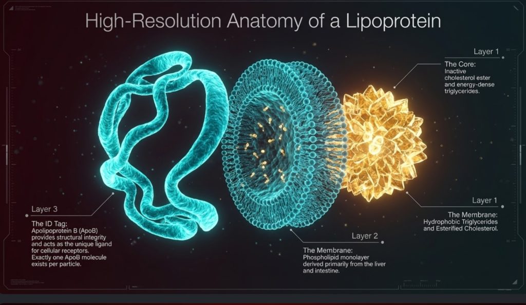
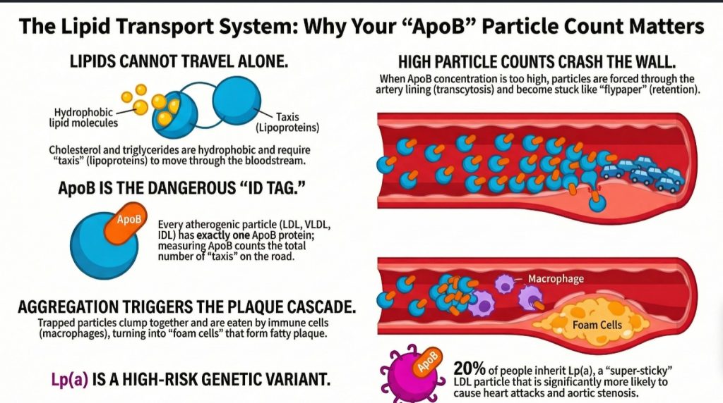
コレステロールは脂質であるため、疎水性(水を嫌う性質)であり、血漿という水分環境の中を自由に漂うことができません。そのため、次のような特殊な運搬体が必要となります。 リポタンパク質リポタンパク質とは、脂肪とコレステロールを血流に乗せて運ぶ小さなカプセルのことです。脂肪は水に溶けないため、移動するにはタンパク質の包みが必要です。.. 私たちはコレステロールばかり気にしていますが、これらの乗り物は本来、エネルギーを供給するために設計されています。 中性脂肪トリグリセリド(中性脂肪)は、血液中および体内の蓄積脂肪の主要な形態です。. そして決定的なことに、リン脂質です。これらのエネルギー輸送「トラック」が荷物を届けながら縮小するにつれて、表面のリン脂質を「脱落」させます。この脱落した断片は、 リン脂質転送タンパク質ホスホリピッド転移タンパク質(PLTP)は、トリグリセリドを豊富に含むリポタンパク質がその積荷を運搬する際に表面から放出したリン脂質を捕捉し、そのリン脂質を新生HDL粒子へと転移させることで、HDL粒子の成熟を可能にする血漿タンパク質である。. そして赤ちゃんの元へ届けられる HDLHDL、すなわち高密度リポタンパク質は、しばしば「善玉コレステロール」と呼ばれる粒子です。組織からコレステロールを回収し、肝臓へ運び戻します。. 粒子であり、それらが成熟することを可能にします。デイスプリング博士が名言しているように:

“リポタンパク質が運ばなければ、人間の体内において脂質はどこへも移動しない。”

テイクアウェイ2:「貨物」ではなく「ボート」である(ApoB革命)

リスクを正確に評価するには、「荷物」の重さを量るのをやめ、「ボート」の数を数え始めなければならない。従来の医学検査が測定しているのは LDL-C, 、すなわち低密度リポタンパク質に含まれるコレステロールの総量である。しかし、デイスプリング博士は、疾患の真の原因は粒子の数、特にアポリポタンパク質B(ApoB)によって特徴づけられる粒子であると主張している。 タンパク質タンパク質は、体内の筋肉や組織の構築と修復に使用される栄養素です。..

ここでの「マスタークラス」の洞察は1:1の比率です:すべての 動脈硬化惹起性粒子動脈硬化惹起性粒子とは、LDL、IDL、VLDL、リポ蛋白(a)といったアポB含有リポ蛋白のことであり、動脈壁に侵入・停滞してプラークの成長を開始・持続させます。本論文では、プラークの退縮を達成するために大幅かつ持続的に低下させなければならない対象を指す言葉としてこの用語を使用しています。.—それが VLDLVLDL(超低密度リポ蛋白)は、肝臓が中性脂肪を体内の他の部位へと送り出すために作り出す粒子です。., 、 IDLIDL(中等密度リポ蛋白)は、トリセリドを多く運ぶ大型の粒子が収縮してLDL粒子へと変化する過程の中間で形成される粒子です。., 、または LDLLDL(低密度リポ蛋白)は、コレステロールを血液中に運ぶ主要な粒子であり、動脈壁に詰まる主原因となるものです。.—はちょうど1分子の アポリポ蛋白BアポBは、動脈の壁に詰まってプラークを引き起こす可能性のあるコレステロール粒子のすべての外側に存在するタンパク質です。それらの粒子はそれぞれ、正確に1個のアポBを運んでいます。.. ApoBの測定は、血液中の「敵」である粒子を文字通り数えることになります。乳児の生理的レベルは20〜30 mg/dLであり、レベルが60 mg/dL未満の人には動脈硬化は基本的に起こりませんが、現代社会における成人のリスク閾値はしばしば80 mg/dLを超えています。「荷物」(LDL-C)の重さを量るよりも、「船」(ApoB)の数を数える方が、動脈での衝突の予測因子として圧倒的に正確です。.

重要ポイント3:「ハエ取り紙」効果と集約のインサイト

構築のプロセス 歯垢プラークとは、動脈の壁の内側にコレステロール、免疫細胞、瘢痕組織、カルシウムが蓄積したものです。., 、または アテローム発生アテローム性動脈硬化形成は、プラークが形成される段階的なプロセスです。., 動脈壁における工学的破綻である。 内皮内皮は、すべての血管の内側にある極めて薄く滑らかな裏地であり、厚さはわずか1細胞分です。. (単層の細胞からなる内層である 動脈動脈は、心臓から全身へ血液を送り出す血管です。.)は、ナイトクラブの専用の「ベルベットロープ(VIP専用の仕切り)」として機能する。しかし、ApoB粒子数が高い場合、これらの粒子は トランスサイトーシストランスサイトーシスとは、細胞が片側で物質を取り込み、反対側へと運び、そして反対側で放出しするプロセスのことである。..

動脈壁に入ると( 内膜内膜は動脈壁の一番内側の層であり、平滑な内壁のすぐ下に位置しています。.)、粒子は生物学的なハエ取り紙のように働く粘着性のある分子「プロテオグリカン」に遭遇します。一般的な「酸化仮説」では、これらの脂質が害を引き起こすためには化学的に損傷を受ける必要があるとされていますが、デイスプリングは、ヒトのデータによって裏付けられた、より重要なステップである「凝集」を指摘しています。ハエ取り紙に付着すると、これらの粒子は固まり合います。この凝集こそが、免疫系による暴食(フィーディング・フレンジー)を引き起こす「不法投棄」なのです。. マクロファージマクロファージは、ゴミや侵入者を飲み込む大きなどん欲な免疫細胞です。その名前は文字通り「大食い」を意味します。" (免疫貪食細胞)が到着して凝集した塊を「食べ」、肥大化して「“泡沫細胞”——文字通りの構成要素 動脈プラーク動脈壁内に脂質、免疫細胞、細胞破片、線維組織が蓄積して形成され、時間の経過とともに血流を狭窄または閉塞させる沈着物。アテローム性動脈硬化性病変またはアテロームとも呼ばれる。..

要点4:Lp(a)—遺伝的な「スリー・プレット(三重の脅威)」“

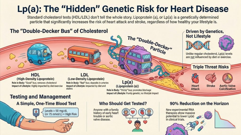
もしApoBがリスクの主な要因であるならば、リポタンパク質(a)、すなわちLp(a)は、それよりもさらに厄介な「親戚」と言えるでしょう。これは、ほとんどの人が聞いたこともない最も一般的な遺伝的異常であり、人口の20%に影響を及ぼしています。 「不運な両親」によって完全に決定されるLp(a)の値は、5歳までに安定し、従来の食事療法や運動では管理することができません。.

Lp(a) 3つの生物学的理由から「トリプルスレット(3つの脅威)」とみなされている:

  1. 動脈硬化誘発性:それはコレロステロールの荷物を血管壁に運び込む。.
  2. 血栓促進性:危険な 血栓血栓は、出血を止めるために形成される血球とタンパク質の塊です。..
  3. 石灰化促進:組織の硬化を刺激し、 大動脈弁狭窄症大動脈弁狭窄症とは、心臓の主要な流出弁である大動脈弁が狭くなったり硬くなったりして、左心室から大動脈への血流を阻害する疾患であり、高Lp(a)血症は石灰化大動脈弁狭窄症の第2の主要な原因として認識されています。. (心臓の主要な弁の硬化)。懸念レベルは >30 mg/dL(質量)または >75〜125 nmol/L(粒子数)から始まり、1回の検査が生死を分ける必要不可欠なものとなります。.

テイクアウト5:「ジャージー・ボーイ」の変身

自らを「ジャージー・ボーイ」と称するデイスプリング医師は、自身の人生の後半における変革を通じて、この科学に人間味を与えている。世界トップクラスの専門家であるにもかかわらず、デイスプリングは苦闘した。 肥満肥満とは、健康に影響を及ぼすほど過剰な体脂肪を蓄えている状態を意味します。. そして インスリン抵抗性インスリン抵抗性とは、細胞がインスリンに対して十分に応答しなくなる状態のことであり、そのため膵臓は同じ働きをするためにますます多くのインスリンを分泌し続けなければならなくなります。. 長年。72歳のとき、「トンネルの向こうに光が見えてきた」と悟った彼は、自分自身を救うために自らの科学を使うことを決意した。.

「スイッチを切り替える」ことによって 間欠的ファスティング間欠的断食は、食事の時間と意識的なカロリー制限の期間を繰り返す食事法であり、この記事の文脈では、デイスプリング医師が大幅な減量と代謝に関する検査値の改善を達成するために用いた糖質制限と併せて説明されています。. そして 炭水化物炭水化物は、パン、米、パスタ、果物、ジャガイモ、お菓子などの、食べ物に含まれる糖分やデンプンです。. 食事制限の結果、彼は67ポンド体重を減らした。さらに重要なことに、検査機関がリスクに応じて色分けしている彼の検査結果が、「赤と黄色」の警告ゾーンから「100% 緑」へと変化した。彼の友人であり、指導を受けているピーター・アッティア博士は次のように述べている:

“「これは、トムをできるだけ長くそばに置いておきたいという、私自身の利己的な動機だ。」”

結論:数字の向こう側

心血管疾患は、加齢に伴う避けられない代償ではありません。適切な指標を用いて早期に介入すれば、その進行を食い止め、さらには予防さえも可能な、管理可能な生物学的プロセスなのです。標準的な脂質プロファイル検査にとどまらず、アポBやLp(a)を測定することで、大惨事につながる前に、動脈内の「違法投棄」を特定することができます。 そのための手段はすでにあり、科学的根拠も確立されており、その結果は人生を変えるものです。なぜまだ医師にアポB検査を依頼していないのでしょうか?

ディープダイブ

分子レベルの血流と動脈疾患

ApoBリポタンパク質とRho GEFシグナル伝達が、アテローム性動脈硬化症のメカニズム解明にどのように寄与するか

動脈硬化症動脈硬化は、ほとんど的心筋梗塞と多くの脳卒中の背景にある病気です。コレステロールの粒子が動脈の壁に入り込み、体がそれを掃除するために免疫細胞を送り込み、何年もかけてその堆積物が硬化してプラークになります。. 多くの場合、あまりにも単純に説明され過ぎています。ほとんどの人は「高」と言われます コレステロールコレステロールは、体が必要とするロウ状の物質です。細胞壁、ホルモン、ビタミンD、そして食べ物を消化する胆汁の材料となります。コレステロールがなければ私たちは生きていけません。.”」は動脈を詰まらせるが、その考え方は正しい方向を指し示しているものの、実際に疾患を引き起こすより深層の生物学的メカニズムを見落としている。コレステロールそのものがすべてではない。より正確な見方としては、アテローム性動脈硬化は、アテローム形成性 リポタンパク質リポタンパク質とは、脂肪とコレステロールを血流に乗せて運ぶ小さなカプセルのことです。脂肪は水に溶けないため、移動するにはタンパク質の包みが必要です。. 粒子が 動脈動脈は、心臓から全身へ血液を送り出す血管です。. 血管壁に付着し、閉じ込められ、長期にわたる炎症の連鎖を引き起こす。それが起こるかどうかは、血液中に循環している粒子の数だけでなく、分子レベルでの動脈壁の挙動にも左右される [1-8].

この後半の部分は、多くの議論で認識されている以上に重要です。動脈壁は単なる受動的な管ではありません。それは能動的で、反応性の高い組織なのです。. 内皮細胞すべての血管の内面を覆う薄い細胞層であり、血管緊張の調節、血液凝固の防止、および動脈壁への物質の通過の制御を行います。また、その機能障害はアテローム性動脈硬化における初期の極めて重要な段階です。. どの粒子が血管壁を通過するかを制御する。免疫細胞は、それらの粒子が捕捉された後、どのように反応するかを決定する。. 平滑筋細胞平滑筋細胞は動脈の中膜を構成しており、血管の収縮や弛緩の程度を調節しています。. そして、Aが~であるかどうかを判断する手助けとなり、 歯垢プラークとは、動脈の壁の内側にコレステロール、免疫細胞、瘢痕組織、カルシウムが蓄積したものです。. 安定化するか、あるいは危険な状態になる。こうした局所的な調節の多くは、いわゆる Rhoグアニンヌクレオチド交換因子, 、または Rho GEF, 、順を追って Rho GTPases RhoA、Rac1、Cdc42など [4].

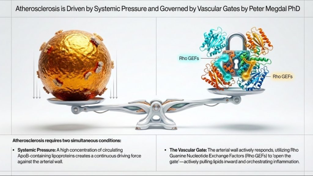
これは、〜について考えるための、より包括的な方法を提供します 心血管疾患心血管疾患とは、心臓発作、脳卒中、下肢の動脈閉塞など、心臓や血管に関する問題の総称です。.. 全身の脂質負荷、とりわけその数は アポB含有リポ蛋白リポタンパク質(LDL、IDL、VLDLおよびそれらの残渣を含む)は、それぞれ表面に1分子のアポリポタンパク質Bを結合している。各粒子が動脈壁に捕捉される可能性があるため、コレステロールの質量そのものではなく、粒子数がアテローム性動脈硬化症の主要な要因となっている。., 、システム内の圧力を設定します。しかし、動脈壁内の局所的なシグナル伝達が、それらの粒子が侵入し、留まり、プラークへと変化するかどうかを決定します。実用的な観点から言うと、, アポリポ蛋白BアポBは、動脈の壁に詰まってプラークを引き起こす可能性のあるコレステロール粒子のすべての外側に存在するタンパク質です。それらの粒子はそれぞれ、正確に1個のアポBを運んでいます。. 道路上の車両数を計測する一方、Rho GEFシグナル伝達は、ゲートがどの程度開いているか、また局所的な応答がどの程度破壊的になるかを決定するのに役立つ [1-4].

この関係を理解することは、高次元の臨床脂質学と現代の血管生物学の架け橋となります。また、この病気を一般の人々に説明しやすくすることにもつながります。動脈硬化は単なる「血中の脂肪」ではありません。それは 微粒子トラフィック、動脈への侵入、滞留、免疫活性化、および組織リモデリング.
そもそもなぜリポタンパク質が存在するのか

人体には、基本的な工学上の問題があります。それは、次のような脂質を輸送する必要があるという点です。 中性脂肪トリグリセリド(中性脂肪)は、血液中および体内の蓄積脂肪の主要な形態です。. やコレステロールも含まれていますが、血漿の大部分は水で構成されています。脂質は疎水性であり、水環境では溶けにくい性質を持っています。もし体内で未処理の脂肪がそのまま血流に放出されたら、水中の油のように分離して凝集してしまうでしょう [1,2].

その答えはリポタンパク質です。リポタンパク質は、疎水性の物質を水分の多い血漿中を運ぶために作られた輸送粒子です。リポタンパク質は、トリグリセリドやコレステロールエステルが蓄えられている疎水性のコアと、リン脂質や遊離コレステロール、そして タンパク質タンパク質は、体内の筋肉や組織の構築と修復に使用される栄養素です。. 呼ばれました アポリポプロテインアポリポ蛋白とは、血液中の脂肪を運ぶ粒子に結合しているタンパク質です。脂肪と水は混ざらないため、これらのタンパク質は脂肪が血流の中を安全に移動できるようにする包みのような役割を果たします。. [1,2,5].

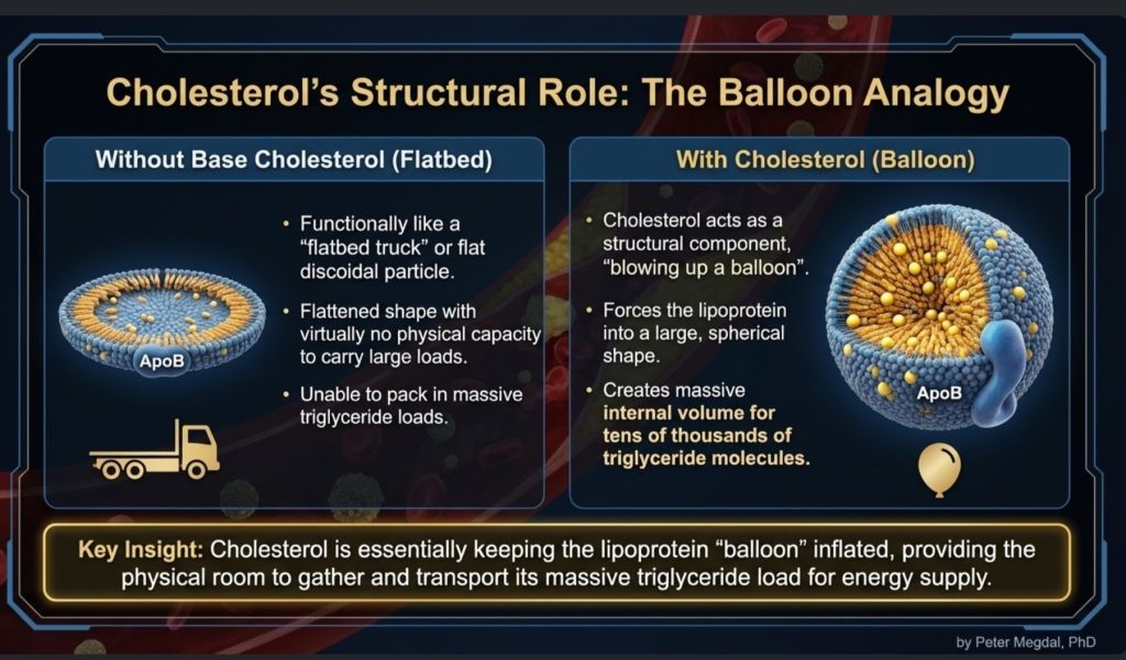
これは重要な点です。なぜなら、リポタンパク質はしばしば抽象的な検査値として扱われがちですが、実際には物質を運搬する物理的な「輸送手段」として理解するほうが適切だからです。リポタンパク質は、エネルギーや構造成分を体内で運搬します。トリグリセリドは、主要なエネルギー源です。コレステロールは、細胞膜の構造的支えとなり、ステロイドホルモンの合成に役立ち、 胆汁酸胆汁酸は、肝臓でコレステロールから生成され、脂肪の消化を助けるために腸内に放出されます。. production. The bloodstream is the highway, and lipoproteins are the fleet [1-3,5].

From that viewpoint, the body did not evolve lipoproteins mainly to move cholesterol. It evolved them to move energy. Cholesterol is necessary, but the transport system is deeply tied to fuel delivery. That distinction matters, because it helps clarify one of the most misunderstood issues in cardiovascular disease: the body does need cholesterol, but it does not need large quantities of cholesterol moving through plasma to keep tissues alive and functioning [2,11-15].

Cholesterol Homeostasis: How Little Is Actually Needed

A central but often overlooked fact in lipid biology is that the body requires only a very small amount of cholesterol at the cellular level. Every cell membrane needs cholesterol for structure and fluidity, and certain specialized tissues need it for steroid hormone synthesis. But that requirement is remarkably small, tightly regulated, and mostly satisfied locally rather than through bulk delivery from circulating LDLLDL(低密度リポ蛋白)は、コレステロールを血液中に運ぶ主要な粒子であり、動脈壁に詰まる主原因となるものです。. [2,11-17].

Cholesterol homeostasis is governed by highly regulated intracellular signaling pathways that maintain sufficiency while preventing toxicity. Contrary to common assumptions, virtually all nucleated cells possess the ability to synthesize their own cholesterol. They do not simply wait for lipoproteins to deliver it. They are genetically equipped to make what they need [11,12].

The central regulatory system is the SREBP-2–SCAP–INSIG axisThe SREBP-2–SCAP–INSIG axis is the cell's master cholesterol-sensing circuit: when intracellular cholesterol falls, the protein SCAP escorts SREBP-2 to the Golgi for activation, switching on genes for cholesterol synthesis and LDL-receptor expression; when cholesterol is sufficient, INSIG retains the complex in the endoplasmic reticulum, shutting the pathway down.. When intracellular cholesterol levels fall, ステロールステロールは、同じ4つの環状構造を基盤とするワックス状の分子のグループです。コレステロールは動物が作るものであり、植物は独自のステロールを作り出します。. regulatory element-binding protein 2 (SREBP-2SREBP-2 (sterol regulatory element-binding protein 2) is a transcription factor that senses low intracellular sterol levels in the liver and responds by increasing the production and surface expression of LDL receptors, thereby accelerating clearance of LDL from the blood.) is escorted by SREBP cleavage-activating protein (SCAP) from the endoplasmic reticulum to the Golgi apparatus. There, SREBP-2 undergoes proteolytic activation. The active transcription factor then enters the nucleus and upregulates genes involved in コレステロール合成Cholesterol synthesis is your body making its own cholesterol, mostly in the liver. Almost every cell can do it. and uptake, including HMG-CoA還元酵素HMG-CoA reductase is the rate-limiting enzyme in the liver's cholesterol biosynthetic (mevalonate) pathway; statins work by competitively blocking it, reducing the liver's own cholesterol production and prompting it to pull more LDL out of the bloodstream. (HMGCRHMGCR is the gene for HMG-CoA reductase, the enzyme that performs the rate-limiting step in making cholesterol. It is the exact target of every statin.) そして、その LDL受容体LDL受容体は、肝細胞の表面にあるドッキングポートであり、血液中からLDL粒子を捕捉して取り込み、分解する。. (LDL受容体LDLR is the gene that builds the LDL receptor, the docking port your liver uses to pull cholesterol particles out of circulation.) [11,12].

This drives the メバロン酸経路The mevalonate pathway is the multi-step biochemical route by which cells, primarily in the liver, synthesize cholesterol and related molecules; statins and bempedoic acid act at two separate nodes of this pathway to reduce cholesterol production., beginning with acetyl-CoA and proceeding through HMG-CoA and mevalonate toward cholesterol synthesis. Because this machinery exists throughout the body, cells do not depend on large plasma pools of cholesterol to preserve membrane integrity or basic survival [11,12].

When intracellular cholesterol rises, the system shuts itself down. Cholesterol and oxysterolsOxysterols are oxidised derivatives of cholesterol formed when cholesterol reacts with oxygen; within cells they act as signalling molecules that activate LXR and promote INSIG-mediated retention of the SCAP–SREBP complex, thereby suppressing further cholesterol synthesis and stimulating its export. promote INSIG-mediated retention of the SCAP–SREBP complex in the endoplasmic reticulum, reducing further transcriptional activation. At the same time, HMG-CoA還元酵素 is targeted for degradation through ubiquitin-proteasome pathways. In other words, cholesterol metabolism is controlled by a built-in feedback loop designed to avoid excess accumulation [11,12].

That excess matters, because free cholesterol is not benign when it builds up inside cells. Too much intracellular cholesterol can destabilize membranes, crystallize in lysosomes, trigger inflammatory signaling such as the NLRP3インフラマソームThe NLRP3 inflammasome is an intracellular protein complex in immune cells that, when activated by cholesterol crystals, oxidized lipids, or other danger signals within an atherosclerotic plaque, triggers the release of the inflammatory cytokines interleukin-1β and interleukin-6, accelerating plaque growth and instability., and promote apoptosis or necrosis [13,14]. This is one reason the body is not designed to stockpile it.

Cells therefore rely not only on synthesis and feedback inhibition, but also on active export. Through Liver X Receptor (LXR)The Liver X Receptor (LXR) is a nuclear receptor activated by oxysterols — oxidised derivatives of cholesterol — that induces expression of cholesterol exporters such as ABCA1 and ABCG1, promoting the removal of excess intracellular cholesterol toward HDL-mediated reverse cholesterol transport. signaling, oxysterols induce transporters such as ABCA1ABCA1は、コレステロールを細胞の外へ汲み出し、肝臓へ戻るためのHDL粒子に受け渡すタンパク質です。. そして ABCG1, which move excess cholesterol out of cells and toward HDL-mediated コレステロール引き抜き転送Reverse cholesterol transport is the process of moving cholesterol out of tissues, including artery walls, and back to the liver for disposal. HDL particles do the hauling. [15].

Taken together, these pathways show something important: cells need cholesterol, but only in small, controlled amounts. The dominant biological problem is not acquiring more cholesterol. It is balancing synthesis, use, storage, and efflux so that excess does not become toxic [11-15].

Tissue-Specific Cholesterol Independence

This principle becomes even clearer when looking at specific organs. Many tissues synthesize cholesterol locally and do not rely heavily on circulating LDL.

その central nervous system is the most striking example. Because apoB-containing lipoproteins do not meaningfully cross the blood-brain barrier, the brain must largely produce its own cholesterol. アストロサイトAstrocytes are a type of support cell (glia) in the brain that act as the primary cholesterol-manufacturing cells of the central nervous system, supplying cholesterol to neurons for membrane maintenance and myelin support. Because lipoproteins in the blood cannot cross the blood–brain barrier, the brain depends almost entirely on astrocyte-derived cholesterol synthesis. synthesize cholesterol and distribute it locally through apoE-containing lipoproteins, a process linked to LXR-regulated pathways [16].

The same is broadly true of steroidogenic tissues, including the adrenal glands and gonads. These tissues can synthesize cholesterol de novo from acetate. Although they express receptors such as LDLR and SR-B1SR-B1 is the receptor on liver cells that takes cholesterol from HDL particles and releases it for disposal in bile., their dependence on circulating LDL under ordinary conditions appears limited, with uptake becoming more relevant in specialized or high-demand states such as acute ACTH stimulation [2,17].

This supports an important physiological point. The body is not dependent on high circulating LDL-C levels for normal endocrine or cellular function. Indeed, mechanistic and clinical discussions in lipidology have emphasized that very low LDL-C levels, even around 10 mg/dL, do not necessarily imply impaired steroidogenesis or membrane failure when intracellular synthesis remains intact [2,11,17].

So while cholesterol is essential, the amount actually required in circulation for cellular survival is far smaller than many people assume. That reality reinforces the view that elevated circulating ApoB particles are not a biological necessity; they are a risk exposure.

The ApoB Fleet: Why Particle Number Matters

Among the many lipoproteins in circulation, the most relevant to atherosclerosis are the ApoB-containing particles. These include カイロミクロンカイロミクロンは、食事由来の脂肪を小腸から血流へと運ぶ非常に大きな粒子です。. and their remnants, very low-density lipoproteins (VLDLVLDL(超低密度リポ蛋白)は、肝臓が中性脂肪を体内の他の部位へと送り出すために作り出す粒子です。.)、中間密度リポタンパク質(IDLIDL(中等密度リポ蛋白)は、トリセリドを多く運ぶ大型の粒子が収縮してLDL粒子へと変化する過程の中間で形成される粒子です。.), low-density lipoproteins (LDL), and リポ蛋白(a)リポタンパク(a)(Lp(a)と表記され、「L-P-リトル-a」と発音される)は、余分な粘着性のあるタンパク質が付着したLDL様粒子です。., or Lp(a) [1,2].

The key clinical insight is that each 動脈硬化惹起性粒子動脈硬化惹起性粒子とは、LDL、IDL、VLDL、リポ蛋白(a)といったアポB含有リポ蛋白のことであり、動脈壁に侵入・停滞してプラークの成長を開始・持続させます。本論文では、プラークの退縮を達成するために大幅かつ持続的に低下させなければならない対象を指す言葉としてこの用語を使用しています。. contains exactly one ApoB molecule. That means ApoB serves as a direct count of the number of atherogenic particles in circulation. By contrast, LDL-C measures only the mass of cholesterol contained within LDL particles. A person can have a relatively normal LDL-C but still have a high number of particles if those particles are small and cholesterol-depleted. In that setting, risk can be underestimated if clinicians look only at LDL-C [1-3].

This is why many lipid experts now view ApoB as the more biologically meaningful metric. It reflects how many opportunities there are for particles to enter the artery wall. Put simply, if every ApoB particle is one potential arterial collision, then ApoB tells us the traffic density [1-3].

That traffic analogy becomes especially useful when explaining risk to non-specialists. The issue is not just how much cargo is in each truck. The issue is how many trucks are on the road, how long they stay there, and how likely they are to crash into the side wall.

This is also where the minimal-cholesterol discussion becomes clinically relevant. If tissues can synthesize the modest cholesterol they need, then high ApoB counts are not best understood as a beneficial delivery reserve. They are better understood as an increased burden of potentially arterial-penetrating particles [2,11-17].

From Fuel Delivery to LDL Persistence

The story begins with triglyceride-rich particles. The intestine produces chylomicrons to transport dietary fat. The liver produces VLDL to transport endogenous triglycerides. These are large particles whose major job is fuel delivery [1,2].

As these particles circulate, they interact with リポ蛋白リパーゼLipoprotein lipase is an enzyme anchored to the walls of small blood vessels that strips triglycerides out of passing particles and hands the fat to muscle and fat tissue. (LPL), an enzyme that unloads triglycerides for use by muscle, heart, and adipose tissue. As triglycerides are removed, the particle shrinks. Surface material is shed. But the ApoB backbone remains. Over time, VLDL is remodeled into IDL and then LDL [1,2,18].

LDL is therefore not some rogue or evil particle by design. It is a downstream product of normal lipoprotein metabolism. What makes LDL especially important is its persistence. LDL stays in circulation much longer than larger triglyceride-rich particles, often for 2 to 5 days, which increases the statistical chance that it will cross into the arterial wall [1,2].

This is where the disease model starts to sharpen. アテローム性動脈硬化形成アテローム性動脈硬化形成は、プラークが形成される段階的なプロセスです。. is not caused simply because LDL exists. It is caused because enough ApoB particles remain in circulation long enough to enter the 内膜内膜は動脈壁の一番内側の層であり、平滑な内壁のすぐ下に位置しています。. and become retained.

At the same time, this reinforces an important conceptual distinction. The presence of LDL in blood is not proof that tissues are desperate for cholesterol. Much of LDL biology reflects the remodeling and persistence of ApoB particles after triglyceride transport. In that sense, LDL is not merely a “cholesterol delivery truck.” It is also a long-circulating remnant of systemic lipid transport with increased opportunity to interact with the vessel wall [1,2,18].

Atherosclerosis Begins With Entry, Not Just Presence

One of the most useful upgrades in modern cardiovascular thinking is the realization that atherosclerosis does not begin merely because particles are in the blood. It begins when they cross the endothelial barrier and lodge in the 内皮下腔内皮下腔は、動脈の内膜のすぐ下、1層の細胞とその下の筋肉の間にある狭い隙間である。., or intima [1,6-8].

That means the artery wall matters enormously. The 内皮内皮は、すべての血管の内側にある極めて薄く滑らかな裏地であり、厚さはわずか1細胞分です。. is only one cell layer thick, but it is biologically active. It senses blood flow, responds to inflammatory signals, regulates barrier function, and controls which materials pass through. Certain arterial regions, especially branch points and curvatures, are more prone to plaque formation because the local flow pattern is disturbed. Those disturbed flow zones create a molecular environment that favors atherogenesis [4,6-8].

Historically, many people imagined LDL simply leaking through a damaged endothelium. The newer picture is more precise: LDL entry is an active, regulated process called トランスサイトーシストランスサイトーシスとは、細胞が片側で物質を取り込み、反対側へと運び、そして反対側で放出しするプロセスのことである。. [6-8].

This matters conceptually. If particle entry is regulated, then atherosclerosis is not just about what is in plasma. It is also about how the arterial wall responds to that plasma environment. Systemic exposure and local permissiveness work together.

DOCK4, SR-B1, and Regulated LDL Transcytosis

One of the most intriguing findings in recent years is that endothelial cells can actively transport LDL across themselves through a mechanism involving SR-B1 and DOCK4 [6-8].

SR-B1, or scavenger receptor class B type 1, has long been discussed in the context of HDLHDL、すなわち高密度リポタンパク質は、しばしば「善玉コレステロール」と呼ばれる粒子です。組織からコレステロールを回収し、肝臓へ運び戻します。. biology, but it also appears to play a role in the uptake and transcytosis of LDL under certain conditions. DOCK4 is a Rho GEF that activates Rac1, which helps drive the cytoskeletal changes and vesicular movement needed for this transport process [4,7,8].

This matters because it reframes the earliest step of atherogenesis. LDL entry into the vessel wall is not just accidental seepage. It is at least partly controlled by a specific signaling system. Even more striking, DOCK4 expression has been observed to increase in atherosclerosis-prone regions before visible 病変循環器学において、病変とは冠動脈を狭窄させるアテローム性動脈硬化プラークの不連続な領域を指し、通常はそれが引き起こす内腔閉塞のパーセンテージによって記述される。この記事では、最も重要な病変が治療された後、血管径が小さすぎてステントを受け入れることができない4つの遺残病変について述べている。. are present, suggesting that the artery wall may become permissive long before disease is obvious on imaging or pathology [4,8].

Clinically, that opens a provocative possibility. ApoB tells us how many particles are available to enter. But the DOCK4/SR-B1/Rac1 system may help determine how easily they can do so. In other words, ApoB measures the pressure at the gate; DOCK4 helps regulate the gate itself.

Retention: The Point of No Return

Entry alone is not enough. Once an ApoB particle reaches the intima, the next key event is retention. These particles bind to matrix molecules called proteoglycans, which act almost like arterial flypaper [1,3].

This step is crucial because a retained particle is now exposed to a very different microenvironment. Inside the arterial wall, it can undergo a series of modifications. It may oxidize, but oxidation is only part of the story. Lipases and other enzymes can alter the particle surface, generating lipids such as sphingomyelin and ceramide that make particles more adhesive and more likely to aggregate [4].

That aggregation step is especially important. A single isolated retained LDL particle is not the same thing biologically as a sticky cluster of modified particles. Once particles aggregate, they become far more likely to provoke マクロファージマクロファージは、ゴミや侵入者を飲み込む大きなどん欲な免疫細胞です。その名前は文字通り「大食い」を意味します。" uptake and 泡沫細胞A foam cell is an immune cell that has eaten so much trapped cholesterol that it swells up and looks foamy under a microscope. formation [1,4].

For clinicians and informed readers alike, this is a useful distinction: the danger is not just circulating LDL or even intimal LDL. The danger is retained, modified, aggregated ApoB-containing lipoprotein.

This is also the point at which “cholesterol toxicity” becomes context-specific. Cholesterol is necessary in tiny, regulated intracellular amounts, but once ApoB particles are trapped and modified in the arterial wall, their lipid cargo becomes part of a highly toxic inflammatory environment. The same sterol that is essential in membranes can become pathologic when delivered into the wrong tissue compartment in the wrong form [13,14].

Leukocyte Recruitment: How the Artery Becomes Inflamed

Once the intima contains retained and modified lipoproteins, the lesion shifts from a lipid problem to an inflammatory one. At this stage, the recruitment of monocytes and other leukocytes becomes central [4].

A Rho GEF called SGEF, also known as Arhgef26, appears to play a major role here. SGEF helps endothelial cells form ICAM-1-dependent docking structures, actin-rich protrusions that allow leukocytes to adhere more effectively under flow conditions. These structures are especially relevant in the high-shear environment of arteries, where immune cells might otherwise be washed away [4].

Another related signaling molecule, Arhgef1, links inflammatory recruitment to Angiotensin II signaling. This creates a mechanistic bridge between 高血圧Hypertension is the medical term for high blood pressure. and arterial 炎症炎症は、怪我や侵入物とみなしたものに対する免疫システムの反応です。これにより腫れや熱、そして浄化細胞がもたらされます。.. High 血圧血圧とは、血液が動脈の壁を押す力ののことです。120/80のように2つの数字で表されます。上の数字は心臓が収縮するときの圧力で、下の数字は弛緩するときの圧力です。. is not merely a hemodynamic burden; it can also amplify leukocyte recruitment through RhoA-mediated pathways [4].

This helps explain why cardiovascular 危険因子危険因子とは、高コレステロール粒子、高血圧、喫煙、糖尿病、家族歴など、病気にかかる可能性を高めるものです。. often cluster biologically. Hypertension, インスリン抵抗性インスリン抵抗性とは、細胞がインスリンに対して十分に応答しなくなる状態のことであり、そのため膵臓は同じ働きをするためにますます多くのインスリンを分泌し続けなければならなくなります。., and high ApoB do not act in isolation. They converge within the arterial wall, where endothelial signaling, immune recruitment, and lipoprotein retention reinforce one another.

Foam Cells and the Macrophage Response

Macrophages are often described as the body’s cleanup crew. In atherosclerosis, however, the cleanup operation can become self-destructive. Macrophages ingest modified and aggregated lipoproteins, become loaded with cholesterol, and transform into foam cells [1,4].

This step is not passive. It depends heavily on cytoskeletal remodeling and receptor signaling. The Vav family of Rho GEFs, especially Vav1, Vav2, and Vav3, helps regulate these processes. Vav proteins influence CD36-mediated uptake of oxidized lipoproteins and support formation of the lysosomal synapse, which allows macrophages to interact with and digest large lipoprotein aggregates [4].

As foam cells accumulate, they release inflammatory mediators, eventually die, and contribute to the 壊死核壊死性コアは、捕捉されたコレステロールを食べてその場で死亡した免疫細胞から形成された、進行したプラークの死滅したドロドロとした中心部である。. of the plaque. This transforms a microscopic lesion into a clinically meaningful plaque. Over time, the lesion can calcify, remain stable, or become vulnerable to rupture [1,3].

This is another point where the molecular framing adds value. The disease is not just about “too much cholesterol.” It is about how immune cells process abnormal lipid material inside a specific tissue environment.

The added cholesterol-homeostasis literature deepens this picture. Foam cell formation is not merely an issue of passive lipid storage. It is a breakdown of normal cholesterol handling. When macrophage uptake overwhelms intracellular control and efflux systems, free cholesterol accumulates, inflammatory pathways are activated, and cell death follows. In that sense, plaque progression can be understood as a localized failure of cholesterol homeostasis inside the arterial wall [13-15].

Smooth Muscle Cells and Plaque Stability

As plaques mature, vascular smooth muscle cells (VSMCs) become major players. These cells normally help maintain vascular tone, but in atherosclerosis they can change phenotype. They may migrate from the media to the intima, proliferate, and contribute to plaque structure [4].

This behavior is influenced by Rho GEF signaling. Molecules such as Kalirin and Vav3 promote migration and proliferation through Rac1- and RhoA-related pathways. Some of this remodeling may be protective, because VSMCs can help build a 線維性被膜The fibrous cap is the tough layer of tissue covering a plaque, separating its greasy core from the bloodstream. over the plaque. But excessive or disordered remodeling can also contribute to luminal narrowing and plaque instability [4].

By contrast, Arhgef7, also called beta-PIX, appears to support barrier integrity and protective cell polarity pathways. This suggests that plaque biology is not just driven by pro-disease signals. It is also shaped by counter-regulatory systems trying to preserve arterial structure [4].

The stability of a plaque therefore depends on a balance between damage, repair, inflammation, and remodeling. That balance is partly governed by the same family of molecular switches that influenced the lesion from the beginning.

Lp(a): The Genetic Risk Multiplier

No modern discussion of lipid-driven cardiovascular disease is complete without lipoprotein(a), or Lp(a). Structurally, Lp(a) is an LDL-like particle with an additional apolipoprotein(a) attached. Clinically, it is one of the most important inherited cardiovascular risk factors [1-3,5,9].

About one in five people has an elevated Lp(a), and levels are largely genetically determined. Unlike standard LDL, Lp(a) is not significantly improved by diet or exercise and is only modestly affected by most conventional lipid therapies [1-3,5,9].

Lp(a) appears to be especially dangerous because it is not only atherogenic but also pro-inflammatory and pro-thrombotic. Elevated levels are linked to premature 心筋梗塞詳しい項目については心臓発作をご覧ください。., 脳卒中脳卒中は、脳の一部への血流が詰まりまたは出血によって止まるときに起こります。., 、および 大動脈弁The aortic valve is the one-way gate between the heart's main pumping chamber and the aorta. 狭窄狭窄とは閉塞のことであり、通常は70%の閉塞といったようにパーセンテージで表されます。.. For this reason, many experts now recommend that every patient have Lp(a) measured at least once in a lifetime [1-3,5,9].

This is a major public health message. Someone can do many things right and still carry substantial inherited risk through elevated Lp(a). Identifying that risk early matters.

Why ApoB Is a Better Clinical Compass Than LDL-C

For everyday practice, the main diagnostic lesson is simple: ApoB is closer to the biology of disease than LDL-C [1-3].

LDL-C estimates how much cholesterol mass is present in LDL particles. ApoB estimates how many atherogenic particles are circulating. Since each particle is one potential entrant into the artery wall, ApoB better captures the opportunity for arterial injury.

Non-HDL cholesterolNon-HDL cholesterol is a simple calculation: your total cholesterol minus your HDL. What's left is the cholesterol riding in all the particles that can harm your arteries. is often a better surrogate than LDL-C, because it includes the cholesterol carried by all ApoB-containing particles. But ApoB remains the cleaner particle-based measure. Coronary artery calcium scoring can help detect established disease later in life, and Lp(a) helps identify inherited risk. But for understanding the causal burden of circulating atherogenic particles, ApoB is central [1-3].

This does not mean LDL-C is useless. It remains clinically valuable and widely available. But when the goal is to align testing with mechanism, ApoB is the stronger marker.

It also fits more cleanly with the physiology described above. If tissues do not require large amounts of circulating cholesterol to survive, and if cholesterol needs are largely met by local synthesis and tightly regulated intracellular pathways, then the clinically important question becomes not “How much cholesterol is present in plasma?” but “How many atherogenic particles are repeatedly interacting with the artery wall?” ApoB answers that better than LDL-C [2,11-17].

Therapeutic Implications: Today and Tomorrow

Current lipid-lowering therapy still matters enormously. スタチンスタチンは、肝臓がコレステロールを作るのに使う酵素の働きを遅らせます。肝臓は血液中からより多くのコレステロールを取り除くことでこれに反応し、そこに真の利益があります。., エゼチミブEzetimibe is a pill that blocks your intestines from absorbing cholesterol., 、および PCSK9阻害薬A PCSK9 inhibitor is a medicine that blocks that cholesterol-destroying protein, leaving more docking ports available to clear particles from the blood. reduce atherogenic 粒子負荷粒子負荷とは、血漿中を循環する動脈硬化性リポ蛋白粒子の総数のことであり、ApoBによって最もよく測定されます。これはコレステロール量とは区別されます。なぜなら、リポ蛋白が動脈壁に浸潤して捕捉される頻度を決定するのは、コレステロールの量ではなく、粒子の物理的な数だからです。. and have strong outcome data. PCSK9PCSK9 is a protein made by your liver that destroys the docking ports your liver uses to pull cholesterol out of your blood. inhibitors also lower Lp(a) modestly, and newer RNA-based therapies targeting Lp(a) are showing dramatic reductions in trials [1,3,9].

Lifestyle remains foundational. Weight lossWeight loss means reducing body fat, whether through food changes, exercise, medication, or surgery., improved インスリン感受性インスリン感受性とは、細胞がインスリンに対してどれだけよく反応するかということです。それはインスリン抵抗性の反対です。., reduced hepatic VLDL production, and lower inflammatory burden all help reduce systemic risk [1]. But the emerging biology suggests that the future of cardiovascular prevention may not stop at lowering plasma particles. It may also involve targeting the arterial wall directly.

That is where Rho GEF biology becomes especially exciting. If specific GEFs such as DOCK4 or SGEF are key regulators of LDL entry or leukocyte recruitment, then direct inhibition of those pathways might offer a more selective way to interfere with plaque formation [4]. Instead of only reducing traffic on the road, clinicians might one day also tighten the gate and blunt the inflammatory response inside the vessel wall.

This is still an emerging area, not a routine clinical strategy. But conceptually it points toward precision medicine: targeting the specific local pathways that convert circulating risk into arterial disease.

The integrated cholesterol-homeostasis framework also helps explain why aggressive lowering of ApoB burden is biologically plausible. If intracellular needs can still be met through endogenous synthesis and tightly regulated tissue handling, then reducing circulating atherogenic particles need not imply cellular deprivation. It may simply reduce unnecessary arterial exposure [2,11-17].

A Better Way to Explain Atherosclerosis

For the general public, the disease can be summarized in a way that is both intuitive and accurate.

ApoB-containing lipoproteins are like transport trucks carrying fuel and structural cargo through the bloodstream. If there are too many trucks on the road, some eventually cross into the artery wall. Once trapped there, they become damaged and sticky. The immune system sends in cleanup cells, but those cells get overloaded and create plaque. Meanwhile, the artery wall itself is not passive. It has molecular gatekeepers that regulate particle entry, immune docking, cholesterol handling, and tissue remodeling [1-8,11-15].

That framework is simple enough for public education but detailed enough to be useful to clinicians. It avoids the misleading oversimplification that cholesterol alone “clogs” arteries while preserving the core truth that cholesterol-containing ApoB particles are causally necessary for disease [1-3].

It also leaves room for an important nuance: the body needs cholesterol, but only in very small, tightly controlled amounts. The real danger is not the existence of cholesterol itself. The danger is the chronic circulation, arterial entry, and retention of ApoB-containing particles in a tissue environment that turns their cargo into inflammation and plaque.

結論

Atherosclerosis is best understood as the intersection of two systems. The first is systemic lipoprotein biology, where ApoB-containing particles circulate as the necessary carriers of triglycerides and cholesterol. The second is local arterial biology, where endothelial cells, leukocytes, macrophages, and smooth muscle cells decide whether those particles become plaque.

ApoB captures the number of circulating atherogenic particles and therefore the statistical opportunity for arterial injury. Rho GEF–Rho GTPase signaling helps explain why and how that injury occurs within the vessel wall. Together, these systems offer a richer model of disease than the old LDL-C-only framework.

At the same time, cholesterol itself is governed by a highly regulated intracellular economy. Through the SREBP-2–SCAP–INSIG system, the mevalonate pathway, LXR-mediated efflux signaling, and tissue-specific synthesis, the body maintains the small amounts of cholesterol it needs while defending itself against excess [11-17]. This means the physiological requirement for cholesterol is real, but the amount required is modest, locally managed, and fundamentally different from the pathological burden imposed by excess circulating ApoB particles.

For clinicians, this supports a stronger emphasis on ApoB, Lp(a), and early prevention. For the public, it clarifies that cardiovascular disease is not simply about eating fat or having “high cholesterol.” It is about how many atherogenic particles circulate, how long they persist, how the artery wall responds to them, and how failures of local cholesterol handling turn retained particles into inflammation and plaque.

The future of lipidology may therefore be dual: lowering the number of dangerous particles in the bloodstream while also targeting the molecular programs that let those particles enter and injure the artery wall in the first place [1-10]. In that broader framework, one of the most important conceptual corrections is this: the body does not need a large circulating surplus of cholesterol to survive. It needs only a very small, carefully regulated amount at the cellular level. Atherosclerosis begins when the transport system overshoots that need and the artery wall pays the price.

参考文献

  1. Perone F, Bernardi M, Spadafora L, et al. Non-Traditional Cardiovascular Risk Factors: Tailored Assessment and Clinical Implications. J Cardiovasc Dev Dis. 2025;12(5):171. Published 2025 Apr 28. doi:10.3390/jcdd12050171
  2. Attia P, Dayspring T. #20 – Tom Dayspring, M.D., FACP, FNLA – Part I of V: an introduction to lipidology.
  3. Hill S. What Causes Cardiovascular Disease? Lipid Series Part 1. The Proof with Simon Hill.
  4. Li M, Jiao Q, Xin W, et al. The Emerging Role of Rho Guanine Nucleotide Exchange Factors in Cardiovascular Disorders: Insights Into Atherosclerosis: A Mini Review. Front Cardiovasc Med. 2022;8:782098. Published 2022 Jan 3. doi:10.3389/fcvm.2021.782098
  5. Heart Matters. Cholesterol Explained: HDL, LDL, and the Hidden Risk of Lp(a).
  6. Hackethal V. Research Suggests LDL Is Actively Transported Into Arterial Walls. NeurologyLive.
  7. Getz GS, Reardon CA. The “HDL receptor” scavenger receptor class B type 1 finesses the uptake of low-density lipoproteins into the subendothelial space of arteries. Biotarget.
  8. Huang L, Chambliss KL, Gao X, et al. SR-B1 drives endothelial cell LDL transcytosis via DOCK4 to promote atherosclerosis. Nature. 2019;569(7757):565-569. doi:10.1038/s41586-019-1140-4
  9. Nutrition Made Simple. Everything you need to know about Lp(a) | ft. Dr. Tom Dayspring.
  10. Heart Fit Clinic. Webinar on Cholesterol from a Lipidologist. Dr. Tom Dayspring.
  11. Brown MS, Goldstein JL. The SREBP pathway: regulation of cholesterol metabolism by proteolysis of a membrane-bound transcription factor. Cell. 1997;89(3):331-340. doi:10.1016/s0092-8674(00)80213-5
  12. Brown MS, Goldstein JL. Sterol regulatory element binding proteins (SREBPs): controllers of lipid synthesis and cellular uptake. Nutr Rev. 1998;56(2 Pt 2):S1-S75. doi:10.1111/j.1753-4887.1998.tb01680.x
  13. Muneer PMA, Alikunju S, Mishra V, et al. Activation of NLRP3 inflammasome by cholesterol crystals in alcohol consumption induces atherosclerotic lesions. Brain Behav Immun. 2017;62:291-305. doi:10.1016/j.bbi.2017.02.014
  14. Feng B, Yao PM, Li Y, et al. The endoplasmic reticulum is the site of cholesterol-induced cytotoxicity in macrophages. Nat Cell Biol. 2003;5(9):781-792. doi:10.1038/ncb1035
  15. Murphy AJ, Akhtari M, Tolani S, et al. ApoE regulates hematopoietic stem cell proliferation, monocytosis, and monocyte accumulation in atherosclerotic lesions in mice. J Clin Invest. 2011;121(10):4138-4149. doi:10.1172/JCI57559
  16. Dietschy JM, Turley SD. Cholesterol metabolism in the brain. Curr Opin Lipidol. 2001;12(2):105-112. doi:10.1097/00041433-200104000-00003
  17. Miller WL. MECHANISMS IN ENDOCRINOLOGY: Rare defects in adrenal steroidogenesis. Eur J Endocrinol. 2018;179(3):R125-R141. doi:10.1530/EJE-18-0279
  18. Ginsberg HN. Lipoprotein metabolism and its relationship to atherosclerosis. Med Clin North Am. 1994;78(1):1-20. doi:10.1016/s0025-7125(16)30174-2


透明性に関する注記: この記事はAIツールの支援を受けて作成されました。最終的なコンテンツは著者によって慎重に確認および編集されており、その正確性については著者が責任を負います。提供される情報は教育目的のみのものであり、医学的なアドバイスを構成するものではありません。.

AIアプリ

心臓病リスク計算ツール

Hスコアのインサイト、視覚的な家系図入力、共有可能なPDFレポートを備えた、教育用の家族歴心臓病リスク計算ツール。.

このアプリが非常に重要である理由をここでご覧ください。.