
Tráfego Molecular e Doença Arterial
Como as Lipoproteínas ApoB e a Sinalização de Rho GEF Ajudam a Explicar a Aterosclerose
Aterosclerose é frequentemente explicado de forma muito simples. A maioria das pessoas ouve que “alto colesterol”entope as artérias, e embora essa ideia aponte na direção certa, ela deixa de fora a biologia mais profunda que realmente impulsiona a doença. O colesterol em si não é toda a história. O retrato mais preciso é que a aterosclerose se desenvolve quando aterogênico lipoproteína partículas entram no artéria parede, ficam presos e desencadeiam uma longa cascata inflamatória. Se isso acontece depende não apenas de quantas partículas estão circulando no sangue, mas também de como a parede arterial se comporta a nível molecular [1-8].
Essa segunda parte importa mais do que a maioria das discussões reconhece. A parede arterial não é um tubo passivo. É um tecido ativo e responsivo. Células endoteliais regulam quais partículas cruzam para a parede do vaso. As células imunológicas decidem como reagir uma vez que essas partículas são retidas. Células musculares lisas então ajude a determinar se um placa estabiliza ou se torna perigoso. Boa parte dessa regulação local é controlada por uma família de interruptores moleculares conhecida como Fatores de troca de nucleotídeos de guanina Rho, ou GEFs para Rho, trabalhando em Rho GTPases tais como RhoA, Rac1 e Cdc42 [4].
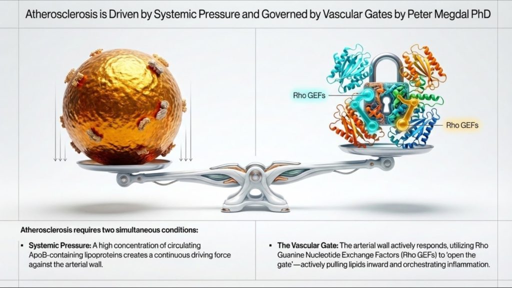
Isso fornece uma maneira mais completa de pensar sobre doença cardiovascular. Carga lipídica sistêmica, especialmente o número de partículas contendo ApoB, define a pressão no sistema. Mas a sinalização local dentro da parede arterial determina se essas partículas têm a entrada permitida, se são retidas e transformadas em placa. Em termos práticos, ApoB mede o número de veículos na estrada, enquanto a sinalização de Rho GEF ajuda a decidir quão aberto o portão está e quão destrutiva será a resposta local1-4].
Compreender essa relação ajuda a unir a lipidologia clínica de alto nível à biologia vascular moderna. Também torna a doença mais fácil de explicar ao público. A aterosclerose não é apenas “gordura no sangue”. É um problema de tráfego de partículas, entrada arterial, retenção, ativação imune e remodelação tecidual.
Por que as lipoproteínas existem para começar
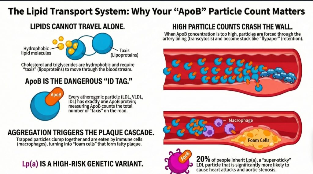
O corpo humano tem um problema básico de engenharia. Ele precisa transportar lipídios, como triglicerídeos e o colesterol, mas o plasma sanguíneo é composto principalmente de água. Os lipídios são hidrofóbicos, o que significa que eles não se dissolveu bem em um ambiente aquoso. Se o corpo simplesmente liberasse gordura pura na corrente sanguínea, ela se separaria e aglomeraria, muito parecido com o óleo na água [1,2].
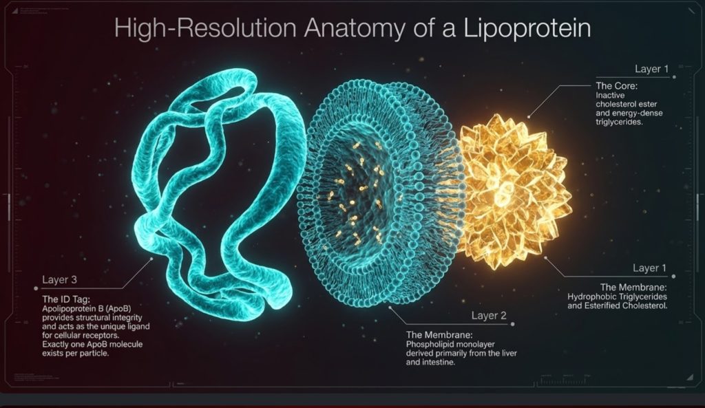
A solução é a lipoproteína. As lipoproteínas são partículas de transporte construídas para carregar carga hidrofóbica através do plasma aquoso. Elas têm um núcleo hidrofóbico, onde triglicerídeos e ésteres de colesterol são armazenados, e uma superfície mais compatível com a água feita de fosfolipídios, colesterol livre e proteínas chamado apolipoproteínas [1,2,5].
Isso é importante porque as lipoproteínas são frequentemente tratadas como valores laboratoriais abstratos, mas são mais bem compreendidas como veículos de transporte físico. Elas movimentam energia e materiais estruturais pelo corpo. Os triglicerídeos são a principal carga de energia. O colesterol fornece suporte estrutural para membranas, síntese de hormônios esteroides e ácido biliar produção. A corrente sanguínea é a rodovia, e as lipoproteínas são a frota1-3,5].
Desse ponto de vista, o corpo não evoluiu lipoproteínas principalmente para mover colesterol. Ele as evoluiu para mover energia. O colesterol é necessário, mas o sistema de transporte está profundamente ligado à entrega de combustível. Essa distinção importa, porque ajuda a esclarecer uma das questões mais mal compreendidas nas doenças cardiovasculares: o corpo precisa de colesterol, mas não precisa de grandes quantidades de colesterol circulando pelo plasma para manter os tecidos vivos e funcionando [2,11-15].
Homeostase do Colesterol: Quão pouco é realmente necessário
Um fato central, mas frequentemente negligenciado na biologia dos lipídios, é que o corpo requer apenas uma quantidade muito pequena de colesterol no nível celular. Cada membrana celular precisa de colesterol para estrutura e fluidez, e certos tecidos especializados precisam dele para a síntese de hormônios esteroides. Mas essa necessidade é notavelmente pequena, rigidamente regulada e, na maior parte, satisfeita localmente, em vez de por meio do fornecimento em massa a partir da circulação LDL [2,11-17].
A homeostase do colesterol é governada por vias de sinalização intracelular altamente reguladas que mantêm a suficiência enquanto evitam a toxicidade. Ao contrário das suposições comuns, praticamente todas as células nucleadas possuem a capacidade de sintetizar seu próprio colesterol. Elas não simplesmente esperam que as lipoproteínas o entreguem. Elas estão geneticamente equipadas para produzir o que precisam [11,12].
O sistema regulatório central é o Eixo SREBP-2–SCAP–INSIG. Quando os níveis intracelulares de colesterol caem, esterol proteína ligadora de elemento regulatório 2 (SREBP-2) é escoltado por Proteína ativadora da clivagem de SREBP (SCAP) do retículo endoplasmático para o aparelho de Golgi. Lá, a SREBP-2 sofre ativação proteolítica. O fator de transcrição ativo então entra no núcleo e regula positivamente os genes envolvidos na síntese de colesterol e absorção, incluindo Redutase de HMG-CoA (HMGCR) e o Receptor de LDL (LDLR) [11,12].
Isso impulsiona o via do mevalonato, começando com acetil-CoA e prosseguindo através do HMG-CoA e do mevalonato em direção à síntese de colesterol. Como essa maquinaria existe em todo o corpo, as células não dependem de grandes estoques plasmáticos de colesterol para preservar a integridade da membrana ou a sobrevivência básica [11,12].
Quando o colesterol intracelular aumenta, o sistema se desliga. Colesterol e oxiesteróis promover Retenção mediada por INSIG do complexo SCAP-SREBP no retículo endoplasmático, reduzindo a ativação transcricional adicional. Ao mesmo tempo, Redutase de HMG-CoA é direcionado para degradação através de vias de ubiquitina-proteassoma. Em outras palavras, o metabolismo do colesterol é controlado por um mecanismo de feedback integrado projetado para evitar o acúmulo excessivo [11,12].
Esse excesso importa, porque o colesterol livre não é benigno quando se acumula dentro das células. Muito colesterol intracelular pode desestabilizar membranas, cristalizar em lisossomos e desencadear sinalização inflamatória, como o Inflamassomo NLRP3, e promover apoptose ou necrose [13,14]. Essa é uma das razões pelas quais o corpo não foi feito para acumulá-lo.
As células, portanto, dependem não apenas da síntese e da inibição por feedback, mas também da exportação ativa. Através Receptor X do Fígado (LXR) sinalização, os oxiesteróis induzem transportadores como ABCA1 e ABCG1, que removem o excesso de colesterol das células em direção ao mediado por HDL transporte reverso de colesterol [15].
Consideradas em conjunto, essas vias mostram algo importante: as células precisam de colesterol, mas apenas em quantidades pequenas e controladas. O principal problema biológico não é adquirir mais colesterol. É equilibrar a síntese, o uso, o armazenamento e o efluxo para que o excesso não se torne tóxico [11-15].
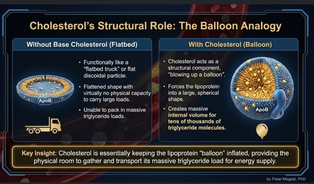
Independência de Colesterol Específica de Tecido
Este princípio torna-se ainda mais claro ao observar órgãos específicos. Muitos tecidos sintetizam colesterol localmente e não dependem fortemente do LDL circulante.
O sistema nervoso central é o exemplo mais marcante. Como as lipoproteínas contendo apoB não cruzam significativamente a barreira hematoencefálica, o cérebro precisa produzir em grande parte seu próprio colesterol. Astrócitos sintetizar colesterol e distribuí-lo localmente através do lipoproteínas contendo apoE, um processo ligado a vias reguladas por LXR [16].
O mesmo vale em grande parte para tecidos esteroidogênicos, incluindo as glândulas adrenais e as gônadas. Esses tecidos podem sintetizar colesterol de novo a partir do acetato. Embora expressem receptores como LDLR e SR-B1, a dependência delas de LDL circulante em condições normais parece limitada, com a captação tornando-se mais relevante em estados especializados ou de alta demanda, como a estimulação aguda por ACTH [2,17].
Isso apoia um ponto fisiológico importante. O corpo não depende de níveis circundantes altos de LDL-C para a função endócrina ou celular normal. De fato, discussões mecanicistas e clínicas na lipidologia têm enfatizado que níveis muito baixos de LDL-C, mesmo em torno de 10 mg/dL, não implicam necessariamente em esteroidogênese prejudicada ou falha de membrana quando a síntese intracelular permanece intacta2,11,17].
Portanto, embora o colesterol seja essencial, a quantidade realmente necessária em circulação para a sobrevivência celular é muito menor do que muitas pessoas assumem. Essa realidade reforça a visão de que partículas de ApoB circulantes elevadas não são uma necessidade biológica; elas são uma exposição ao risco.
A Frota de ApoB: Por que a Contagem de Partículas Importa
Entre as muitas lipoproteínas em circulação, as mais relevantes para a aterosclerose são as partículas contendo ApoB. Estas incluem quilomícrons e seus remanescentes, lipoproteínas de muito baixa densidade (VLDL), lipoproteínas de densidade intermediária (IDL), lipoproteínas de baixa densidade (LDL) e lipoproteína(a), ou Lp(a) [1,2].
O principal insight clínico é que cada partícula aterogênica contém exatamente uma molécula de ApoB. Isso significa que a ApoB serve como uma contagem direta do número de partículas aterogênicas em circulação. Em contraste, o LDL-C mede apenas a massa de colesterol contida nas partículas de LDL. Uma pessoa pode ter um LDL-C relativamente normal, mas ainda assim apresentar um alto número de partículas se essas partículas forem pequenas e pobres em colesterol. Nesse cenário, o risco pode ser subestimado se os clínicos olharem apenas para o LDL-C [1-3].
É por isso que muitos especialistas em lipídios agora veem a ApoB como a métrica biologicamente mais significativa. Ela reflete quantas oportunidades existem para as partículas entrarem na parede arterial. Em termos simples, se cada partícula de ApoB é uma potencial colisão arterial, então a ApoB nos diz a densidade do tráfego [1-3].
Essa analogia do trânsito se torna especialmente útil ao explicar o risco para não especialistas. A questão não é apenas quanta carga há em cada caminhão. A questão é quantos caminhões estão na estrada, quanto tempo eles ficam lá e qual a probabilidade de baterem na parede lateral.
É também aqui que a discussão sobre o colesterol mínimo se torna clinicamente relevante. Se os tecidos podem sintetizar o colesterol modesto de que precisam, então as contagens elevadas de ApoB não são melhor compreendidas como uma reserva de entrega benéfica. Elas são mais bem compreendidas como uma carga aumentada de partículas potencialmente penetrantes nas artérias [2,11-17].
Do Fornecimento de Combustível à Persistência de LDL
A história começa com partículas ricas em triglicerídeos. O intestino produz quilomícrons para transportar gordura da dieta. O fígado produz VLDL para transportar triglicerídeos endógenos. Estas são partículas grandes cuja principal função é a entrega de combustível [1,2].
À medida que essas partículas circulam, elas interagem com lipase lipoproteica (LPL), uma enzima que descarrega triglicerídeos para uso pelo tecido muscular, cardíaco e adiposo. Conforme os triglicerídeos são removidos, a partícula encolhe. O material de superfície é eliminado. Mas o arcabouço de ApoB permanece. Com o tempo, a VLDL é remodelada em IDL e depois em LDL [1,2,18].
O LDL, portanto, não é alguma partícula rebelde ou maligna por design. Ele é um produto a jusante do metabolismo normal das lipoproteínas. O que torna o LDL especialmente importante é a sua persistência. O LDL permanece em circulação muito mais tempo do que as partículas maiores ricas em triglicerídeos, frequentemente de 2 a 5 dias, o que aumenta a chance estatística de ele atravessar para a parede arterial [1,2].
É aqui que o modelo da doença começa a ficar mais nítido. Aterogênese não é causado simplesmente porque o LDL existe. É causado porque partículas de ApoB suficientes permanecem em circulação por tempo suficiente para entrar na íntima e ser retido.
Ao mesmo tempo, isso reforça uma importante distinção conceitual. A presença de LDL no sangue não é prova de que os tecidos estão desesperados por colesterol. Boa parte da biologia da LDL reflete a remodelação e a persistência de partículas de ApoB após o transporte de triglicerídeos. Nesse sentido, a LDL não é meramente um “caminhão de entrega de colesterol”. Ela também é um remanescente de longa circulação do transporte lipídico sistêmico com maior oportunidade de interagir com a parede arterial [1,2,18].
A aterosclerose começa com a entrada, e não apenas com a presença
Uma das atualizações mais úteis no pensamento cardiovascular moderno é a constatação de que a aterosclerose não começa apenas porque há partículas no sangue. Ela começa quando elas cruzam a barreira endotelial e se alojam na espaço subendotelial, ou íntima [1,6-8].
Isso significa que a parede da artéria tem uma importância enorme. A endotélio tem apenas uma camada celular de espessura, mas é biologicamente ativa. Ela detecta o fluxo sanguíneo, responde a sinais inflamatórios, regula a função de barreira e controla quais materiais passam por ela. Certas regiões arteriais, especialmente pontos de ramificação e curvaturas, são mais propensas à formação de placa, pois o padrão local de fluxo é perturbado. Essas zonas de fluxo perturbado criam um ambiente molecular que favorece a aterogênese [4,6-8].
Historicamente, muitas pessoas imaginavam que o LDL simplesmente vazava através de um endotélio danificado. O panorama mais recente é mais preciso: a entrada do LDL é um processo ativo e regulado chamado transcitose [6-8].
Isso é importante do ponto de vista conceitual. Se a entrada de partículas é regulada, então a aterosclerose não se resume apenas ao que está presente no plasma. Tem a ver também com a forma como a parede arterial responde a esse ambiente plasmático. A exposição sistêmica e a permissividade local atuam em conjunto.
DOCK4, SR-B1 e transcitosis regulada do LDL
Uma das descobertas mais intrigantes dos últimos anos é que as células endoteliais podem transportar ativamente o LDL através de si mesmas por meio de um mecanismo que envolve SR-B1 e DOCK4 [6-8].
O SR-B1, ou receptor scavenger da classe B, tipo 1, vem sendo discutido há muito tempo no contexto de HDL biologia, mas também parece desempenhar um papel na captação e transcitose do LDL sob certas condições. O DOCK4 é um Rho GEF que ativa o Rac1, o que ajuda a impulsionar as alterações no citoesqueleto e o movimento vesicular necessários para esse processo de transporte [4,7,8].
Isso é importante porque redefine a compreensão da etapa inicial da aterogênese. A entrada do LDL na parede vascular não se trata apenas de uma infiltração acidental. Ela é, pelo menos em parte, controlada por um sistema de sinalização específico. Mais impressionante ainda, observou-se que a expressão do DOCK4 aumenta em regiões propensas à aterosclerose antes mesmo que sejam visíveis lesões estão presentes, sugerindo que a parede arterial pode se tornar vulnerável muito antes que a doença se torne evidente em exames de imagem ou na análise patológica [4,8].
Do ponto de vista clínico, isso abre uma possibilidade instigante. A ApoB nos indica quantas partículas estão disponíveis para entrar. Mas o sistema DOCK4/SR-B1/Rac1 pode ajudar a determinar com que facilidade elas conseguem fazê-lo. Em outras palavras, a ApoB mede a pressão no portão; o DOCK4 ajuda a regular o próprio portão.
Retenção: O ponto sem volta
A entrada, por si só, não é suficiente. Quando uma partícula de ApoB atinge a íntima, o próximo evento fundamental é a retenção. Essas partículas se ligam a moléculas da matriz chamadas proteoglicanos, que atuam quase como uma papelada adesiva arterial [1,3].
Essa etapa é crucial, pois a partícula retida passa a estar exposta a um microambiente muito diferente. Dentro da parede arterial, ela pode sofrer uma série de modificações. Ela pode se oxidar, mas a oxidação é apenas parte do processo. Lipases e outras enzimas podem alterar a superfície da partícula, gerando lipídios como a esfingomielina e a ceramida, que tornam as partículas mais aderentes e mais propensas à agregação [4].
Essa etapa de agregação é especialmente importante. Uma única partícula de LDL retida e isolada não é, do ponto de vista biológico, a mesma coisa que um aglomerado aderente de partículas modificadas. Uma vez que as partículas se agregam, elas passam a ter uma probabilidade muito maior de provocar macrófago absorção e célula de espuma formação [1,4].
Tanto para os médicos quanto para os leitores bem informados, essa é uma distinção útil: o perigo não está apenas no LDL circulante, nem mesmo no LDL intimal. O perigo está na lipoproteína contendo ApoB retida, modificada e agregada.
É também nesse ponto que a “toxicidade do colesterol” passa a depender do contexto. O colesterol é necessário em quantidades minúsculas e reguladas no interior das células, mas, uma vez que as partículas de ApoB ficam retidas e modificadas na parede arterial, sua carga lipídica passa a fazer parte de um ambiente inflamatório altamente tóxico. O mesmo esterol que é essencial nas membranas pode se tornar patológico quando transportado para o compartimento tecidual errado na forma errada [13,14].
Recrutamento de leucócitos: como a artéria fica inflamada
Quando a íntima passa a conter lipoproteínas retidas e modificadas, a lesão deixa de ser um problema lipídico para se tornar um problema inflamatório. Nessa fase, o recrutamento de monócitos e outros leucócitos passa a ser fundamental [4].
Um Rho GEF chamado SGEF, também conhecido como Arhgef26, parece desempenhar um papel importante nesse contexto. O SGEF auxilia as células endoteliais a formar estruturas de acoplamento dependentes de ICAM-1, protuberâncias ricas em actina que permitem que os leucócitos adiram de forma mais eficaz em condições de fluxo. Essas estruturas são especialmente relevantes no ambiente de alto cisalhamento das artérias, onde, de outra forma, as células imunológicas poderiam ser arrastadas [4].
Outra molécula de sinalização relacionada, a Arhgef1, associa o recrutamento inflamatório à sinalização da angiotensina II. Isso cria uma ponte mecanística entre hipertensão e arterial inflamação. Alto pressão arterial não é apenas uma sobrecarga hemodinâmica; também pode intensificar o recrutamento de leucócitos por meio de vias mediadas pela RhoA [4].
Isso ajuda a explicar por que as doenças cardiovasculares fatores de risco costumam se agrupar biologicamente. Hipertensão, resistência à insulina, e os níveis elevados de ApoB não atuam isoladamente. Eles convergem na parede arterial, onde a sinalização endotelial, o recrutamento imunológico e a retenção de lipoproteínas se reforçam mutuamente.
Células espumosas e a resposta dos macrófagos
Os macrófagos são frequentemente descritos como a equipe de limpeza do corpo. Na aterosclerose, no entanto, essa operação de limpeza pode se tornar autodestrutiva. Os macrófagos ingerem lipoproteínas modificadas e agregadas, ficam carregados de colesterol e se transformam em células espumosas [1,4].
Essa etapa não é passiva. Ela depende fortemente da remodelação do citoesqueleto e da sinalização dos receptores. A família Vav de Rho GEFs, especialmente Vav1, Vav2 e Vav3, ajuda a regular esses processos. As proteínas Vav influenciam a captação mediada por CD36 de lipoproteínas oxidadas e favorecem a formação da sinapse lisossômica, o que permite que os macrófagos interajam com grandes agregados de lipoproteínas e os digiram [4].
À medida que as células espumosas se acumulam, elas liberam mediadores inflamatórios, acabam morrendo e contribuem para o núcleo necrótico da placa. Isso transforma uma lesão microscópica em uma placa clinicamente significativa. Com o tempo, a lesão pode se calcificar, permanecer estável ou tornar-se vulnerável à ruptura [1,3].
Esse é outro ponto em que a abordagem molecular agrega valor. A doença não se resume apenas ao “excesso de colesterol”. Trata-se de como as células imunológicas processam o material lipídico anormal dentro de um ambiente tecidual específico.
A literatura adicional sobre a homeostase do colesterol aprofunda esse quadro. A formação de células espumosas não é meramente uma questão de armazenamento passivo de lipídios. Trata-se de uma falha no processamento normal do colesterol. Quando a captação pelos macrófagos sobrecarrega os sistemas de controle e efluxo intracelulares, o colesterol livre se acumula, as vias inflamatórias são ativadas e ocorre a morte celular. Nesse sentido, a progressão da placa pode ser entendida como uma falha localizada da homeostase do colesterol dentro da parede arterial [13-15].
Células musculares lisas e estabilidade da placa
À medida que as placas se desenvolvem, as células musculares lisas vasculares (VSMCs) passam a desempenhar um papel fundamental. Essas células normalmente ajudam a manter o tônus vascular, mas, na aterosclerose, podem sofrer alterações fenotípicas. Elas podem migrar da média para a íntima, proliferar e contribuir para a estrutura da placa [4].
Esse comportamento é influenciado pela sinalização do Rho GEF. Moléculas como a kalirina e a Vav3 promovem a migração e a proliferação por meio de vias relacionadas ao Rac1 e ao RhoA. Parte dessa remodelação pode ter um efeito protetor, pois as células musculares lisas da parede vascular (VSMCs) podem ajudar a formar uma cápula fibrosa sobre a placa. No entanto, uma remodelação excessiva ou desordenada também pode contribuir para o estreitamento da luz do vaso e para a instabilidade da placa [4].
Em contrapartida, o Arhgef7, também chamado de beta-PIX, parece contribuir para a integridade da barreira e para as vias de polaridade celular protetoras. Isso sugere que a biologia da placa não é impulsionada apenas por sinais pró-doença. Ela também é moldada por sistemas contrarreguladores que tentam preservar a estrutura arterial [4].
A estabilidade de uma placa depende, portanto, de um equilíbrio entre dano, reparo, inflamação e remodelação. Esse equilíbrio é, em parte, regulado pela mesma família de interruptores moleculares que influenciou a lesão desde o início.
Lp(a): O multiplicador do risco genético
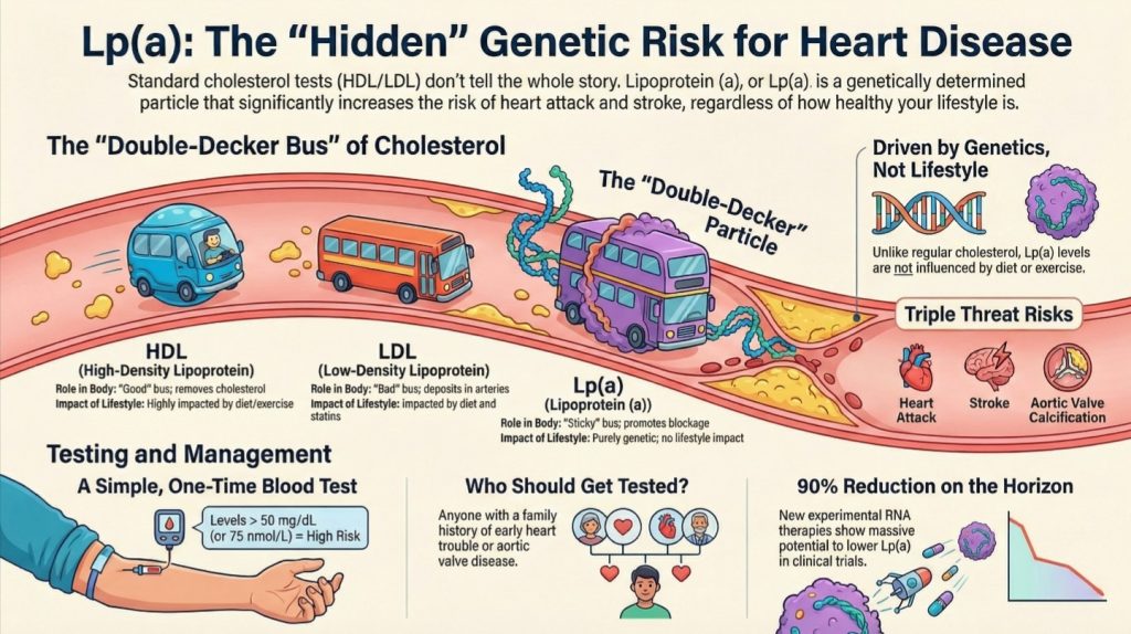
Nenhuma discussão moderna sobre doenças cardiovasculares relacionadas aos lipídios está completa sem a menção à lipoproteína (a), ou Lp(a). Estruturalmente, a Lp(a) é uma partícula semelhante ao LDL, à qual está ligada uma apolipoproteína (a) adicional. Clinicamente, ela é um dos mais importantes fatores de risco cardiovasculares hereditários [1-3,5,9].
Cerca de uma em cada cinco pessoas apresenta níveis elevados de Lp(a), e esses níveis são, em grande parte, determinados geneticamente. Ao contrário do LDL comum, os níveis de Lp(a) não melhoram significativamente com dieta ou exercícios físicos e são afetados apenas de forma modesta pela maioria das terapias lipídicas convencionais [1-3,5,9].
A Lp(a) parece ser especialmente perigosa, pois não é apenas aterogênica, mas também pró-inflamatória e pró-trombótica. Níveis elevados estão associados à morte prematura infarto do miocárdio, acidente vascular cerebral, e valva aórtica estenose. Por esse motivo, muitos especialistas recomendam agora que todos os pacientes façam a medição da Lp(a) pelo menos uma vez na vida [1-3,5,9].
Esta é uma mensagem importante de saúde pública. Uma pessoa pode ter muitos hábitos saudáveis e, mesmo assim, apresentar um risco genético significativo devido aos níveis elevados de Lp(a). É fundamental identificar esse risco precocemente.
Por que a ApoB é um indicador clínico mais confiável do que o LDL-C
Para a prática clínica cotidiana, a principal lição diagnóstica é simples: a ApoB está mais intimamente ligada à biologia da doença do que o LDL-C [1-3].
O LDL-C estima a quantidade de massa de colesterol presente nas partículas de LDL. A ApoB estima quantas partículas aterogênicas estão circulando. Como cada partícula representa um potencial agente de penetração na parede arterial, a ApoB reflete melhor o risco de lesão arterial.
Colesterol não-HDL costuma ser um indicador substituto melhor do que o LDL-C, pois inclui o colesterol transportado por todas as partículas que contêm ApoB. Mas a ApoB continua sendo a medida mais precisa baseada nas partículas. A pontuação de cálcio nas artérias coronárias pode ajudar a detectar a doença já estabelecida em idades mais avançadas, e a Lp(a) ajuda a identificar o risco hereditário. No entanto, para compreender o impacto causal das partículas aterogênicas circulantes, a ApoB é fundamental [1-3].
Isso não significa que o LDL-C seja inútil. Ele continua sendo clinicamente valioso e amplamente disponível. Mas, quando o objetivo é alinhar o exame ao mecanismo, a ApoB é o marcador mais confiável.
Isso também se encaixa de forma mais coerente com a fisiologia descrita acima. Se os tecidos não necessitam de grandes quantidades de colesterol circulante para sobreviver, e se as necessidades de colesterol são atendidas em grande parte pela síntese local e por vias intracelulares rigidamente reguladas, então a questão clinicamente importante deixa de ser “Quanto colesterol está presente no plasma?”, mas sim “Quantas partículas aterogênicas estão interagindo repetidamente com a parede arterial?”. A ApoB responde a essa pergunta melhor do que o LDL-C [2,11-17].
Implicações terapêuticas: hoje e amanhã
A terapia hipolipemiante atual ainda é de extrema importância. Estatinas, ezetimiba, e Inibidores da PCSK9 reduzir a aterogênese carga de partículas e apresentem dados sólidos sobre os resultados. PCSK9 Os inibidores também reduzem moderadamente a Lp(a), e as terapias mais recentes baseadas em RNA, direcionadas à Lp(a), estão apresentando reduções drásticas em ensaios clínicos [1,3,9].
O estilo de vida continua sendo fundamental. Perda de peso, aprimorado sensibilidade à insulina, a redução da produção hepática de VLDL e a diminuição da carga inflamatória contribuem para reduzir o risco sistêmico [1]. No entanto, os recentes avanços na biologia sugerem que o futuro da prevenção cardiovascular pode não se limitar à redução das partículas plasmáticas. Ele também pode envolver a ação direta sobre a parede arterial.
É aí que a biologia das proteínas GEF da família Rho se torna especialmente interessante. Se proteínas GEF específicas, como a DOCK4 ou a SGEF, forem reguladores-chave da entrada do LDL ou do recrutamento de leucócitos, então a inibição direta dessas vias poderia oferecer uma maneira mais seletiva de interferir na formação de placas [4]. Em vez de apenas reduzir o tráfego na via, os médicos poderão, um dia, também restringir o acesso e atenuar a resposta inflamatória dentro da parede do vaso.
Essa ainda é uma área emergente, não uma estratégia clínica de rotina. Mas, conceitualmente, ela aponta para a medicina de precisão: atuar diretamente nas vias locais específicas que transformam o risco circulante em doença arterial.
O modelo integrado de homeostase do colesterol também ajuda a explicar por que a redução agressiva da carga de ApoB é biologicamente plausível. Se as necessidades intracelulares ainda puderem ser atendidas por meio da síntese endógena e do manejo tecidual rigidamente regulado, então a redução das partículas aterogênicas circulantes não precisa implicar em privação celular. Isso pode simplesmente reduzir a exposição arterial desnecessária [2,11-17].
Uma maneira melhor de explicar a aterosclerose
Para o público em geral, a doença pode ser resumida de uma forma que seja ao mesmo tempo intuitiva e precisa.
As lipoproteínas que contêm ApoB são como caminhões de transporte que levam combustível e carga estrutural pela corrente sanguínea. Se houver caminhões demais na estrada, alguns acabam atravessando a parede arterial. Uma vez presos ali, eles ficam danificados e pegajosos. O sistema imunológico envia células de limpeza, mas essas células ficam sobrecarregadas e formam a placa. Enquanto isso, a própria parede arterial não fica passiva. Ela possui “guardiões” moleculares que regulam a entrada de partículas, a ligação do sistema imunológico, o manejo do colesterol e a remodelação tecidual [1-8,11-15].
Esse modelo é simples o suficiente para a educação do público, mas detalhado o bastante para ser útil aos profissionais da área médica. Ele evita a simplificação exagerada e enganosa de que o colesterol, por si só, “obstrui” as artérias, ao mesmo tempo em que preserva a verdade fundamental de que as partículas de ApoB contendo colesterol são causalmente necessárias para o desenvolvimento da doença [1-3].
Isso também abre espaço para uma nuance importante: o corpo precisa do colesterol, mas apenas em quantidades muito pequenas e rigidamente controladas. O verdadeiro perigo não é a existência do colesterol em si. O perigo está na circulação crônica, na entrada nas artérias e na retenção de partículas contendo ApoB em um ambiente tecidual que transforma seu conteúdo em inflamação e placa.
Conclusão
A aterosclerose é melhor compreendida como a interseção de dois sistemas. O primeiro é a biologia sistêmica das lipoproteínas, na qual as partículas contendo ApoB circulam como transportadoras essenciais de triglicerídeos e colesterol. O segundo é a biologia arterial local, na qual as células endoteliais, os leucócitos, os macrófagos e as células musculares lisas determinam se essas partículas se transformam em placa.
A ApoB reflete o número de partículas aterogênicas circulantes e, portanto, o risco estatístico de lesão arterial. A via de sinalização Rho GEF–Rho GTPase ajuda a explicar por que e como essa lesão ocorre na parede vascular. Juntos, esses sistemas oferecem um modelo mais abrangente da doença do que o antigo paradigma baseado exclusivamente no LDL-C.
Ao mesmo tempo, o próprio colesterol é regido por um equilíbrio intracelular altamente regulado. Por meio do sistema SREBP-2–SCAP–INSIG, da via do mevalonato, da sinalização de efluxo mediada pelo LXR e da síntese específica de cada tecido, o corpo mantém as pequenas quantidades de colesterol de que necessita, ao mesmo tempo em que se defende contra o excesso [11-17]. Isso significa que a necessidade fisiológica de colesterol é real, mas a quantidade necessária é modesta, regulada localmente e fundamentalmente diferente da carga patológica imposta pelo excesso de partículas de ApoB em circulação.
Para os profissionais da área médica, isso reforça a importância de dar maior ênfase à ApoB, à Lp(a) e à prevenção precoce. Para o público em geral, isso esclarece que as doenças cardiovasculares não se resumem simplesmente ao consumo de gordura ou ao fato de ter “colesterol alto”. Trata-se de quantas partículas aterogênicas circulam, por quanto tempo elas persistem, como a parede arterial responde a elas e como falhas no processamento local do colesterol transformam as partículas retidas em inflamação e placa.
O futuro da lipidologia pode, portanto, ter duas vertentes: reduzir o número de partículas perigosas na corrente sanguínea e, ao mesmo tempo, atuar sobre os mecanismos moleculares que permitem que essas partículas entrem na parede arterial e a danifiquem [1-10]. Nesse contexto mais amplo, uma das correções conceituais mais importantes é a seguinte: o corpo não precisa de um grande excedente de colesterol em circulação para sobreviver. Ele precisa apenas de uma quantidade muito pequena, cuidadosamente regulada no nível celular. A aterosclerose começa quando o sistema de transporte excede essa necessidade e a parede arterial acaba pagando o preço.
Referências
- Perone F, Bernardi M, Spadafora L, et al. Non-Traditional Cardiovascular Risk Factors: Tailored Assessment and Clinical Implications. J Cardiovasc Dev Dis. 2025;12(5):171. Published 2025 Apr 28. doi:10.3390/jcdd12050171
- Attia P, Dayspring T. #20 – Tom Dayspring, M.D., FACP, FNLA – Part I of V: an introduction to lipidology.
- Hill S. What Causes Cardiovascular Disease? Lipid Series Part 1. The Proof with Simon Hill.
- Li M, Jiao Q, Xin W, et al. The Emerging Role of Rho Guanine Nucleotide Exchange Factors in Cardiovascular Disorders: Insights Into Atherosclerosis: A Mini Review. Front Cardiovasc Med. 2022;8:782098. Published 2022 Jan 3. doi:10.3389/fcvm.2021.782098
- Heart Matters. Cholesterol Explained: HDL, LDL, and the Hidden Risk of Lp(a).
- Hackethal V. Research Suggests LDL Is Actively Transported Into Arterial Walls. NeurologyLive.
- Getz GS, Reardon CA. The “HDL receptor” scavenger receptor class B type 1 finesses the uptake of low-density lipoproteins into the subendothelial space of arteries. Biotarget.
- Huang L, Chambliss KL, Gao X, et al. SR-B1 drives endothelial cell LDL transcytosis via DOCK4 to promote atherosclerosis. Nature. 2019;569(7757):565-569. doi:10.1038/s41586-019-1140-4
- Nutrition Made Simple. Everything you need to know about Lp(a) | ft. Dr. Tom Dayspring.
- Heart Fit Clinic. Webinar on Cholesterol from a Lipidologist. Dr. Tom Dayspring.
- Brown MS, Goldstein JL. The SREBP pathway: regulation of cholesterol metabolism by proteolysis of a membrane-bound transcription factor. Cell. 1997;89(3):331-340. doi:10.1016/s0092-8674(00)80213-5
- Brown MS, Goldstein JL. Sterol regulatory element binding proteins (SREBPs): controllers of lipid synthesis and cellular uptake. Nutr Rev. 1998;56(2 Pt 2):S1-S75. doi:10.1111/j.1753-4887.1998.tb01680.x
- Muneer PMA, Alikunju S, Mishra V, et al. Activation of NLRP3 inflammasome by cholesterol crystals in alcohol consumption induces atherosclerotic lesions. Brain Behav Immun. 2017;62:291-305. doi:10.1016/j.bbi.2017.02.014
- Feng B, Yao PM, Li Y, et al. The endoplasmic reticulum is the site of cholesterol-induced cytotoxicity in macrophages. Nat Cell Biol. 2003;5(9):781-792. doi:10.1038/ncb1035
- Murphy AJ, Akhtari M, Tolani S, et al. ApoE regulates hematopoietic stem cell proliferation, monocytosis, and monocyte accumulation in atherosclerotic lesions in mice. J Clin Invest. 2011;121(10):4138-4149. doi:10.1172/JCI57559
- Dietschy JM, Turley SD. Cholesterol metabolism in the brain. Curr Opin Lipidol. 2001;12(2):105-112. doi:10.1097/00041433-200104000-00003
- Miller WL. MECHANISMS IN ENDOCRINOLOGY: Rare defects in adrenal steroidogenesis. Eur J Endocrinol. 2018;179(3):R125-R141. doi:10.1530/EJE-18-0279
- Ginsberg HN. Lipoprotein metabolism and its relationship to atherosclerosis. Med Clin North Am. 1994;78(1):1-20. doi:10.1016/s0025-7125(16)30174-2

