このページは自動翻訳されています。相違がある場合は、英語版が優先されます。.

改訂日:2026年7月16日

目に見えない梗塞:MINOCAにおけるメカニズム、診断、管理に関する詳細な研究検証

著:ピーター・メグダル博士

この記事の使い方

医療上の免責事項: この記事は教育目的のものであり、医学的な助言ではありません。個別の指導については、必ずかかりつけの医師にご相談ください。.

読みやすい

病院のベッドに横たわっていると想像してください。胸が締め付けられ、まるで重い石が乗っかっているようです。息をするのも苦しいです。自分が間違いなく起こしているという確信があるため、恐怖を感じています。 心臓発作. 医師たちはすばやく動きます。心臓のパイプを見るために、あなたを特別な部屋へ連れて行きます。この検査の名前は 血管造影. それは医師が心房細動を調べる標準的な方法です。.

少しして、医師が戻ってきます。彼は驚いたような顔をしています。良いニュースのはずなのに、何かがおかしく感じられる知らせをあなたに告げます。「あなたの動脈はきれいに見えます。大きな詰まりはありません。心臓のパイプは開いています」と彼は言います。“

あなたは安心なんてしていません。自分の身体がどう感じたか、あなた自身が分かっているからです。痛みが本物だったことも知っています。これは多くの患者にとって恐怖の瞬間です。「血管(パイプ)」が詰まっていないのに、どうして心臓発作が起きるのでしょうか?

長い間、医師たちも患者と同じように混乱していました。今日、科学にはその答えがあります。この疾患の名前は MINOCA. 。それは次のような意味を持つ長い名前です。“心筋梗塞 閉塞性を伴わない 冠状動脈.。」簡単に言えば、これは実際に心筋梗塞を起こしたものの、主要な冠動脈が少なくとも50%開いている状態を指します。これを「見えない心筋梗塞」と呼んでいます。”

テイクアウト 1:あなたの心は単純なキッチンの配管ではない

多くの人は心臓を家の配管のようなものだと考えています。医師はこれを「配管モデル」と呼ぶことがよくあります。この考え方では、心臓のパイプは台所の流し台のようなものです。長年の間に、油やゴミがたまっていきます。このゴミは次のように呼ばれます 歯垢. やがてパイプにごみが詰まりすぎて、水が通らなくなります。かつては、すべての心臓発作もそのように起こるのだと考えられていました。.

しかし、心臓は生きていて動いている体のパーツです。金属のパイプよりもはるかに複雑です。「配管モデル」はあまりにも単純すぎます。それは人々権に、パイプが開いていれば心臓は健康に違いないと思い込ませます。MINOCAはこれが間違いである証明しています。.

シンクのパイプではなく、庭用のホースを思い浮かべてみてください。ホースの中を覗いても、大きな詰まりは見当たらないかもしれません。しかし、ホースが突然鋭く折れ曲がったらどうでしょう?あるいは、ホースの内側が剥がれてめくれてきたらどうでしょう?それでも水の流れは止まってしまいます。もし油の大きな固まりのような詰まりだけを探していたら、本当の問題は見逃されてしまうでしょう。.

循環器医学は現在、大きな転換期を迎えています。かつて医師たちはあなたのような症例を単なる奇妙な偶然と考えていましたが、現在では、独自の特別なケアを必要とする特定型の心臓疾患として捉えています。.

近代心臓病学の大部分において、急性心筋梗塞(AMI)は「配管モデル」に支配されてきた。すなわち、動脈硬化性プラークが何年もかけて蓄積し、冠動脈を徐々に狭窄させるというものである。 ルーメン, 、そして最終的には破裂する……。しかし、現代の心血管科学により、大きな修正が迫られている。つまり、冠動脈造影で閉塞が認められない場合でも、真の心筋梗塞は発生し得るのである。 狭窄.

この「パラダイムシフト」は、医師が単純な「詰まったシンク」という発想から脱却しつつあることを意味します。これは患者であるあなたにとって重要なステップです。つまり、管に異常が見当たらない場合でも、医師はそこでの探索をやめず、さらに深く調べ始めるべきだということです。.

テイクアウェイ 2:MINOCAは最終的な答えではなく、手がかりに過ぎない

医師からMINOCA(冠動脈非閉塞性心筋梗塞)と告げられると、まるで謎が解けたように感じられるかもしれません。しかし、重要なのは、MINOCAが「“作業診断.それはスタートラインであり、ゴールではありません。.

刑事は事件現場にいる。窓が割られていないのを見ても、彼はまだ泥棒を捕まえていない。彼はただ泥棒がどのようにして侵入したかを知っているだけだ。 しなかった 家の中に入る。それがMINOCAだ。この検査結果により、医師は、心臓発作の原因が、大動脈の巨大な「100%」という閉塞ではなかったことを知ることができる。.

MINOCAと呼ぶのは、医師が「私は知っている 起きたんだ――心臓発作を起こしたんだ――だから今、調べないと なぜ.”

医師から「異常なし」と言われ、そこで終わってしまうのは危険です。これでは本当の原因究明が止まってしまいます。それはまるで、泥棒がまだ家にいるのに、窓に異常がないという理由で探偵が捜査を諦めるようなものです。MINOCAという診断名を、さらに調査を続けるためのきっかけとして活用しなければなりません。真の原因が残した「指紋」を探し出さなければならないのです。.

ポイント3:「5%クラブ」は、あなたが思っているよりも規模が大きい

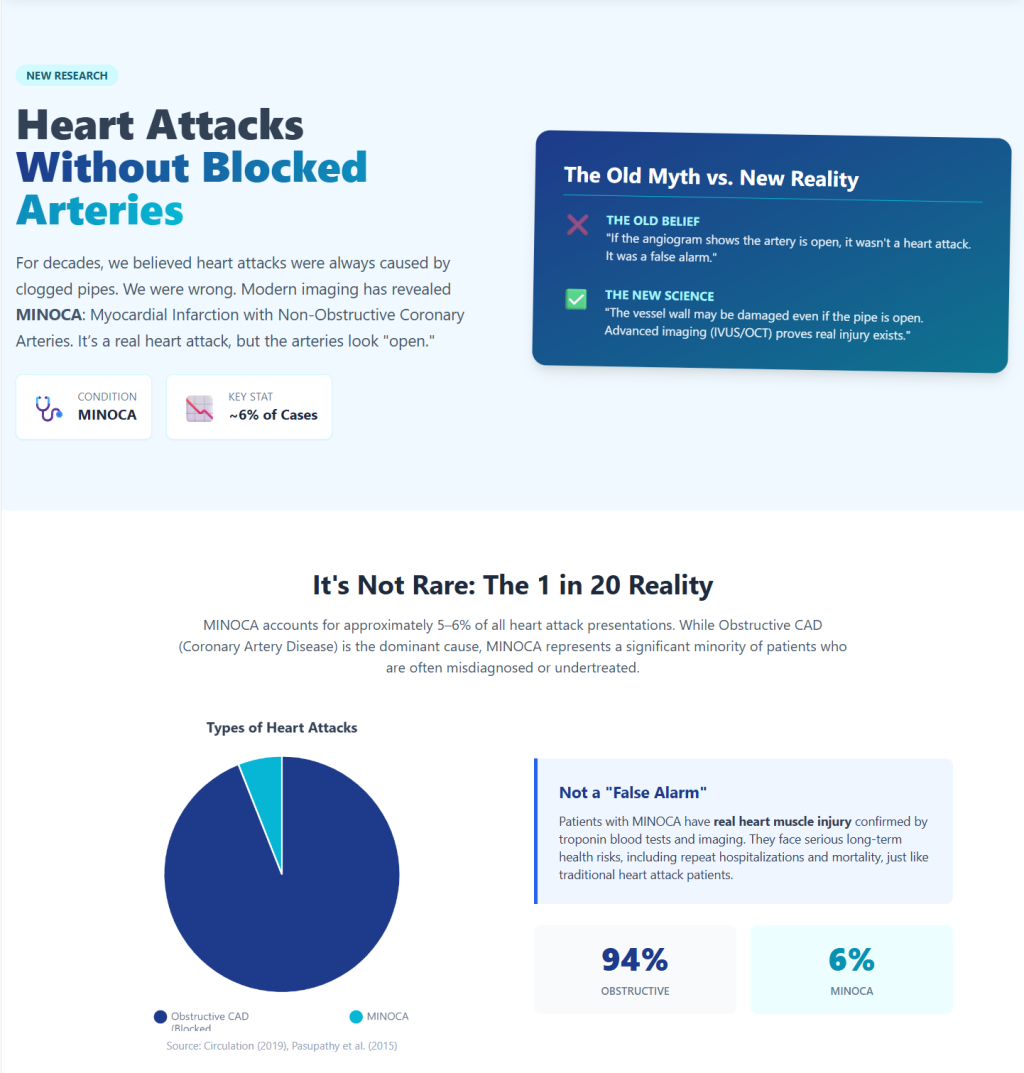
「自分だけがこんな目に遭った」と感じるかもしれませんが、決して一人ではありません。心筋梗塞全体の約5%~6%がMINOCA症例です。.

それは少ない数字に聞こえるかもしれませんが、毎年どれほどの人が心臓発作を起こしているかを考えてみてください。5%か6%ということは、何千人もの人々が痛みを感じているにもかかわらず、動脈は「正常(異常なし)」だと言われているということです。.

この「5%クラブ」には、非常に特定の条件を満たす患者たちがいます。男性よりも女性に多く見られます。また、若い患者にも多く見られます。 かつて、こうした患者の多くは、胸の痛みは単なるストレスや「神経のせい」だと言われていました。「血管の詰まり」がないという理由で、何の説明も受けずに帰宅させられていたのです。.

これは重大な健康上の問題です。もし自分の声が届いていないと感じるなら、何千人もの他の人々もあなたと全く同じ境遇にあることを思い出してください。こうした数字を知ることは、私たちがより良いケアを求めて声を上げる力になります。これが、医師が真剣に受け止めなければならない現実の病状であることを証明しているのです。.

Takeaway 4: 「プラーク」があっても「閉塞」がない場合がある“

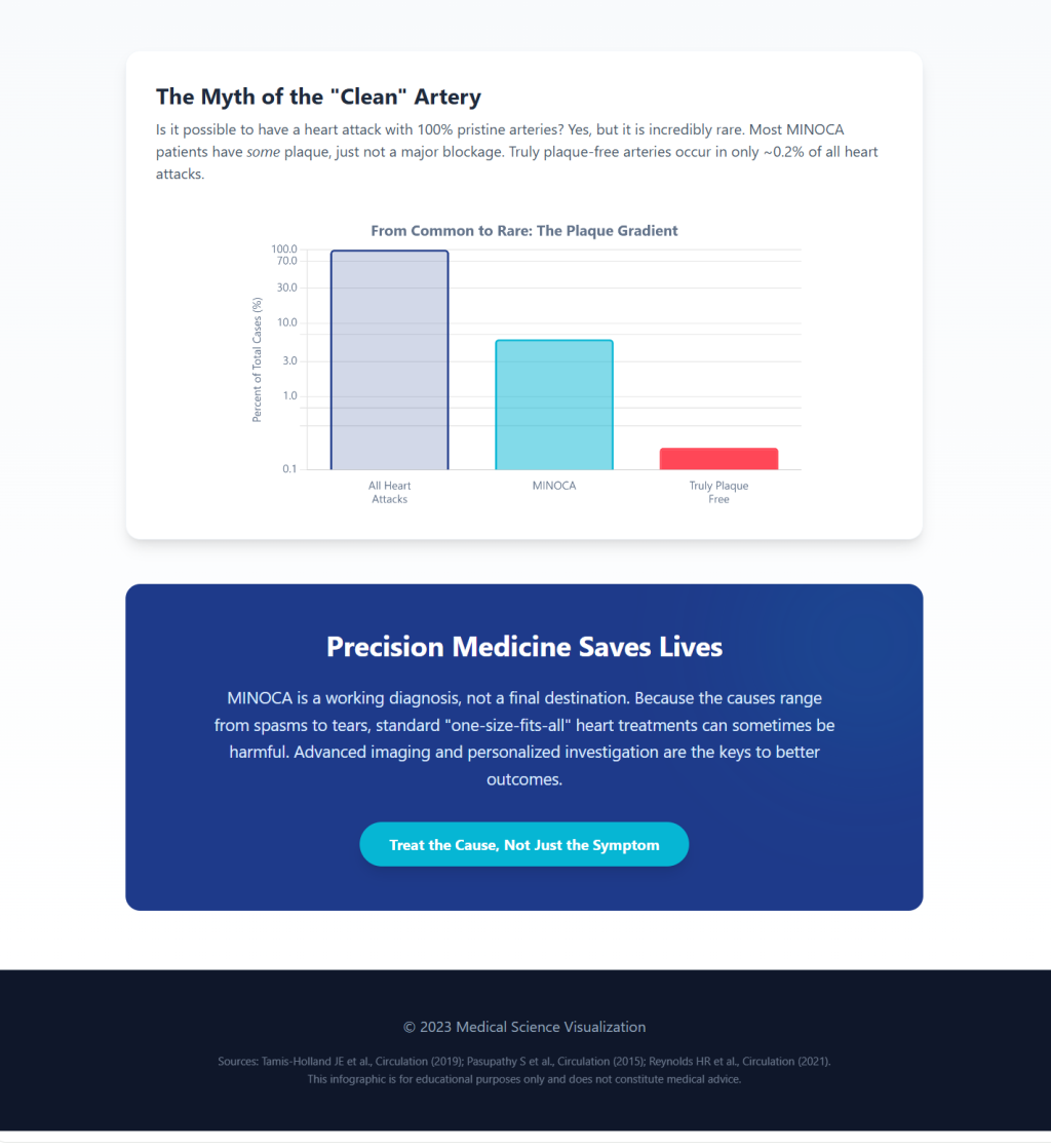
「きれいな動脈」が「完璧な動脈」を意味するという大きな誤解が存在します。MINOCAにおいては、これが事実であることはほとんどありません。医師があなたの動脈は「閉塞性ではない」と言うとき、それは専門用語を使っているのです。.

医療の世界では、「閉塞」とは、管が50%以上塞がっている場合にのみそう呼ばれます。管が20%や40%ほどゴミで詰まっている場合、医師はしばしばそれを「非閉塞性」だと言います。 しかし、40%まで詰まっている状態は、それでも「正常」とは言えません。“

皮膚にできたにきびを思い浮かべてください。にきびは小さく、腕の動きを妨げることはありません。しかし、そのにきびが「つぶれる」か破れると、小さな傷ができ、かさぶたになります。これと同じことが心臓の血管の中でも起こりうるのです。血流をほとんど妨げないような、小さなプラークの隆起があるかもしれません。しかし、その小さな隆起がつぶれると、微小な 血栓. その血栓が心臓発作を引き起こす可能性があります。.

医師があなたの心臓の写真を撮る頃には、その小さな血栓は流れ去っているかもしれない。血管は再び開いているように見える。しかし、「ニキビ」のような病変はまだそこにある。これが、「きれいだからといって安全とは限らない」理由である。たとえ少量のプラークであっても、不安定であれば大きな問題を引き起こす可能性がある。.

テイクアウト5:ハイテクカメラが暴く「目に見えない」真実

心臓の一般的な検査は、部屋の向こう側からパイプを見ているようなものです。パイプが開いているかどうかは分かりますが、パイプの壁の内側で何が起きているのかまでは分かりません。真実を知るために、医師にはより優れたカメラが必要です。.

HARP研究と呼ばれる有名な研究では、2つのハイテクツールが使用されました。

  1. OCT(光干渉断層計): これは心臓の血管の中に直接入る、超小型の高解像度カメラのようなもので、血管の壁を驚くほど詳細に映し出すことができます。.
  2. 心臓MRI(心臓磁気共鳴画像法) これは心臓専用のMRIです。心筋がどこを損傷したのか正確にわかります。.

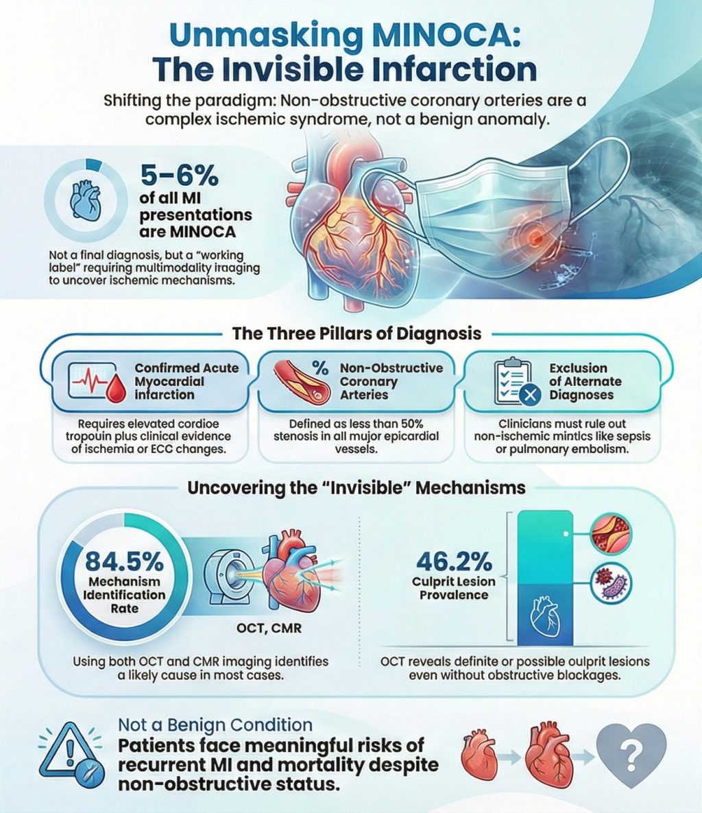
医師たちがこの2つの検査を併用したところ、調査対象となった女性の84.5%において、心臓発作の真の原因を突き止めることができたのです!これは大きな成果です。.

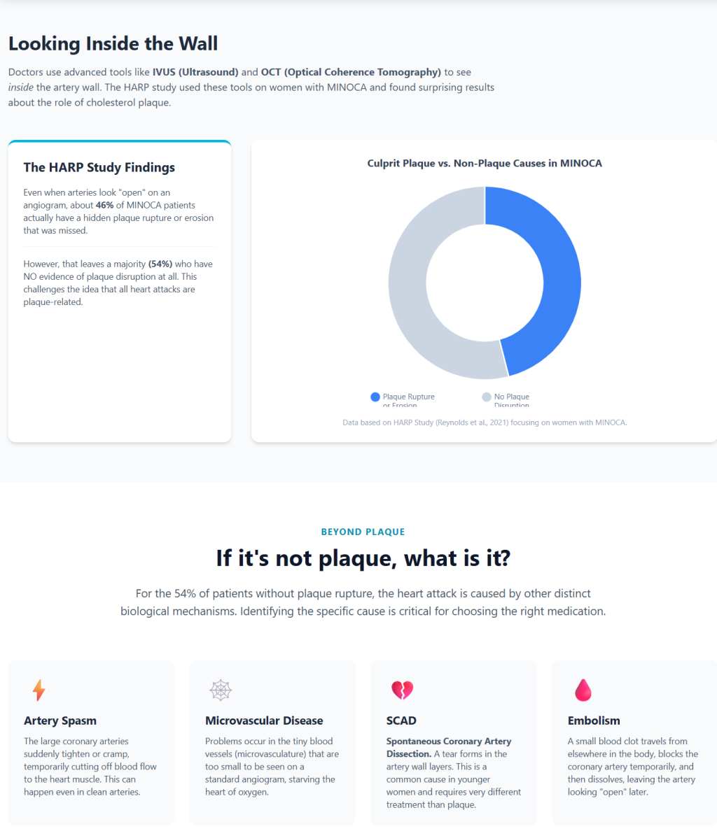
この研究からの大きな発見の一つは、「“責任病変.。」これは複雑に聞こえるかもしれませんが、単にトラブルの「出発点」を意味するだけです。カメラによって、患者の46.2%において、こうした出発点が特定されました。 この数値を誤解する人もいます。プラークの一部が破裂しただけだと考えるのです。しかし、この研究により、それ以上の意味があることが示されました。つまり、心筋梗塞を引き起こした「特定の場所」そのものを指すのです。これらの検査技術により、医師はこれまで「見えなかった」ものを確認できるようになりました。”

テイクアウェイ 6:「透明」は「安全」を意味するわけではない“

過去に、一部の医師はMINOCAを説明する際に「良性」という言葉を使っていました。「良性」は通常、大したことではない、あるいは害を及ぼさないという意味です。これは非常に大きな間違いでした。.

MINOCAは重大な医療イベントです。それは本物の心臓発作です。研究によると、MINOCAの心臓発作を起こした人は、将来も依然としてリスクに直面することが示されています。再び胸の痛みを起こす可能性があります。再び心臓発作を起こす可能性もあります。さらに、血管が「詰まった」人たちと同等の死亡リスクさえも抱えています。.

これらの症例を「安全」と呼ぶことは、多くの患者が本当に必要としている薬や検査を受けられなかったことを意味していた。彼らは自宅に帰され、心配ないと言われた。しかし、あなた べき 用心してください。動脈に異常はないと言われたにもかかわらず心臓発作を起こした場合、その事実を重く受け止めなければなりません。それは誤報ではありません。心臓が助けを求めている本当の警告サインです。.

テイクアウト 7:あなたの心臓が止まる4つの秘密の方法

心臓発作の原因が大きな詰まりではないとしたら、何なのだろうか?医師たちは、これが起こる4つの主な「隠れた」原因を発見した。標準的な検査では永久的な詰まりしか探さないため、これらを見逃すことがよくある。.

  • プラーク破綻: これが私たちが話していた「ニキビ」です。プラークの小さな破片が亀裂を起こしたり、破裂したりします。それが一時的な血栓を作り、心臓に痛みを引き起こした後、血栓は消えます。. 標準テストではこれが見落とされる 内視鏡で確認するころには、血栓がすでに消失していることが多いため.
  • 心臓のけいれん(冠動脈攣縮): 心臓の管は筋肉です。時々、突然の激しいけいれんを起こします。きつく縮み上がって、血液がまったく通れなくなってしまいます。. 標準テストではこれが見落とされる 配管はほんの数分間だけ収縮し、その後また元通りに戻って正常に見えるためです。.
  • 小型船舶のトラブル微小血管障害): 心臓には太い管と細い管があります。通常の検査では、太い管しか見えません。時々、細い管が働かなくなります。. 標準テストではこれが見落とされる 心臓の最も小さな血管を見るには十分な能力がないため、.
  • 突然の涙(SCAD): これは内壁が 動脈 実際には剥けたり切れたりします。若い女性に非常に多く見られます。. 標準テストではこれが見落とされる 裂傷が壁の内部にあるため、基本的なスキャンでは表面が滑らかで正常に見えることがあるからです。.

結論:さらに深く見つめるための招待

ここから得られる最も重要な教訓は単純です。心臓スキャン結果がクリアであることは、物語の終わりではありません。それは新しい物語の始まりなのです。.

もしあなたの血管が「きれい」に見えても、体は心筋梗塞を起こしたと告げているなら、そのまま家に帰って忘れてしまわないでください。これは、より深く探求するよう促されているサインです。「いつもの」原因が見当たらなかったということですから、医師は「通常とは異なる」原因を探さなければなりません。.

私たちは自分自身のために声を上げなければなりません。医師から「異常はない」と言われても、自分ではそうではないと感じる場合は、OCTやCMRといったより詳細な検査を依頼しましょう。これらの検査法を用いれば、80%以上の症例において真実を突き止めることができます。私たちの心臓は、単なる台所の配管のような単純なものではなく、生き生きとした複雑なシステムであることを理解しなければなりません。.

標準的な検査の結果がどうであれ、自分の体が別の何かを訴えているときは、常に自分の体に耳を傾けてください。クリアな検査結果は良いニュースですが、真の原因を見つけることこそが、長期的に健康を保つ唯一の方法です。あなたの心が求めている探偵になってください。.

ディープダイブ

エグゼクティブ・サマリー:急性冠症候群におけるパラダイムシフト

現代の心臓病学の多くにおいて、急性 心筋梗塞 (AMI)は、アテローム性動脈硬化という「配管モデル」に支配されてきた。 歯垢 長年にわたって蓄積し、徐々に冠動脈を狭窄させる ルーメン, 、そして最終的に破裂し、引き起こす 血栓症 そして突然の、血流を制限する 閉塞. このパラダイムは、依然として大部分の心筋梗塞の中心をなしている。しかし、現代の心血管科学により、大幅な修正が迫られている: 冠動脈造影で閉塞が認められない場合でも、真の心筋梗塞が発生することがある。 狭窄. この臨床的実体は—非閉塞性心筋梗塞 冠動脈 MINOCAもはや異常なものや排他的な珍奇なものとしてではなく、むしろ heterogeneous ischemic syndrome that requires structured evaluation and mechanism-directed therapy.¹

This report verifies the key mechanisms, definitions, and landmark evidence supporting modern MINOCA care, with particular focus on the 2019 American Heart Association (AHA) Scientific Statement¹ and the Women’s 心臓発作 Research Program (HARP) multimodality imaging study.² The core evidence supports several conclusions with high confidence:

  1. MINOCA is not rare and accounts for approximately 5–6% of MI presentations, with reported ranges varying by population and operational definition¹,⁴;
  2. MINOCA is explicitly framed as a 作業診断, not a final label¹;
  3. Intravascular imaging (光干渉断層計, OCT) そして 心臓磁気共鳴画像法 identify a likely mechanism in a high proportion of women with MINOCA (≈84.5% among those undergoing both modalities in HARP)²; and
  4. the frequently cited “46%” statistic from HARP refers to OCT-identified definite or possible 責任病変, not “46% プラーク破裂.”²

Chapter 1: Historical Context and the Modern Definition

1.1 From diagnostic dismissal to structured clinical framework

Historically, patients—particularly women—presenting with chest pain, バイオマーカー elevation, and non-obstructive coronary angiography were variably labeled with ambiguous or dismissive terms. This contributed to under-recognition, inconsistent management, and inadequate secondary prevention. Contemporary consensus has reversed this approach: MINOCA is now recognized as a genuine MI phenotype encompassing multiple ischemic mechanisms and several clinically important non-ischemic mimics.

The most influential U.S. consensus document is the 2019 AHA Scientific Statement chaired by Tamis-Holland, which formalized a practical definition of MINOCA and emphasized its use as a working diagnosis requiring further investigation rather than a diagnostic endpoint.¹

1.2 The three diagnostic pillars (AHA + Universal Definition alignment)

The AHA statement explicitly aligns MINOCA with the Fourth Universal Definition of Myocardial Infarction.¹,³ Diagnosis requires fulfillment of three pillars:

Pillar 1: A true acute myocardial infarction.
This requires detection of a rise and/or fall in cardiac トロポニン with at least one value above the 99th percentile upper reference limit, plus evidence of myocardial 虚血, including ischemic symptoms, new ischemic ECG changes, pathological Q waves, imaging evidence of new ischemic myocardial injury, or identification of coronary thrombus.¹,³ The Universal Definition underscores the critical distinction between myocardial injury (non-ischemic mechanisms) and 心筋梗塞 (ischemic mechanisms).³

Pillar 2: Non-obstructive epicardial coronary arteries.
Non-obstructive disease is pragmatically defined as no coronary stenosis ≥50% in any major epicardial vessel on angiography.¹ The statement acknowledges the limitations of visual angiographic estimation and notes that physiologic assessment (e.g., fractional flow reserve) may be considered in selected cases, but the ≥50% angiographic threshold remains the operational definition.¹

Pillar 3: No alternate diagnosis explaining the presentation.
MINOCA is a label applied after angiography but before exclusion of non-ischemic mimics or systemic causes of troponin elevation. The AHA statement emphasizes ruling out overt alternative diagnoses (e.g., sepsis, pulmonary embolism) and recognizing subtler mimics such as myocarditis または takotsubo syndrome that can resemble MI at presentation.¹,³

Chapter 2: Epidemiology, Demographics, and Why Definitions Matter

2.1 General prevalence and variability

MINOCA prevalence varies substantially depending on cohort selection and diagnostic rigor. The AHA Scientific Statement summarizes MINOCA as occurring in approximately 5–6% of MI patients undergoing angiography, while acknowledging broader ranges across studies.¹ A widely cited システマティックレビュー by Pasupathy et al. highlights substantial heterogeneity in reported prevalence and outcomes across international cohorts.⁴

2.2 Sex distribution and clinical implications

MINOCA is consistently reported to occur more frequently in women than in men. The AHA statement notes a disproportionate representation of women among MINOCA cases compared with obstructive MI.¹ Large U.S. registry analyses, including the ACTION Registry–Get With The Guidelines, demonstrate that MINOCA is more common among younger patients and women and provide comparative outcome data by sex and obstructive disease status.⁵

2.3 Clarifying misconceptions about “normal arteries”

A persistent misconception is that MINOCA implies “no plaque” or “completely normal coronary arteries.” This is incorrect. MINOCA explicitly includes patients with mild to moderate 動脈硬化, provided no 病変 meets the ≥50% stenosis threshold.¹

Importantly, the HARP study does ではない report a prevalence of “completely normal arteries.” Its primary focus is on imaging-detected mechanisms, not angiographic smoothness rates.² The AHA statement is explicit: MINOCA does not mean normal arteries

Chapter 3: HARP—Unmasking the Invisible With OCT and CMR

3.1 Why HARP was pivotal

The HARP study fundamentally advanced MINOCA research by applying two complementary imaging modalities: OCT to visualize coronary 動脈 wall pathology and CMR to characterize myocardial injury patterns. This design addressed the central limitation of angiography, which visualizes only the lumen and frequently misses plaque surface disruption, small thrombi, subtle dissections, or transient ischemic mechanisms.²

3.2 Verified enrollment and imaging pipeline

According to the HARP publication:

  • 301 women were enrolled across 16 sites
  • 170 were diagnosed with MINOCA at angiography
  • 145 had OCT images adequate for analysis
  • 116 of those underwent CMR
    ²

3.3 Correct interpretation of the “46.2%” statistic

HARP reports that a definite or possible culprit lesion was identified by OCT in 46.2% (67/145) of participants.² This refers to any OCT-defined culprit lesion, not plaque rupture alone. Treating this number as “46% plaque rupture” is incorrect and unsupported by the primary publication.

3.4 Multimodality diagnostic yield

Among participants who underwent both OCT and CMR, a likely mechanism for the MI presentation was identified in 84.5% (98/116).² This finding strongly supports the AHA’s framing of MINOCA as a diagnosis that benefits from layered evaluation rather than diagnostic closure.¹,²

Chapter 4: Mechanisms of the “Invisible” Infarction

MINOCA is a phenotype encompassing multiple ischemic mechanisms, as outlined in the AHA Scientific Statement.¹

4.1 Plaque disruption without obstructive stenosis

Plaque rupture or erosion can occur in lesions that never produced ≥50% stenosis. Resulting thrombus may be transient, partially lysed, or embolize distally, leaving a non-obstructive 血管造影 despite true ischemic injury. The AHA statement highlights plaque disruption as a common mechanism and supports intracoronary imaging when appropriate.¹ Classic IVUS studies provide conceptual support for this mechanism.⁶

4.2 Coronary vasospasm

Intense, transient vasoconstriction of an epicardial artery can produce ischemia and infarction, then resolve before angiography. The AHA statement discusses vasospasm mechanisms and the selective use of provocative testing.¹

4.3 Coronary 微小血管機能障害

Microvascular dysfunction involves impaired regulation of small intramyocardial vessels not visualized on angiography. The AHA statement acknowledges CMD as a potential mechanism and emphasizes uncertainty in determining causality versus consequence in individual patients.¹,³

4.4 Coronary embolism or thrombosis

Embolic or thrombotic events can cause transient obstruction or distal microvascular occlusion with minimal residual epicardial disease. The AHA statement outlines selective evaluation, including thrombophilia testing when appropriate.¹

4.5 Spontaneous coronary artery dissection (SCAD)

SCAD is a critical non-atherosclerotic cause of MI, particularly in younger and middle-aged women. Angiographic appearances may be subtle, and intracoronary imaging can be required in selected cases. The AHA SCAD Scientific Statement provides authoritative guidance, and the MINOCA statement emphasizes careful angiographic review for overlooked SCAD.¹,⁷

Chapter 5: Prognosis—Dispelling the “Benign” Myth

MINOCA is not benign. Although outcomes may be better than in extensive obstructive CAD, patients face meaningful risks of recurrent symptoms, recurrent MI, and mortality.¹ Registry analyses, including ACTION Registry–GWTG studies, provide comparative outcome data and reinforce the need for structured follow-up.⁵ Contemporary clinical reviews emphasize that inconsistent definitions historically contributed to conflicting prognostic estimates.⁸

Chapter 6: Management—From Uniform Treatment to Cause-Directed Care

Randomized trial evidence is limited because many classic MI trials excluded patients without obstructive CAD. The AHA statement therefore emphasizes mechanism-directed therapy supported by physiology and observational data.¹

その SWEDEHEART registry analysis demonstrated associations between スタチン and renin–angiotensin system blockers and improved long-term outcomes in MINOCA patients, while routine dual 抗血小板療法院 showed no clear overall benefit across the heterogeneous cohort.⁹,¹⁰ These findings are observational and hypothesis-generating, consistent with AHA guidance.¹

Conclusion

MINOCA has evolved from diagnostic confusion to a defined clinical framework. The 2019 AHA Scientific Statement establishes MINOCA as an MI phenotype requiring exclusion of mimics and pursuit of a specific ischemic mechanism.¹ The HARP study demonstrates that when OCT and CMR are applied, a likely mechanism can be identified in the majority of women studied (≈84.5%), and that nearly half have an OCT-defined culprit lesion despite non-obstructive angiography.²

The core implication is clear: **a non-obstructive angiogram is not a dismissal—it is an invitation to look deeper.**¹,²,³

参考文献

  1. Tamis-Holland JE, Jneid H, Reynolds HR, et al. Contemporary Diagnosis and Management of Patients With Myocardial Infarction in the Absence of Obstructive Coronary Artery Disease: A Scientific Statement From the American Heart Association. Circulation. 2019;139(18):e891-e908. doi:10.1161/CIR.0000000000000670
  2. Reynolds HR, Maehara A, Kwong RY, et al. Coronary Optical Coherence Tomography and Cardiac Magnetic Resonance Imaging to Determine Underlying Causes of Myocardial Infarction With Nonobstructive Coronary Arteries in Women. Circulation. 2021;143(7):624-640. doi:10.1161/CIRCULATIONAHA.120.052008
  3. Thygesen K, Alpert JS, Jaffe AS, et al. Fourth universal definition of myocardial infarction (2018). Eur Heart J. 2019;40(3):237-269. doi:10.1093/eurheartj/ehy462
  4. Pasupathy S, Air T, Dreyer RP, Tavella R, Beltrame JF. Systematic review of patients presenting with suspected myocardial infarction and nonobstructive coronary arteries. Circulation. 2015;131(10):861-870. doi:10.1161/CIRCULATIONAHA.114.011201
  5. Smilowitz NR, Mahajan AM, Roe MT, et al. Mortality of Myocardial Infarction by Sex, Age, and Obstructive Coronary Artery Disease Status in the ACTION Registry-GWTG (Acute Coronary Treatment and Intervention Outcomes Network Registry-Get With the Guidelines). Circ Cardiovasc Qual Outcomes. 2017;10(12):e003443. doi:10.1161/CIRCOUTCOMES.116.003443
  6. Gerbaud E, Arabucki F, Nivet H, et al. OCT and CMR for the Diagnosis of Patients Presenting With MINOCA and Suspected Epicardial Causes. JACC Cardiovasc Imaging. 2020;13(12):2619-2631. doi:10.1016/j.jcmg.2020.05.045
  7. Hayes SN, Kim ESH, Saw J, et al. Spontaneous Coronary Artery Dissection: Current State of the Science: A Scientific Statement From the American Heart Association. Circulation. 2018;137(19):e523-e557. doi:10.1161/CIR.0000000000000564
  8. Buda KG, Mallick S, Kohl LP. Myocardial infarction with nonobstructive coronary arteries: Current management strategies. Cleve Clin J Med. 2024;91(12):743-753. Published 2024 Dec 2. doi:10.3949/ccjm.91a.19127
  9. Lindahl B, Baron T, Erlinge D, et al. Medical Therapy for Secondary Prevention and Long-Term Outcome in Patients With Myocardial Infarction With Nonobstructive Coronary Artery Disease. Circulation. 2017;135(16):1481-1489. doi:10.1161/CIRCULATIONAHA.116.026336
  10. Smilowitz NR, Dubner R, Hellkamp AS, Widmer RJ, Reynolds HR. Variability of discharge medical therapy for secondary prevention among patients with myocardial infarction with non-obstructive coronary arteries (MINOCA) in the United States. PLoS One. 2021;16(8):e0255462. Published 2021 Aug 2. doi:10.1371/journal.pone.0255462

透明性に関する注記: この記事はAIツールの支援を受けて作成されました。最終的なコンテンツは著者によって慎重に確認および編集されており、その正確性については著者が責任を負います。提供される情報は教育目的のみのものであり、医学的なアドバイスを構成するものではありません。.

AIアプリ

心臓病リスク計算ツール

Hスコアのインサイト、視覚的な家系図入力、共有可能なPDFレポートを備えた、教育用の家族歴心臓病リスク計算ツール。.

このアプリが非常に重要である理由をここでご覧ください。.