
慢性運動と冠動脈アテロームの構造的進化:退縮、リモデリング、およびアスリートのパラドックスに関する批判的評価
肉体的な労力が進行を根本的に逆転させることができるかどうかを突き止める探求 冠動脈疾患 初期の疫学的観察から、血管壁の微小構造を中心とした高度に専門的な議論へと移行した。人工知能を活用した定量的冠動脈の急速な進歩を特徴とする現代の心臓病学において、 コンピュータ断層撮影 血管造影(AI-QCTA)および高解像度血管内イメージングにより、内腔の従来の指標である 狭窄 はるかに複雑な生物学的プロセスの不十分な輪郭であるとますますみなされている1]. 予防 cardiology における現在の根本的な緊張関係は、運動のもたらす疑う余地のない心血管系の利益が、実際の低減を通じて実現されるのかどうかにある。 歯垢 質量—真の退縮—なのか、それとも運動が血管リモデリングの主たる調節因子として機能しているのか、そして プラーク安定化, 基礎疾患の負荷を必ずしも排除することなく、臨床イベントのリスクを効果的に軽減する2].
この報告書は、この緊張関係を取り巻く証拠を批判的に評価し、以下の高忠実度イメージングデータを優先している。 血管内超音波検査 (IVUS)、, 光干渉断層計 (OCT)、および冠動脈CT血管撮影(CCTA)。マッチドコホート、縦断的アスリート登録、およびずり応力のメカニズムモデルからのデータを統合することにより、本分析は、運動が主に冠動脈の主要な原動力ではなく、その安定化因子およびリモデリング因子として機能するという仮説を検証する。 プラーク退縮.
プラーク容積対内腔サイズ:グラゴフの文脈
運動誘発性の変化の正確な評価 冠動脈 船体の外側の境界とその内部の関係についての微妙な理解が求められる ルーメン. 数十年にわたり、冠動脈疾患の診断におけるゴールドスタンダード(黄金律)は 動脈 疾患であり、内腔の二次元マップである「ルミノグラム」を提供する侵襲的冠動a造影であった。しかし、このアプローチは本質的に制限されているのは グラゴフ現象, 、または ポジティブリモデリング, ここで、冠動脈は進行性の拡大をきたす 外部弾性膜(EEM) プラークの蓄積に対する反応として3].
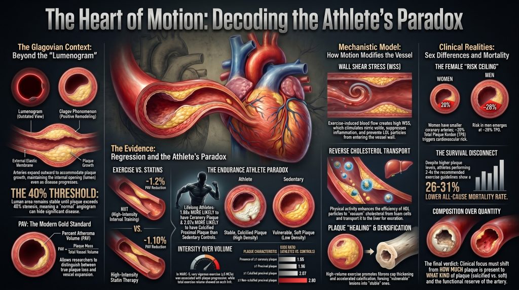
この代償性拡大により、血管は、著しい(病変・プラークなど)が存在する場合であっても、その内腔断面積を維持することが可能になる。 粉瘤 開発する。イン グラゴフの 136例の解剖された左冠状動脈に関するパイオニア的な組織学的研究において、管腔断面積は0〜40%の狭窄率の関数としては減少しなかったが、その閾値を超えると著明に減少した(r = −0.73, P < 0.001)3]。これは、造影検査で正常に見える血管にも、実質的かつ潜在的に脆弱な, プラーク負荷. PROSPECT IVUSのサブ解析により、552例の左冠動脈主幹部において、EEM断面積がプラーク・中膜断面積に比例して増大することが生体内で確認された(全体でr = 0.61、P < 0.0001、プラーク負荷が20%以下の場合はr = 0.88まで上昇した)[4].
運動は血管リモデリングの強力な刺激因子である。慢性的な有酸素運動は血流とずり応力の持続的な増加を引き起こし、それが内皮経路を活性化することで一酸化窒素の生体利用能を高め、送血動脈の構造的口径を拡大させる。5]。その結果、運動が〜を減少させると主張するいかなる研究も、 動脈硬化 内腔の直径のみに基づいた場合、実際のプラークの縮小と代償性の血管拡大の両方が組み合わさって測定されている可能性が高い。これらの効果を区別するために、研究者は次のような手法に依存している。 プラーク体積百分率 (PAV)。これはプラークの質量を血管の総容積で正規化するものである。PAVは、(外弾性板[EEM]面積-内腔面積)の総和をEEM面積の総和で割ったものとして計算され、パーセンテージで表される。この指標により、血管が太くなり(EEMの増加)、プラーク面積が一定のままであっても、PAVは低下し、絶対的な 総アテローマ容積 横ばいで推移している6].
真のプラーク退縮のエビデンス:薬物療法とのベンチマーク
運動による退縮誘導能を評価する際には、PAVおよびTAVの減少に関して最も強固なデータを持つ脂質低下療法(LLT)を用いて基準を設定する必要がある。包括的な メタ分析 成人9,113名を対象とした51件の研究の分析結果によると、LLTsは全体としてPAVを平均−1.10%(95% 信頼区間 −1.63~−0.56、 P < 0.01)でPAVを減少させ、TAVを−5.84 mm³(95%信頼区間 −8.64 ~ −3.04、P < 0.01)減少させることが示された;; 高強度スタチン TAV効果の最大値(−7.60 mm³;95% 信頼区間 −11.89~−3.31、P < 0.01)をもたらした [6GLAGOVランダム化試験からの直接的な頭部対頭部プラークイメージングの証拠は、さらに次のように実証している。 PCSK9 追加された抑制 スタチン 治療は、確立された冠動脈疾患患者において漸進的なPAV退縮をもたらす2].
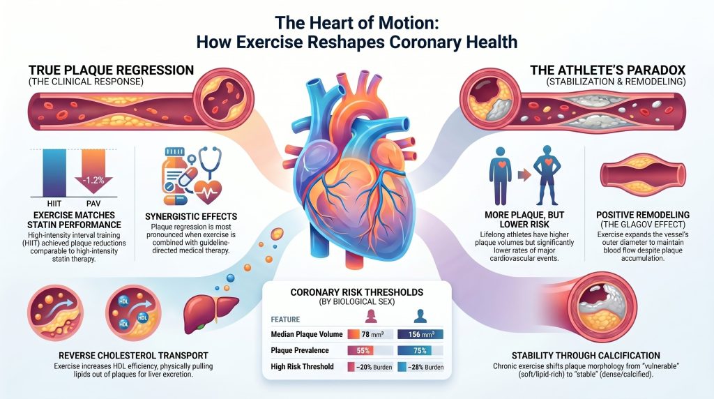
比較すると、運動誘発性の退縮に関する直接的な証拠は出つつあるものの、依然として控えめである。 CENIT試験 (冠動脈疾患と高強度インターバルトレーニングの効果)は、経皮的冠動脈インターベンションを受けた安定狭心症患者60名を無作為に割り付け、6か月の管理された 高強度インターバルトレーニング ピーク時の85〜95パーセントで 心拍数 または現代の予防ガイドラインに7本研究では、IVUSを用いて残存冠動脈セグメントにおけるPAVおよびTAVの変化を測定した。.
| パラメータ | HIIT群(n=30) | コントロールグループ (n=30) | 群間差 |
| PAV(%)の変化 | −1.2(95% 信頼区間:−2.1~−0.2;P=0.017) | +0.2(95% 信頼区間:−0.7~1.1;P=0.616) | −1.4(95% 信頼区間:−2.7~−0.1;P=0.036) [7] |
| TAVnormの変化(mm³) | −9.0(95% 信頼区間:−14.7~−3.4、P=0.002) | — | −12.0(95% 信頼区間:−19.9~−4.2;P=0.003)[7] |
The CENIT results are notable because they demonstrate that exercise can achieve PAV reductions of a magnitude comparable to lipid-lowering therapy (−1.2% vs. −1.10%) [6], [7]. Importantly, this regression was observed in a population already receiving guideline-directed medical therapy, making it difficult to isolate the independent contribution of exercise from a synergistic interaction with statins.
The Endurance Athlete Paradox
While interventional trials in patients with established coronary artery disease suggest the potential for regression, observational data from healthy veteran athletes present a challenging paradox. Large registries, including MARC (Measuring Athlete’s Risk of Cardiovascular Events) and Master@Heart, consistently report that lifelong endurance athletes have a higher prevalence of coronary atherosclerotic plaque and higher 冠動脈石灰化スコア scores than less active individuals with comparable cardiovascular risk profiles [8], [9].
その Master@Heart研究, a prospective observational cohort analysis of 191 lifelong master endurance athletes, 191 late-onset athletes (initiating endurance sports after age 30), and 176 healthy non-athletic controls (all male, low cardiovascular risk profile, median age 55 years), reported the following adjusted associations for lifelong endurance sports relative to a healthy non-athletic lifestyle [9]:
- ≥1 coronary plaque: OR 1.86 (95% CI 1.17–2.94)
- ≥1 proximal plaque: OR 1.96 (95% CI 1.24–3.11)
- ≥1 石灰化プラーク: OR 1.58 (95% CI 1.01–2.49)
- ≥1 calcified proximal plaque: OR 2.07 (95% CI 1.28–3.35)
- ≥1 非石灰化プラーク: OR 1.95 (95% CI 1.12–3.40)
- ≥1 non-calcified proximal plaque: OR 2.80 (95% CI 1.39–5.65)
- ≥1 mixed plaque: OR 1.78 (95% CI 1.06–2.99)
The finding of higher odds of non-calcified (lipid-rich) plaques in proximal segments is particularly notable, as such plaques are typically associated with higher near-term risk than purely calcified 病変 [9].
その MARC-2試験 added longitudinal granularity by distinguishing exercise volume from intensity in 289 middle-aged and older male athletes (median age 54 years) followed for a mean of 6.3 years [8]. Total exercise volume (MET-hours/week) showed no association with progression of CAC or plaque. By contrast, exercise intensity stratified by metabolic equivalents — vigorous (6–9 METs) versus very vigorous (≥9 METs) — was significantly associated with imaging outcomes:
| Exercise Intensity | Effect on CAC Score (β per 10% increase) | Plaque Progression |
| Vigorous (6–9 METs) | −0.05 (95% CI −0.09 to −0.01; P=0.02) [8] | Less progression |
| Very Vigorous (≥9 METs) | +0.05 (95% CI 0.01 to 0.09; P=0.01) [8] | aOR 1.09 (95% CI 1.01–1.18) per 10% increase [8] |
| Total Volume | No significant association [8] | No significant association [8] |
That intensity rather than volume drives plaque and CAC progression in athletes suggests a 生理的閾値 beyond which exercise may exert pro-atherogenic effects. Proposed mechanisms include exercise 収縮期血圧 that can exceed 200 mmHg in well-trained athletes during near-maximal effort [25], repetitive mechanical stress on the coronary wall, and inflammatory signaling pathways implicated in athlete-related vascular 石灰化 [10].
The defining feature of the athlete’s paradox, however, is that despite higher plaque burden, endurance athletes have lower rates of 主要心血管イベント (MACE) than the general population. Although the rank order of plaque morphology (calcified > mixed > non-calcified) is broadly similar across active and sedentary populations, lifelong endurance athletes who harbor plaque are more likely than sedentary controls to harbor exclusively calcified lesions, which are considered more stable [12]. This phenotypic enrichment for dense, calcified (stable) lesions — rather than for mixed or non-calcified vulnerable lesions — suggests that high-volume exercise may accelerate the natural healing or densification process of atherosclerosis [10], [11].
Matched Cohort Evidence and Socioeconomic Confounders
A critical requirement for validating the impact of exercise is the control of social and economic confounders. Higher socioeconomic status (SES) is independently associated with both greater 運動耐容能 and improved cardiovascular outcomes [13]. In the Heart and Soul Study, an analysis of 943 men and women with stable 冠動脈疾患, exercise capacity decreased in a graded fashion across categories of income, education, housing, and occupation. After multivariable adjustment, the difference in exercise capacity between the highest and lowest categories was 2.4 METs for household income, 1.8 METs for education, 2.3 METs for housing, and 1.3 METs for occupation (P < 0.001 for all trends) [13].
Spousal Concordance and Partner Studies
Cohabiting partners share a micro-environment encompassing diet, socioeconomic stress, and access to healthcare. In a US nationwide cohort of 5,364 married or domestic-partner couples, within-couple concordance exceeded 50 percent for every component of the American Heart Association’s Life’s Simple 7 framework, ranging from 53 percent for 総コレステロール to 95 percent for healthy diet score; concordance for メタボリックシンドローム reached 73 percent [14]. Despite this strong concordance for behaviors and 危険因子, marital status itself, when adjusted for cardiovascular risk factors, was not independently associated with subclinical coronary atherosclerosis on CCTA in a study of 9,288 asymptomatic individuals [15]. Together, these findings suggest that the shared environment of a partnership produces a background risk level, but the trajectory of plaque burden is more heavily influenced by individual health behaviors — including the choice to exercise regularly — than by partnership status itself.
Occupational Activity as a Controlled Environment
The London Transport Workers Study remains the canonical example of an occupational analog [16]. By comparing conductors and drivers within the same transport system — men of similar age and social class — Jeremy Morris was able to isolate occupational physical activity from most confounders. Morris and colleagues studied tens of thousands of London bus and postal workers and reported that physically active conductors, who climbed and descended hundreds of stairs per shift, experienced approximately half the rate of acute and sudden coronary deaths observed among the sedentary drivers [16], [17]. This natural experiment provides some of the earliest robust evidence that occupational physical activity independently modifies the clinical expression of coronary disease, even when the macro-environment is held constant.
Dose-Response Relationships and the Mortality Curve
The relationship between exercise dose and mortality is widely regarded as curvilinear, with the most substantial risk reductions occurring as individuals move from sedentary to moderately active. The システマティックレビュー and meta-analysis by Blond and colleagues, encompassing 48 前向きコホート studies, used random-effects restricted cubic spline 用量反応 modeling to estimate hazard ratios across the full exposure range. Compared with the recommended physical activity dose (750 MET-min/week), risk continued to decrease beyond recommendations: at 5,000 MET-min/week, hazard ratios were approximately 0.86 for 全因死亡率 and 0.73 for cardiovascular mortality. No excess risk was observed at the highest exposure levels examined [18].
The Lee et al. Circulation analysis of 116,221 adults pooled from the Nurses’ Health Study and the Health Professionals Follow-up Study, with up to 30 years of follow-up and approximately 47,000 deaths, examined long-term physical activity intensity in detail [19]. Compared with no leisure-time vigorous physical activity (VPA), participants meeting the guideline (75–149 min/week of VPA) had hazard ratios of 0.81 (95% CI 0.76–0.87) for all-cause mortality, 0.69 (95% CI 0.60–0.78) for CVD mortality, and 0.85 (95% CI 0.79–0.92) for non-CVD/non-cancer mortality. Participants performing two to four times the recommended minimum had a 21–23 percent lower all-cause mortality for VPA (150–299 min/week) and a 26–31 percent lower all-cause mortality for moderate physical activity (300–599 min/week) [19]. Higher long-term doses (≥300 min/week VPA or ≥600 min/week moderate activity) did not provide further mortality reduction, but no excess risk was observed [19].
This mortality benefit persists despite the athlete’s paradox of increased CAC, supporting the conclusion that the plaques found in highly active individuals are compositionally more benign in their clinical manifestation [10], [11].
Mechanistic Model: How Exercise Modifies Plaque
The biological mechanisms by which exercise potentially reduces plaque burden or facilitates stabilization act both systemically and locally.
Fluid Mechanics and Wall Shear Stress
Plaque development is governed by patterns of blood flow. Laminar flow produces high wall shear stress (WSS), which maintains endothelial homeostasis through the mechanosensitive transcription-factor axis (KLF2 and KLF4) and downstream activation of endothelial 一酸化窒素 synthase. Disturbed or oscillatory flow, common at coronary bifurcations, produces low WSS that promotes an atherogenic endothelial phenotype, including upregulation of NF-κB-driven adhesion molecules, monocyte recruitment, and increased endothelial トランスサイトーシス の LDL [20]. Exercise-induced increases in flow rate directly enhance the protective frictional force at the vessel wall. Direct molecular evidence in human coronary arteries has shown that regular physical activity in patients with coronary artery disease increases phosphorylation of endothelial nitric oxide synthase at Ser1177, the key regulatory site governing nitric oxide production [5].
Reverse Cholesterol Transport and HDL Functionality
Exercise facilitates the removal of lipids from plaque via the コレステロール引き抜き転送 pathway, beginning with コレステロール efflux from 泡沫細胞 へ HDL particles, which transport lipids to the liver for hepatobiliary excretion. A randomized exercise-dosing trial measuring multiple HDL functional metrics has shown that increasing both exercise intensity and dose produces dose-responsive improvements in HDL cholesterol efflux capacity, anti-oxidant capacity, and anti-inflammatory function — effects that are largely independent of changes in HDL cholesterol concentration alone [22].
Plaque Stabilization, Matrix Metalloproteinases, and the Fibrous Cap
Beyond regression, exercise promotes structural stabilization through fibrous-cap thickening, mediated in part by reduced intra-plaque matrix metalloproteinase activity. Animal studies in アポリポ蛋白 E knockout mice show that 有酸素運動 training reduces intra-plaque MMP-8 and MMP-9 activity, increases tissue inhibitor of metalloproteinases (TIMP-1, TIMP-2), increases fibrous-cap thickness, and reduces the relative size of the necrotic lipid core [23]. Human systematic-review evidence links chronic physical activity to favorable changes in circulating MMP-9, oxidized LDL, and inflammatory mediators relevant to plaque vulnerability [21]. Concurrent reductions in systemic 炎症, including C反応性タンパク質, may further contribute to plaque stabilization [11], [21]. The net effect in chronically active individuals is a transition from compositionally vulnerable to compositionally stable plaque morphology, even when total プラーク体積 increases [10], [12].
Sex-Specific Differences in Plaque Risk
A crucial refinement in modern preventive cardiology is the recognition that plaque burden carries different prognostic implications in men and women. In a recent analysis of the PROMISE試験 CCTA arm (4,267 patients, approximately 51 percent women, median follow-up 26 months), women had lower coronary plaque prevalence than men (55 percent versus 75 percent; P < 0.001) and lower median total plaque volume, yet a similar incidence of major adverse cardiovascular events (2.3 percent versus 3.4 percent) [24]. Critically, when MACE risk was modeled against total plaque burden using sex-stratified spline Cox regression, the hazard ratio crossed unity at a substantially lower plaque-burden threshold in women than in men.
| 特徴 | Women | Men | Implication |
| Plaque prevalence | 55% | 75% | Lower prevalence in women [24] |
| Plaque volume | Lower (median) | Higher (median) | Smaller absolute burden in women [24] |
| Total plaque burden at HR=1.0 | ≈20% | ≈28% | Risk emerges at a lower burden in women [24] |
| Total plaque burden at HR=1.5 | ≈32% | ≈42% | Steeper risk trajectory in women [24] |
These data indicate that uniform thresholds for high-risk plaque underestimate risk in women: because women have smaller coronary arteries, a smaller absolute plaque volume produces a larger relative plaque burden and a more rapid acceleration of MACE risk [24]. This implies that for female athletes, the ceiling for compositionally benign subclinical plaque may be considerably lower than for their male counterparts.
Testing the Hypothesis: Remodeling or Regression?
The hypothesis under examination is that exercise does not meaningfully reduce total coronary plaque burden but rather improves vessel caliber and plaque stability. The evidence supports a population-dependent answer:
In patients with established CAD on guideline-directed therapy: Exercise can meaningfully reduce total plaque burden. The CENIT trial demonstrated a between-group PAV difference of −1.4 percent and a TAVnorm reduction of −12 mm³ after six months of HIIT [7].
In healthy lifelong endurance athletes: Exercise does not reduce total plaque burden but is associated with higher prevalence and burden across all plaque types [9]. In this population, the hypothesis is largely supported: the cardiovascular benefit of exercise is realized through vascular remodeling and stabilization (calcification) rather than removal.
Apparent versus true benefits: Increased lumen size in trained individuals reflects a real physiological adaptation (positive remodeling) that confers significant clinical protection through enhanced 冠血流予備能 [3], [4]. This remodeling benefit is distinct from the discrete benefit of true plaque regression, and the two effects operate on different timescales.
Final Verdict: Regression, Progression, or Stabilization?
The totality of the evidence indicates that chronic exercise is a multi-modal modifier of coronary atherosclerosis. True regression — measurable reductions in PAV and TAV — is achievable through high-intensity exercise interventions, particularly in populations with existing coronary artery disease, where the magnitude approaches that of intensive lipid-lowering therapy [2], [6], [7]. Among individuals without known disease, moderate exercise appears to slow the progression of soft, vulnerable, lipid-rich plaques through favorable lipid metabolism, enhanced HDL cholesterol efflux capacity, and reduced systemic inflammation [21], [22]. In high-volume endurance athletes, exercise functions primarily as a stabilizing agent, paradoxically accelerating calcification while expanding vessel caliber through positive remodeling — yielding a compositionally benign atherosclerotic burden that does not translate into elevated clinical risk [9], [10], [12].
Critically, the survival benefit of exercise is decoupled from absolute plaque volume: extreme athletes have more plaque yet exhibit substantially lower all-cause and CVD mortality than the general population [18], [19]. The clinical emphasis should therefore shift from quantifying how much plaque is present toward characterizing what kind of plaque is present and what functional reserve the individual’s coronary tree retains.
参考文献
- Narula J, Stuckey TD, Nakazawa G, et al. Prospective deep learning-based quantitative assessment of coronary plaque by computed tomography angiography compared with intravascular ultrasound: the REVEALPLAQUE study. Eur Heart J Cardiovasc Imaging. 2024;25(9):1287-1295. doi:10.1093/ehjci/jeae115
- Nicholls SJ, Puri R, Anderson T, et al. Effect of Evolocumab on Progression of Coronary Disease in Statin-Treated Patients: The GLAGOV Randomized Clinical Trial. JAMA. 2016;316(22):2373-2384. doi:10.1001/jama.2016.16951
- Glagov S, Weisenberg E, Zarins CK, Stankunavicius R, Kolettis GJ. Compensatory enlargement of human atherosclerotic coronary arteries. N Engl J Med. 1987;316(22):1371-1375. doi:10.1056/NEJM198705283162204
- Inaba S, Mintz GS, Shimizu T, et al. Compensatory enlargement of the left main coronary artery: insights from the PROSPECT study. Coron Artery Dis. 2014;25(2):98-103. doi:10.1097/MCA.0000000000000074
- Hambrecht R, Adams V, Erbs S, et al. Regular physical activity improves endothelial function in patients with coronary artery disease by increasing phosphorylation of endothelial nitric oxide synthase. Circulation. 2003;107(25):3152-3158. doi:10.1161/01.CIR.0000074229.93804.5C
- Rivera FB, Cha SW, Varona MC, et al. Atherosclerotic coronary plaque regression from lipid-lowering therapies: A meta-analysis and meta-regression. Am J Prev Cardiol. 2024;18:100645. Published 2024 Mar 11. doi:10.1016/j.ajpc.2024.100645
- Vesterbekkmo EK, Aksetøy IA, Follestad T, et al. High-intensity interval training induces beneficial effects on coronary atheromatous plaques: a randomized trial. Eur J Prev Cardiol. 2023;30(5):384-392. doi:10.1093/eurjpc/zwac309
- Aengevaeren VL, Mosterd A, Bakker EA, et al. Exercise Volume Versus Intensity and the Progression of Coronary Atherosclerosis in Middle-Aged and Older Athletes: Findings From the MARC-2 Study. Circulation. 2023;147(13):993-1003. doi:10.1161/CIRCULATIONAHA.122.061173
- De Bosscher R, Dausin C, Claus P, et al. Lifelong endurance exercise and its relation with coronary atherosclerosis. Eur Heart J. 2023;44(26):2388-2399. doi:10.1093/eurheartj/ehad152
- Hsu JJ, Tintut Y, Demer LL. Paradox of Exercise and Coronary Artery Calcification: Potential Underlying Mechanisms. Circ Res. 2025;137(2):335-349. doi:10.1161/CIRCRESAHA.125.326011
- Aengevaeren VL, Mosterd A, Sharma S, et al. Exercise and Coronary Atherosclerosis: Observations, Explanations, Relevance, and Clinical Management. Circulation. 2020;141(16):1338-1350. doi:10.1161/CIRCULATIONAHA.119.044467
- Aengevaeren VL, Mosterd A, Braber TL, et al. Relationship Between Lifelong Exercise Volume and Coronary Atherosclerosis in Athletes. Circulation. 2017;136(2):138-148. doi:10.1161/CIRCULATIONAHA.117.027834
- Cohen B, Vittinghoff E, Whooley M. Association of socioeconomic status and exercise capacity in adults with coronary heart disease (from the Heart and Soul Study). Am J Cardiol. 2008;101(4):462-466. doi:10.1016/j.amjcard.2007.09.093
- Shiffman D, Louie JZ, Devlin JJ, Rowland CM, Mora S. Concordance of Cardiovascular Risk Factors and Behaviors in a Multiethnic US Nationwide Cohort of Married Couples and Domestic Partners. JAMA Netw Open. 2020;3(10):e2022119. Published 2020 Oct 1. doi:10.1001/jamanetworkopen.2020.22119
- Ann SH, Lee H, Park KS, et al. Marital Status and Subclinical Coronary Atherosclerosis in Asymptomatic Individuals. J Am Heart Assoc. 2022;11(11):e024942. doi:10.1161/JAHA.121.024942
- Morris JN, Heady JA, Raffle PA, Roberts CG, Parks JW. Coronary heart-disease and physical activity of work. Lancet. 1953;262(6795):1053-1057. doi:10.1016/s0140-6736(53)90665-5
- Morris JN, Kagan A, Pattison DC, Gardner MJ. Incidence and prediction of ischaemic heart-disease in London busmen. Lancet. 1966;2(7463):553-559. doi:10.1016/s0140-6736(66)93034-0
- Blond K, Brinkløv CF, Ried-Larsen M, Crippa A, Grøntved A. Association of high amounts of physical activity with mortality risk: a systematic review and meta-analysis. Br J Sports Med. 2020;54(20):1195-1201. doi:10.1136/bjsports-2018-100393
- Lee DH, Rezende LFM, Joh HK, et al. Long-Term Leisure-Time Physical Activity Intensity and All-Cause and Cause-Specific Mortality: A Prospective Cohort of US Adults. Circulation. 2022;146(7):523-534. doi:10.1161/CIRCULATIONAHA.121.058162
- Niu N, Xu S, Xu Y, Little PJ, Jin ZG. Targeting Mechanosensitive Transcription Factors in Atherosclerosis. Trends Pharmacol Sci. 2019;40(4):253-266. doi:10.1016/j.tips.2019.02.004
- Palmefors H, DuttaRoy S, Rundqvist B, Börjesson M. The effect of physical activity or exercise on key biomarkers in atherosclerosis–a systematic review. Atherosclerosis. 2014;235(1):150-161. doi:10.1016/j.atherosclerosis.2014.04.026
- Sarzynski MA, Ruiz-Ramie JJ, Barber JL, et al. Effects of Increasing Exercise Intensity and Dose on Multiple Measures of HDL (High-Density Lipoprotein) Function. Arterioscler Thromb Vasc Biol. 2018;38(4):943-952. doi:10.1161/ATVBAHA.117.310307
- Kadoglou NP, Moustardas P, Kapelouzou A, et al. The anti-inflammatory effects of exercise training promote atherosclerotic plaque stabilization in apolipoprotein E knockout mice with diabetic atherosclerosis. Eur J Histochem. 2013;57(1):e3. Published 2013 Mar 25. doi:10.4081/ejh.2013.e3
- Brendel JM, Mayrhofer T, Karády J, et al. Risk in Women Emerges at Lower Coronary Plaque Burden Than in Men: PROMISE Trial. Circ Cardiovasc Imaging. 2026;19(3):e019011. doi:10.1161/CIRCIMAGING.125.019011
- Currie KD, Floras JS, La Gerche A, Goodman JM. Exercise Blood Pressure Guidelines: Time to Re-evaluate What is Normal and Exaggerated?. Sports Med. 2018;48(8):1763-1771. doi:10.1007/s40279-018-0900-x

