
This page has been automatically translated. If there is any discrepancy, the English version is authoritative.

Revised: July 16, 2026

Exercise Doesn’t Erase Arterial Plaque – It Turns It to Stone!

By: Peter Megdal PhD

How to Use This Article

Medical disclaimer: This article is for education only and is not medical advice. Always consult your clinician for personal guidance.

Easy Read

The Surprising Truth About Exercise and Clogged Arteries

Introduction: The Heart of the Matter

Many people feel a deep fear when they hear the words “clogged arteries.” We often think of our heart like the plumbing in an old house. We imagine that over time, the pipes get filled with thick gunk. In this simple view, if the pipes are clear, you are healthy. If the pipes are full of gunk, you are in big trouble. We assume that people who exercise a lot will have the cleanest pipes of all. But science is showing us a very strange and surprising truth. It is called the “Athlete’s Paradox.” Researchers have found that people who exercise the most—like lifelong runners or cyclists—sometimes have much more plaquePlaque is the buildup of cholesterol, immune cells, scar tissue, and calcium inside an artery wall. in their heart than people who sit on the couch.

If you just look at a heart scan, it might look like the athlete is in danger. However, I want to show you why this is not as scary as it sounds. Your heart is much smarter than a metal pipe. It can change its shape and its chemistry to stay safe. This news is actually very encouraging for anyone who loves to move. Let’s look at why your heart scan might be lying to you.

Takeaway 1: The “Expanding Pipe” Trick (Vascular Remodeling)

In the past, doctors looked at the heart using a “lumenogram.” This is like taking a picture of the water flowing through a pipe. If the water path looks wide, doctors thought the pipe was clean. However, a famous scientist named Dr. GlagovGLAGOV added a PCSK9 inhibitor to statin therapy and measured coronary plaque with intravascular ultrasound before and after. proved that this doesn’t tell the whole story. He discovered something called the “Glagov phenomenonThe Glagov phenomenon (also called compensatory or outward remodeling) is the tendency of an arterial wall to expand outward as atherosclerotic plaque accumulates, thereby preserving the inner lumen diameter until plaque burden becomes very large. Because lumen size stays normal during this compensatory phase, standard stress tests and angiograms can appear normal even in the presence of substant….”

Imagine a garden hose. If dirt starts building up on the inside walls, you would expect the water path to get smaller right away. But our arteries are alive and very clever. They perform a trick called “positive remodeling.” When gunk builds up, the arteryAn artery is a blood vessel that carries blood away from the heart to the rest of the body. actually stretches and grows larger on the outside. This keeps the water path on the inside wide and clear. Dr. Glagov found that if the buildup is between 0 and 40 percent, the inside path doesn’t shrink at all! A scan might show a “normal” pipe, even if there is a lot of plaque hidden inside the walls.

Exercise is the “master regulator” of this stretching process. Constant movement and fast-flowing blood tell your arteries to expand. This means that even if an athlete has some plaque, their “pipes” have grown so large that the blood can still zoom through without any problems. The vessel adapts to the gunk by making more room.

Takeaway 2: HIIT Can Be as Strong as Medicine

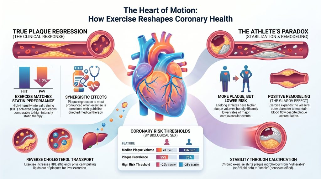
We usually think of statinsA statin slows the enzyme your liver uses to make cholesterol. Your liver responds by pulling more cholesterol out of your blood, which is where the real benefit comes from., which are common cholesterolCholesterol is a waxy substance your body needs. It goes into cell walls, hormones, vitamin D, and the bile that digests your food. You would die without it. pills, as the only way to fix our arteries. Medicine is very good at its job. On average, these pills can reduce the amount of plaque by about 1.1 percent. But new research called the CENIT trialThe CENIT (Coronary Disease and the Effect of High-Intensity Interval Training) trial was a randomized study of 60 patients with stable coronary artery disease that used intravascular ultrasound to measure whether six months of supervised HIIT at 85–95% of peak heart rate changed plaque volume compared with standard care. It found HIIT reduced percent atheroma volume by 1.2% and total normalized… shows that high-intensity exercise can be just as strong as medicine.

The CENIT trial looked at people who already had heart disease. They put one group through High-Intensity Interval Training (HIIT)High-intensity interval training is a structured exercise method that alternates short bouts of near-maximal effort (typically 85–95% of peak heart rate) with periods of active recovery or rest. In the cardiovascular context, HIIT protocols have been studied as a non-pharmacological intervention to reduce plaque burden and improve heart function.. This means doing short bursts of very hard work, like running fast for a minute and then walking for a minute. They found that HIIT reduced plaque volumePlaque volume is the total physical amount of plaque in a stretch of artery, measured in cubic millimeters. by 1.2 percent. It also reduced the total amount of “gunk” (called TAVnorm) by a huge 12.0 cubic millimeters. This is amazing because it shows exercise can work just as well as—or even better than—a pill.

The Comparison: Standard Care vs. Adding HIIT

  • Before (Standard Medicine Only): Plaque is reduced by about 1.1 percent. This is good, but the “pipes” stay mostly the same size.
  • After (Medicine PLUS HIIT Exercise): Plaque is reduced by 1.2 percent, and the total volume of gunk drops significantly. The heart becomes much cleaner.

The most important part is that these people were already taking their heart medicine. The HIIT was a “bonus” that made their hearts even healthier. As the researchers noted:

“The CENIT results are notable because they demonstrate that exercise can achieve plaque reductions of a magnitude comparable to medicine, even in people already receiving top-tier medical care.”

Takeaway 3: The Athlete’s Paradox (More Plaque, Less Risk)

A study called Master@Heart looked at lifelong athletes and compared them to healthy people who did not exercise much. You would think the athletes would have the cleanest hearts. But the data showed something very weird. Lifelong athletes actually had more plaque and higher “calcium scoresA calcium score (coronary artery calcium score) is a number derived from a CT scan that quantifies the total amount of calcified plaque in the coronary arteries; a score of zero indicates no detectable calcified plaque, while higher scores reflect greater plaque burden and elevated cardiovascular risk.” than the people who sat still.

Scientists use “Odds Ratios” (OR) to explain risk. If a score is 1.0, the risk is normal. If it is higher than 1.0, the risk is higher. Lifelong athletes had an OR of 1.86 for having any plaque. They had an OR of 1.96 for having plaque in the most important “proximal” parts of the heart. In simple terms, these athletes were nearly twice as likely to have plaque as the non-athletes.

This sounds like bad news, but it isn’t! Even though these athletes have more plaque on their scans, they have fewer heart attacksA heart attack happens when blood flow to part of the heart muscle is cut off and that muscle starts to die. and live much longer. This is the “Athlete’s Paradox.” The scan shows more “gunk,” but the person is actually much safer. The amount of plaque on a screen doesn’t tell you how strong the heart really is.

Takeaway 4: Soft Plaque vs. Hard Shells

Why are athletes safer if they have more plaque? It is because of the type of plaque they have. There are two main kinds: “vulnerable” and “stable.” Vulnerable plaqueAtherosclerotic deposits within artery walls that have not yet undergone calcification; sometimes called 'soft' plaque, these lesions are lipid-rich and structurally unstable, making them more prone to rupture and acute thrombosis than calcified plaque. is soft and filled with fat. This is the dangerous kind that can pop like a pimple and cause a heart attack. Stable plaque is hard and “calcified.”

Think of calcificationCalcification is when calcium gets deposited into a plaque, turning part of it hard and bony. like a “scab” inside your artery. When you scrape your knee, your body makes a hard scab to protect the skin while it heals. Your heart does the same thing. It takes soft, dangerous plaque and covers it in a hard shell. This makes the plaque “stable” so it won’t pop.

We must be honest about the data: the Master@Heart studyThe Master@Heart study is a controlled study of lifelong male endurance athletes that found they carried more total coronary plaque than healthy, active non-athletes—including more calcified and non-calcified plaque in proximal artery segments—raising questions about whether extreme lifelong endurance training promotes atherosclerosis even in the absence of traditional risk factors. showed athletes actually have higher odds of having soft plaque too (an OR of 2.80). However, the total effect of exercise is to turn the heart into a fortress. Even if they have more soft plaque, they have even more hard plaque to balance it out. The study explains:

“The net effect in chronically active individuals is a transition from compositionally vulnerable to compositionally stable plaque morphology.”

Exercise helps the body “heal” the arteries by turning the gunk into hard, safe shells.

Takeaway 5: Why Speed Matters More than Distance (The 9 MET Threshold)

How hard you push yourself changes how your heart looks. A study called MARC-2 looked at “exercise volume” (how long you work out) versus “exercise intensity” (how hard you work).

Scientists measure intensity using “METs.”

  • Vigorous Exercise (6 to 9 METs): This is like a fast walk or a light jog. This level was actually linked to less plaque growth. It slowed down the “rusting” of the pipes.
  • Very Vigorous Exercise (Over 9 METs): This is “redlining” the engine. It is pushing your heart to the absolute limit. This was linked to more plaque and more calcification.

Think of it like a car. Driving at a steady, fast speed is great for the engine. It keeps everything running smooth. But if you push the car as fast as it can possibly go every single day, the engine will start to show some wear and tear. For athletes, pushing very hard makes the plaque build up and harden faster. This isn’t necessarily bad, but it explains why the scans of very fast runners look different than those of casual walkers.

Takeaway 6: The “Small Pipe” Problem for Women

Heart health is not the same for everyone. A study called the PROMISE trialA large randomized trial that evaluated CT angiography versus functional stress testing in symptomatic patients with stable chest pain; findings cited here showed that 16.5% of zero-CAC participants had non-obstructive coronary artery disease and 1.5% had obstructive disease, illustrating residual risk despite a zero calcium score. found that women’s hearts face a unique challenge. The reason is simple: women usually have smaller coronary arteriesThe coronary arteries are the small vessels that wrap around the outside of your heart and feed the heart muscle itself. than men.

Because the “pipes” are smaller, even a little bit of gunk takes up more space. Imagine a small pebble. If you drop that pebble into a wide drainpipe, the water still flows fine. But if you drop that same pebble into a thin drinking straw, the straw gets blocked.

  • For Men: Their risk starts to go up when plaque fills about 42% of the vessel.
  • For Women: Their risk starts to go up much earlier, at only 32%.

This means the “safe ceiling” for plaque is lower for female athletes. A woman might have less total plaque than a man, but it might be more dangerous for her because her vessels are smaller. Female athletes need to be extra careful and keep an eye on their heart health sooner than men do.

Takeaway 7: It’s Not Just Your Zip Code or Your Partner

We often think our health is decided by where we live or who we marry. It is true that couples share a lot. In fact, 95% of partners share the same eating habits! If your partner eats junk food, you probably do too. Also, people with more money or better jobs often have better heart health.

But your own choice to move is more powerful than your environment. We know this because of a famous study of 31,000 London transport workers. Researchers compared bus drivers to bus conductors. These men were the same age, had the same pay, and lived in the same neighborhoods. But the drivers sat in a seat all day. The conductors walked up and down the stairs of the double-decker buses hundreds of times every shift.

The results were shocking. The conductors—the ones who moved—had half the number of heart attacks as the drivers. This shows that movement itself is the magic ingredient. Even if you live in a certain “zip code” or share a kitchen with someone who doesn’t eat well, your choice to be active can rewrite the future of your heart.

Takeaway 8: The Body’s Internal “Garbage Trucks”

Exercise doesn’t just change the shape of your heart; it changes the chemistry. Your body has a special cleanup crew called “Reverse Cholesterol TransportReverse cholesterol transport is the process of moving cholesterol out of tissues, including artery walls, and back to the liver for disposal. HDL particles do the hauling..”

Think of HDLHDL, or high-density lipoprotein, is the particle often called "good cholesterol." It picks up cholesterol from tissues and carries it back to the liver. particles as “garbage trucks.” Their job is to drive through your arteries and pick up the “trash” (fat and cholesterol). Then they take that trash to the “dump” (your liver) to be thrown away. Exercise makes these garbage trucks much better at their job. It gives you more trucks and makes them faster at picking up the trash.

Movement also creates “Wall Shear StressWall shear stress is the frictional force exerted by flowing blood on the inner surface of an artery; low, oscillatory, or multidirectional shear stress at arterial bends and bifurcations promotes endothelial dysfunction and plaque initiation, whereas high, uniform shear stress in straight segments is generally protective..” This is the friction of blood rubbing against the walls of your arteries. While friction sounds like it might be bad, this specific kind is very good. It triggers a chemical called “nitric oxideNitric oxide is a gas your blood vessel lining makes to tell the vessel to relax and widen..” I like to think of nitric oxide as a magic spray that keeps the pipe walls slippery and relaxed. It stops inflammationInflammation is your immune system's response to injury or something it treats as an invader. It brings swelling, heat, and cleanup cells. and makes it very hard for “gunk” to stick to the walls. It’s like having a self-cleaning oven inside your chest!

Conclusion: A New Way to Look at Your Heart

For a long time, people thought that any plaque on a heart scan was a sign of failure. We believed that the goal of exercise was to keep our arteries as clean and shiny as a brand-new metal pipe. But modern science has shown us a much more hopeful and interesting story. Your heart is a living, breathing thing that knows how to protect itself.

The “Athlete’s Paradox” teaches us that the amount of plaque on a scan is not the most important number. What matters more is the type of plaque and the strength of the vessel. If you exercise, your heart learns to grow wider. It learns to turn soft, dangerous gunk into hard, stable “scabs” that won’t cause trouble. It builds a better cleanup crew of garbage trucks and keeps its walls slippery and healthy.

Instead of worrying about a single number on a scan, we should focus on “functional reserve.” This is a fancy way of asking: “What can your heart actually do?” Is it strong? Can it handle a tough workout? Does it have the wide, flexible pipes that only movement can provide? The real question isn’t “How much plaque do I have?” but rather: “How well-built is my heart for the long journey ahead?”

Deep Dive

Chronic Exercise and the Structural Evolution of Coronary Atheroma: A Critical Appraisal of Regression, Remodeling, and the Athlete’s Paradox

The quest to determine whether physical exertion can fundamentally reverse the progression of coronary artery diseaseCoronary artery disease is plaque buildup in the arteries feeding the heart muscle. has transitioned from early epidemiological observations to a highly technical discourse centered on the micro-architecture of the vessel wall. In the modern era of cardiology, characterized by rapid advancements in artificial intelligence–enabled quantitative coronary computed tomographyComputed tomography, or CT, takes X-ray images from many angles and reconstructs them into cross-sections of the body. angiography (AI-QCTA) and high-resolution intravascular imaging, the traditional metric of luminal stenosisStenosis is narrowing — usually described as a percentage, like a 70 percent blockage. is increasingly viewed as an insufficient silhouette of a far more complex biological process [1]. The fundamental tension in preventive cardiology today lies in whether the undisputed cardiovascular benefits of exercise are realized through the actual reduction of plaquePlaque is the buildup of cholesterol, immune cells, scar tissue, and calcium inside an artery wall. mass — true regression — or whether exercise serves as a master regulator of vascular remodeling and plaque stabilizationPlaque stabilization is making an existing plaque less likely to crack open — thickening its cap, shrinking its greasy core, and calming the inflammation inside it., effectively mitigating the risk of clinical events without necessarily eliminating the underlying disease burden [2].
This report critically evaluates the evidence surrounding this tension, prioritizing high-fidelity imaging data from intravascular ultrasoundIntravascular ultrasound, or IVUS, uses a tiny ultrasound probe threaded inside a coronary artery to photograph the wall from within. (IVUS), optical coherence tomographyOptical coherence tomography, or OCT, threads a light-based probe into a coronary artery. It sees roughly ten times finer detail than ultrasound. (OCT), and coronary computed tomography angiography (CCTA). By synthesizing data from matched cohorts, longitudinal athlete registries, and mechanistic models of shear stress, this analysis tests the hypothesis that exercise primarily functions as a stabilizer and remodeler of the coronary tree rather than a primary agent of plaque regressionPlaque regression means existing plaque actually gets smaller, rather than just growing more slowly..

Plaque Volume versus Lumen Size: The Glagovian Context

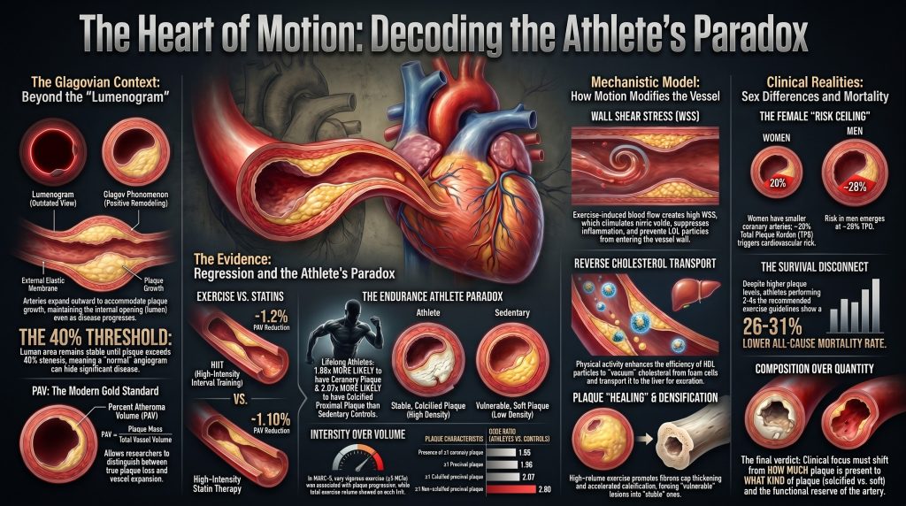
The accurate assessment of exercise-induced changes in the coronary arteriesThe coronary arteries are the small vessels that wrap around the outside of your heart and feed the heart muscle itself. requires a nuanced understanding of the relationship between the vessel’s external boundaries and its internal lumenThe lumen is the open channel inside a blood vessel where blood actually flows.. For decades, the gold standard for diagnosing coronary arteryAn artery is a blood vessel that carries blood away from the heart to the rest of the body. disease was invasive coronary angiography, which provides a two-dimensional map of the lumen — a “lumenogram.” However, this approach is fundamentally limited by the Glagov phenomenonThe Glagov phenomenon (also called compensatory or outward remodeling) is the tendency of an arterial wall to expand outward as atherosclerotic plaque accumulates, thereby preserving the inner lumen diameter until plaque burden becomes very large. Because lumen size stays normal during this compensatory phase, standard stress tests and angiograms can appear normal even in the presence of substant…, or positive remodelingAn outward expansion of the arterial wall that accommodates growing atherosclerotic plaque while preserving the inner lumen diameter; the artery appears unobstructed on tests that only assess lumen narrowing, masking a structurally vulnerable plaque., wherein the coronary artery undergoes a progressive enlargement of its external elastic membrane (EEM)The external elastic membrane is the boundary between an artery's muscular wall and surrounding connective tissue; in intravascular imaging, the area enclosed by the EEM is used to quantify total vessel size including both the open channel and the plaque within the wall. in response to the accumulation of plaque [3].

This compensatory expansion allows the vessel to maintain its luminal cross-sectional area even as a significant atheromaAtheroma is another word for the fatty deposit inside an artery wall — essentially a synonym for plaque, used more often in research writing. develops. In Glagov’sGLAGOV added a PCSK9 inhibitor to statin therapy and measured coronary plaque with intravascular ultrasound before and after. seminal histologic study of 136 autopsied left main coronary arteries, lumen area did not decrease as a function of percent stenosis between 0 and 40 percent, but diminished markedly above that threshold (r = −0.73, P < 0.001) [3]. This implies that an angiographically normal vessel may harbor a substantial, and potentially vulnerable, plaque burdenPlaque burden is the total amount of plaque in your arteries, everywhere — not just at the single worst spot.. The PROSPECT IVUS sub-analysis confirmed in vivo that EEM cross-sectional areas increase in proportion to plaque-and-media area in 552 left main coronary arteries (r = 0.61, P < 0.0001 overall, rising to r = 0.88 when plaque burden was at or below 20 percent) [4].

Exercise is a potent stimulus for vascular remodeling. Chronic aerobic training induces sustained increases in blood flow and shear stress, which activate endothelial pathways that increase nitric-oxide bioavailability and the structural caliber of conduit arteries [5]. Consequently, any study claiming that exercise reduces atherosclerosisAtherosclerosis is the disease behind most heart attacks and many strokes. Cholesterol particles get stuck in the wall of an artery, the body sends immune cells to clean up, and over years that mess hardens into plaque. based solely on luminal diameter is likely measuring a combination of actual plaque loss and compensatory vessel expansion. To distinguish between these effects, researchers rely on percent atheroma volumePercent atheroma volume, or PAV, is the share of an artery segment taken up by plaque rather than open channel. (PAV), which normalizes plaque mass to the total volume of the vessel. PAV is calculated as the sum of (EEM area − lumen area) divided by the sum of EEM area, expressed as a percentage. This metric ensures that if a vessel grows larger (increased EEM) but the plaque area remains constant, PAV decreases, reflecting a relative reduction in the occluded proportion of the artery, even when absolute total atheroma volume (TAV)Total atheroma volume is an IVUS-derived absolute measure of the total three-dimensional volume of plaque within an imaged coronary segment, reported in cubic millimeters. Unlike PAV, TAV is not normalized to vessel size, so it captures the raw amount of disease removed or added over time. remains stable [6].

Evidence for True Plaque Regression: Benchmarking against Pharmacotherapy

When evaluating the capacity of exercise to induce regression, it is necessary to establish a benchmark using lipid-lowering therapies (LLTs), which have the most robust data for reducing PAV and TAV. A comprehensive meta-analysisA meta-analysis statistically combines the results of many separate studies into one overall estimate. of 51 studies involving 9,113 adults demonstrated that LLTs as a class reduce PAV by a mean of −1.10 percent (95% CI −1.63 to −0.56, P < 0.01) and reduce TAV by −5.84 mm³ (95% CI −8.64 to −3.04, P < 0.01); high-intensity statinsA high-intensity statin is a dose expected to cut LDL by 50 percent or more — in practice, higher doses of atorvastatin or rosuvastatin. drove the largest TAV effect (−7.60 mm³; 95% CI −11.89 to −3.31, P < 0.01) [6]. Direct head-to-head plaque-imaging evidence from the GLAGOV randomized trial further demonstrates that PCSK9PCSK9 is a protein made by your liver that destroys the docking ports your liver uses to pull cholesterol out of your blood. inhibition added to statinA statin slows the enzyme your liver uses to make cholesterol. Your liver responds by pulling more cholesterol out of your blood, which is where the real benefit comes from. therapy produces incremental PAV regression in patients with established coronary artery disease [2].

In comparison, the direct evidence for exercise-induced regression is emerging but remains modest. The CENIT trialThe CENIT (Coronary Disease and the Effect of High-Intensity Interval Training) trial was a randomized study of 60 patients with stable coronary artery disease that used intravascular ultrasound to measure whether six months of supervised HIIT at 85–95% of peak heart rate changed plaque volume compared with standard care. It found HIIT reduced percent atheroma volume by 1.2% and total normalized… (Coronary Disease and the Effect of High-Intensity Interval Training) provided critical longitudinal data by randomizing 60 patients with stable coronary artery disease, all of whom underwent percutaneous coronary intervention, to either six months of supervised high-intensity interval training (HIIT)High-intensity interval training is a structured exercise method that alternates short bouts of near-maximal effort (typically 85–95% of peak heart rate) with periods of active recovery or rest. In the cardiovascular context, HIIT protocols have been studied as a non-pharmacological intervention to reduce plaque burden and improve heart function. at 85–95 percent of peak heart rateHeart rate is how many times your heart beats per minute. or to contemporary preventive guidelines [7]. The study used IVUS to measure changes in PAV and TAV in residual coronary segments.

Parameter HIIT Group (n=30) Control Group (n=30) Between-Group Difference
Change in PAV (%) −1.2 (95% CI: −2.1 to −0.2; P=0.017) +0.2 (95% CI: −0.7 to 1.1; P=0.616) −1.4 (95% CI: −2.7 to −0.1; P=0.036) [7]
Change in TAVnorm (mm³) −9.0 (95% CI: −14.7 to −3.4; P=0.002) −12.0 (95% CI: −19.9 to −4.2; P=0.003) [7]

The CENIT results are notable because they demonstrate that exercise can achieve PAV reductions of a magnitude comparable to lipid-lowering therapy (−1.2% vs. −1.10%) [6], [7]. Importantly, this regression was observed in a population already receiving guideline-directed medical therapy, making it difficult to isolate the independent contribution of exercise from a synergistic interaction with statins.

The Endurance Athlete Paradox

While interventional trials in patients with established coronary artery disease suggest the potential for regression, observational data from healthy veteran athletes present a challenging paradox. Large registries, including MARC (Measuring Athlete’s Risk of Cardiovascular Events) and Master@Heart, consistently report that lifelong endurance athletes have a higher prevalence of coronary atherosclerotic plaque and higher Coronary Artery Calcium (CAC)Coronary artery calcium is a measure of calcified plaque deposits in the walls of the coronary arteries, quantified by CT scan and expressed as an Agatston score; higher scores indicate greater cumulative plaque burden and predict future cardiovascular events. scores than less active individuals with comparable cardiovascular risk profiles [8], [9].

The Master@Heart studyThe Master@Heart study is a controlled study of lifelong male endurance athletes that found they carried more total coronary plaque than healthy, active non-athletes—including more calcified and non-calcified plaque in proximal artery segments—raising questions about whether extreme lifelong endurance training promotes atherosclerosis even in the absence of traditional risk factors., a prospective observational cohort analysis of 191 lifelong master endurance athletes, 191 late-onset athletes (initiating endurance sports after age 30), and 176 healthy non-athletic controls (all male, low cardiovascular risk profile, median age 55 years), reported the following adjusted associations for lifelong endurance sports relative to a healthy non-athletic lifestyle [9]:

  • ≥1 coronary plaque: OR 1.86 (95% CI 1.17–2.94)
  • ≥1 proximal plaque: OR 1.96 (95% CI 1.24–3.11)
  • ≥1 calcified plaqueCalcified plaque is the hardened, calcium-filled part of a plaque. It shows up brightly on a CT scan, which is what a calcium scan measures.: OR 1.58 (95% CI 1.01–2.49)
  • ≥1 calcified proximal plaque: OR 2.07 (95% CI 1.28–3.35)
  • ≥1 non-calcified plaqueNon-calcified plaque is the soft, fatty portion of a plaque that has not hardened with calcium. It shows up dark on a CT scan.: OR 1.95 (95% CI 1.12–3.40)
  • ≥1 non-calcified proximal plaque: OR 2.80 (95% CI 1.39–5.65)
  • ≥1 mixed plaque: OR 1.78 (95% CI 1.06–2.99)

The finding of higher odds of non-calcified (lipid-rich) plaques in proximal segments is particularly notable, as such plaques are typically associated with higher near-term risk than purely calcified lesionsIn cardiology, a lesion refers to a discrete area of atherosclerotic plaque narrowing a coronary artery, typically described by the percentage of luminal obstruction it causes. The article describes four residual lesions too small in vessel diameter to accept a stent after the most critical one was treated. [9].

The MARC-2 studyMARC-2 (Male Athlete's Risk of Cardiovascular disease) is a clinical study that examined the relationship between vigorous exercise habits and coronary plaque burden in male athletes, finding an association between high-intensity training and greater calcified plaque. added longitudinal granularity by distinguishing exercise volume from intensity in 289 middle-aged and older male athletes (median age 54 years) followed for a mean of 6.3 years [8]. Total exercise volume (MET-hours/week) showed no association with progression of CAC or plaque. By contrast, exercise intensity stratified by metabolic equivalents — vigorous (6–9 METs) versus very vigorous (≥9 METs) — was significantly associated with imaging outcomes:

Exercise Intensity Effect on CAC Score (β per 10% increase) Plaque Progression
Vigorous (6–9 METs) −0.05 (95% CI −0.09 to −0.01; P=0.02) [8] Less progression
Very Vigorous (≥9 METs) +0.05 (95% CI 0.01 to 0.09; P=0.01) [8] aOR 1.09 (95% CI 1.01–1.18) per 10% increase [8]
Total Volume No significant association [8] No significant association [8]

That intensity rather than volume drives plaque and CAC progression in athletes suggests a physiological thresholdThe exercise intensity beyond which the body can no longer sustain aerobic energy production and begins to accumulate fatigue products rapidly, making it impossible to maintain that effort level; exceeding it early in a race causes premature collapse in pace. beyond which exercise may exert pro-atherogenic effects. Proposed mechanisms include exercise systolic blood pressuresSystolic blood pressure is the top number — the pressure in your arteries while your heart is squeezing. that can exceed 200 mmHg in well-trained athletes during near-maximal effort [25], repetitive mechanical stress on the coronary wall, and inflammatory signaling pathways implicated in athlete-related vascular calcificationCalcification is when calcium gets deposited into a plaque, turning part of it hard and bony. [10].

The defining feature of the athlete’s paradox, however, is that despite higher plaque burden, endurance athletes have lower rates of major adverse cardiovascular eventsA major adverse cardiovascular event, or MACE, is a bundle of bad outcomes counted together in a study — typically cardiovascular death, heart attack, and stroke. (MACE) than the general population. Although the rank order of plaque morphology (calcified > mixed > non-calcified) is broadly similar across active and sedentary populations, lifelong endurance athletes who harbor plaque are more likely than sedentary controls to harbor exclusively calcified lesions, which are considered more stable [12]. This phenotypic enrichment for dense, calcified (stable) lesions — rather than for mixed or non-calcified vulnerable lesions — suggests that high-volume exercise may accelerate the natural healing or densification process of atherosclerosis [10], [11].

Matched Cohort Evidence and Socioeconomic Confounders

A critical requirement for validating the impact of exercise is the control of social and economic confoundersA confounder is a variable that is associated with both the exposure being studied (such as TMAO) and the outcome (such as heart disease), making it appear as though one causes the other when a third factor is actually responsible. The article lists renal function, insulin resistance, systemic inflammation, and age as major confounders that inflate the apparent cardiovascular risk of high TMAO in…. Higher socioeconomic status (SES) is independently associated with both greater exercise capacityExercise capacity is a quantitative measure of the maximum physical work a person can perform, typically assessed during a graded stress test as peak workload in watts, peak oxygen consumption (VO2 max), or metabolic equivalents (METs). In the article, a decline in exercise capacity four months after stenting indicated that the procedure alone had not resolved the underlying disease. and improved cardiovascular outcomes [13]. In the Heart and Soul Study, an analysis of 943 men and women with stable coronary heart diseaseCoronary heart disease is the narrowing or blockage of the arteries that supply blood to the heart muscle, caused by the buildup of atherosclerotic plaque; it is the leading cause of heart attack and cardiac death worldwide., exercise capacity decreased in a graded fashion across categories of income, education, housing, and occupation. After multivariable adjustment, the difference in exercise capacity between the highest and lowest categories was 2.4 METs for household income, 1.8 METs for education, 2.3 METs for housing, and 1.3 METs for occupation (P < 0.001 for all trends) [13].

Spousal Concordance and Partner Studies

Cohabiting partners share a micro-environment encompassing diet, socioeconomic stress, and access to healthcare. In a US nationwide cohort of 5,364 married or domestic-partner couples, within-couple concordance exceeded 50 percent for every component of the American Heart Association’s Life’s Simple 7 framework, ranging from 53 percent for total cholesterolTotal cholesterol adds together the cholesterol in all your particles, harmful and helpful alike. to 95 percent for healthy diet score; concordance for metabolic syndromeMetabolic syndrome is a cluster of five problems that tend to travel together: a large waist, high triglycerides, low HDL, high blood pressure, and high blood sugar. Having three or more counts. reached 73 percent [14]. Despite this strong concordance for behaviors and risk factorsA risk factor is something that raises your chance of developing a disease — high cholesterol particles, high blood pressure, smoking, diabetes, family history., marital status itself, when adjusted for cardiovascular risk factors, was not independently associated with subclinical coronary atherosclerosis on CCTA in a study of 9,288 asymptomatic individuals [15]. Together, these findings suggest that the shared environment of a partnership produces a background risk level, but the trajectory of plaque burden is more heavily influenced by individual health behaviors — including the choice to exercise regularly — than by partnership status itself.

Occupational Activity as a Controlled Environment

The London Transport Workers Study remains the canonical example of an occupational analog [16]. By comparing conductors and drivers within the same transport system — men of similar age and social class — Jeremy Morris was able to isolate occupational physical activity from most confounders. Morris and colleagues studied tens of thousands of London bus and postal workers and reported that physically active conductors, who climbed and descended hundreds of stairs per shift, experienced approximately half the rate of acute and sudden coronary deaths observed among the sedentary drivers [16], [17]. This natural experiment provides some of the earliest robust evidence that occupational physical activity independently modifies the clinical expression of coronary disease, even when the macro-environment is held constant.

Dose-Response Relationships and the Mortality Curve

The relationship between exercise dose and mortality is widely regarded as curvilinear, with the most substantial risk reductions occurring as individuals move from sedentary to moderately active. The systematic reviewA systematic review searches for every study on a question using a pre-declared method, then assesses them by consistent criteria. and meta-analysis by Blond and colleagues, encompassing 48 prospective cohortA prospective cohort enrolls healthy people, records their characteristics, and then waits to see what happens. studies, used random-effects restricted cubic spline dose-responseA dose-response relationship means more of something produces more of an effect, in a consistent gradient. modeling to estimate hazard ratiosA hazard ratio compares how quickly events happen in two groups. A ratio of 0.75 means events occurred at three-quarters the rate in the treated group. across the full exposure range. Compared with the recommended physical activity dose (750 MET-min/week), risk continued to decrease beyond recommendations: at 5,000 MET-min/week, hazard ratios were approximately 0.86 for all-cause mortalityAll-cause mortality means death from any cause at all, not just heart disease — the broadest, hardest-to-game outcome a study can measure. and 0.73 for cardiovascular mortality. No excess risk was observed at the highest exposure levels examined [18].

The Lee et al. Circulation analysis of 116,221 adults pooled from the Nurses’ Health Study and the Health Professionals Follow-up Study, with up to 30 years of follow-up and approximately 47,000 deaths, examined long-term physical activity intensity in detail [19]. Compared with no leisure-time vigorous physical activity (VPA), participants meeting the guideline (75–149 min/week of VPA) had hazard ratios of 0.81 (95% CI 0.76–0.87) for all-cause mortality, 0.69 (95% CI 0.60–0.78) for CVD mortality, and 0.85 (95% CI 0.79–0.92) for non-CVD/non-cancer mortality. Participants performing two to four times the recommended minimum had a 21–23 percent lower all-cause mortality for VPA (150–299 min/week) and a 26–31 percent lower all-cause mortality for moderate physical activity (300–599 min/week) [19]. Higher long-term doses (≥300 min/week VPA or ≥600 min/week moderate activity) did not provide further mortality reduction, but no excess risk was observed [19].

This mortality benefit persists despite the athlete’s paradox of increased CAC, supporting the conclusion that the plaques found in highly active individuals are compositionally more benign in their clinical manifestation [10], [11].

Mechanistic Model: How Exercise Modifies Plaque

The biological mechanisms by which exercise potentially reduces plaque burden or facilitates stabilization act both systemically and locally.

Fluid Mechanics and Wall Shear Stress

Plaque development is governed by patterns of blood flow. Laminar flow produces high wall shear stressWall shear stress is the frictional force exerted by flowing blood on the inner surface of an artery; low, oscillatory, or multidirectional shear stress at arterial bends and bifurcations promotes endothelial dysfunction and plaque initiation, whereas high, uniform shear stress in straight segments is generally protective. (WSS), which maintains endothelial homeostasis through the mechanosensitive transcription-factor axis (KLF2 and KLF4) and downstream activation of endothelial nitric oxideNitric oxide is a gas your blood vessel lining makes to tell the vessel to relax and widen. synthase. Disturbed or oscillatory flow, common at coronary bifurcations, produces low WSS that promotes an atherogenic endothelial phenotype, including upregulation of NF-κB-driven adhesion molecules, monocyte recruitment, and increased endothelial transcytosisTranscytosis is the process by which a cell picks something up on one side, carries it across, and releases it on the other. of LDLLDL, or low-density lipoprotein, is the main particle that carries cholesterol through your blood — and the main one that gets stuck in artery walls. [20]. Exercise-induced increases in flow rate directly enhance the protective frictional force at the vessel wall. Direct molecular evidence in human coronary arteries has shown that regular physical activity in patients with coronary artery disease increases phosphorylation of endothelial nitric oxide synthase at Ser1177, the key regulatory site governing nitric oxide production [5].

Reverse Cholesterol Transport and HDL Functionality

Exercise facilitates the removal of lipids from plaque via the reverse cholesterol transportReverse cholesterol transport is the process of moving cholesterol out of tissues, including artery walls, and back to the liver for disposal. HDL particles do the hauling. pathway, beginning with cholesterolCholesterol is a waxy substance your body needs. It goes into cell walls, hormones, vitamin D, and the bile that digests your food. You would die without it. efflux from foam cellsA foam cell is an immune cell that has eaten so much trapped cholesterol that it swells up and looks foamy under a microscope. to HDLHDL, or high-density lipoprotein, is the particle often called "good cholesterol." It picks up cholesterol from tissues and carries it back to the liver. particles, which transport lipids to the liver for hepatobiliary excretion. A randomized exercise-dosing trial measuring multiple HDL functional metrics has shown that increasing both exercise intensity and dose produces dose-responsive improvements in HDL cholesterol efflux capacityCholesterol efflux capacity is a laboratory test of how well someone's HDL actually pulls cholesterol out of cells — a measure of function rather than quantity., anti-oxidant capacity, and anti-inflammatory function — effects that are largely independent of changes in HDL cholesterol concentration alone [22].

Plaque Stabilization, Matrix Metalloproteinases, and the Fibrous Cap

Beyond regression, exercise promotes structural stabilization through fibrous-cap thickening, mediated in part by reduced intra-plaque matrix metalloproteinaseMatrix metalloproteinases are enzymes that cut through the collagen scaffolding of tissue. Immune cells inside plaques release them. activity. Animal studies in apolipoproteinAn apolipoprotein is a protein attached to a fat-carrying particle in your blood. Fat and water don't mix, so these proteins act like a wrapper that lets fat travel safely through the bloodstream. E knockout mice show that aerobic exerciseAerobic exercise is steady activity that gets you breathing harder for a while, like walking fast, cycling, swimming, or jogging. training reduces intra-plaque MMP-8 and MMP-9 activity, increases tissue inhibitor of metalloproteinases (TIMP-1, TIMP-2), increases fibrous-cap thickness, and reduces the relative size of the necrotic lipid coreThe lipid core is the soft, greasy center of a plaque, made of cholesterol and the debris of dead immune cells. [23]. Human systematic-review evidence links chronic physical activity to favorable changes in circulating MMP-9, oxidized LDLOxidized LDL is an LDL particle that has been chemically damaged after getting stuck in an artery wall., and inflammatory mediators relevant to plaque vulnerability [21]. Concurrent reductions in systemic inflammationInflammation is your immune system's response to injury or something it treats as an invader. It brings swelling, heat, and cleanup cells., including C-reactive proteinC-reactive protein, or CRP, is a substance your liver makes when there is inflammation somewhere in your body. A sensitive version of the test, hs-CRP, is used to estimate heart risk., may further contribute to plaque stabilization [11], [21]. The net effect in chronically active individuals is a transition from compositionally vulnerable to compositionally stable plaque morphology, even when total plaque volumePlaque volume is the total physical amount of plaque in a stretch of artery, measured in cubic millimeters. increases [10], [12].

Sex-Specific Differences in Plaque Risk

A crucial refinement in modern preventive cardiology is the recognition that plaque burden carries different prognostic implications in men and women. In a recent analysis of the PROMISE trialA large randomized trial that evaluated CT angiography versus functional stress testing in symptomatic patients with stable chest pain; findings cited here showed that 16.5% of zero-CAC participants had non-obstructive coronary artery disease and 1.5% had obstructive disease, illustrating residual risk despite a zero calcium score. CCTA arm (4,267 patients, approximately 51 percent women, median follow-up 26 months), women had lower coronary plaque prevalence than men (55 percent versus 75 percent; P < 0.001) and lower median total plaque volume, yet a similar incidence of major adverse cardiovascular events (2.3 percent versus 3.4 percent) [24]. Critically, when MACE risk was modeled against total plaque burden using sex-stratified spline Cox regression, the hazard ratio crossed unity at a substantially lower plaque-burden threshold in women than in men.

Feature Women Men Implication
Plaque prevalence 55% 75% Lower prevalence in women [24]
Plaque volume Lower (median) Higher (median) Smaller absolute burden in women [24]
Total plaque burden at HR=1.0 ≈20% ≈28% Risk emerges at a lower burden in women [24]
Total plaque burden at HR=1.5 ≈32% ≈42% Steeper risk trajectory in women [24]

These data indicate that uniform thresholds for high-risk plaque underestimate risk in women: because women have smaller coronary arteries, a smaller absolute plaque volume produces a larger relative plaque burden and a more rapid acceleration of MACE risk [24]. This implies that for female athletes, the ceiling for compositionally benign subclinical plaque may be considerably lower than for their male counterparts.

Testing the Hypothesis: Remodeling or Regression?

The hypothesis under examination is that exercise does not meaningfully reduce total coronary plaque burden but rather improves vessel caliber and plaque stability. The evidence supports a population-dependent answer:

In patients with established CAD on guideline-directed therapy: Exercise can meaningfully reduce total plaque burden. The CENIT trial demonstrated a between-group PAV difference of −1.4 percent and a TAVnorm reduction of −12 mm³ after six months of HIIT [7].

In healthy lifelong endurance athletes: Exercise does not reduce total plaque burden but is associated with higher prevalence and burden across all plaque types [9]. In this population, the hypothesis is largely supported: the cardiovascular benefit of exercise is realized through vascular remodeling and stabilization (calcification) rather than removal.

Apparent versus true benefits: Increased lumen size in trained individuals reflects a real physiological adaptation (positive remodeling) that confers significant clinical protection through enhanced coronary flow reserveCoronary flow reserve compares blood flow through the heart's arteries at rest with flow when the heart is working hard. [3], [4]. This remodeling benefit is distinct from the discrete benefit of true plaque regression, and the two effects operate on different timescales.

Final Verdict: Regression, Progression, or Stabilization?

The totality of the evidence indicates that chronic exercise is a multi-modal modifier of coronary atherosclerosis. True regression — measurable reductions in PAV and TAV — is achievable through high-intensity exercise interventions, particularly in populations with existing coronary artery disease, where the magnitude approaches that of intensive lipid-lowering therapy [2], [6], [7]. Among individuals without known disease, moderate exercise appears to slow the progression of soft, vulnerable, lipid-rich plaquesAn atherosclerotic lesion whose core is dominated by cholesterol esters and inflammatory lipids rather than calcium or fibrous tissue; the article notes that such plaques are highly responsive to intensive treatment and that the dramatic 65-percentage-point regression at the diagonal branch origin is consistent with reversal of a lipid-rich lesion. through favorable lipid metabolism, enhanced HDL cholesterol efflux capacity, and reduced systemic inflammation [21], [22]. In high-volume endurance athletes, exercise functions primarily as a stabilizing agent, paradoxically accelerating calcification while expanding vessel caliber through positive remodeling — yielding a compositionally benign atherosclerotic burden that does not translate into elevated clinical risk [9], [10], [12].

Critically, the survival benefit of exercise is decoupled from absolute plaque volume: extreme athletes have more plaque yet exhibit substantially lower all-cause and CVD mortality than the general population [18], [19]. The clinical emphasis should therefore shift from quantifying how much plaque is present toward characterizing what kind of plaque is present and what functional reserve the individual’s coronary tree retains.

References

  1. Narula J, Stuckey TD, Nakazawa G, et al. Prospective deep learning-based quantitative assessment of coronary plaque by computed tomography angiography compared with intravascular ultrasound: the REVEALPLAQUE study. Eur Heart J Cardiovasc Imaging. 2024;25(9):1287-1295. doi:10.1093/ehjci/jeae115
  2. Nicholls SJ, Puri R, Anderson T, et al. Effect of Evolocumab on Progression of Coronary Disease in Statin-Treated Patients: The GLAGOV Randomized Clinical Trial. JAMA. 2016;316(22):2373-2384. doi:10.1001/jama.2016.16951
  3. Glagov S, Weisenberg E, Zarins CK, Stankunavicius R, Kolettis GJ. Compensatory enlargement of human atherosclerotic coronary arteries. N Engl J Med. 1987;316(22):1371-1375. doi:10.1056/NEJM198705283162204
  4. Inaba S, Mintz GS, Shimizu T, et al. Compensatory enlargement of the left main coronary artery: insights from the PROSPECT study. Coron Artery Dis. 2014;25(2):98-103. doi:10.1097/MCA.0000000000000074
  5. Hambrecht R, Adams V, Erbs S, et al. Regular physical activity improves endothelial function in patients with coronary artery disease by increasing phosphorylation of endothelial nitric oxide synthase. Circulation. 2003;107(25):3152-3158. doi:10.1161/01.CIR.0000074229.93804.5C
  6. Rivera FB, Cha SW, Varona MC, et al. Atherosclerotic coronary plaque regression from lipid-lowering therapies: A meta-analysis and meta-regression. Am J Prev Cardiol. 2024;18:100645. Published 2024 Mar 11. doi:10.1016/j.ajpc.2024.100645
  7. Vesterbekkmo EK, Aksetøy IA, Follestad T, et al. High-intensity interval training induces beneficial effects on coronary atheromatous plaques: a randomized trial. Eur J Prev Cardiol. 2023;30(5):384-392. doi:10.1093/eurjpc/zwac309
  8. Aengevaeren VL, Mosterd A, Bakker EA, et al. Exercise Volume Versus Intensity and the Progression of Coronary Atherosclerosis in Middle-Aged and Older Athletes: Findings From the MARC-2 Study. Circulation. 2023;147(13):993-1003. doi:10.1161/CIRCULATIONAHA.122.061173
  9. De Bosscher R, Dausin C, Claus P, et al. Lifelong endurance exercise and its relation with coronary atherosclerosis. Eur Heart J. 2023;44(26):2388-2399. doi:10.1093/eurheartj/ehad152
  10. Hsu JJ, Tintut Y, Demer LL. Paradox of Exercise and Coronary Artery Calcification: Potential Underlying Mechanisms. Circ Res. 2025;137(2):335-349. doi:10.1161/CIRCRESAHA.125.326011
  11. Aengevaeren VL, Mosterd A, Sharma S, et al. Exercise and Coronary Atherosclerosis: Observations, Explanations, Relevance, and Clinical Management. Circulation. 2020;141(16):1338-1350. doi:10.1161/CIRCULATIONAHA.119.044467
  12. Aengevaeren VL, Mosterd A, Braber TL, et al. Relationship Between Lifelong Exercise Volume and Coronary Atherosclerosis in Athletes. Circulation. 2017;136(2):138-148. doi:10.1161/CIRCULATIONAHA.117.027834
  13. Cohen B, Vittinghoff E, Whooley M. Association of socioeconomic status and exercise capacity in adults with coronary heart disease (from the Heart and Soul Study). Am J Cardiol. 2008;101(4):462-466. doi:10.1016/j.amjcard.2007.09.093
  14. Shiffman D, Louie JZ, Devlin JJ, Rowland CM, Mora S. Concordance of Cardiovascular Risk Factors and Behaviors in a Multiethnic US Nationwide Cohort of Married Couples and Domestic Partners. JAMA Netw Open. 2020;3(10):e2022119. Published 2020 Oct 1. doi:10.1001/jamanetworkopen.2020.22119
  15. Ann SH, Lee H, Park KS, et al. Marital Status and Subclinical Coronary Atherosclerosis in Asymptomatic Individuals. J Am Heart Assoc. 2022;11(11):e024942. doi:10.1161/JAHA.121.024942
  16. Morris JN, Heady JA, Raffle PA, Roberts CG, Parks JW. Coronary heart-disease and physical activity of work. Lancet. 1953;262(6795):1053-1057. doi:10.1016/s0140-6736(53)90665-5
  17. Morris JN, Kagan A, Pattison DC, Gardner MJ. Incidence and prediction of ischaemic heart-disease in London busmen. Lancet. 1966;2(7463):553-559. doi:10.1016/s0140-6736(66)93034-0
  18. Blond K, Brinkløv CF, Ried-Larsen M, Crippa A, Grøntved A. Association of high amounts of physical activity with mortality risk: a systematic review and meta-analysis. Br J Sports Med. 2020;54(20):1195-1201. doi:10.1136/bjsports-2018-100393
  19. Lee DH, Rezende LFM, Joh HK, et al. Long-Term Leisure-Time Physical Activity Intensity and All-Cause and Cause-Specific Mortality: A Prospective Cohort of US Adults. Circulation. 2022;146(7):523-534. doi:10.1161/CIRCULATIONAHA.121.058162
  20. Niu N, Xu S, Xu Y, Little PJ, Jin ZG. Targeting Mechanosensitive Transcription Factors in Atherosclerosis. Trends Pharmacol Sci. 2019;40(4):253-266. doi:10.1016/j.tips.2019.02.004
  21. Palmefors H, DuttaRoy S, Rundqvist B, Börjesson M. The effect of physical activity or exercise on key biomarkers in atherosclerosis–a systematic review. Atherosclerosis. 2014;235(1):150-161. doi:10.1016/j.atherosclerosis.2014.04.026
  22. Sarzynski MA, Ruiz-Ramie JJ, Barber JL, et al. Effects of Increasing Exercise Intensity and Dose on Multiple Measures of HDL (High-Density Lipoprotein) Function. Arterioscler Thromb Vasc Biol. 2018;38(4):943-952. doi:10.1161/ATVBAHA.117.310307
  23. Kadoglou NP, Moustardas P, Kapelouzou A, et al. The anti-inflammatory effects of exercise training promote atherosclerotic plaque stabilization in apolipoprotein E knockout mice with diabetic atherosclerosis. Eur J Histochem. 2013;57(1):e3. Published 2013 Mar 25. doi:10.4081/ejh.2013.e3
  24. Brendel JM, Mayrhofer T, Karády J, et al. Risk in Women Emerges at Lower Coronary Plaque Burden Than in Men: PROMISE Trial. Circ Cardiovasc Imaging. 2026;19(3):e019011. doi:10.1161/CIRCIMAGING.125.019011
  25. Currie KD, Floras JS, La Gerche A, Goodman JM. Exercise Blood Pressure Guidelines: Time to Re-evaluate What is Normal and Exaggerated?. Sports Med. 2018;48(8):1763-1771. doi:10.1007/s40279-018-0900-x


Transparency Note: This blog post was created with assistance from AI tools. The final content has been carefully reviewed and edited by the author, who is responsible for its accuracy. The information provided is for educational purposes only and does not constitute medical advice.

AI App

Heart Risk Calulator

Educational family-history heart risk calculator with H-score insights, visual family tree input and shareable PDF reports.

Read why this app is so important here.