اللوحة التي لم يعالجها أحد
تخيل مريضًا بعد بضعة أيام من إجراء نوبة قلبية. المحجوب شريان تم افتتاحه، أ دعامة يثبته في مكانه، ويختفي الألم في الصدر. ومن الطبيعي أن نعتقد أن المشكلة قد حُلّت. لكن هذا ليس صحيحًا. فقد عالج الدعامة بقعة واحدة في شريان واحد. أما في بقية الشجرة التاجية، فتنتشر لوحات, ، ومعظمها صغيرة جدًّا بحيث لا تبدو مقلقة على تصوير الأوعية الدموية, ، وهذه هي الأماكن التي ستنشأ منها العديد من النوبات القلبية القادمة.
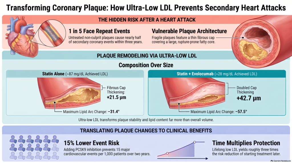
هذا ليس مجرد تكهن. إن دراسة PROSPECT تمت متابعة 697 مريضًا لمدة ثلاث سنوات بعد إجراء متلازمة الشريان التاجي الحادة. على مدى ثلاث سنوات، بلغ المعدل التراكمي لمؤشر الدراسة المركب لـ الأحداث القلبية الوعائية السلبية الكبرى كان 20.4%، أي حوالي واحد من كل خمسة؛ وكان هذا الرقم يمثل مجموع حالات الوفاة القلبية،, سكتة قلبية, ، أو نوبة قلبية، أو إعادة الدخول إلى المستشفى بسبب حالة غير مستقرة أو متفاقمة ذبحة صدرية, ، وليس النوبات القلبية وحدها. فقد وقعت الأحداث التي تُعزى إلى المواقع المسببة الأصلية لدى 12.9% من المرضى، بينما وقعت الأحداث التي تُعزى إلى لويحات لم تُعالج سابقًا لدى 11.6%؛ وكان بعض المرضى يعانون من كلا النوعين، لذا فإن مجموع النسبتين لا يساوي الإجمالي، كما أن ليس كل حدث ناتجًا عن لويحة لم تُعالج سابقًا. كانت معظم اللويحات غير المعالجة التي تسببت في مشاكل لاحقًا تبدو ضيقة بشكل طفيف فقط في تصوير الأوعية الدموية الأصلي [8].
لذا فإن السؤال الحقيقي بعد الإصابة بنوبة قلبية لا يقتصر على “هل يعمل الدعامة؟” فحسب، بل هو “هل يمكننا معالجة بقية جوانب المرض قبل أن يتسبب في حدوث نوبة أخرى؟” يتناول هذا المقال هذا السؤال من خلال الأدلة العلمية: ما الذي يحدث داخل اللويحة عندما الكوليسترول الضار يتم خفضه إلى مستوى منخفض جدًّا، وما إذا كان ذلك سيؤدي إلى انخفاض عدد النوبات القلبية، وما هو المستوى المنخفض الكافي، ولماذا لا يصل الخطر أبدًا إلى الصفر.
الإجابة المختصرة. في دراسات التصوير العشوائية التي أُجريت بعد الأحداث التاجية الحادة، أدى الخفض المكثف لمستويات الكوليسترول الضار (LDL-C) إلى تحسن كبير في مؤشرات محددة لقابلية اللويحات للتكسر، بما في ذلك سماكة الغلاف ومحتوى الدهون، مع انخفاضات متوسطة نسبياً في حجم اللويحة. تُظهر تجارب عشوائية منفصلة تركز على النتائج انخفاضًا في عدد الأحداث القلبية الوعائية مع خفض مستويات الكوليسترول الضار (LDL-C)، لكنها لا تثبت أن التغيرات الملحوظة في تصوير اللويحات هي التي تتوسط هذه الفائدة. ولا يختفي مرض الشريان التاجي.
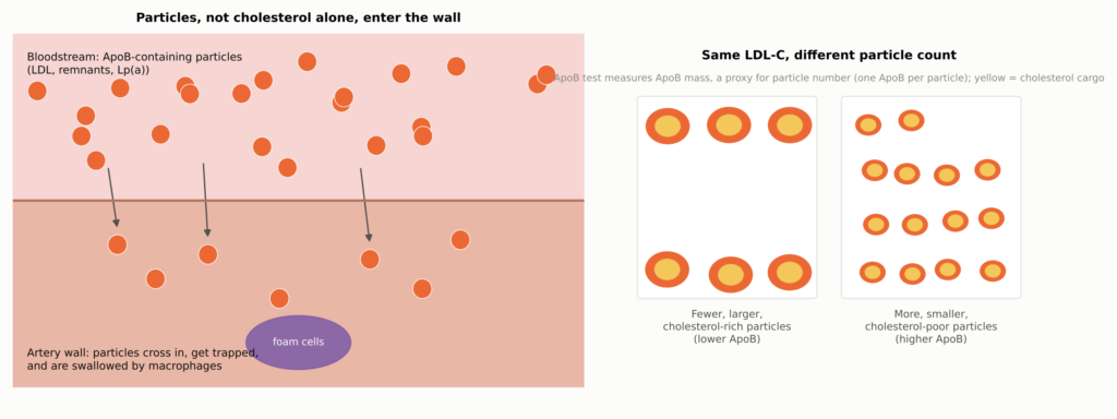
انظر الشكل 1 في الجزء الأول.
نظرة سريعة على الأدلة
| سؤال | ما تظهره الأدلة | ما مدى يقيننا؟ |
| هل يؤدي انخفاض مستوى الكوليسترول الضار (LDL-C) بشكل كبير إلى تغيير في اللويحات؟ | نعم. تزداد سماكة الطبقة العلوية، وتتقلص الإشارات الدهنية، وينخفض حجم اللويحات بشكل طفيف. | تم إثباتها في تجارب تصوير عشوائية بالنسبة لمقاييس تصويرية محددة؛ وليست بديلاً مُثبت الفعالية عن الأحداث |
| هل يقلل خفض مستوى الكوليسترول الضار (LDL-C) من خطر الإصابة بالنوبات القلبية و السكتات الدماغية? | نعم. تُظهر الدراسات السريرية الكبيرة انخفاضًا في عدد النوبات القلبية،, السكتات الدماغية الإقفارية, ، والإجراءات. | أثبتت فعاليتها في تجارب عشوائية واسعة النطاق |
| هل يقلل ذلك من عدد الوفيات؟ | نعم لـ ستاتين في التجارب المجمعة؛ أما بالنسبة للأدوية المضافة، ففي التحليلات الاسمية أو المفتوحة فقط. | أثبتت فعاليتها بالنسبة للستاتينات؛ وتشير إلى فعاليتها بالنسبة للعلاجات المكمّلة |
| هل يعتبر المستوى الأقل من 20 أو حتى 10 ملغ/ديسيلتر أفضل؟ | وقد ارتبطت المستويات المنخفضة التي تم تحقيقها بانخفاض المخاطر، وهو ما يتوافق مع الفوائد ولكنه يخضع لـ ربط مربك وعدم الدقة؛ ولم يتم رصد أي مؤشر رئيسي متسق يتعلق بالسلامة في المجموعات السكانية ودورات المتابعة التي شملتها الدراسة. | تلميحية (لم يتم توزيع المشاركين عشوائيًا على تلك المستويات) |
| هل للوقت أهمية؟ | نعم. تتزايد الفوائد مع مرور السنين، ويكون معدل الإصابة بأمراض الشريان التاجي أقل بكثير لدى الأشخاص الذين يحافظون على مستويات منخفضة من الكوليسترول الضار (LDL-C) طوال حياتهم. | تحظى بدعم قوي |
| هل يمكن أن تصل المخاطر إلى الصفر؟ | لا. لا تزال الأحداث تحدث حتى عند مستويات منخفضة للغاية من الكوليسترول الضار (LDL-C). | متسقة عبر جميع البيانات |
| هل التغيرات التي تطرأ على اللويحات هي السبب وراء انخفاض معدل حدوث هذه الحالات؟ | هذا احتمال وارد، لكنه لم يثبت. | الاستدلال |
ما يحدث داخل الجسم أثناء نوبة قلبية
لفهم ما هويغنس عند قياسها، من المفيد معرفة الشكل الذي تبدو عليه اللويحات الخطيرة.
انظر الشكل 2 في الجزء الأول.
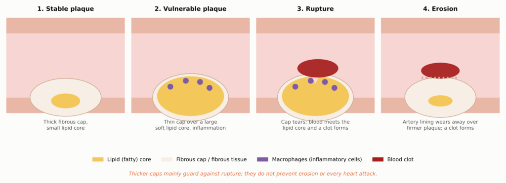
اللويحة هي تراكم داخل جدار الشريان، وليست ترسبًا عالقًا على الجدار الداخلي للأنبوب. وتتكون من عدة أجزاء:
- الغنية بالدهون أو اللب النخري: مجموعة من كوليسترول, بلورات الكوليسترول, ، وحطام الخلايا الميتة. وهو ناعم، وضعيف من الناحية الميكانيكية، ويؤدي إلى تكوين جلطات بشكل كبير في حال وصول الدم إليه.
- غطاء ليفي: طبقة من الكولاجين، تتكون من خلايا العضلات الملساء, ، والتي تعزل النواة عن مجرى الدم. وفي دراسات التشريح التي أجريت على حالات الوفاة القلبية المفاجئة، تبين أن الأغلفة الممزقة كانت عادةً أرق من حوالي 65 ميكرومترًا (μm)، وهو ما يعادل تقريبًا عرض شعرة بشرية رفيعة [25].
- البالعات الكبيرة: الخلايا المناعية التي تمتص الكوليسترول وتصبح “خلايا رغوية.” فهي تفرز إنزيمات قادرة على هضم الكولاجين الموجود في الغطاء.
- الكالسيوم: الرواسب المعدنية. فقد تؤدي ترسبات الكالسيوم الصغيرة والمتفرقة في اللويحة النشطة إلى تركيز الضغط، في حين أن الطبقات الكبيرة والكثيفة غالبًا ما تشير إلى مرض أقدم وأكثر ليفيّة. ولا تزال ترسبات الكالسيوم الواسعة النطاق مؤشرًا على عبء اللويحات والمخاطر.
تبدأ معظم النوبات القلبية عندما يتمزق (ينفجر) غشاء رقيق، فيتلامس الدم مع اللب الذي يشجع على تكوين الجلطات. وتبدأ نسبة أقل منها بـ التآكل, ، حيث تتآكل البطانة الداخلية للشريان لتكشف عن لويحة أكثر صلابة وأقل دهنية [26]. وسيكون لهذا الاختلاف أهمية لاحقًا، لأن زيادة سماكة الغطاء توفر الحماية بشكل أساسي من التمزق.
كيف يدخل الكوليسترول إلى جدار الشريان
البروتين الدهني منخفض الكثافة لا ينتقل الكوليسترول إلى جدار الشريان على شكل دهون حرة. بل ينتقل داخل جسيمات، وهذه الجسيمات هي التي تعبر بطانة الشريان، فتعلق فيها، ثم تبتلعها الخلايا البلعمية. وكل جسيم من هذه الجسيمات، سواء كان LDL، أو بقايا غنية بالدهون الثلاثية، أو Lp(a)، يحمل جزيءًا واحدًا من بروتين المسمى أبروليپوبروتين بأبوبروتين ب): أبو بروتين-100 على الجسيمات التي ينتجها الكبد، وكلما كان أبو بروتين دهني-48 على كيلوكرون بقايا من الأمعاء.
انظر الشكل 3 في الجزء الأول.
بعبارة بسيطة: بروتين ApoB هو عدد المركبات؛ أما LDL-C فهو حمولة الكوليسترول. يقيس المؤشر المختبري القياسي، LDL-C، كمية الكوليسترول التي تحملها جزيئات LDL. أما اختبار ApoB فيقيس تركيز كتلة ApoB، والذي يُعتبر مؤشراً بديلاً لعدد الجزيئات، لأن كل جزيء رئيسي جزيء مُتصلِّب الشرايين يحمل جزيءًا واحدًا من ApoB-100؛ فهو لا يقوم بحساب الجسيمات بالمعنى الحرفي للكلمة. وعادةً ما يتحرك الاثنان معًا. وعندما يختلفان، كما قد يحدث في حالة ارتفاع الدهون الثلاثية, مرض السكري, السمنة, ، أو جزيئات LDL الصغيرة، قد يعكس بروتين ApoB بشكل أفضل عدد الجزيئات التي يمكنها دخول جدار الشريان.
بالنسبة للقراء الذين يستخدمون الوحدات الدولية، فيما يلي التحويلات التقريبية لمستويات الكوليسترول الضار (LDL-C) التي تمت مناقشتها في هذا المقال (القيم مقربة؛ على سبيل المثال، 40 مجم/ديسيلتر تساوي حوالي 1.03 مليمول/لتر، لذا فإن القيم المقربة ليست عتبات متكافئة تمامًا):
| الكوليسترول الضار (LDL-C) (ملغم/ديسيلتر) | 70 | 55 | 40 | 30 | 20 | 10 |
| LDL-C (مليمول/لتر) | 1.8 | 1.4 | 1.0 | 0.78 | 0.52 | 0.26 |
نظرة داخل شريان حي
طوال معظم تاريخ طب القلب، لم يكن بإمكان أحد رؤية الغطاء الليفي لدى شخص حي. فالتصوير الوعائي لا يُظهر سوى الصورة الظلية للقناة المفتوحة. وقد تغير هذا الوضع بفضل القسطرة التي تتيح تصوير الشريان من الداخل.
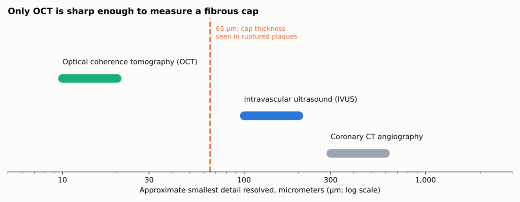
الشكل 10. الدقة التقريبية لـ تصوير الشريان التاجي الطرق (النطاقات النموذجية؛ تختلف حسب النظام). ولا يمكن إلا لتقنية OCT تحديد تفاصيل أدق من الغطاء الليفي الهش، لذا فإن نتائج سماكة الغطاء تُستمد من تقنية OCT، بينما يُستمد حجم اللويحة من تقنية IVUS، التي ترى عبر الجدار بالكامل. يكتشف التحليل الطيفي بالأشعة تحت الحمراء القريبة (NIRS، غير موضح) البصمة الكيميائية للدهون بدلاً من هيكلها. ولا يُعد أي من هذه الأجهزة مجهرًا: فالتصنيفات النسيجية هي استنتاجات.
| أداة التصوير | ما الذي يجيده | ما لا يجيده |
| OCT (ضوء) | يقيس سماكة الغطاء،, قوس الدهون, ، إشارات شبيهة بالبلاعم، الكالسيوم، الجلطة | لا يمكن رؤية الحجم الإجمالي للوحة الترسبات خلف الدهون |
| IVUS (الموجات فوق الصوتية) | يقيس حجم اللويحات وكيفية إعادة تشكيل الشريان | خشنة جدًّا بحيث لا يمكن قياس سعة الغطاء |
| NIRS (التحليل الطيفي) | يكتشف البصمة الكيميائية للدهون | لا يمكنه قياس السعة أو الحجم بمفرده |
| التصوير المقطعي المحوسب للشرايين التاجية | يُظهر تراكم اللويحات في جميع أنحاء القلب دون استخدام قسطرة | لا يمكن رؤية الأحرف الكبيرة أو التفاصيل الدقيقة |
لا يُعد أي من هذه الأجهزة مجهرًا. فالعلامات النسيجية التي تنتجها هي استنتاجات مستمدة من سلوك الضوء أو الصوت. فزيادة سماكة الأوعية الدموية التي تظهرها تقنية التصوير المقطعي البصري التوافقي (OCT)، وانخفاض نسبة الدهون الذي تظهره تقنية التصوير بالضوء القريب من الأشعة تحت الحمراء (NIRS)، وانخفاض حجم اللويحات الذي تظهره تقنية التصوير داخل الأوعية الدموية (IVUS)، هي نتائج متكاملة وليست قابلة للتبادل.
تجربة هيغنز
HUYGENS (تقييم عالي الدقة لللويحات التاجية في دراسة عالمية) إيفولوكوماب أُجريت هذه الدراسة العشوائية للإجابة على سؤال واحد: إذا تم خفض مستوى الكوليسترول الضار (LDL) إلى مستويات منخفضة جدًّا فور الإصابة بنوبة قلبية، فهل تتغير اللويحات التي تبدو خطيرة الموجودة في أجزاء أخرى من القلب؟ وكانت هذه الدراسة عبارة عن تجربة تصويرية من المرحلة الثالثة، متعددة المراكز، عشوائية، مزدوجة التعمية، ومضبوطة بالغفل [9, 1]. قامت شركة «أمجين»، المصنعة لعقار «إيفولوكوماب»، بتمويل الدراسة، وأفاد العديد من الباحثين بوجود علاقات مالية تربطهم بالشركة [9].
من شارك
اختار الباحثون عن عمد أشخاصًا بدت اللويحات المتبقية لديهم هشة بالفعل. وكان المرضى قد أصيبوا بنوبة قلبية دون ارتفاع مقطع ST (NSTEMI) عولجت عن طريق التصوير الوعائي وإجراء جراحي على الشريان المسبب للنوبة. وكان من الضروري أن يكون مستوى الكوليسترول الضار (LDL-C) لديهم أعلى من عتبة معينة تعتمد على استخدامهم لعقاقير الستاتين (130 ملغ/ديسيلتر على الأقل في حالة عدم تناول الستاتين، و80 ملغ/ديسيلتر في حالة تناول ستاتين منخفض أو متوسط القوة، أو 60 ملغ/ديسيلتر في حالة تناول ستاتين عالي الفعالية)، وجزء منفصل من الشريان لم يخضع للعلاج يبلغ طوله 40 ملم على الأقل ويظهر لويحة غنية بالدهون في فحص OCT: غطاء لا يزيد سمكه عن 120 ميكرومتر وقوس دهني يزيد عن 90 درجة [9].
تناول الجميع جرعة من الستاتين هي أقصى جرعة يمكن تحملها. كما تم توزيع نصف المشاركين عشوائياً على إيفولوكوماب 420 ملغ, ، محقون مثبط بروكاتين كونسيرتيز سابتايليسين/كيكسين من النوع 9, ، والنصف الآخر إلى دواء موهم حقنة، تُعطى شهريًّا حتى الأسبوع 48، مع إعادة إجراء فحص OCT وفحص IVUS في الأسبوع 50 إلى 52 تقريبًا [9]. وقد أدرج «هويغنز» 161 مريضًا تم توزيعهم عشوائيًا وتلقيهم العلاج (80 منهم تلقوا إيفولوكوماب، و81 تلقوا الدواء الوهمي). من بين المرضى الذين تلقوا العلاج، كان لدى 135 مريضًا صورًا قابلة للتقييم عند المتابعة، وكان لدى مجموعة فرعية أصغر قياسات متزاوجة باستخدام التصوير بالموجات فوق الصوتية داخل الأوعية (IVUS)، تشير نتائج السجل إلى مؤشرات OCT لمجموعة التحليل الكاملة التي تضم جميع المرضى الـ 161 الذين خضعوا للعلاج، ومن بينهم 119 مريضًا خضعوا لقياسات OCT مزدوجة للمؤشر الرئيسي، لذا لم يتم إعادة تصوير جميع المرضى الـ 161 بشكل مباشر في التحليل؛ أما نتائج التصوير الداخلي للأوعية الدموية (IVUS) فهي مستمدة من مجموعة فرعية أصغر [1]. بلغ متوسط عمر المشاركين 60.5 سنة، وبلغت نسبة النساء 28.6%.
إلى أي مدى انخفض مستوى الكوليسترول الضار (LDL)
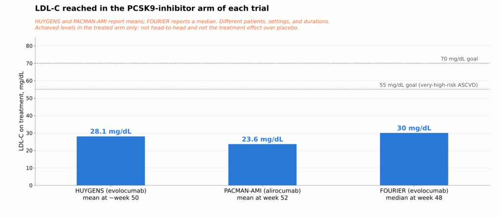
كان متوسط مستوى الكوليسترول الضار (LDL-C) عند التسجيل هو 141.3 ملغ/ديسيلتر. وعند المتابعة، تبين أن 28.1 ملغ/ديسيلتر مع إيفولوكوماب مقابل 87.2 ملغ/ديسيلتر مع الدواء الوهمي [1]. لم تكن المجموعة التي تلقت الدواء الوهمي غير معالجة: فقد دخل العديد من المرضى الدراسة دون تناول أي من عقاقير الستاتين أو بتناول جرعة منخفضة منها، لذا فإن انخفاض مستويات الكوليسترول لديهم من حوالي 141 إلى 87 ملغ/ديسيلتر يعكس إلى حد كبير بدء تناول عقار الستاتين أو زيادة جرعته. كل مقارنة في دراسة HUYGENS هي الستاتين مقابل الستاتين مع إيفولوكوماب.
ماذا حدث للوحة التذكارية؟
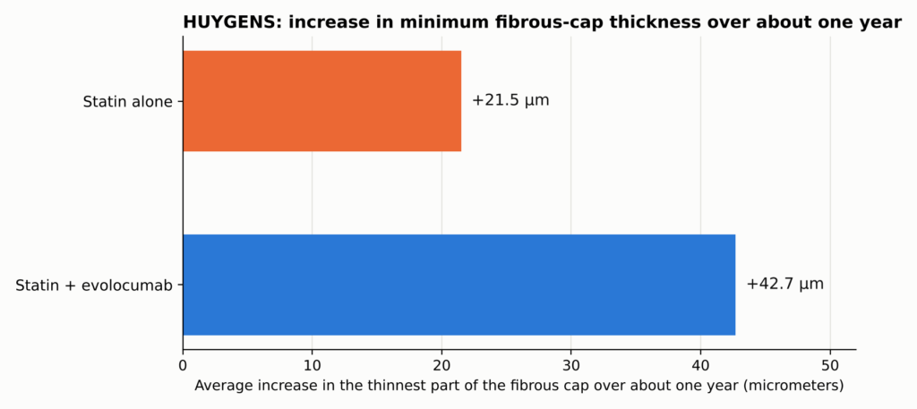
انظر الشكل 4 في الجزء الأول.
زاد سمك الغطاء الواقي بمعدل +42.7 ميكرومتر مع عقار إيفولوكوماب مقابل +21.5 ميكرومتر مع الدواء الوهمي, متوسطات أقل المربعات استنادًا إلى تحليل التباين المشترك الذي أُجري في إطار التجربة على جميع المرضى الـ 161 الذين تلقوا العلاج (مجموعة التحليل الكاملة، وفقًا لنتائج سجل التجربة)؛ ظهر فرق قدره 21.2 ميكرومتر (95% CI 4.7–37.7؛ P=0.015) [1]. تقلص قوس الدهون بشكل أكبر (−57.5 مقابل −31.4 درجة)، وانخفضت إشارة البلاعم بشكل أكبر. أما حجم اللويحات، الذي تم قياسه بواسطة التصوير بالموجات فوق الصوتية داخل الأوعية (IVUS) في مجموعة فرعية استكشافية، فلم يتغير إلا بشكل طفيف: حجم العصيدة النخامية المئوي التغير −2.29 مقابل −0.61 نقطة مئوية، وهو فرق حسابي يبلغ −1.68 نقطة (ليس تباينًا معدلًا، ولا يمكن مقارنته مباشرةً بـ PACMAN-AMI (التقدير القائم على النموذج).
بعبارات بسيطة. بلغ متوسط الزيادة في سماكة الغشاء القلبي حوالي ضعف ما سُجل مع عقار إيفولوكوماب. ويُقارن هذا بين حجم الزيادات، وليس بين الأغشية نفسها، ولا يعني ذلك أن خطر الإصابة بنوبة قلبية قد انخفض إلى النصف. كان التغيير الكبير في المادة التي صُنعت منها اللوحة التذكارية، وليس في حجمها. تصف هذه النتائج المقاطع المختارة غير المسببة للمشكلة التي تم تصويرها، وليس كل لويحات الشرايين التاجية.
كما أشارت مراجعة ثانوية أجراها الباحثون في التجربة إلى انخفاض عدد المرضى الذين عولجوا بعقار إيفولوكوماب والذين ظهرت لديهم أغشية رقيقة جدًّا (أقل من 65 ميكرومتر) بعد العلاج [27]. يرد جدول نتائج HUYGENS الكامل في الملحق الفني.
بحث تحليلان لاحقان أجراهما «هويغنز» عن المرضى الذين أظهروا استجابات مختلفة. وأظهر المرضى الذين يعانون من ارتفاع مستوى Lp(a) فرقًا أكبر في سماكة الغشاء بين عقار إيفولوكوماب والعلاج الوهمي [28]، وكان لدى المرضى الذين وصلوا إلى مستويات أقل من بروتين ApoB نتائج فحص اللويحات أكثر إيجابية مقارنةً بالمرضى الذين لم يصلوا إلى تلك المستويات [29]. وكلاهما عبارة عن تحليلات فرعية صغيرة أُجريت بعد إجراء الدراسة: فلم يتم توزيع المشاركين عشوائيًا على تلك المستويات، وهما بحاجة إلى إعادة إجرائهما، كما أنهما لا يحددان هدفًا أو آلية علاجية مستقلة. وترد التفاصيل في الملحق الفني.
ما لا يستطيع «هويغنز» أن يخبرنا به
يقدم دراسة «هويجنز» (HUYGENS) أدلة عشوائية على أن إضافة عقار إيفولوكوماب إلى العلاج بالستاتينات بعد الإصابة بنوبة قلبية حادة بدون ارتفاع مقطع ST (NSTEMI) يحسّن العديد من الخصائص المستمدة من التصوير المقطعي البصري (OCT) المرتبطة بهشاشة اللويحات في شرايين تاجية مختارة غير مسؤولة عن النوبة آفات. ولا تثبت هذه الدراسة أن التغيرات التي ظهرت في الصور التشخيصية قد حالت بشكل مباشر دون حدوث أحداث قلبية وعائية لاحقة.
هويغنس لم يُصمم هذا الدواء لتقليل حالات النوبات القلبية أو السكتات الدماغية أو الوفيات. وهذا يتطلب عشرات الآلاف من المرضى، وليس 161 مريضًا فقط. ولم تُجرَ التصويرات إلا في بداية الدراسة وبعد مرور عام واحد، لذا لا يمكنها تحديد مدى سرعة تغير طبقة الكاب. وقد شملت الدراسة أشخاصًا تم اختيارهم لأن لويحاتهم كانت تبدو هشة بالفعل. كما أنها لا تثبت أن زيادة سماكة طبقة الكاب أدت إلى أي انخفاض لاحق في معدل حدوث الأحداث. ولتحقيق ذلك، نحتاج إلى أنواع أخرى من التجارب.
ليست حالة فردية: فقد شهدت تجارب أخرى الأمر نفسه
قد يكون من السهل تجاهل تجربة واحدة شملت 161 مريضًا. لكن تجربة «هويجنز» (HUYGENS) مقنعة لأن تجارب أخرى، استخدمت أدوية وأدوات تصوير مختلفة، أظهرت النمط نفسه.
باكمان-آمي عشوائي 300 مريض أصيبوا بنوبة قلبية إلى أليروكوماب, ، آخر PCSK9 الأجسام المضادة، أو الدواء الوهمي، وكلاهما بالإضافة إلى روزوفراتاستين, ، ابتداءً من في غضون 24 ساعة بعد إجراء تدخل القسطرة التاجية العاجل بالنسبة للنوبة القلبية، وقاموا بتقييم اللويحات غير المسؤولة عن النوبة باستخدام طرق تصوير تكميلية. من بين المشاركين الـ300 الذين تم توزيعهم عشوائياً، توفرت بيانات متزاوجة من التصوير الداخلي للأوعية الدموية (IVUS) لـ265 مشاركاً؛ وشمل تحليل الدهون باستخدام تقنية NIRS 263 مريضاً، بينما شمل تحليل سماكة الغشاء باستخدام تقنية OCT 221 مريضاً (105 يتناولون أليروكوماب، و116 يتناولون الدواء الوهمي) ممن كان لديهم تليف عصيدي في كلتا النقطتين الزمنيتين. وقد استُمدت القياسات الثلاث جميعها من نفس التجربة العشوائية. وانخفض مستوى الكوليسترول الضار (LDL-C) إلى 23.6 ملغ/ديسيلتر مقابل 74.4 ملغ/ديسيلتر. أظهرت ثلاث طرق تصوير متكاملة، تم قياسها لدى مرضى متداخلين ضمن التجربة نفسها، تغييرات إيجابية متوافقة: النسبة المئوية عصيدي انخفض الحجم بدرجة أكبر (الفرق −1.21 نقطة مئوية)، وانخفضت إشارة الدهون المقاسة بتقنية NIRS بدرجة أكبر (الفرق −41.24)، وارتفع سمك الغطاء بدرجة أكبر (+62.67 مقابل +33.19 ميكرومتر؛ الفرق +29.65 ميكرومتر) [7]. هذه الاختلافات هي التقديرات المستندة إلى النموذج في التجربة. وعندما تتوافق العوامل الضوئية والصوتية والكيميائية في تجربة واحدة، تكون النتيجة أكثر إقناعًا من أي مقياس منفرد، على الرغم من أنها لا تزال مجرد تجربة واحدة.
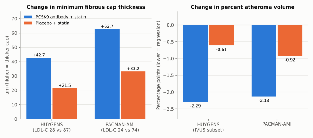
الشكل 11. دراستا «HUYGENS» و«PACMAN-AMI»: التغيير على مدار حوالي عام واحد في كل مجموعة علاجية. أضافت كلتا الدراستين جسمًا مضادًا لبروتين PCSK9 (إيفولوكوماب في دراسة «HUYGENS»، وأليلوكوماب في دراسة «PACMAN-AMI») إلى العلاج بالستاتينات بعد احشاء عضلة القلب. تمثل الأعمدة التغيرات المتوسطة داخل كل ذراع كما وردت في التقرير؛ وبلغت الفروق بين المجموعتين +21.2 ميكرومتر (P=0.015) و+29.65 ميكرومتر (11.75–47.55) بالنسبة لسمك الغطاء، و−1.68 (حسابي) و−1.21 (−1.78 إلى −0.65) نقطة مئوية بالنسبة لنسبة حجم اللويحات. بالنسبة لدراسة PACMAN-AMI، فإن الطرح البسيط لمتوسطات المجموعتين يعطي 29.48 ميكرومتر، وهو يختلف قليلاً عن القيمة المعدلة وفقًا للنموذج في التجربة البالغة 29.65 ميكرومتر؛ وذلك لأن نموذج التأثيرات المختلطة في التجربة يأخذ في الاعتبار القياسات المتكررة والشرايين المتعددة التي تم تصويرها في كل مريض؛ ويستخدم النص الرقم المعدل. يستخدم اللوحان وحدات قياس مختلفة ولا ينبغي مقارنتهما ببعضهما البعض. كانت النسبة المئوية لحجم التصلب الشرياني في دراسة HUYGENS نقطة نهاية استكشافية في مجموعة فرعية من صور التصوير الموجي الداخلي للأوعية الدموية (IVUS). تختلف مجموعات التحليل حسب المقياس: نتائج غطاء HUYGENS هي تقديرات نموذجية في مجموعة التحليل الكاملة المكونة من 161 مريضًا خضعوا للعلاج (119 منهم مع تصوير OCT مقترن؛ و135 مع أي تصوير متابعة قابل للتقييم)، بينما يأتي حجم اللويحات (PAV) الخاص بها من مجموعة فرعية أصغر من IVUS؛ يأتي حجم التصلب الشرياني (PAV) في دراسة PACMAN-AMI من 265 مريضًا لديهم فحوصات IVUS مقترنة، بينما يأتي سمك الغطاء من 221 مريضًا لديهم تصلب ليفي في كلا النقطتين الزمنيتين. المصادر: [1, 7].
غلاغوف عشوائي 968 مريضًا (846 مريضًا خضعوا لفحص IVUS قابل للتقييم خلال فترة المتابعة) يعانون من مرض الشريان التاجي المستقر، حيث تلقوا إيفولوكوماب أو دواءً وهميًا لمدة 76 أسبوعًا، مع إجراء الفحص الرئيسي بواسطة IVUS في الأسبوع 78. وبلغ متوسط مستوى الكوليسترول الضار (LDL-C) 36.6 مقابل 93.0 ملغ/ديسيلتر. تغيرت النسبة المئوية لحجم اللويحات التصلبية بمقدار −0.95 نقطة مئوية مع عقار إيفولوكوماب مقابل +0.05 مع الدواء الوهمي (الفرق بين المجموعتين −1.0 نقطة مئوية؛ نطاق الثقة 95٪ −1.8 إلى −0.64، مقتبس حرفياً من التجربة المنشورة)، و 64% مقابل 47% من المرضى أظهروا تراجعًا قابلًا للقياس [30]. هذه هي التغيرات داخل كل ذراع بالإضافة إلى التباين المبلغ عنه بين المجموعات في التجربة، وليست نسب انخفاض في كتلة اللويحات. وفي مجموعة فرعية استكشافية بلغت مستوياتها حوالي 24 مجم/ديسيلتر، تراجعت مستويات 81%. كما تُظهر دراسة GLAGOV حدود هذه النتائج: لم يحدث تراجع في حوالي ثلث المرضى الذين تلقوا العلاج، ولم تجد دراسة فرعية لتحليل الأنسجة أي اختلافات ذات دلالة إحصائية في مكونات اللويحات [31].
البدء المبكر في تناول الستاتين و إزيتيمايب وأظهرت التجارب نفس النتيجة عند مستويات أعلى من الكوليسترول الضار (LDL-C): فكلما انخفض مستوى الكوليسترول الضار (LDL-C)، قل نمو اللويحات أو حدث تراجع طفيف فيها [32, 33, 34, 35]، وظهور طبقات أكثر سمكًا في صور التصوير المقطعي البصري (OCT) [36]. أظهرت دراسة «YELLOW III» تغيرات إيجابية داخل الفرد في قياسات تصوير اللويحات على مدار 26 أسبوعًا؛ ونظرًا لكونها دراسة غير مراقبة، فإنها لا تستطيع تحديد مدى ارتباط هذا التغير بعقار إيفولوكوماب [37]. أشارت دراسة أجريت عام 2025 على عقار إيفولوكوماب باستخدام التصوير المقطعي المحوسب (CT) والتصوير المقطعي بالإصدار البوزيتروني (PET) في مجموعة واحدة إلى انخفاض لويحة غير متكلسة وأقل من ذلك التكلس الدقيق النشاط، في حين أن لويحة متكلسة ارتفع الحجم؛ وتقيس مؤشرات التصوير الخاصة به عناصر مختلفة، لذا ينبغي تقييمها كل على حدة [38]. لم تكن جميع التجارب ناجحة: أوديسي لم تجد دراسة J-IVUS أي فرق ذي دلالة إحصائية في مؤشر اللويحات على مدى 36 أسبوعًا [39].
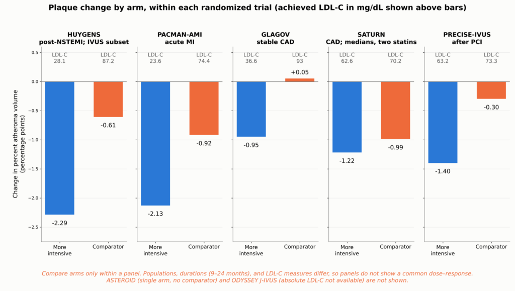
الشكل 12. التغير في النسبة المئوية لحجم اللويحات التصلبية وفقًا لتصوير الأوعية الدموية داخل الأوعية (IVUS) في كل ذراع من أذرع خمس تجارب تصوير عشوائية، بلوحة واحدة لكل تجربة، مع مستويات الكوليسترول الضار من النوع LDL التي تم تحقيقها في كل ذراع. قارن المجموعات فقط داخل كل لوحة: تختلف المجموعات السكانية، والمدد الزمنية (من 9 إلى 24 شهرًا)، وقراءات LDL-C (تُحسب قراءة GLAGOV على أساس مرجح زمنيًا)، لذا فإن اللوحات لا تشكل دليلًا على وجود علاقة مشتركة بين الجرعة والاستجابة. تأتي قيم HUYGENS من مجموعة فرعية استكشافية من بيانات التصوير بالموجات فوق الصوتية داخل الأوعية الدموية (IVUS). زحل أوردت هذه الدراسات التغيرات في القيمة الوسيطة ومستوى الكوليسترول الضار (LDL-C) المرجح زمنياً، في حين أوردت التجارب الأخرى القيم المتوسطة. كويكب تم استبعاده لعدم وجود مجموعة مقارنة، كما تم استبعاد دراسة «ODYSSEY J-IVUS» لعدم توفر القيم المطلقة لمستوى الكوليسترول الضار (LDL-C) الذي تم تحقيقه في المصادر التي تمت مراجعتها. المصادر: [34, 35, 30, 1, 7].
ماذا يعني انخفاض حجم اللويحات بمقدار نقطة واحدة؟ النسبة المئوية لحجم اللويحات (PAV) هي النسبة التي تشغلها اللويحات من مساحة الوعاء الدموي. ويُعتبر التغير من 40% إلى 39% انخفاضًا بمقدار نقطة مئوية واحدة. وهذا لا يعني أن 1% من إجمالي اللويحات قد اختفت، ولا يعني أن التضيق قد انفتح بنفس المقدار.
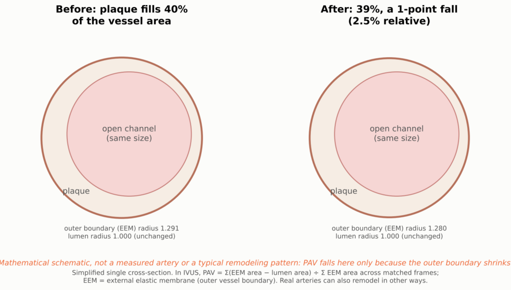
الشكل 13. رسم توضيحي مفاهيمي لحجم التغير بمقدار نقطة مئوية واحدة في النسبة المئوية لحجم اللويحات (PAV)؛ القناة المفتوحة (لومن) مرسومة بنفس الحجم في كلا اللوحتين، بينما يتقلص الحد الخارجي للوعاء الدموي بشكل طفيف. ويُقصد بـ PAV النسبة المئوية لمساحة الوعاء الدموي التي تشغلها اللويحات، محسوبة كمتوسط على طول مقطع معين. الانخفاض من 40% إلى 39% هو مقياس التغيير في تجارب التصوير بالموجات فوق الصوتية داخل الأوعية (IVUS). وهذا لا يعني اختفاء 1% من إجمالي اللويحات، ولا يعني أن القناة المفتوحة قد اتسعت بنفس القدر، لأن الشرايين تتغير هيكلها أيضًا.
يوجد في الملحق الفني جدول مقارنة بين جميع تجارب التصوير.
مختلف، وليس أصغر بكثير
إذا كان حجم اللويحة لا يتغير إلا قليلاً، فكيف يمكن أن تقل احتمالية تمزقها؟ تقدم ميكانيكا المواد إجابة معقولة. يحدث التمزق عندما يتركز الضغط في غطاء رقيق ممتد فوق قلب لين. وينبغي أن يؤدي انخفاض نسبة الدهون وزيادة سماكة الغطاء إلى خفض هذا الضغط حتى لو كان حجم اللويحة متماثلاً تقريباً. وهذا يقدم تفسيراً معقولاً لكيفية إظهار التجارب السريرية انخفاضات ملموسة في الأحداث القلبية، في حين لا تظهر التجارب التصويرية سوى تغيرات طفيفة في الحجم، على الرغم من أن هذا يظل تفسيراً وليس سبباً مثبتاً.
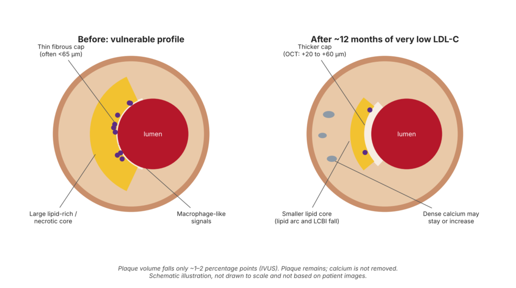
الشكل 14. رسم تخطيطي توضيحي — لا يمثل مريضًا حقيقيًّا ولا استجابة مضمونة للعلاج. رسم تخطيطي لمفهوم استقرار اللويحة: لم يتم رسمها وفقًا للمقياس الحقيقي ولا تستند إلى صور حقيقية للمرضى. تُظهر التجارب التصويرية العشوائية سماكة القشرة، وانخفاضًا في نسبة الدهون، وانخفاضًا في الإشارات الشبيهة بالبلاعم، وانخفاضًا طفيفًا في حجم اللويحة. وتُعد التغيرات في مستويات الكالسيوم وتشريح الآفة أمثلة توضيحية، وليست تغيرات مقاسة في لويحة واحدة. ولا تُظهر التجارب العشوائية إزالة اللويحة، أو إزالة الكالسيوم، أو توسعًا روتينيًا في تجويف الشريان.
تتألف سلسلة الاستدلال التي تربط بين انخفاض مستوى الكوليسترول الضار (LDL-C) وانخفاض عدد النوبات القلبية من عدة حلقات. بعضها يُقاس بشكل مباشر، بينما يُستنتج البعض الآخر.
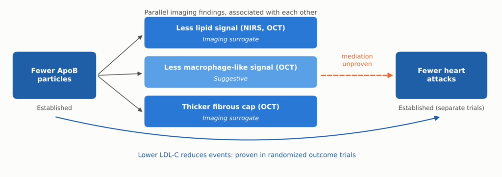
الشكل 15. العلاقة بين انخفاض مستوى الكوليسترول الضار (LDL-C) وتصوير اللويحات والأحداث السريرية. وقد ثبت أن انخفاض عدد جزيئات البروتين ApoB وانخفاض عدد الأحداث السريرية مرتبطان ببعضهما، وقد تم إثبات هذا الارتباط الأخير في تجارب كبيرة تركز على النتائج. وتُعد نتائج تحاليل الدهون، والخلية الشبيهة بالبلاعم، والقبعات مؤشرات تصويرية بديلة تُقاس معًا، وليست تسلسلًا مثبتًا. ولم يتم إثبات ما إذا كانت هذه التغيرات التصويرية تتوسط في انخفاض عدد الأحداث السريرية أم لا.
تدعم الصور المأخوذة من الأبحاث الرصدية فكرة أن هذه السمات ذات أهمية. ففي دراسة CLIMA، كانت اللويحات غير المعالجة التي تجمع بين أربع سمات عالية الخطورة وفقًا لفحص OCT تنطوي على خطر حدوث أحداث خلال عام واحد أعلى بعدة أضعاف [40]. غير أن ربط سمة ما بالمخاطر لا يعني بالضرورة إثبات أن تغيير تلك السمة يؤدي إلى خفض المخاطر.
هناك ثلاثة أمور يجب أخذها في الاعتبار للحفاظ على التوازن في هذا الشأن:
- الغطاء الأكثر سمكًا ليس ضمانًا. يستند الحد الفاصل البالغ 65 ميكرومترًا إلى دراسات تشريح الجثث [25]. قد تتعرض الأغطية الأكثر سمكًا للتمزق أيضًا، كما أن قياسات OCT لهذه الهياكل الرقيقة تتفاوت، ويتفاوت سمك الغطاء على طول مساره.
- أما التآكل فمختلف. تحدث النوبات القلبية الناتجة عن التآكل بشكل أساسي بسبب تلف بطانة الشريان و التهاب أكثر من تخفيف الغطاء [26]. ويهدف تكثيف العلاج في الغالب إلى معالجة حالات التمزق، وهو أحد الأسباب التي تجعل هذه الأحداث لا تزال تحدث حتى عند مستويات منخفضة جدًّا من الكوليسترول الضار (LDL-C).
- قد يرتفع مستوى الكالسيوم، لا ينخفض. أدت الستاتينات عالية الفعالية إلى زيادة قياسات الكالسيوم في التصوير بالموجات فوق الصوتية داخل الأوعية الدموية (IVUS) مع تقليل حجم اللويحات [41]، وقد لوحظ لدى مستخدمي الستاتين في مجموعة PARADIGM CT نمو أبطأ للوحة غير المتكلسة، في حين لوحظ نمو أسرع للوحة المتكلسة [42]. ارتفاع نقاط الكالسيوم إن توقف الاستجابة للعلاج لا يعني، في حد ذاته، أن العلاج قد فشل، كما أن قياسات الكالسيوم المتتالية ليست طريقة معتمدة لتقييم فعالية العلاج. ولا توجد أدلة تدعم الادعاءات التي تزعم أن خفض مستويات الكوليسترول الضار (LDL) “يذيب الكالسيوم”.
كما أن تراجع اللويحات لا يؤدي بالضرورة إلى إعادة فتح الشريان. فانخفاض النسبة المئوية لحجم اللويحات لا يعني بالضرورة زيادة مماثلة في قطر التجويف. الشرايين التاجية يمكن إعادة تصميمها نحو الخارج أو نحو الداخل، و تراجع اللويحة قد يحدث ذلك دون حدوث تغيير ذي دلالة سريرية في التضيق الملحوظ في صورة الأوعية الدموية. ولا تثبت الدراسات التصويرية التي نناقشها هنا إمكانية عكس التضيق التاجي الثابت بشكل روتيني.
من تقليل تراكم البلاك إلى خفض معدل الإصابة بالنوبات القلبية
تُظهر الدراسات التصويرية ما يحدث للوحة التصلب الشرياني. أما الدراسات السريرية التي تقيس النتائج، والتي تشمل آلاف المرضى وتسجل حالات النوبات القلبية والسكتات الدماغية والوفيات الفعلية، فتُظهر ما إذا كان لذلك أهمية أم لا. وهذا هو الدليل الأقوى في هذا السياق.
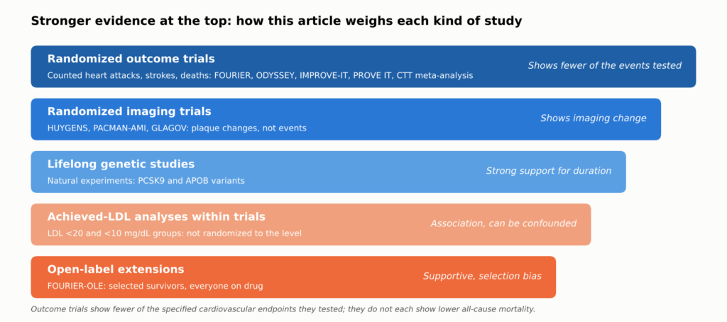
الشكل 16. كيفية تقييم الأدلة الواردة في هذه المقالة. تثبت التجارب العشوائية التي تركز على النتائج أن خفض مستوى الكوليسترول الضار (LDL-C) يمنع حدوث الأحداث التي تم اختبارها؛ وتُظهر التجارب العشوائية التي تعتمد على التصوير أن ذلك يؤدي إلى تغيير في قياسات التصوير الخاصة باللويحات؛ أما المستويات المتبقية فتدعم الادعاءات المتعلقة بالأهداف المنخفضة جدًّا، والآثار طويلة المدى، والفوائد مدى الحياة، ولكنها لا تستطيع إثباتها بمفردها. وقد تم تصنيف كل ادعاء في النص حسب نوع الدليل الذي يستند إليه.
The Numbers That Matter to Patients
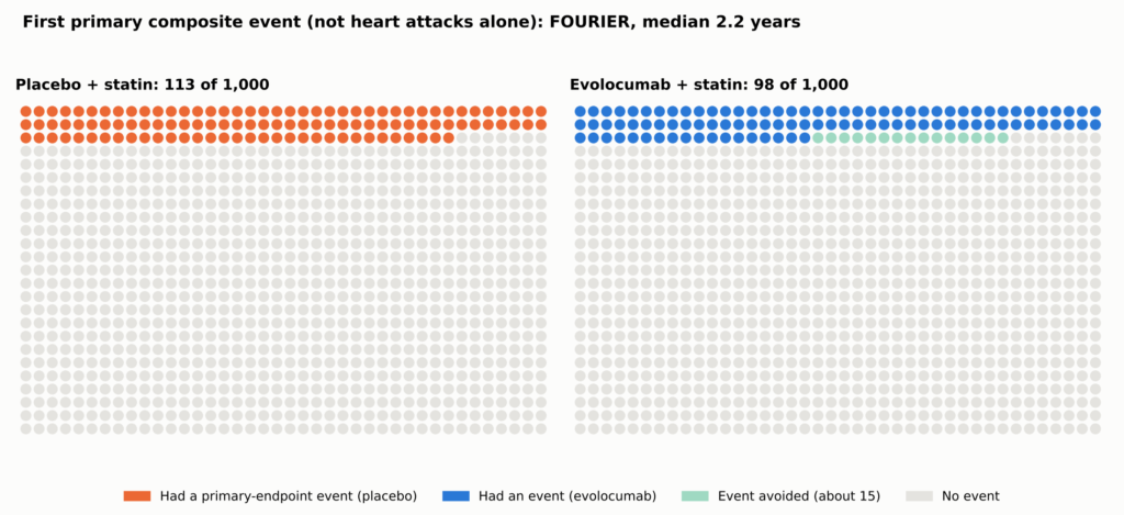
Relative reductions sound large; absolute reductions show what a person gains. In فورييه, the primary composite (cardiovascular death, heart attack, stroke, hospitalization for ذبحة صدرية غير مستقرة, أو التاجي إعادة التروية) occurred in 9.8% with evolocumab versus 11.3% with placebo over a median 2.2 years: a 15% relative hazard reduction and an absolute difference of about 1.5 percentage points, or about 15 fewer people per 1,000 [2]. That composite is not the chance of a heart attack alone, and the original تجربة فورير did not establish a reduction in death.
See Figure 5 in Part 1.
| المحاكمة | What was tested | المتابعة | Event rate, treatment vs control | Difference per 1,000 (people; فورييه-أولي: events per 1,000 person-years) |
| MIRACL [43] | High-dose statin vs placebo, started in hospital | 16 weeks | 14.8% vs 17.4% | About 26 |
| PROVE IT–TIMI 22 [10] | Intensive vs moderate statin | سنتان | 22.4% vs 26.3% | About 39 |
| IMPROVE-IT [4] | Adding ezetimibe to a statin | 7 years | 32.7% vs 34.7% | About 20 |
| نتائج أوديسي [3] | Adding alirocumab after ACS | Median 2.8 years | 9.5% vs 11.1% | About 16 |
| فورييه [2] | Adding evolocumab, stable disease | Median 2.2 years | 9.8% vs 11.3% | About 15 |
| FOURIER-OLE [14] (open-label extension; not a randomized trial endpoint) | Earlier vs later evolocumab (open label) | Median 5 more years | CV death, MI, or stroke 2.05 vs 2.58 per 100 person-years | About 5.3 fewer events per 1,000 person-years (rate difference, not people) |
Each trial counted its own combination of events in its own population over its own follow-up, so the rows are not a ranking. FOURIER-OLE compares original randomized assignment in selected survivors, all on open-label evolocumab. A detailed version of this table, with relative effects, LDL-C levels, and mortality, is in the Technical Appendix.
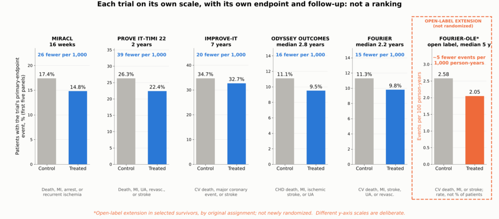
Figure 17. Absolute benefit in the major outcome trials, shown as small multiples: each panel gives one trial’s control and treated primary-endpoint event rates on its own y-axis scale, with the difference expressed per 1,000 patients treated. Each trial used a different نقطة نهاية مركبة, population, comparator, and follow-up period, so the panels cannot be compared with one another or read as a ranking; they show the scale of benefit within each trial. *FOURIER-OLE shows cardiovascular death, MI, or stroke during the open-label extension, comparing patients originally assigned evolocumab with those originally assigned placebo (2.58 vs 2.05 events per 100 person-years); because this is a rate, not a percentage of patients, its panel has its own y-axis label; it is an exploratory comparison in selected survivors, not a new randomized trial. Sources: [43, 10, 4, 3, 2, 14].
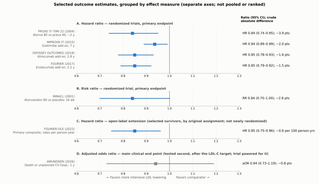
Figure 18. Selected outcome estimates from the trials discussed: primary endpoints for the randomized trials, the open-label extension comparison for FOURIER-OLE, and the main clinical end point for AMUNDSEN. Each trial used a different composite endpoint, population, and follow-up, and reported a different effect measure (risk ratio, نسبة الخطر, or adjusted odds ratio), so each effect measure is shown in its own panel with its own axis, for orientation, not pooled or ranked. Absolute differences are crude differences in each trial’s reported event rates over its own follow-up (for FOURIER-OLE, per 100 person-years). FOURIER-OLE is not a newly randomized outcome comparison: it compares selected surviving participants by original assignment during an open-label extension in which all received evolocumab. AMUNDSEN (gray) is shown for its main clinical end point, which was tested second in the trial’s prespecified hierarchy, after the LDL-C target endpoint; the trial was powered for it. PROVE IT–TIMI 22’s interval is derived from its reported 16% (5%–26%) relative reduction. MIRACL’s الخطر النسبي is shown as the trial reported it, estimated from a Cox proportional hazards model. Sources: [43, 10, 4, 3, 2, 14, 24].
Fewer Heart Attacks and Strokes; Death Is Harder to Move
The benefit is concentrated in nonfatal events. In FOURIER, heart attacks fell from 4.6% to 3.4%, strokes from 1.9% to 1.5%, and coronary procedures from 7.0% to 5.5% [2]. ODYSSEY OUTCOMES and IMPROVE-IT showed similar reductions in heart attack and ischemic stroke [3, 4]. These are secondary analyses that support the main findings.
Among the add-on trials (IMPROVE-IT, ODYSSEY OUTCOMES, and FOURIER), none showed a reduction in death that was statistically significant under its prespecified rules. ODYSSEY OUTCOMES’ lower all-cause death rate was nominal only. This does not prove that there is no mortality benefit; it means one should not be claimed as proven for these drugs individually.
Counting Every Event
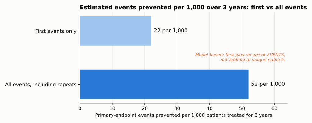
Trials usually count only each patient’s first event. But a patient who has one heart attack often has another, or a stroke, or a stent. When FOURIER counted every event, the estimated number of events prevented more than doubled, although the relative risk reduction did not: about 22 first events but 52 total events prevented per 1,000 patients treated for three years, counting first and repeat events rather than additional people helped (total events: rate ratio 0.82, 95% CI 0.75–0.90; first events: hazard ratio 0.85) [11].
See Figure 6 in Part 1.
The Big Picture
The Cholesterol Treatment Trialists pooled individual data from 26 statin trials and about 170,000 people. Each 38.7 mg/dL (1 mmol/L) reduction in LDL-C lowered major vascular events by about 22%, and all-cause death fell by about 10% per mmol/L, with no increase in cancer or other non-cardiovascular deaths [12]. A meta-regression of randomized trials found broadly similar proportional risk reductions per unit of LDL-C lowering across statins, ezetimibe, and PCSK9 inhibitors, which act through LDL-receptor pathways [13]. That supports a common LDL-mediated mechanism, though it does not mean every drug has identical clinical effects or safety profiles.
بعبارات بسيطة. The absolute LDL-C reduction and how long it lasts are major determinants of benefit, but therapies differ in evidence, timing, off-target effects, and tolerability.
Do the plaque changes explain this benefit? Plausibly, in part. The same drugs at similar LDL-C levels improve plaque in imaging trials and reduce events in outcome trials. But the imaging and outcome trials enrolled different people, and no trial has shown that the imaging changes account for the event reduction. The plaque findings make the benefit biologically coherent; the outcome trials establish the benefit for the endpoints they tested.
Time Is the Hidden Ingredient
Perhaps the most important lesson in the newer data is that benefit builds over time.
The Lag Effect
In FOURIER, the relative reduction in cardiovascular death, heart attack, or stroke was about 16% in the first year and about 25% after the first year [2]. Earlier statin trials showed the same pattern: benefit grows the longer treatment continues. That is consistent with, though it does not prove, plaque gradually becoming more stable.
The Legacy Effect
FOURIER did not demonstrate a significant cardiovascular mortality benefit during its median 2.2-year randomized phase. Afterward, 6,635 surviving participants who chose to continue entered an open-label extension, FOURIER-OLE, in which everyone received evolocumab; median extension follow-up was 5.0 years, and maximum evolocumab exposure across the parent trial and extension reached about 8.4 years; median LDL-C during the extension was about 30 mg/dL [14].
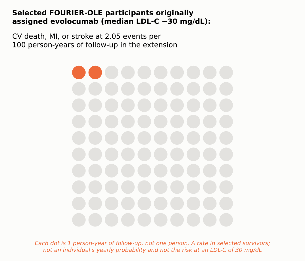
Even though both groups were now on the same drug at similar LDL-C, those originally assigned evolocumab, about two years earlier, had fewer subsequent events. Cardiovascular death, heart attack, or stroke occurred at 2.05 versus 2.58 events per 100 person-years (HR 0.80; 95% CI 0.68–0.93), and cardiovascular death at 0.68 versus 0.90 per 100 person-years (HR 0.77; 95% CI 0.60–0.99), as reported in the results table of the primary FOURIER-OLE publication, where events were counted during extension follow-up by original randomized assignment [14]. This is an exploratory comparison among selected survivors in open-label follow-up, not a new blinded randomized trial, so it cannot establish a mortality benefit by itself. It is compatible with a legacy effect from earlier, longer LDL lowering, but selection into the extension and open-label treatment mean it cannot show that early treatment caused the difference, or what carried it forward.
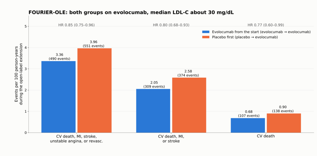
Figure 19. FOURIER-OLE: event rates per 100 person-years during the open-label extension, when all 6,635 participants received evolocumab and median LDL-C was about 30 mg/dL. Patients first randomized to evolocumab had 2.2 more years of low LDL-C exposure. Even in that group, cardiovascular death, MI, or stroke occurred at about 2 per 100 person-years. Extension participants were selected survivors and treatment was open label. Source: [14]. These are event rates in a selected-survivor, open-label extension; they cannot prove a randomized mortality benefit.
How Fast Things Change
- البروتين الدهني منخفض الكثافة (LDL-C) falls within days with a PCSK9 antibody and within two to four weeks with a statin. In AMUNDSEN, median LDL-C was 16 mg/dL six weeks after evolocumab was started before stenting, versus 56 mg/dL with standard care [24].
- لوحة changes in the randomized imaging trials were measured after roughly 9 to 24 months. YELLOW III documented within-person changes at 26 weeks, but it was uncontrolled, so it cannot establish the size of the drug’s effect or how early it began [37]. A trial that imaged only at the start and at one year cannot show that caps began thickening within days.
- Clinical events: MIRACL showed fewer recurrent ischemic events within 16 weeks [43]; FOURIER’s curves separated within about six months [2]; FOURIER-OLE’s exploratory cardiovascular-death difference emerged only during the open-label extension [14].
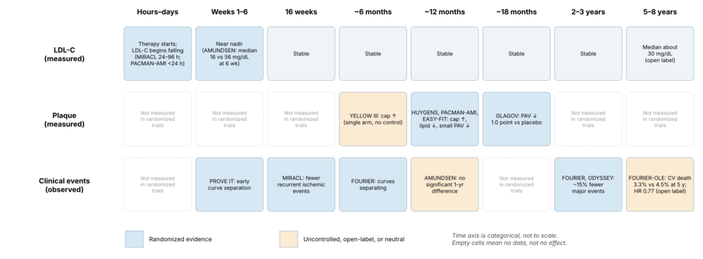
Figure 20. Timeline separating measured LDL-C changes, measured plaque changes, and observed clinical benefit. Each cell shows when a study made a measurement, not when a biological effect began; the figure cannot show when stabilization started, and no unmeasured time points are extrapolated. The time of a study measurement is not necessarily the time a biological benefit began; clinical benefit can begin before imaging is repeated, and no fixed biological lag is implied. A blank cell means not measured, not no effect.
Exposure Over a Lifetime
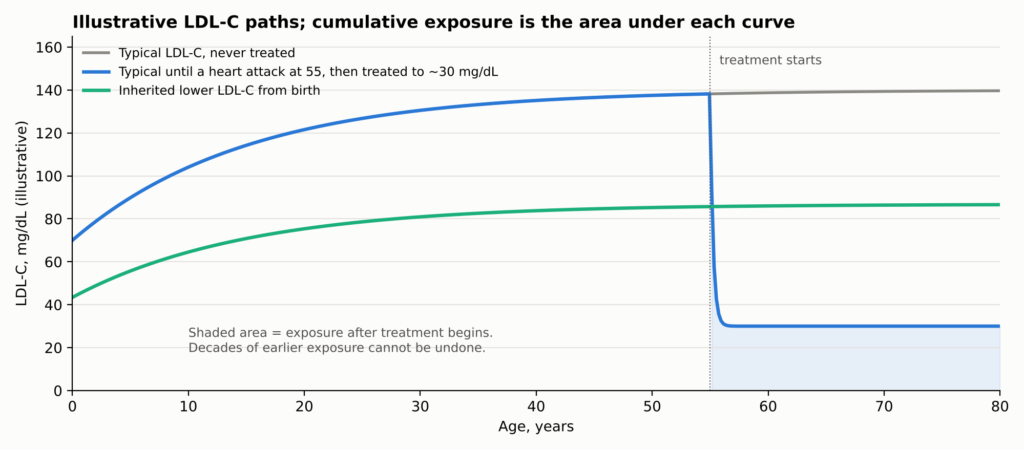
Why should time matter so much? Because تصلب الشرايين reflects the total, accumulated exposure of the artery wall to ApoB particles over a lifetime. Genetic studies indicate that التعرض التراكمي drives risk [44]. A person treated after a heart attack lowers their future exposure but carries decades of past exposure and the plaque it built.
See Figure 7 in Part 1.
How much a patient gains depends on three things together: baseline risk, how much LDL-C falls, and how long it stays down. The same LDL reduction prevents far more events in a high-risk patient, and larger sustained reductions prevent more.
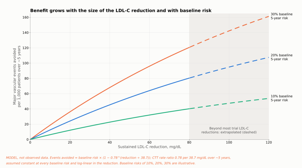
Figure 21. Evidence summary, not a patient-specific prediction. Lines apply the Cholesterol Treatment Trialists’ estimate of a 22% lower risk of major vascular events per 38.7 mg/dL (1 mmol/L) of LDL-C lowering over about five years [12] to three illustrative baseline five-year risks. The same LDL-C reduction prevents more events in higher-risk patients, and larger sustained reductions prevent more. Longer duration adds further benefit beyond what a five-year estimate shows (see the lifetime-exposure figure). Reductions above about 80 mg/dL extend beyond much of the trial data. Assumptions: the CTT rate ratio (a relative risk from a تحليل تلوي, not a trial hazard ratio) is applied uniformly at every baseline risk and scales log-linearly with LDL-C reduction. This is not a patient حاسبة المخاطر.
إلى أي مستوى يجب أن ينخفض مستوى الكوليسترول الضار (LDL)؟
Patients often ask whether 40 is good enough, or whether 20 or 10 is better. The honest answer depends on what kind of evidence you accept, because the lower one goes, the more the evidence shifts from randomized comparisons to observational ones.
| LDL-C level | What the evidence looks like | الخلاصة |
| أقل من 70 ملغ/ديسيلتر | Randomized trials (PROVE IT, statin imaging trials) | Established for drug regimens that reached these levels (not randomized to the threshold) |
| أقل من 55 ملغ/ديسيلتر | Randomized trials (IMPROVE-IT, ODYSSEY, FOURIER) | Established for drug regimens that reached these levels (not randomized to the threshold) |
| Below 40 mg/dL | Randomized trial arms reached it (FOURIER about 30; GLAGOV 36.6) | تحظى بدعم قوي |
| Below 30 mg/dL | Imaging trials (HUYGENS 28, PACMAN-AMI 24); achieved-level analyses | Supported for plaque; outcomes observational |
| أقل من 20 ملغ/ديسيلتر | Thousands of patients in FOURIER and FOURIER-OLE reached it, but were not randomized to it | Lower risk, no clear safety signal; observational |
| أقل من 10 ملغ/ديسيلتر | Small subsets of FOURIER, FOURIER-OLE, and ODYSSEY | Consistent but statistically fragile |
No trial cited here randomized post-heart-attack patients between specific LDL-C targets below 55 mg/dL. People in the drug trials were randomized to drugs, not to “20 versus 40.” That is the most important caveat in this area.
Within those limits, the data point one way. In FOURIER, 2,669 participants reached LDL-C below 20 mg/dL (0.5 mmol/L) by week 4; lower achieved LDL-C was associated with steadily lower cardiovascular risk across the range studied, and serious adverse events were not related to achieved LDL-C over the randomized period [17]. In FOURIER-OLE, about 1,604 participants maintained LDL-C below 20 mg/dL; lower levels remained associated with lower risk and no detected increase in serious adverse events over the extension’s follow-up [18]. In ODYSSEY OUTCOMES, patients who transiently reached below 15 mg/dL had fewer subsequent events in an adjusted post hoc comparison (HR 0.72; 95% CI 0.51–0.997), which does not establish that aiming below 15 mg/dL is better [45].
بعبارات بسيطة. Lower achieved LDL-C has generally been linked to lower event rates across the ranges examined, including very low levels. The studies reviewed have not identified a clear lower threshold; evidence becomes sparse at extreme LDL-C, and achieved-level analyses cannot identify the best target. No randomized trial has shown that aiming for 10 or 20 mg/dL beats aiming for 30 or 40 mg/dL.
How Low Can Risk Go?
There is a different question hiding inside “how low should LDL go.” Suppose LDL-C is pushed nearly to zero. How low can the rate of heart attack, stroke, and cardiovascular death fall? The answer differs sharply between people who already have coronary disease and people who have had low LDL-C since birth.
In People Who Already Have Atherosclerosis
This is not a randomized comparison of LDL-C targets: patients sorted themselves into achieved-LDL groups, so selection and confounding can only partly be adjusted for, and the analysis does not prove there is no biological floor. Within that limit, the peer-reviewed FOURIER-OLE achieved-LDL analysis found risk falling steadily down to the lowest levels studied, below 20 mg/dL [18].
Two points follow. Risk was lowest at the lowest achieved levels studied, an observational association. And the floor is well above zero: among selected participants entering FOURIER-OLE, cardiovascular death, heart attack, or stroke occurred at 2.05 events per 100 person-years of follow-up in those originally assigned evolocumab, the group treated longest; this is a rate by original randomized assignment, not the risk at an LDL-C of 30 mg/dL [14].
These are observational comparisons within trials; patients who reach the lowest levels may differ in ways that statistical adjustment cannot fully remove.
Why the Floor Exists
- Plaque already built. These patients carried decades of accumulated plaque, and IVUS shows plaque burden falling by only a few percentage points.
- Short exposure. Two to eight years of very low LDL-C cannot erase forty or more years of higher exposure.
- Other pathways. Lp(a), triglyceride-rich remnants, and residual inflammation persist. In كانتوس, COLCOT, and LoDoCo2, anti-inflammatory therapy reduced events in patients already on lipid-lowering drugs [19, 20, 46].
- Other causes. Some strokes come from the heart or tiny vessels, and some heart attacks from erosion, artery tears, or clots carried from elsewhere. التدخين, ضغط الدم, diabetes, and مرض الكلى also continue to drive risk.
Nature’s Experiment: People Born With Low LDL
Some people inherit متغيرات الجينات that lower LDL-C from birth. Because those variants are assigned at conception, largely independent of lifestyle, they act like natural randomized experiments. Their results are dramatic.
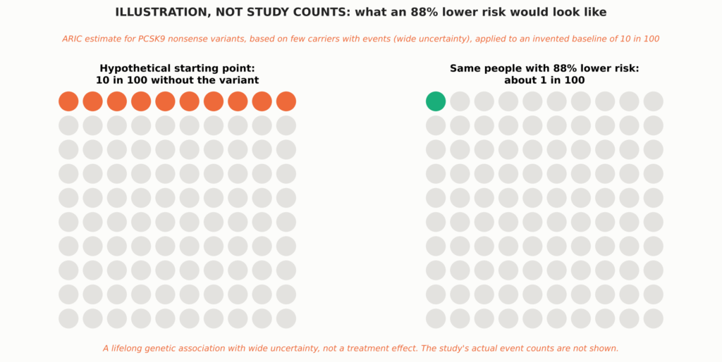
See Figure 8 in Part 1.
- PCSK9 nonsense variants. في اريك study, Black participants carrying inactivating PCSK9 variants had about 28% lower LDL-C and an 88% lower risk of مرض الشريان التاجي over 15 years. White participants with a milder variant had about 15% lower LDL-C and 47% lower risk [5]. Few carriers had events, so the estimate is imprecise (hazard ratio 0.11; 95% CI 0.02–0.81).
- APOB truncating variants. Carriers had 43 mg/dL lower LDL-C and 72% lower odds of early coronary disease [47].
- PCSK9 R46L in large studies. This common variant lowers LDL-C only modestly, yet it was associated with less coronary or مرض القلب الإشكيمي [48, 15] and lower cardiovascular mortality [16].
- Across nine variants in six genes (312,321 people), each 38.7 mg/dL of lifelong lower LDL-C was associated with 5% lower coronary risk, about three times the effect per unit of statins started later in life; this is an illustrative comparison across different study designs, not causal equivalence [6].
The genetic data also mark limits. The same R46L variant that lowered coronary risk was not associated with lower ischemic stroke [15], and lifelong lower LDL-C from PCSK9 variants was associated with lower cardiovascular but not الوفيات لجميع الأسباب [16]. Across these datasets, reductions in ischemic stroke or all-cause mortality were not consistently demonstrated; estimates differ by variant, population, endpoint, and statistical precision. These are observational genetic associations with population-specific uncertainty. Effect sizes differ across variants, populations, and endpoints, and because they reflect lifelong exposure, their scale cannot be equated with a drug treatment effect started in adulthood; quantitative comparisons with trials are illustrative.
People who inherit two inactivating PCSK9 variants are extremely rare. The best-documented case is a 32-year-old woman with no detectable PCSK9 and LDL-C of 14 mg/dL who was healthy, fertile, and working as an aerobics instructor [49]. She was described at a single point in time; nobody knows her long-term cardiovascular outcome.
Putting the Two Together
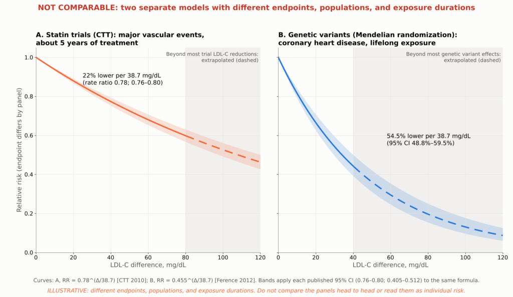
Figure 22. Illustrative model, not trial data or a patient risk calculator. Lines show two published log-linear associations with different endpoints, populations, and exposure durations: the Cholesterol Treatment Trialists’ rate ratio of 0.78 (95% CI 0.76–0.80) for major vascular events per 38.7 mg/dL over about five years of statin treatment [12], and the الوراثة العشوائية المندلية estimate of 54.5% (95% CI 48.8%–59.5%) lower coronary heart disease risk per 38.7 mg/dL of lifelong exposure, from 312,321 participants and 9 variants in 6 genes [6]. Each curve is ratio^(LDL-C difference/38.7); shaded bands carry each published 95% فترة الثقة through the same formula and are not observed trial confidence bands. Extrapolated segments are dashed: in panel B beyond about 40 mg/dL, past the range of most genetic variants, and in panel A beyond about 80 mg/dL, past most trial LDL-C reductions, and the model assumes the علاقة لوغاريتمية خطية holds at extreme exposures, which has not been shown. The two estimates are shown in separate panels, without individual study or trial points, because their endpoints and populations differ; they must not be read as a head-to-head comparison of statin therapy with lifelong genetic LDL reduction or as a prediction of individual lifetime risk.
The figure compares two published estimates. Applied to a difference of about 75 mg/dL, the statin-trial estimate predicts roughly 38% fewer major vascular events over about five years, while the genetic estimate predicts roughly 78% fewer coronary events over a lifetime. Both are extrapolations, especially the genetic one. They illustrate why duration matters, not what any individual should expect.
The defensible conclusion. Early, sustained, very low exposure to ApoB particles probably drives atherosclerotic coronary events toward a low floor. But no human data show near-zero cardiovascular events, the genetic data suggest stroke and total mortality would remain above zero, and in people who already have coronary disease, events still occur even at extremely low LDL-C.
هل يعتبر انخفاض مستوى الكوليسترول الضار (LDL) إلى درجة منخفضة جدًّا أمرًا آمنًا؟
A natural worry is that the body needs cholesterol. It does, but cells make their own, and the evidence so far is reassuring, with honest limits.
- Memory and thinking. In EBBINGHAUS, 1,204 FOURIER patients underwent formal cognitive testing over a median 19 months, with no difference between evolocumab and placebo and no link to achieved LDL-C [21]. Longer follow-up found no excess of reported cognitive events [18], though that relies on adverse-event reports.
- Bleeding strokes. جرعة عالية أتورفاستاتين after stroke modestly increased سكتة دماغية نزفية in SPARCL [50]. FOURIER, ODYSSEY OUTCOMES, and the very-low-LDL analyses showed no significant excess [2, 17, 18], though such strokes are uncommon.
- Statins modestly raise the risk of new diabetes, about 9% in relative terms, mostly in people already near the threshold [22]. PCSK9 inhibitors and very low LDL-C have not shown a comparable increase [2, 17].
- The adrenal glands and gonads can make their own cholesterol and also take it up from circulating البروتينات الدهنية, and large PCSK9 programs have not shown a clinically important hormonal signal.
Achieved-LDL analyses are observational within trials and subject to selection and التشويش المتبقي; any isolated nominal findings are hypothesis-generating, and very long-term safety at extreme levels remains incompletely characterized.
Drug side effects are a separate matter: PCSK9 antibodies cause slightly more injection-site reactions (2.1% vs 1.6% in FOURIER) [2], and statins can cause muscle symptoms, though true muscle injury is rare. Randomized safety data for PCSK9 antibodies cover about two to three years; open-label exposure reached about 8.4 years [14]. In selected FOURIER extension participants, very low achieved LDL-C was not associated with a detected increase in several prespecified safety outcomes over the parent trial and its open-label extension [18]. That is reassuring but limited by participant selection, observational comparisons, and the inability to exclude rare or lifelong effects. Unlimited lifelong safety at extremely low levels has not been, and cannot yet be, established.
What Guidelines Recommend
Guidelines turn many trials into practical goals. The 2026 U.S. multisociety اضطراب شحميات الدم guideline reintroduced explicit LDL-C goals and divides people with atherosclerotic أمراض القلب والأوعية الدموية (ASCVD) into two groups [23]. Very-high-risk ASCVD means multiple major ASCVD events, or one major event plus multiple high-risk conditions; a recent heart attack alone does not automatically qualify.
| Who | What the 2026 U.S. guideline recommends |
| ASCVD, not very high risk | High-intensity statin; LDL-C below 70 mg/dL and non-HDL-C below 100 mg/dL (recommended) |
| ASCVD, not very high risk | Add ezetimibe, a PCSK9 antibody, or حمض البيمبيدويك if needed to reach below 70 mg/dL |
| ASCVD, not very high risk | Going further, to below 55 mg/dL (non-HDL-C below 85) (reasonable) |
| Very-high-risk ASCVD | High-intensity statin; LDL-C below 55 mg/dL and non-HDL-C below 85 mg/dL (recommended) |
| Very-high-risk ASCVD | Add ezetimibe and/or a PCSK9 antibody if needed to reach those goals |
| Very-high-risk ASCVD | Bempedoic acid is a further add-on option |
“Recommended” and “reasonable” are shown only for the LDL-C and non-HDL-C goals, where they follow the guideline’s own wording for stronger and moderate recommendations. For add-on drugs, the guideline’s recommendation tables give the formal classes, evidence levels, and eligibility criteria, and separate recommendations apply in special situations such as فرط كوليстеロール الدم الشديد [23].
The 2025 U.S. acute coronary syndrome guideline recommends high-intensity statin therapy and timely addition of nonstatin therapy for those above recommended levels [51]. European guidance after a heart attack recommends at least a 50% reduction and LDL-C below 55 mg/dL [52], and allows a goal below 40 mg/dL after a second vascular event within two years [53]. None of these specific targets was directly tested against another in a randomized target trial; they are expert judgments built on the evidence above.
Should a Second Drug Start in the Hospital?
AMUNDSEN randomized 2,161 heart attack patients and demonstrated that a strategy of giving the first evolocumab injection before PCI substantially improved attainment of the combined LDL-C target at 12 months (82% versus 40%). However, the difference in the trial’s main clinical end point (tested second in the prespecified order, after the LDL-C target), death or unplanned cardiovascular hospitalization, was not statistically significant at one year (14.6% versus 15.4%; adjusted OR 0.94, 95% CI 0.73–1.19). The trial therefore supports faster, more reliable target attainment, but does not demonstrate an additional short-term clinical-event benefit from this initiation strategy [24]. Standard care permitted a PCSK9 inhibitor when indicated, but only 3.8% of the standard-care group received one. The trial was powered (80%) to detect an odds ratio of 0.68 for this end point, so it argues against a large early clinical effect of starting a PCSK9 inhibitor in the catheterization laboratory; it does not exclude a smaller benefit or one that emerges with longer treatment. A prespecified per-protocol analysis suggested fewer events with evolocumab (10.2% versus 14.3%), but the investigators caution that this is likely affected by selection bias. A one-year result does not refute the longer outcome trials. EVOLVE-MI, testing evolocumab within 10 days of a heart attack, had not reported by the search cutoff. For now, early combination therapy is مثبت to reach lower LDL-C sooner and, in PACMAN-AMI, to improve one-year plaque changes; it is not yet proven to prevent more events than adding the same drug weeks later.
Where ApoB Fits
The guideline gives ApoB a role in assessing المخاطر المتبقية. ApoB is most useful when it and LDL-C disagree, as in people with high triglycerides, diabetes, or obesity, where LDL-C can look “at goal” while particle number remains high. No randomized trial reviewed here has compared ApoB treatment targets with LDL-C targets, so ApoB is best used as an added measure of remaining عبء الجسيمات rather than as a proven separate target.
الخلاصة
If someone has already had a heart attack, how much can intensive خافض للدهون change the disease and lower the chance of another event?
The disease: Within about a year, lowering LDL-C to roughly 25 to 30 mg/dL produces about twice the cap thickening seen with a statin alone, reduces imaging markers of lipid in untreated plaques, and adds roughly a 1.2 to 1.7 percentage-point reduction in percent atheroma volume compared with control groups. The plaque does not disappear, calcium does not dissolve, and the artery is not restored to normal.
The risk: In randomized trials after an acute coronary syndrome, relative reductions in each trial’s main composite endpoint were roughly 15%, with absolute benefits that differ by trial and follow-up. Across the pooled statin trials, each 38.7 mg/dL of LDL-C lowering reduced major vascular events by about 22%. Heart attacks and ischemic strokes are clearly reduced; a reduction in death is proven for statins in pooled analyses, but for add-on therapies only in nominal or open-label analyses.
How low can risk go: The studies reviewed have not identified a clear lower threshold, evidence becomes sparse at extreme LDL-C, and achieved-level analyses cannot define the optimal target; but events still occur even at extremely low LDL-C. People with lifelong low LDL-C have far less coronary disease, which points to duration of exposure as a major lever, but reductions in ischemic stroke and total mortality were not consistently shown. No evidence shows that cardiovascular events can be driven to zero.
| Well established | مثير للخيال | Still unresolved |
| Lowering LDL-C after a heart attack reduces repeat heart attack, ischemic stroke, and procedures, in proportion to the LDL reduction | Benefit and safety at sustained LDL-C below 20 and even below 10 mg/dL | Whether starting a PCSK9 inhibitor in the hospital prevents more events than starting it weeks later |
| The benefit continues below 70 and 55 mg/dL in randomized comparisons | A lasting legacy benefit from earlier treatment (open-label, selected survivors) | Whether any target below 55 mg/dL beats another in a randomized trial |
| Intensive LDL lowering improves plaque composition and modestly reduces plaque volume over about a year | Far greater protection from lifelong low LDL-C | The true floor of risk with lifelong near-zero LDL-C |
| For statins, ezetimibe, and PCSK9 antibodies, benefits generally outweigh recognized harms in the populations studied | A cardiovascular-mortality benefit from adding PCSK9 inhibition | Lifelong safety at extremely low LDL-C |
| That plaque changes explain part of the benefit; that ApoB and Lp(a) modify the plaque response | How to prevent heart attacks caused by erosion; whether imaging-guided therapy helps |
Practical Points to Discuss With Your Cardiologist
- Start or continue high-intensity statin therapy during the hospital stay after an acute coronary syndrome, unless it is contraindicated or not tolerated.
- Know your numbers and your risk category. The 2026 U.S. guideline recommends an LDL-C goal below 55 mg/dL for people with ASCVD classified as very high risk and below 70 mg/dL for those who are not, with a goal below 55 mg/dL reasonable in selected patients who are not at very high risk; European ACS guidance generally recommends at least a 50% reduction and LDL-C below 55 mg/dL after an acute coronary syndrome. Ask your clinician which category you are in and what your goal is. If you are not at goal on a statin, ask about adding ezetimibe or a PCSK9 inhibitor.
- Ask for a one-time Lp(a) test, and consider ApoB, especially if your triglycerides are high or you have diabetes.
- Do not read a rising calcium score as failure on its own. Discuss what it means in your case.
- Treat the whole picture. Stop smoking, control blood pressure and سكر الدم, attend إعادة التأهيل القلبي, and take prescribed علاج مضاد للصفيحات.
- Stay on therapy. LDL-C rises when treatment stops, and the benefit depends on how long it stays low.
إخلي المسؤولية الطبية: This article is for educational purposes and summarizes published research. It is not medical advice and does not replace individualized care from your physician. Do not start, stop, or change any medication without talking to your doctor.
Technical Appendix
About This Review
هذا مراجعة سردية selectively summarizes landmark peer-reviewed studies available through October 2026 and is not a مراجعة منهجية or meta-analysis. Where full text was not available, I used the peer-reviewed abstract only when it contained enough detail to support the specific statement. Press releases, registry pages, and news coverage were not used as evidence. Numbers not supported by the sources consulted were excluded. Every value plotted in a data figure was checked against the published abstract or primary report, as listed in the companion figure register; supplementary tables were not used. The bibliographic details of all 54 references (authors, title, journal, year, volume, and pages) were checked against the Crossref registry.
A1. HUYGENS Results
| HUYGENS measure (change over about one year) | إيفولوكوماب + الستاتين | Placebo + statin | Difference* | P value |
| Achieved LDL-C, mg/dL | 28.1 | 87.2 | about −59 | <٠٫٠٠١ |
| Minimum fibrous cap thickness, μm (primary endpoint) | +42.7 | +21.5 | +21.2 | 0.015 |
| Maximum lipid arc, degrees (secondary) | −57.5 | −31.4 | −26.1 | 0.04 |
| Macrophage index, mm (exploratory) | −3.17 | −1.45 | −1.72 | 0.04 |
| Percent atheroma volume, percentage points (IVUS subset; exploratory) | −2.29 | −0.61 | −1.68 | 0.009 |
| Microchannels and calcium (exploratory) | لا يوجد فرق ذو دلالة إحصائية | NS |
*Values and P values are as reported in the published HUYGENS abstract; 135 treated patients had evaluable imaging at follow-up. The cap-thickness values are least-squares means from the trial’s analysis of covariance in the full analysis set of 161 treated patients, 119 with paired OCT (registry results: difference 21.2 μm, 95% CI 4.7–37.7); IVUS values come from a smaller subset. Other differences are simple arithmetic on the reported group means; the P values come from the trial’s own models. Source: [1].
The protocol prespecified the absolute change in minimum cap thickness as the primary endpoint; secondary endpoints were the percent change in minimum cap thickness, the change in average minimum cap thickness, and the change in maximum lipid arc; IVUS plaque burden, macrophages, and microchannels were exploratory. The trial planned about 150 patients so that 125 would complete paired imaging, with 90% power to detect a 51 μm benefit; the observed adjusted benefit, 21.2 μm, was less than half that assumption yet still statistically significant [9]. Imaging trials can only measure patients who survive, stay in the study, and undergo a second catheterization with usable images, so missing follow-up imaging can introduce selection bias.
A2. HUYGENS Subgroup Analyses
These small, post hoc findings are hypothesis-generating and do not establish separate treatment thresholds or independently causal mechanisms. They were based on subsets and not adjusted for multiple comparisons.
- بروتين دهني (أ). In HUYGENS, the difference in cap thickening between evolocumab and placebo appeared larger in patients with high Lp(a) than in those with lower Lp(a), partly because the placebo-plus-statin group with high Lp(a) showed a smaller response [28]. Evolocumab lowered Lp(a) only modestly, and the analysis does not show that lowering Lp(a) caused the benefit. In PACMAN-AMI, baseline Lp(a) did not modify the plaque-volume or cap-thickness response to alirocumab, although patients with the highest Lp(a) had a smaller reduction in the plaque lipid signal [54].
- Patients who reached lower ApoB levels showed more favorable OCT findings than those who did not [29]. Achieved ApoB was not randomized, and the full adjustment model could not be examined, so this does not establish an optimal ApoB target or an independent causal effect.
- Achieved LDL-C. Pooling both arms, lower achieved LDL-C and larger LDL-C reductions were associated with greater cap thickening [1]; also non-randomized.
A3. Plaque-Imaging Trials
| المحاكمة | السكان | Treatment vs comparator | Achieved LDL-C, mg/dL (mmol/L) | Imaging, duration | Main plaque finding | Key limitations |
| HUYGENS (2022) [1] | 161 post-NSTEMI | Evolocumab vs placebo, both + statin | 28.1 vs 87.2 (0.73 vs 2.25) | OCT; IVUS subset; 50 wk | Cap +42.7 vs +21.5 μm; lipid arc −57.5° vs −31.4°; PAV −2.29 vs −0.61 | 135 with evaluable follow-up imaging; denominators vary by endpoint; selected plaque |
| PACMAN-AMI (2022) [7] | 300 acute MI, started <24 h after PCI | Alirocumab vs placebo, both + rosuvastatin 20 | 23.6 vs 74.4 (0.61 vs 1.92) | IVUS + NIRS + OCT; 52 wk | PAV diff −1.21; LCBI diff −41.24; cap diff +29.65 μm | 265 paired IVUS; 81% men |
| GLAGOV (2016) [30] | 968 stable CAD on statin | Evolocumab vs placebo | 36.6 vs 93.0 (0.95 vs 2.41) | IVUS; 76 wk treatment, week-78 IVUS | PAV −0.95 vs +0.05; regression 64% vs 47% | Not post-MI; composition substudy neutral |
| REVERSAL (2004) [32] | 654 stable CAD | Atorvastatin 80 vs برافاستاتين 40 | 79 vs 110 (2.0 vs 2.8) | IVUS; 18 mo | Progression halted vs continued | Moderate LDL levels |
| ASTEROID (2006) [33] | 507 CAD | Rosuvastatin 40, single arm | 60.8 (1.57) | IVUS; 24 mo | PAV −0.98 | No control; attrition |
| SATURN (2011) [34] | 1,385 CAD randomized; 1,039 with follow-up IVUS | Rosuvastatin 40 vs atorvastatin 80 | 62.6 vs 70.2 (1.62 vs 1.82) | IVUS; 104 wk | Median PAV −1.22 vs −0.99 (P=0.17) | No significant difference between the two statins; both regressed; no placebo arm |
| PRECISE-IVUS (2015) [35] | 246 after PCI, Japan | Atorvastatin + ezetimibe vs atorvastatin | 63.2 vs 73.3 (1.63 vs 1.90) | IVUS; 9–12 mo | PAV −1.4 vs −0.3; regression 78% vs 58% | Open label |
| EASY-FIT (2014) [36] | 70 unstable angina | Atorvastatin 20 vs 5 | 69 vs 78 (1.78 vs 2.02) | OCT; 12 mo | Cap +69% vs +17% | Small; low-dose comparator |
| ODYSSEY J-IVUS (2019) [39] | 206 recent ACS, Japan | Alirocumab vs standard care | Not shown | IVUS; 36 wk | No significant difference in primary plaque endpoint | Neutral; short duration |
| YELLOW III (2026) [37] | 110 stable CAD, lipid-rich plaque | Evolocumab + statin, single arm | Not reported in sources reviewed | OCT + NIRS-IVUS; 26 wk | Cap 70.9 → 97.7 μm; maxLCBI4mm 306.8 → 213.1; PAV −1.38 points | No control group |
| CCTA/PET study (2025) [38] | 47 stable, noncalcified plaque | Evolocumab, single arm | Not reported in sources reviewed | CCTA + PET; 18 mo | Noncalcified plaque fell; calcified plaque rose | No control group |
Values are not directly comparable across trials. PACMAN-AMI “diff” values are model-based between-group differences; other entries are within-arm changes as reported. PAV, percent atheroma volume; TAV, total atheroma volume; LCBI, لب لبني burden index.
A4. Major Outcome Trials in Detail
| المحاكمة | Population, timing, follow-up | Treatment vs comparator | LDL-C, mg/dL (baseline; achieved) | Primary endpoint | النتيجة | Mortality |
| MIRACL (2001) [43] | 3,086 unstable angina or non-Q-wave MI; 24–96 h; 16 wk | Atorvastatin 80 vs placebo | ~124; 72 vs 135 at 16 wk | Death, MI, resuscitated arrest, or recurrent نقص الدم with rehospitalization | 14.8% vs 17.4%; RR 0.84 (0.70–1.00) | No significant reduction in death or MI; benefit driven mainly by fewer rehospitalizations for symptomatic ischemia |
| PROVE IT–TIMI 22 (2004) [10] | 4,162 ACS; within 10 days; mean 24 mo | Atorvastatin 80 vs pravastatin 40 | 106; 62 vs 95 | Death, MI, UA rehospitalization, revascularization, or stroke | 22.4% vs 26.3%; 16% reduction (5%–26%) | Death 2.2% vs 3.2%; P=0.07 |
| IMPROVE-IT (2015) [4] | 18,144 ACS; within 10 days; median ~6 y | Simvastatin + ezetimibe vs simvastatin | 93.8; 53.7 vs 69.5 | CV death, major coronary event, or nonfatal stroke | 32.7% vs 34.7% at 7 y; HR 0.936 (0.89–0.99) | No difference |
| ODYSSEY OUTCOMES (2018) [3] | 18,924 ACS 1–12 mo earlier; median 2.8 y | Alirocumab vs placebo + statin | ~92 (mean); ~40 vs ~93 at 4 mo | CHD death, MI, ischemic stroke, or UA hospitalization | 9.5% vs 11.1%; HR 0.85 (0.78–0.93) | All-cause death 3.5% vs 4.1%, nominal only |
| FOURIER (2017) [2] | 27,564 stable ASCVD; median 2.2 y | Evolocumab vs placebo + statin | 92 (median); 30 with evolocumab at 48 wk, 56 mg/dL lower than placebo | CV death, MI, stroke, UA hospitalization, or revascularization | 9.8% vs 11.3%; HR 0.85 (0.79–0.92) | CV death HR 1.05 (0.88–1.25) |
| FOURIER-OLE (2022) [14] | 6,635 FOURIER completers; median 5 more years | Original evolocumab vs original placebo, all on open-label drug | Median ~30 at extension week 12, on open-label evolocumab | Same as FOURIER | 3.36 vs 3.96 per 100 person-years; HR 0.85 (0.75–0.96) | CV death 0.68 vs 0.90 per 100 person-years, HR 0.77 (0.60–0.99); exploratory |
| AMUNDSEN (2026) [24] | 2,161 STEMI/NSTEMI (1,087 vs 1,074); open label; 12 mo | Evolocumab before PCI + standard care vs standard care (PCSK9 inhibitor permitted; used in 3.8%) | Median 16 vs 56 at 6 wk | LDL-C <55 and ≥50% reduction; main clinical end point tested second in a prespecified hierarchy (80% power for OR 0.68) | 792/970 (82%) vs 370/934 (40%); aOR 5.54 (4.50–6.82). Clinical composite 159/1,087 (14.6%) vs 165/1,074 (15.4%); aOR 0.94 (0.73–1.19) | Not significant |
Event rates are Kaplan–Meier estimates for PROVE IT–TIMI 22 and IMPROVE-IT and observed proportions for the others. Endpoints, populations, and follow-up differ, so these figures should not be compared across trials. AMUNDSEN was checked against the full primary JAMA article. CV, cardiovascular; CHD, coronary heart disease; UA, unstable angina.
| Endpoint (treatment vs control) | IMPROVE-IT (7 y) [4] | ODYSSEY OUTCOMES (median 2.8 y) [3] | FOURIER (median 2.2 y) [2] |
| احشاء عضلة القلب | 13.1% vs 14.8% | 6.6% vs 7.6% | 3.4% vs 4.6% (HR 0.73) |
| Stroke (trial-specific definition) | Ischemic stroke 3.4% vs 4.1% | Ischemic stroke 1.2% vs 1.6% | Any stroke 1.5% vs 1.9% (HR 0.79) |
| Coronary revascularization | 21.8% vs 23.4% (not significant) | Ischemia-driven, lower with alirocumab | 5.5% vs 7.0% (HR 0.78) |
| Cardiovascular death | لا يوجد فرق ذو دلالة إحصائية | Not significant (HR 0.88) | No difference (HR 1.05) |
| الوفيات من جميع الأسباب | لا يوجد فرق ذو دلالة إحصائية | 3.5% vs 4.1% (HR 0.85), nominal only | No difference |
These components are secondary or nominal analyses, not multiplicity-controlled results.
A5. Limitations of the Evidence
- Small imaging trials. HUYGENS randomized 161 patients, 135 of whom had evaluable follow-up imaging; PACMAN-AMI randomized 300, with 265 having paired IVUS. Both selected particular segments, had missing follow-up imaging, and enrolled mostly men. HUYGENS selected patients with already vulnerable-looking plaque.
- Imaging is not histology. OCT can mistake fibrous tissue for lipid, and macrophage signals are indirect.
- تمويل القطاع. Amgen funded HUYGENS, GLAGOV, and FOURIER; Sanofi and Regeneron supported ODYSSEY OUTCOMES, and Sanofi, Regeneron, and Infraredx funded PACMAN-AMI; Merck funded IMPROVE-IT; AstraZeneca funded SATURN. The major outcome trials used blinded treatment and independent event حكم قضائي, which limits but does not eliminate the concern.
- Different populations. The outcome trials differ in timing, background therapy, and endpoint definitions.
- Achieved-LDL and open-label analyses are vulnerable to selection and confounding.
- Genetic studies estimate lifelong effects of specific variants; their effect sizes cannot be transferred directly to drugs started in adulthood.
- Abstract-level sources. The following were reviewed at the abstract or published-summary level rather than in full text: the HUYGENS primary report (supplemented by its registry results) and ApoB analysis, Cohen 2006 (PCSK9 variants), Peloso 2019 (APOB variants), Benn 2010 and Benn 2019 (PCSK9 R46L), and Ference 2012 (Mendelian عشوائية). These are also marked in the reference list.
- Sources of figure data. Each data figure plots values reported in the study cited in its caption; conceptual and model figures are labeled as illustrative.
- Review method. This narrative review is based on selected peer-reviewed studies and guidelines rather than a registered systematic search. Some findings were extracted from peer-reviewed abstracts or published summaries. Numbers not supported by the sources consulted were excluded. Figure values were checked against the published abstracts or primary reports listed in the companion figure register, and each statement in the text is supported by its cited source at the level indicated above (abstract or full text).
المراجع
- Nicholls SJ, Kataoka Y, Nissen SE, et al. Effect of evolocumab on coronary plaque phenotype and burden in statin-treated patients following myocardial infarction (HUYGENS). JACC Cardiovasc Imaging. 2022;15(7):1308-1321. doi:10.1016/j.jcmg.2022.03.002. PubMed: https://pubmed.ncbi.nlm.nih.gov/35431172/
- Sabatine MS, Giugliano RP, Keech AC, et al. Evolocumab and clinical outcomes in patients with cardiovascular disease (FOURIER). N Engl J Med. 2017;376:1713-1722. doi:10.1056/NEJMoa1615664. PubMed: https://pubmed.ncbi.nlm.nih.gov/28304224/
- Schwartz GG, Steg PG, Szarek M, et al. Alirocumab and cardiovascular outcomes after acute coronary syndrome (ODYSSEY OUTCOMES). N Engl J Med. 2018;379:2097-2107. doi:10.1056/NEJMoa1801174. PubMed: https://pubmed.ncbi.nlm.nih.gov/30403574/
- Cannon CP, Blazing MA, Giugliano RP, et al. Ezetimibe added to statin therapy after acute coronary syndromes (IMPROVE-IT). N Engl J Med. 2015;372:2387-2397. doi:10.1056/NEJMoa1410489. PubMed: https://pubmed.ncbi.nlm.nih.gov/26039521/
- Cohen JC, Boerwinkle E, Mosley TH Jr, Hobbs HH. Sequence variations in PCSK9, low LDL, and protection against coronary heart disease. N Engl J Med. 2006;354:1264-1272. doi:10.1056/NEJMoa054013 [abstract reviewed]
- Ference BA, Yoo W, Alesh I, et al. Effect of long-term exposure to lower low-density lipoprotein cholesterol beginning early in life on the risk of coronary heart disease: a Mendelian randomization analysis. J Am Coll Cardiol. 2012;60:2631-2639. doi:10.1016/j.jacc.2012.09.017 [abstract summary reviewed]
- Räber L, Ueki Y, Otsuka T, et al. Effect of alirocumab added to high-intensity statin therapy on coronary atherosclerosis in patients with acute myocardial infarction: the PACMAN-AMI randomized clinical trial. JAMA. 2022;327:1771-1781. doi:10.1001/jama.2022.5218
- Stone GW, Maehara A, Lansky AJ, et al. A prospective natural-history study of coronary atherosclerosis (PROSPECT). N Engl J Med. 2011;364:226-235. doi:10.1056/NEJMoa1002358
- Nicholls SJ, Nissen SE, Prati F, et al. Assessing the impact of PCSK9 inhibition on coronary plaque phenotype with optical coherence tomography: rationale and design of the randomized, placebo-controlled HUYGENS study. Cardiovasc Diagn Ther. 2021;11(1):120-129. doi:10.21037/cdt-20-684. PMCID: PMC7944215. PubMed: https://pubmed.ncbi.nlm.nih.gov/33708484/
- Cannon CP, Braunwald E, McCabe CH, et al. Intensive versus moderate lipid lowering with statins after acute coronary syndromes (PROVE IT–TIMI 22). N Engl J Med. 2004;350:1495-1504. doi:10.1056/NEJMoa040583. PubMed: https://pubmed.ncbi.nlm.nih.gov/15007110/
- Murphy SA, Pedersen TR, Gaciong ZA, et al. Effect of the PCSK9 inhibitor evolocumab on total cardiovascular events in patients with cardiovascular disease: a prespecified analysis from the FOURIER trial. JAMA Cardiol. 2019;4:613-619. doi:10.1001/jamacardio.2019.0886
- Cholesterol Treatment Trialists’ (CTT) Collaboration. Efficacy and safety of more intensive lowering of LDL cholesterol: a meta-analysis of data from 170,000 participants in 26 randomised trials. Lancet. 2010;376:1670-1681. doi:10.1016/S0140-6736(10)61350-5. PubMed: https://pubmed.ncbi.nlm.nih.gov/21067804/
- Silverman MG, Ference BA, Im K, et al. Association between lowering LDL-C and cardiovascular risk reduction among different therapeutic interventions: a systematic review and meta-analysis. JAMA. 2016;316:1289-1297. doi:10.1001/jama.2016.13985
- O’Donoghue ML, Giugliano RP, Wiviott SD, et al. Long-term evolocumab in patients with established atherosclerotic cardiovascular disease (FOURIER-OLE). Circulation. 2022;146:1109-1119. doi:10.1161/CIRCULATIONAHA.122.061620
- Hopewell JC, Malik R, Valdés-Márquez E, Worrall BB, Collins R; METASTROKE Collaboration of the ISGC. Differential effects of PCSK9 variants on risk of coronary disease and ischaemic stroke. Eur Heart J. 2018;39:354-359. doi:10.1093/eurheartj/ehx373
- Benn M, Tybjærg-Hansen A, Nordestgaard BG. Low LDL cholesterol by PCSK9 variation reduces cardiovascular mortality. J Am Coll Cardiol. 2019;73:3102-3114. doi:10.1016/j.jacc.2019.03.517 [abstract reviewed]
- Giugliano RP, Pedersen TR, Park JG, et al. Clinical efficacy and safety of achieving very low LDL-cholesterol concentrations with the PCSK9 inhibitor evolocumab: a prespecified secondary analysis of the FOURIER trial. Lancet. 2017;390:1962-1971. doi:10.1016/S0140-6736(17)32290-0
- Gaba P, O’Donoghue ML, Park JG, et al. Association between achieved low-density lipoprotein cholesterol levels and long-term cardiovascular and safety outcomes: an analysis of FOURIER-OLE. Circulation. 2023;147:1192-1203. doi:10.1161/CIRCULATIONAHA.122.063399
- Ridker PM, Everett BM, Thuren T, et al. Antiinflammatory therapy with canakinumab for atherosclerotic disease (CANTOS). N Engl J Med. 2017;377:1119-1131. doi:10.1056/NEJMoa1707914
- Tardif JC, Kouz S, Waters DD, et al. Efficacy and safety of low-dose colchicine after myocardial infarction (COLCOT). N Engl J Med. 2019;381:2497-2505. doi:10.1056/NEJMoa1912388
- Giugliano RP, Mach F, Zavitz K, et al. Cognitive function in a randomized trial of evolocumab (EBBINGHAUS). N Engl J Med. 2017;377:633-643. doi:10.1056/NEJMoa1701131. PubMed: https://pubmed.ncbi.nlm.nih.gov/28813214/
- Sattar N, Preiss D, Murray HM, et al. Statins and risk of incident diabetes: a collaborative meta-analysis of randomised statin trials. Lancet. 2010;375:735-742. doi:10.1016/S0140-6736(09)61965-6
- Blumenthal RS, et al. 2026 ACC/AHA/AACVPR/ABC/ACPM/ADA/AGS/APhA/ASPC/NLA/PCNA guideline on the management of dyslipidemia. Circulation. 2026;153(17). doi:10.1161/CIR.0000000000001423. Copublished in J Am Coll Cardiol. 2026;87(19):2624-2757. doi:10.1016/j.jacc.2025.11.016. PubMed: https://pubmed.ncbi.nlm.nih.gov/41824552/
- Montalescot G, Ferrari E, Souteyrand G, et al. LDL cholesterol lowering with evolocumab before percutaneous coronary intervention for acute myocardial infarction: the AMUNDSEN randomized clinical trial. JAMA. Published online August 29, 2026. doi:10.1001/jama.2026.17302. PubMed: https://pubmed.ncbi.nlm.nih.gov/42666092/
- Burke AP, Farb A, Malcom GT, et al. Coronary risk factors and plaque morphology in men with coronary disease who died suddenly. N Engl J Med. 1997;336:1276-1282. doi:10.1056/NEJM199705013361802
- Jia H, Abtahian F, Aguirre AD, et al. In vivo diagnosis of plaque erosion and calcified nodule in patients with acute coronary syndrome by intravascular optical coherence tomography. J Am Coll Cardiol. 2013;62:1748-1758. doi:10.1016/j.jacc.2013.05.071
- Fujino M, Nicholls SJ. PCSK9 inhibitors and coronary atherosclerosis: insights from recent intravascular imaging clinical trials. Cardiometab Syndr J. 2023;3(2):103-109. doi:10.51789/cmsj.2023.3.e14
- Di Giovanni G, et al. Impact of evolocumab on plaque phenotypic changes in patients with acute coronary syndrome and elevated lipoprotein(a) levels: a HUYGENS secondary analysis. Eur J Prev Cardiol. 2026;33(11):2047-2054. doi:10.1093/eurjpc/zwaf211
- Fujino M, et al. Achieved levels of apolipoprotein B and plaque composition after acute coronary syndromes: insights from HUYGENS. Atherosclerosis. 2025;403:119145. doi:10.1016/j.atherosclerosis.2025.119145 [abstract reviewed]
- Nicholls SJ, Puri R, Anderson T, et al. Effect of evolocumab on progression of coronary disease in statin-treated patients: the GLAGOV randomized clinical trial. JAMA. 2016;316:2373-2384. doi:10.1001/jama.2016.16951. PubMed: https://pubmed.ncbi.nlm.nih.gov/27846344/
- Nicholls SJ, et al. Effect of evolocumab on coronary plaque composition. J Am Coll Cardiol. 2018;72:2012-2021. doi:10.1016/j.jacc.2018.06.078
- Nissen SE, Tuzcu EM, Schoenhagen P, et al. Effect of intensive compared with moderate lipid-lowering therapy on progression of coronary atherosclerosis: a randomized controlled trial (REVERSAL). JAMA. 2004;291:1071-1080. doi:10.1001/jama.291.9.1071
- Nissen SE, Nicholls SJ, Sipahi I, et al. Effect of very high-intensity statin therapy on regression of coronary atherosclerosis: the ASTEROID trial. JAMA. 2006;295:1556-1565. doi:10.1001/jama.295.13.jpc60002
- Nicholls SJ, Ballantyne CM, Barter PJ, et al. Effect of two intensive statin regimens on progression of coronary disease (SATURN). N Engl J Med. 2011;365:2078-2087. doi:10.1056/NEJMoa1110874. PubMed: https://pubmed.ncbi.nlm.nih.gov/22085316/
- Tsujita K, Sugiyama S, Sumida H, et al. Impact of dual lipid-lowering strategy with ezetimibe and atorvastatin on coronary plaque regression in patients with percutaneous coronary intervention: the multicenter randomized controlled PRECISE-IVUS trial. J Am Coll Cardiol. 2015;66:495-507. doi:10.1016/j.jacc.2015.05.065. PubMed: https://pubmed.ncbi.nlm.nih.gov/26227186/
- Komukai K, Kubo T, Kitabata H, et al. Effect of atorvastatin therapy on fibrous cap thickness in coronary atherosclerotic plaque as assessed by optical coherence tomography: the EASY-FIT study. J Am Coll Cardiol. 2014;64:2207-2217. doi:10.1016/j.jacc.2014.08.045
- Kini AS, Vengrenyuk Y, Liu J, et al. Intracoronary imaging and transcriptomic characteristics before and after maximum lipid-lowering therapy: YELLOW-III study of evolocumab and maximum statin therapy in chronic coronary disease. JACC Cardiovasc Imaging. 2026;19(4):516-534. doi:10.1016/j.jcmg.2025.12.013. PubMed: https://pubmed.ncbi.nlm.nih.gov/41758107/
- Han D, et al. Effects of evolocumab on coronary plaque composition and microcalcification activity by coronary PET and CT angiography. JACC Cardiovasc Imaging. 2025;18:589-599. doi:10.1016/j.jcmg.2025.01.005. PubMed: https://pubmed.ncbi.nlm.nih.gov/40178463/
- Ako J, Hibi K, Tsujita K, et al. Effect of alirocumab on coronary atheroma volume in Japanese patients with acute coronary syndrome: the ODYSSEY J-IVUS trial. Circ J. 2019;83(10):2025-2033. doi:10.1253/circj.CJ-19-0412
- Prati F, Romagnoli E, Gatto L, et al. Relationship between coronary plaque morphology of the left anterior descending artery and 12 months clinical outcome: the CLIMA study. Eur Heart J. 2020;41:383-391. doi:10.1093/eurheartj/ehz520
- Puri R, Nicholls SJ, Shao M, et al. Impact of statins on serial coronary calcification during atheroma progression and regression. J Am Coll Cardiol. 2015;65:1273-1282. doi:10.1016/j.jacc.2015.01.036
- Lee SE, Chang HJ, Sung JM, et al. Effects of statins on coronary atherosclerotic plaques: the PARADIGM study. JACC Cardiovasc Imaging. 2018;11:1475-1484. doi:10.1016/j.jcmg.2018.04.015
- Schwartz GG, Olsson AG, Ezekowitz MD, et al. Effects of atorvastatin on early recurrent ischemic events in acute coronary syndromes: the MIRACL study. JAMA. 2001;285:1711-1718. doi:10.1001/jama.285.13.1711. PubMed: https://pubmed.ncbi.nlm.nih.gov/11277825/
- Ference BA, Ginsberg HN, Graham I, et al. Low-density lipoproteins cause atherosclerotic cardiovascular disease. 1. Evidence from genetic, epidemiologic, and clinical studies. A consensus statement from the European Atherosclerosis Society Consensus Panel. Eur Heart J. 2017;38:2459-2472. doi:10.1093/eurheartj/ehx144
- Schwartz GG, Szarek M, Bittner VA, et al. Transiently achieved very low LDL-cholesterol levels by statin and alirocumab after acute coronary syndrome are associated with cardiovascular risk reduction: the ODYSSEY OUTCOMES trial. Eur Heart J. 2023;44:1408-1417. doi:10.1093/eurheartj/ehad144. PubMed: https://pubmed.ncbi.nlm.nih.gov/36879424/
- Nidorf SM, Fiolet ATL, Mosterd A, et al. Colchicine in patients with chronic coronary disease (LoDoCo2). N Engl J Med. 2020;383:1838-1847. doi:10.1056/NEJMoa2021372
- Peloso GM, Nomura A, Khera AV, et al. Rare protein-truncating variants in APOB, lower low-density lipoprotein cholesterol, and protection against coronary heart disease. Circ Genom Precis Med. 2019;12:e002376. doi:10.1161/CIRCGEN.118.002376 [abstract reviewed]
- Benn M, Nordestgaard BG, Grande P, Schnohr P, Tybjaerg-Hansen A. PCSK9 R46L, low-density lipoprotein cholesterol levels, and risk of ischemic heart disease: 3 independent studies and meta-analyses. J Am Coll Cardiol. 2010;55:2833-2842. doi:10.1016/j.jacc.2010.02.044 [abstract reviewed]
- Zhao Z, Tuakli-Wosornu Y, Lagace TA, et al. Molecular characterization of loss-of-function mutations in PCSK9 and identification of a compound heterozygote. Am J Hum Genet. 2006;79(3):514-523. doi:10.1086/507488. PMCID: PMC1559532
- Amarenco P, Bogousslavsky J, Callahan A 3rd, et al. High-dose atorvastatin after stroke or transient ischemic attack (SPARCL). N Engl J Med. 2006;355:549-559. doi:10.1056/NEJMoa061894
- Rao SV, O’Donoghue ML, Ruel M, et al. 2025 ACC/AHA/ACEP/NAEMSP/SCAI guideline for the management of patients with acute coronary syndromes. J Am Coll Cardiol. 2025;85:2135-2237. doi:10.1016/j.jacc.2024.11.009. PubMed: https://pubmed.ncbi.nlm.nih.gov/40013746/
- Byrne RA, Rossello X, Coughlan JJ, et al. 2023 ESC guidelines for the management of acute coronary syndromes. Eur Heart J Acute Cardiovasc Care. 2024;13:55-161. doi:10.1093/ehjacc/zuad107. PubMed: https://pubmed.ncbi.nlm.nih.gov/37740496/
- Mach F, Baigent C, Catapano AL, et al. 2019 ESC/EAS guidelines for the management of dyslipidaemias: lipid modification to reduce cardiovascular risk. Atherosclerosis. 2019;290:140-205. doi:10.1016/j.atherosclerosis.2019.08.014. PubMed: https://pubmed.ncbi.nlm.nih.gov/31591002/
- Koskinas KC, Häner J, Ueki Y, et al. Association of lipoprotein(a) with changes in coronary atherosclerosis in patients treated with alirocumab. Circ Cardiovasc Imaging. 2024;17:e016683. doi:10.1161/CIRCIMAGING.124.016683. PubMed: https://pubmed.ncbi.nlm.nih.gov/39561225/