वह पट्टिका जिसे किसी ने नहीं देखा
एक मरीज की कल्पना करें, एक के कुछ दिनों बाद दिल का दौरा. अवरुद्ध धमनी एक खोला गया है, एक स्टेंट यह इसे अपनी जगह पर बनाए रखता है, और सीने का दर्द गायब हो जाता है। यह सोचना स्वाभाविक है कि समस्या ठीक हो गई है। ऐसा नहीं है। स्टेंट ने एक धमनी में एक स्थान का ही उपचार किया। शेष कोरोनरी धमनियों में अन्य बिखरे हुए हैं। पट्टिकाएँ, उनमें से ज़्यादातर इतने छोटे कि किसी पर चिंताजनक न दिखें एंजियोग्राम, और वहीं से अगले कई दिल के दौरे आएंगे।.
यह अटकलें नहीं हैं। प्रॉस्पेक्ट अध्ययन एक के बाद 697 रोगियों का तीन वर्षों तक अनुसरण किया गया तीव्र कोरोनरी सिंड्रोम. तीन वर्षों में, अध्ययन के समग्र सूचकांक की संचयी दर प्रमुख प्रतिकूल हृदय संबंधी घटनाएं 20.4% था, लगभग पाँच में से एक; यह हृदय संबंधी मृत्यु का एक मिश्रित आंकड़ा था, हार्ट अटैक, हृदयाघात, या अस्थिर या प्रगतिशील के लिए पुनः अस्पताल में भर्ती एनजाइना, केवल हृदयाघात ही नहीं। मूल दोषी स्थलों से जुड़ी घटनाएँ 12.9% रोगियों में हुईं, और पहले से अनुपचारित प्लाक से जुड़ी घटनाएँ 11.6% में; कुछ रोगियों में दोनों थीं, इसलिए ये दोनों मिलकर कुल नहीं बनतीं, और हर घटना पहले से अनुपचारित प्लाक से नहीं हुई। बाद में समस्या पैदा करने वाले अधिकांश बिना उपचारित धब्बों ने मूल एंजियोग्राम पर केवल हल्की संकीर्णता दिखाई थी [8].
तो दिल के दौरे के बाद असली सवाल सिर्फ “क्या स्टेंट काम कर रहा है?” नहीं है। असली सवाल यह है: “क्या हम अगले घटना होने से पहले बाकी बीमारी को बदल सकते हैं?” यह लेख उस सवाल का साक्ष्यों के आधार पर अनुसरण करता है: जब प्लाक के अंदर क्या होता है एलडीएल कोलेस्ट्रॉल जो बहुत कम कर दिया जाता है, क्या इसका मतलब दिल के दौरे में कमी है, कितना कम पर्याप्त है, और जोखिम कभी शून्य क्यों नहीं होता।.
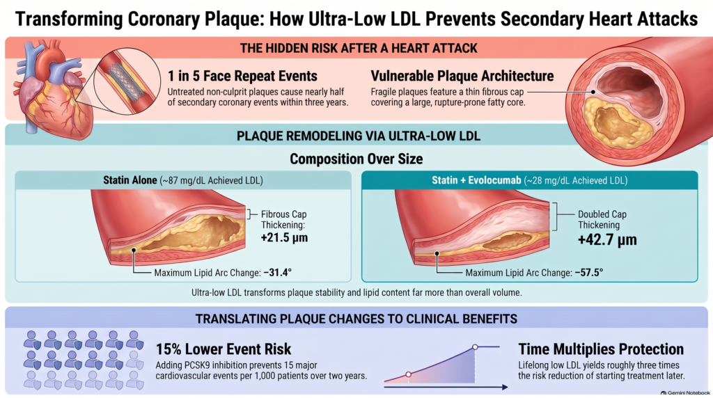
संक्षिप्त उत्तर।. तीव्र कोरोनरी घटनाओं के बाद यादृच्छिक इमेजिंग अध्ययनों में, एलडीएल-सी में तीव्र कमी ने चुनिंदा प्लाक भेद्यता संकेतकों में महत्वपूर्ण सुधार किए, जिनमें कैप की मोटाई और लिपिड सामग्री शामिल हैं, जबकि औसत कमी अपेक्षाकृत मामूली रही। तख्ती की मात्रा. अलग-अलग यादृच्छिक परिणाम परीक्षणों से एलडीएल-सी कम करने पर हृदय संबंधी घटनाओं में कमी देखी गई है, लेकिन ये यह स्थापित नहीं करते कि देखी गई प्लाक-इमेजिंग परिवर्तनों के माध्यम से यह लाभ होता है। कोरोनरी रोग समाप्त नहीं होता।.
भाग 1 में आकृति 1 देखें।.
एक नजर में सबूत
| प्रश्न | साक्ष्य क्या दिखाते हैं | हम कितने निश्चित हैं? |
| क्या बहुत कम एलडीएल-सी प्लाक को बदलता है? | हाँ। कैप्स मोटे हो जाते हैं, लिपिड संकेत सिकुड़ जाते हैं, और प्लाक की मात्रा थोड़ी कम हो जाती है।. | विशिष्ट इमेजिंग मापदंडों के लिए यादृच्छिक इमेजिंग परीक्षणों में दिखाया गया है; घटनाओं के लिए यह एक मान्य विकल्प नहीं है। |
| क्या एलडीएल-सी को कम करने से दिल के दौरे रोके जा सकते हैं और स्ट्रोक्स? | हाँ। बड़े परीक्षण कम दिल के दौरे दिखाते हैं।, इस्केमिक स्ट्रोक, और प्रक्रियाएँ।. | बड़े यादृच्छिक परीक्षणों में सिद्ध |
| क्या यह मौतों को कम करता है? | हाँ के लिए स्टैटिन पूल किए गए परीक्षणों में; अतिरिक्त दवाओं के लिए, केवल नाममात्र या ओपन-लेबल विश्लेषणों में।. | स्टैटिन के लिए सिद्ध; सहायक दवाओं के लिए संकेतात्मक |
| क्या 20 या यहां तक कि 10 मिलीग्राम/डेली से भी कम स्तर बेहतर है? | निम्न प्राप्त स्तर निम्न जोखिम से जुड़े थे, लाभ के अनुरूप लेकिन के अधीन भ्रम में डालने वाला और अनिश्चितता; अध्ययन की गई आबादी और अनुवर्ती अध्ययन में कोई सुसंगत प्रमुख सुरक्षा संकेत नहीं मिला।. | संकेतात्मक (उन स्तरों पर यादृच्छिक नहीं) |
| क्या समय मायने रखता है? | हाँ। लाभ वर्षों के साथ बढ़ता है, और जिन लोगों का जीवनभर LDL-C स्तर कम रहता है, उनमें कोरोनरी रोग बहुत कम होता है।. | मजबूती से समर्थन |
| क्या जोखिम शून्य तक पहुँच सकता है? | नहीं। अत्यंत निम्न एलडीएल-सी स्तर पर भी घटनाएँ होती रहती हैं।. | सभी डेटा में सुसंगत |
| क्या प्लेट परिवर्तन घटनाओं में गिरावट का कारण बनते हैं? | संभावित, लेकिन सिद्ध नहीं।. | निष्कर्ष |
दिल का दौरा अंदर से
यह समझने के लिए कि क्या हाइगेंस मापा गया, यह जानने में मदद करता है कि एक खतरनाक पट्टिका कैसी दिखती है।.
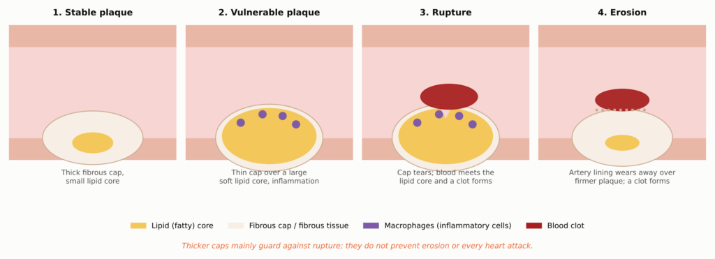
भाग 1 में आकृति 2 देखें।.
प्लाक धमनी की दीवार के अंदर जमाव है, न कि पाइप के अंदर अटका हुआ अवशेष। इसके कई भाग हैं:
- लिपिड-युक्त या नेक्रोटिक कोर: का एक समूह कोलेस्ट्रॉल, कोलेस्ट्रॉल क्रिस्टल, और मृत कोशिकाओं के मलबे। यह नरम, यांत्रिक रूप से कमजोर, और यदि रक्त इसमें पहुँच जाए तो यह रक्त के थक्के जमने को दृढ़ता से बढ़ावा देता है।.
- तंतुमय टोपी: एक कोलेजन परत, निर्मित द्वारा चिकनी पेशी कोशिकाएं, जो कोर को रक्तप्रवाह से अलग कर देती है। अचानक कोरोनरी मृत्यु के शव-परीक्षण अध्ययनों में, फटे कैप्स आमतौर पर लगभग 65 माइक्रोमीटर (μm) से पतले होते थे, जो मोटे तौर पर एक मानव के महीन बालों की चौड़ाई के बराबर है।25].
- मैक्रोफेज: कोलेस्ट्रॉल को निगलने वाले प्रतिरक्षा कोशिकाएं और बन जाती हैं “फ़ोम कोशिकाएं.वे ऐसे एंजाइम छोड़ते हैं जो टोपी में मौजूद कोलेजन को पचा सकते हैं।.
- कैल्शियम: खनिज जमाव। सक्रिय प्लाक में छोटा, छिटपुट कैल्शियम तनाव को केंद्रित कर सकता है, जबकि बड़े, घने पत्र अक्सर पुराने, अधिक रेशेदार रोग का संकेत देते हैं। व्यापक कैल्शियम एक मार्कर बना रहता है। प्लाक का बोझ और जोखिम।.
अधिकांश हृदयाघात तब शुरू होते हैं जब एक पतली परत फट जाती है (rupture) और रक्त थक्के को बढ़ावा देने वाले केंद्र से मिलता है। एक छोटा हिस्सा तब शुरू होता है जब क्षरण, जिसमें धमनी की भीतरी परत एक अधिक सख्त, कम वसायुक्त पट्टिका पर घिस जाती है [26]. यह अंतर बाद में मायने रखेगा, क्योंकि टोपी को मोटा करने से मुख्य रूप से फटने से बचाव होता है।.
कोलेस्ट्रॉल दीवार में कैसे प्रवेश करता है
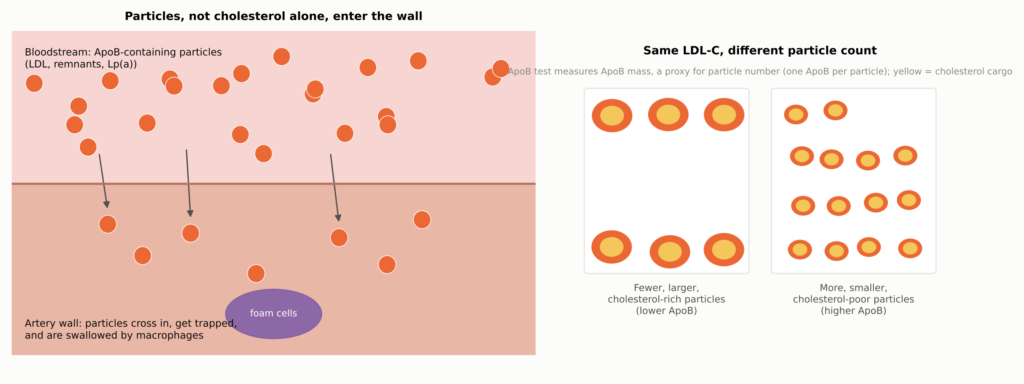
एलडीएल कोलेस्ट्रॉल धमनियों की दीवार में ढीले वसा के रूप में तैरकर नहीं जाता। यह कणों के भीतर यात्रा करता है, और यही कण धमनियों की परत को पार करते हैं, फँस जाते हैं, और मैक्रोफेज द्वारा निगल लिए जाते हैं। इन प्रत्येक कण में, चाहे वह एलडीएल हो, ट्राइग्लिसराइड-समृद्ध अवशेष हो, या एलपी(ए) हो, एक अणु का वहन होता है। प्रोटीन कहा एपोलिपोप्राटीन Bअपोबी): एपोबी-100 जिगर द्वारा बनाए गए कणों पर, और छोटा ApoB-48 पर काइलोमाइक्रोन आंत के अवशेष।.
भाग 1 में आकृति 3 देखें।.
सरल शब्दों में: ApoB यातायात की संख्या है; LDL-C कोलेस्ट्रॉल का माल है।. मानक प्रयोगशाला मान, एलडीएल-सी, यह मापता है कि एलडीएल कण कितनी कोलेस्ट्रॉल ले जाते हैं। एपोबी परीक्षण एपोबी द्रव्यमान सांद्रता को मापता है, जो कणों की संख्या का प्रतिनिधि होता है क्योंकि प्रत्येक प्रमुख एथोजेनिक कण एक ApoB-100 अणु वहन करता है; यह शाब्दिक रूप से कणों की गिनती नहीं करता। आमतौर पर दोनों साथ-साथ चलते हैं। जब वे असहमत होते हैं, जैसा कि उच्च के साथ हो सकता है ट्राइग्लिसराइड्स, मधुमेह, मोपापन, या छोटे एलडीएल कणों में, एपोबी बेहतर रूप से यह दर्शा सकता है कि कितने कण धमनी की दीवार में प्रवेश कर सकते हैं।.
अंतर्राष्ट्रीय इकाइयों का उपयोग करने वाले पाठकों के लिए, इस लेख में चर्चा किए गए एलडीएल-सी स्तरों पर ये अनुमानित रूपांतरण हैं (गोल किए गए; उदाहरण के लिए, 40 मिलीग्राम/डेली लगभग 1.03 मिमोल/लीटर है, इसलिए गोल किए गए मान बिल्कुल समान सीमाएँ नहीं हैं):
| एलडीएल-सी (एमजी/डीएल) | 70 | 55 | 40 | 30 | 20 | 10 |
| एलडीएल-सी (मिलीमोल/लीटर) | 1.8 | 1.4 | 1.0 | 0.78 | 0.52 | 0.26 |
एक जीवित धमनी की झलक
हृदय रोग विज्ञान के अधिकांश इतिहास में, किसी भी जीवित व्यक्ति में फाइब्रोस कैप देखना संभव नहीं था। एंजियोग्राम केवल खुले मार्ग की रूपरेखा दिखाते थे। यह तब बदल गया जब कैथेटर ने धमनी की आंतरिक छवि ली।.
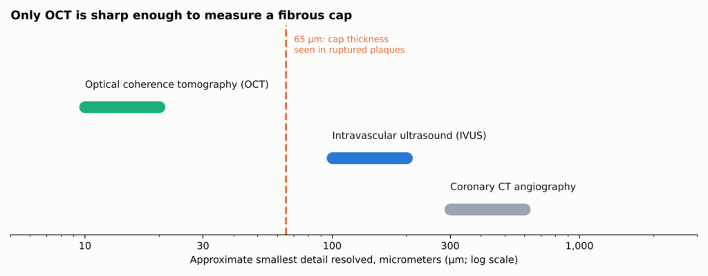
आकृति 10. का अनुमानित संकल्प कोरोनरी इमेजिंग विधि (आम सीमाएँ; प्रणाली के अनुसार भिन्न)। केवल OCT एक संवेदनशील रेशेदार टोपी से भी महीन विवरण को स्पष्ट कर सकता है, इसलिए टोपी की मोटाई के परिणाम OCT से आते हैं, जबकि पट्टिका का आयतन IVUS से आता है, जो पूरी दीवार के पार देख सकता है। निकट-अवरक्त स्पेक्ट्रोस्कोपी (NIRS, नहीं दिखाया गया) संरचना की बजाय लिपिड के रासायनिक हस्ताक्षर का पता लगाती है। इनमें से कोई भी माइक्रोस्कोप नहीं है: ऊतक लेबल अनुमान हैं।.
| इमेजिंग उपकरण | इसमें क्या अच्छा है | यह जो अच्छी तरह नहीं कर सकता |
| ओसीटी (प्रकाश) | माप मोटाई को सीमित करता है, लिपिड चाप, मैक्रोफेज-सदृश संकेत, कैल्शियम, थक्का | लिपिड के पीछे कुल प्लाक आयतन नहीं देखा जा सकता। |
| आईवीयूएस (अल्ट्रासाउंड) | प्लाक की मात्रा और धमनी के पुनर्गठन को मापता है। | टोपी का माप लेने के लिए बहुत मोटा |
| एनआईआरएस (स्पेक्ट्रोस्कोपी) | लिपिड के रासायनिक हस्ताक्षर का पता लगाता है | यह स्वयं कैप्स या वॉल्यूम माप नहीं सकता। |
| कोरोनरी सीटी | बिना कैथेटर के पूरे हृदय में प्लाक दिखाता है | बड़े अक्षर या बारीक विवरण नहीं दिख रहे हैं। |
इनमें से कोई भी माइक्रोस्कोप नहीं है। ये जो ऊतक लेबल उत्पन्न करते हैं, वे प्रकाश या ध्वनि के व्यवहार से निकाले गए अनुमान हैं। OCT में कैप की मोटाई में वृद्धि, NIRS में लिपिड में कमी, और IVUS में प्लाक की मात्रा में कमी पूरक निष्कर्ष हैं, परस्पर प्रतिस्थापन योग्य नहीं।.
ह्यूगेन्स प्रयोग
ह्यूगेन्स (एक वैश्विक में कोरोनरी प्लाक का उच्च-रिज़ॉल्यूशन आकलन) इवोलocumab रैंडमाइज्ड अध्ययन) एक प्रश्न का उत्तर देने के लिए तैयार किया गया था: यदि हृदयाघात के तुरंत बाद एलडीएल कोलेस्ट्रॉल को बहुत कम कर दिया जाए, तो क्या हृदय के अन्य हिस्सों में दिखने वाले खतरनाक-दिखने वाले प्लाक बदलते हैं? यह एक चरण 3, बहु-केंद्र, रैंडमाइज्ड, डबल-ब्लाइंड, प्लेसबो-नियंत्रित इमेजिंग परीक्षण था [9, 1]. एम्जेन, जो इवोलोकुमाब बनाती है, ने इसे वित्त पोषित किया, और कई शोधकर्ताओं ने कंपनी के साथ वित्तीय संबंधों की सूचना दी [9].
किसने भाग लिया
शोधकर्ताओं ने जानबूझकर उन लोगों को चुना जिनकी बची हुई प्लाक पहले से ही कमजोर दिख रही थीं। मरीजों को नॉन-एसटी-एलिवेशन हार्ट अटैक (एनएसटीईएमआई) हुआ था, जिसका इलाज एंजियोग्राफी और दोषी धमनी पर एक प्रक्रिया के साथ किया गया। उन्हें एक ऐसी सीमा से ऊपर LDL-C की आवश्यकता थी जो उनके स्टेटिन उपयोग पर निर्भर करती थी (स्टेटिन न लेने पर कम से कम 130 mg/dL, कम या मध्यम तीव्रता वाले स्टेटिन पर 80 mg/dL, या 60 mg/dL पर उच्च-तीव्रता वाली स्टैटिन), और एक अलग, बिना उपचारित धमनी खंड कम से कम 40 मिमी लंबा दिखाता हुआ लिपिड-युक्त प्लाक ओसीटी पर: 120 माइक्रोमीटर से अधिक मोटी नहीं एक कैप और 90 डिग्री से अधिक का एक लिपिड आर्क [9].
सभी ने अधिकतम सहनशीलता वाली स्टैटिन ली। आधे को भी यादृच्छिक रूप से आवंटित किया गया था। एवोलोकुमाब 420 मिलीग्राम, एक इंजेक्ट किया हुआ PCSK9 अवरोधक, और आधा एक को प्लैसबो सप्ताह 48 तक मासिक इंजेक्शन, और लगभग सप्ताह 50 से 52 में दोबारा OCT और IVUS के साथ [9]. ह्यूगेन्स शामिल 161 यादृच्छिक रूप से चयनित और उपचारित रोगी (80 इवोलोकुमाब, 81 प्लेसबो). उपचारित रोगियों में से, 135 का फॉलो-अप पर मूल्यांकन योग्य इमेजिंग हुई, और एक छोटे उपसमूह में युग्मित IVUS, रजिस्ट्री परिणामों में सभी 161 उपचारित रोगियों के पूर्ण विश्लेषण सेट के लिए OCT अंतबिंदु रिपोर्ट किए गए हैं, जिनमें से 119 के पास प्राथमिक अंतबिंदु के लिए युग्मित OCT माप थे, इसलिए विश्लेषण ने सीधे सभी 161 की पुनः इमेजिंग नहीं की; IVUS परिणाम एक छोटे उपसमूह से प्राप्त हुए हैं [1प्रतिभागियों की औसत आयु 60.5 वर्ष थी, और 28.6% महिलाएँ थीं।.
एलडीएल कैसे कम हुआ
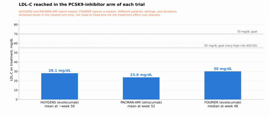
भर्ती के समय औसत एलडीएल-सी था 141.3 मिलीग्राम/डेसीलीटर. फॉलो-अप पर यह था इवोलोकुमाब के साथ 28.1 mg/dL बनाम प्लेसबो के साथ 87.2 mg/dL [1]. प्लेसबो समूह को बिना उपचार के नहीं छोड़ा गया था: कई मरीजों ने बिना किसी स्टैटिन के या कम खुराक पर शुरुआत की थी, इसलिए लगभग 141 से 87 mg/dL तक उनकी गिरावट मुख्यतः स्टैटिन शुरू करने या उसकी खुराक बढ़ाने का परिणाम है। प्रत्येक HUYGENS तुलना है स्टैटिन बनाम स्टैटिन प्लस इवोलोकुमाब.
प्लैक का क्या हुआ
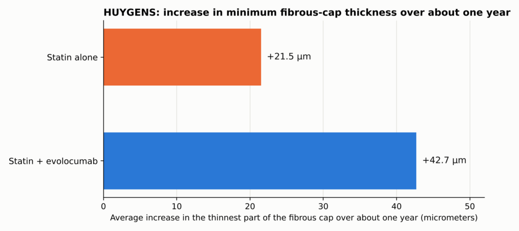
भाग 1 में आकृति 4 देखें।.
संरक्षणात्मक टोपी औसतन मोटी हो गई इवोलोकुमाब के साथ +42.7 माइक्रोमीटर बनाम प्लेसबो के साथ +21.5 माइक्रोमीटर, न्यूनतम वर्गों का अर्थ सभी 161 उपचारित रोगियों में परीक्षण के सह-विविधान विश्लेषण से (परीक्षण के रजिस्ट्री परिणामों के अनुसार, पूर्ण विश्लेषण सेट); 21.2 μm का अंतर (95% विश्वास अंतराल 4.7–37.7; P=0.015) [1]. लिपिड आर्क और अधिक सिकुड़ गया (−57.5 बनाम −31.4 डिग्री), और मैक्रोफेज संकेत और अधिक घटा। एक अन्वेषणात्मक उपसमूह में IVUS द्वारा मापा गया प्लेक आयतन केवल मामूली रूप से बदला: एथेरोमा आयतन प्रतिशत −2.29 बनाम −0.61 प्रतिशत अंक का परिवर्तन, −1.68 अंक का अंकगणितीय अंतर (एक समायोजित तुलना नहीं, और सीधे तौर पर तुलनीय नहीं) पैकमैन-एएमआई के मॉडल-आधारित अनुमान.
सरल शब्दों में।. एवोलोकुमाब के साथ कैप की मोटाई में औसत वृद्धि लगभग दोगुनी अधिक थी। यह तुलना कैप्स के आकार की वृद्धि की है, न कि कैप्स स्वयं की, और इसका यह मतलब नहीं है कि हृदयघात का जोखिम आधा हो गया था।. बड़ी बदलाव इस बात में था कि पट्टिका किस सामग्री से बनी है, न कि यह कितनी बड़ी है।. ये परिणाम उन चयनित गैर-दोषी खंडों का वर्णन करते हैं जिन्हें इमेज किया गया था, न कि कोरोनरी वृक्ष की हर प्लाक का।.
ट्रायल जांचकर्ताओं द्वारा एक द्वितीयक समीक्षा में भी उपचार के बाद बहुत पतली कैप्स (65 μm से कम) वाले इवोलोकुमाब-उपचारित रोगियों की संख्या कम होने की सूचना दी गई [27]. पूर्ण HUYGENS परिणाम तालिका तकनीकी परिशिष्ट में है।.
दो बाद के ह्यूजेन्स विश्लेषणों ने उन रोगियों की तलाश की जिन्होंने अलग तरह से प्रतिक्रिया दी। उच्च एलपी(ए) वाले रोगियों में इवोलोकुमाब और प्लेसबो के बीच कैप मोटाई में अधिक अंतर दिखा।28], और जो रोगी निम्न ApoB स्तर तक पहुँचे, उनके प्लैक के निष्कर्ष उन रोगियों की तुलना में अधिक अनुकूल थे जो ऐसा नहीं कर पाए [29दोनों छोटे, पश्चात्-आधारित उपसमूह विश्लेषण हैं: इन्हें उन स्तरों पर यादृच्छिक रूप से विभाजित नहीं किया गया था, इन्हें पुनरावृत्ति की आवश्यकता है, और ये कोई स्वतंत्र उपचार लक्ष्य या तंत्र स्थापित नहीं करते। विवरण तकनीकी परिशिष्ट में हैं।.
ह्यूगेन्स हमें क्या नहीं बता सकता
ह्यूजेन्स यादृच्छिक साक्ष्य प्रदान करता है कि NSTEMI के बाद स्टेटिन चिकित्सा में इवोलोकुमाब जोड़ने से चुनिंदा गैर-दोषी कोरोनरी धमनियों में प्लाक की भेद्यता से जुड़ी कई OCT-आधारित विशेषताएँ बेहतर होती हैं। घाव. यह स्थापित नहीं करता कि इन इमेजिंग परिवर्तनों ने सीधे बाद में होने वाली हृदय संबंधी घटनाओं को रोका।.
हाइगेंस कम दिल के दौरे, स्ट्रोक, या मौतें दिखाने के लिए डिज़ाइन नहीं किया गया था।. इसके लिए 161 नहीं, बल्कि दसियों हज़ार मरीज़ों की आवश्यकता होगी। इसने केवल शुरुआत में और एक साल बाद ही इमेजिंग की, इसलिए यह नहीं बता सकता कि कैप्स कितनी तेजी से बदलते हैं। इसने उन लोगों का अध्ययन किया जिन्हें इसलिए चुना गया था क्योंकि उनकी पट्टिकाएँ पहले से ही कमजोर दिख रही थीं। और यह नहीं दिखा सकता कि मोटे कैप्स ने बाद में घटनाओं में कोई कमी लाई। इसके लिए हमें अन्य प्रकार के परीक्षणों की आवश्यकता है।.
एकलौती घटना नहीं: अन्य परीक्षणों में भी यही देखा गया
एकल 161-रोगियों का परीक्षण खारिज करना आसान होगा। HUYGENS विश्वसनीय है क्योंकि अन्य परीक्षणों में, विभिन्न दवाओं और विभिन्न इमेजिंग उपकरणों का उपयोग करते हुए, समान पैटर्न देखा गया।.
पैकमान-एमी यादृच्छिक 300 दिल का दौरा पड़े मरीज़ को एलिरोकूमाब, एक और PCSK9 एंटीबॉडी, या प्लेसबो, दोनों के ऊपर रोसुवास्टेटिन, शुरू करते हुए तात्कालिक पीसीआई के 24 घंटों के भीतर हार्ट अटैक के लिए, और पूरक इमेजिंग विधियों से गैर-दोषी प्लाक का आकलन किया गया। 300 यादृच्छिक प्रतिभागियों में से, 265 में युग्मित IVUS उपलब्ध था; NIRS लिपिड विश्लेषण में 263 रोगी शामिल थे, और OCT कैप-मोटाई विश्लेषण में 221 रोगी (105 एलिरोकोमाब, 116 प्लेसबो) थे, जिन्हें फाइब्रोएथेरोमा दोनों समय बिंदुओं पर। तीनों माप एक ही यादृच्छिक परीक्षण से लिए गए थे। एलडीएल-सी गिरकर 23.6 mg/dL बनाम 74.4 mg/dL. तीन पूरक इमेजिंग विधियाँ, जिन्हें एक ही परीक्षण में ओवरलैपिंग रोगियों में मापा गया, ने सुसंगत अनुकूल परिवर्तन दिखाए: प्रतिशत एथेरोमा आयतन में अधिक गिरावट आई (अंतर −1.21 प्रतिशत अंक), NIRS लिपिड संकेत में अधिक गिरावट आई (अंतर −41.24), और टोपी की मोटाई में अधिक वृद्धि हुई (+62.67 बनाम +33.19 μm; अंतर +29.65 μm) [7]. ये अंतर परीक्षण के मॉडल-आधारित अनुमान हैं। जब प्रकाश, ध्वनि और रसायनशास्त्र एक ही परीक्षण में सहमत होते हैं, तो यह निष्कर्ष किसी भी एकल माप से अधिक विश्वसनीय होता है, हालांकि यह फिर भी केवल एक परीक्षण ही है।.
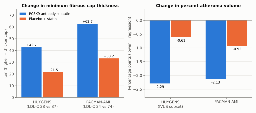
आकृति 11. HUYGENS और PACMAN-AMI: प्रत्येक उपचार शाखा में लगभग एक वर्ष के बाद स्विच किया गया। दोनों परीक्षणों में स्टैटिन चिकित्सा के बाद PCSK9 एंटीबॉडी (HUYGENS में इवोलोकुमाब, PACMAN-AMI में एलिरोकुमाब) जोड़ी गई। मायोकार्डियल इंफार्क्शन. बार रिपोर्ट किए गए अनुसार भुजा के भीतर औसत परिवर्तन हैं; समूहों के बीच अंतर +21.2 μm (P=0.015) और +29.65 μm थे (11.75–47.55) कैप मोटाई के लिए, और −1.68 (गणितीय) और −1.21 (−1.78 से −0.65) प्रतिशत अंक एथेरोमा आयतन के लिए। PACMAN-AMI के लिए, बांह के माध्यों को सीधे घटाने पर 29.48 μm प्राप्त होता है, जो परीक्षण के मॉडल-समायोजित 29.65 μm से थोड़ा अलग है क्योंकि परीक्षण के मिश्रित-प्रभाव मॉडल में बार-बार किए गए मापों और प्रत्येक रोगी में इमेज की गई कई धमनियों को शामिल किया जाता है; पाठ में समायोजित आंकड़े का उपयोग किया गया है। दोनों पैनल विभिन्न इकाइयों का उपयोग करते हैं और एक-दूसरे से तुलना नहीं की जानी चाहिए। HUYGENS एथेरोमा की प्रतिशत मात्रा IVUS उपसमूह में एक अन्वेषणात्मक समाप्ति बिंदु था। विश्लेषण आबादी माप के अनुसार भिन्न होती हैं: HUYGENS कैप के परिणाम 161 उपचारित रोगियों के पूर्ण विश्लेषण सेट में मॉडल अनुमान हैं (119 युग्मित OCT के साथ; 135 किसी भी मूल्यांकन योग्य फॉलो-अप इमेजिंग के साथ) और इसका PAV एक छोटे IVUS उपसमूह से आता है; PACMAN-AMI का PAV युग्मित IVUS वाले 265 रोगियों से आता है और इसकी कैप मोटाई दोनों समय बिंदुओं पर फाइब्रोएथेरोमा वाले 221 रोगियों से आती है। स्रोत: [1, 7].
GLAGOV यादृच्छिक ९६८ रोगी (846 जिनका मूल्यांकन योग्य फॉलो-अप IVUS था) स्थिर कोरोनरी रोग वाले रोगियों को 76 सप्ताह तक इवोलोकुमाब या प्लेसबो दिया गया, मुख्य IVUS स्कैन सप्ताह 78 में किया गया। एलडीएल-सी का औसत 36.6 बनाम 93.0 मिलीग्राम/डेसीलीटर. एथरोमा आयतन में प्रतिशत परिवर्तन evolocumab के साथ −0.95 प्रतिशत अंक और प्लेसबो के साथ +0.05 (समूहों के बीच अंतर −1.0 प्रतिशत अंक; 95% CI −1.8 से −0.64, प्रकाशित परीक्षण से शब्दशः पुनरुत्पादित), और 64% बनाम 47% मरीज़ों में मापनीय प्रतिगमन हुआ था [30]. ये बांह के भीतर के परिवर्तन और परीक्षण द्वारा रिपोर्ट किए गए समूहों के बीच का अंतर हैं, न कि प्लाक द्रव्यमान में प्रतिशत कमी। एक अन्वेषणात्मक उपसमूह में, जो लगभग 24 mg/dL तक पहुँचा, 81% में प्रतिगमन हुआ। GLAGOV भी सीमाएँ दिखाता है: लगभग एक तिहाई उपचारित रोगियों में प्रतिगमन नहीं हुआ, और एक ऊतक-विश्लेषण उप-अध्ययन में प्लाक घटकों में कोई महत्वपूर्ण अंतर नहीं मिला [31].
पहले स्टैटिन और एज़ेटिमाइब उच्च एलडीएल-सी स्तरों पर भी परीक्षणों ने यही कहानी बताई: कम एलडीएल-सी का मतलब था कम प्लाक वृद्धि या मामूली कमी [32, 33, 34, 35], और OCT पर मोटे कैप्स [36YELLOW III ने 26 सप्ताह के दौरान व्यक्ति-स्तर पर प्लाक-इमेजिंग मापों में अनुकूल परिवर्तन दर्ज किए; चूंकि यह अनियंत्रित अध्ययन था, इसलिए यह स्थापित नहीं कर सकता कि परिवर्तन में से कितना हिस्सा एवोलोकुमाब के कारण था।37]. 2025 के एकल-बांह इवोलोकुमाब सीटी/पीईटी अध्ययन ने कम रिपोर्ट किया गैर-कैल्सीकृत पट्टिका और नीचे माइкроकैल्सीफिकेशन गतिविधि, जबकि कैल्सीफाइड प्लाक वॉल्यूम बढ़ा; इसके इमेजिंग एंडपॉइंट्स अलग-अलग चीज़ों को मापते हैं और इन्हें अलग-अलग पढ़ा जाना चाहिए।38]. हर परीक्षण सकारात्मक नहीं था: ओडिसी जे-आईवीयूएस में 36 सप्ताह के दौरान इसके प्लाक एंडपॉइंट में कोई महत्वपूर्ण अंतर नहीं पाया गया [39].
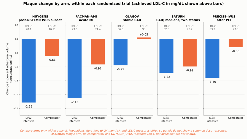
आकृति 12. पाँच यादृच्छिक इमेजिंग परीक्षणों के प्रत्येक शाखा में IVUS प्रतिशत एथेरोमा आयतन में परिवर्तन, प्रत्येक परीक्षण का एक पैनल, प्रत्येक शाखा द्वारा प्राप्त LDL-C के साथ। केवल एक पैनल के भीतर शाखाओं की तुलना करें: आबादी, अवधि (9 से 24 महीने), और एलडीएल-सी माप (GLAGOV समय-भारित है) भिन्न हैं, इसलिए ये पैनल सामान्य खुराक-प्रतिक्रिया संबंध के प्रमाण नहीं हैं। HUYGENS मान एक अन्वेषणात्मक IVUS उपसमूह से प्राप्त हैं।. शनि माध्यिका परिवर्तन और समय-भारित एलडीएल-सी रिपोर्ट किए गए, जबकि अन्य परीक्षण औसत रिपोर्ट करते हैं।. क्षुद्रग्रह को छोड़ दिया गया है क्योंकि इसमें कोई तुलनात्मक समूह नहीं था, और ODYSSEY J-IVUS क्योंकि समीक्षा किए गए स्रोतों में प्राप्त पूर्ण LDL-C उपलब्ध नहीं था। स्रोत: [34, 35, 30, 1, 7].
प्लाक की मात्रा में एक बिंदु की कमी का क्या मतलब है? एथेरोमा आयतन का प्रतिशत (PAV) धमनी के क्षेत्रफल में प्लाक द्वारा घिरे हिस्से का अनुपात है। 40% से 39% में परिवर्तन एक प्रतिशत-बिंदु की गिरावट है। इसका यह मतलब नहीं है कि सभी प्लाक में से 1% गायब हो गया, और इसका यह भी मतलब नहीं है कि संकीर्णता उतनी ही मात्रा में खुल गई।.
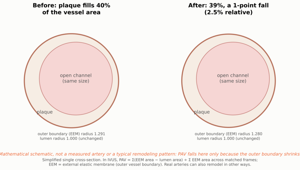
आकृति 13. एथेरोमा आयतन प्रतिशत (PAV) में एक प्रतिशत-बिंदु परिवर्तन के पैमाने का वैचारिक चित्रण; खुला चैनल (ल्युमेन) दोनों पैनलों में समान आकार में दर्शाया गया है, और बाहरी रक्तवाहिनी की सीमा थोड़ी सिकुड़ जाती है। PAV उस खंड में औसतन प्लाक द्वारा कब्जा किए गए रक्तवाहिनी क्षेत्र का हिस्सा है। 40% से 39% तक की गिरावट IVUS परीक्षणों में परिवर्तन का पैमाना है। यह सभी प्लाक का 1% गायब होना नहीं है, और इसका यह मतलब नहीं कि खुली नली समान मात्रा में चौड़ी हो गई, क्योंकि धमनियाँ भी पुनर्निर्माण करती हैं।.
सभी इमेजिंग परीक्षणों की एक साथ-साथ तालिका तकनीकी परिशिष्ट में है।.
अलग, बहुत छोटा नहीं
यदि प्लाक का आयतन लगभग अपरिवर्तित रहता है, तो प्लाक फटने की संभावना कैसे कम हो सकती है? यांत्रिकी एक संभावित उत्तर देती है। फटना तब होता है जब तनाव एक नरम कोर पर फैली पतली परत में केंद्रित हो जाता है। कम लिपिड और मोटी परत उस तनाव को कम कर देगी, भले ही प्लाक का आकार लगभग वही रहे। यह एक संभावित व्याख्या प्रदान करता है कि कैसे परिणाम परीक्षण घटनाओं में सार्थक कमी दिखा सकते हैं, जबकि इमेजिंग परीक्षण केवल आयतन में मामूली परिवर्तन दिखाते हैं, हालांकि यह एक व्याख्या है, सिद्ध कारण नहीं।.
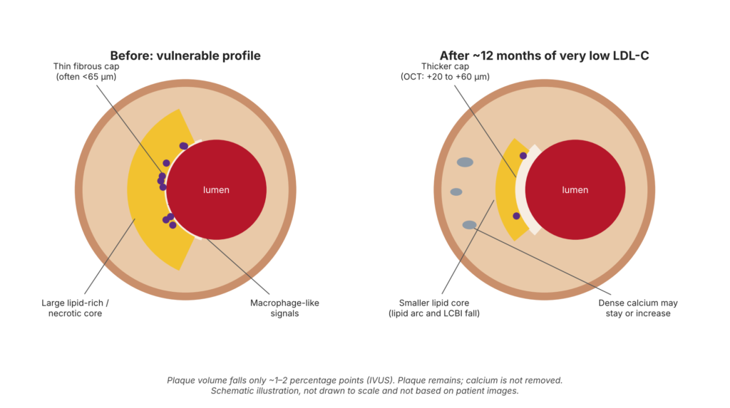
आकृति 14. प्रतीकात्मक आरेख—यह किसी देखे गए रोगी या उपचार की गारंटीकृत प्रतिक्रिया नहीं है। की वैचारिक आरेख पट्टिका स्थिरीकरण: पैमाने पर नहीं बनाया गया और रोगी की छवियों से व्युत्पन्न नहीं है। रैंडमाइज्ड इमेजिंग परीक्षणों में कैप की मोटाई बढ़ना, कम लिपिड, मैक्रोफेज-सदृश संकेतों की संख्या में कमी और प्लाक आयतन में थोड़ी कमी देखी गई है। कैल्शियम में परिवर्तन और घाव की संरचना केवल उदाहरण के तौर पर हैं, न कि एक ही प्लाक में मापे गए परिवर्तन। रैंडमाइज्ड परीक्षणों में प्लाक का उन्मूलन, कैल्शियम का निष्कासन या नियमित रूप से लुमेन का विस्तार नहीं देखा गया है।.
निम्न एलडीएल-सी से कम दिल के दौरे तक के तर्क की श्रृंखला में कई कड़ियाँ हैं। कुछ को सीधे मापा जाता है; अन्य का अनुमान लगाया जाता है।.
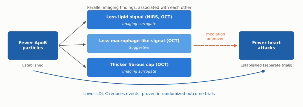
आकृति 15. निम्न एलडीएल-सी का प्लाक इमेजिंग और नैदानिक घटनाओं से संबंध कैसे होता है। कम एपोबी कण और कम घटनाएँ दोनों स्थापित हैं, दूसरी बड़ी परिणाम परीक्षणों में। लिपिड, मैक्रोफेज-सदृश और कैप निष्कर्ष समान रूप से मापे जाने वाले इमेजिंग प्रतिनिधि हैं, न कि सिद्ध अनुक्रम। क्या ये इमेजिंग परिवर्तन घटनाओं में कमी को मध्यस्थता करते हैं, यह स्थापित नहीं है।.
पर्यवेक्षणात्मक इमेजिंग इस विचार का समर्थन करती है कि ये विशेषताएँ महत्वपूर्ण हैं। CLIMA अध्ययन में, उन बिना उपचारित प्लाक में जिनमें चार उच्च-जोखिम वाली OCT विशेषताएँ संयुक्त थीं, घटनाओं का एक-वर्षीय जोखिम कई गुना अधिक पाया गया।40हालांकि किसी विशेषता को जोखिम से जोड़ना यह साबित करने के समान नहीं है कि उस विशेषता को बदलने से जोखिम कम होता है।.
तीन चेतावनियाँ इसे संतुलित बनाए रखती हैं:
- एक मोटी टोपी कोई गारंटी नहीं है।. 65 μm का कटऑफ शव-परीक्षा अध्ययनों से आता है [25]. मोटे कैप्स फिर भी फट सकते हैं, ऐसे पतले संरचनाओं का OCT मापन भिन्न होता है, और कैप की मोटाई इसकी लंबाई के साथ बदलती रहती है।.
- क्षरण अलग है।. क्षरण से होने वाले हृदयाघात अधिकतर धमनी की परत को लगी चोट के कारण होते हैं और सूजन कैप थिनिंग की तुलना में [26]. कैप मोटाई बढ़ना मुख्यतः फाड़ को संबोधित करता है, जो एक कारण है कि घटनाएँ अभी भी बहुत कम एलडीएल-सी पर होती हैं।.
- कैल्शियम बढ़ सकता है, कम नहीं हो सकता।. उच्च-तीव्रता वाले स्टेटिनों ने IVUS कैल्शियम मापों को बढ़ाया, जबकि पट्टिका की मात्रा को कम किया।41], और PARADIGM CT समूह में स्टैटिन उपयोगकर्ताओं में गैर-कैल्सीफाइड प्लाक का विकास धीमा था लेकिन कैल्सीफाइड प्लाक का विकास तेज़ था [42]. एक बढ़ता हुआ कैल्शियम स्कोर उपचार जारी होने का स्वतः यह अर्थ नहीं कि उपचार विफल हो गया है, और सीरियल कैल्शियम स्कोर उपचार का मूल्यांकन करने का मान्य तरीका नहीं हैं। एलडीएल कम करने से “कैल्शियम घुल जाता है” जैसे दावे समर्थित नहीं हैं।.
रिट्रोग्रेशन जरूरी नहीं कि धमनी को फिर से खोल दे। एथेरोमा की मात्रा में कमी जरूरी नहीं कि ल्यूमेन के व्यास में समानुपाती वृद्धि में बदल जाए।. कोरोनरी धमनियाँ बाहर की ओर या अंदर की ओर पुनर्निर्माण कर सकता है, और पट्टिका प्रतिगमन एंजियोग्राम में देखी गई संकीर्णता में चिकित्सकीय दृष्टि से महत्वपूर्ण परिवर्तन के बिना भी यह हो सकता है। यहां चर्चा किए गए इमेजिंग परीक्षण स्थायी कोरोनरी संकीर्णता के नियमित उलटफेर को स्थापित नहीं करते हैं।.
बेहतर दिखने वाले प्लाक से लेकर कम दिल के दौरे तक
इमेजिंग परीक्षण दिखाते हैं कि प्लाक के साथ क्या होता है। परिणाम परीक्षण, जिनमें हजारों मरीजों को शामिल किया जाता है और वास्तविक हृदयाघात, स्ट्रोक और मौतों की गिनती की जाती है, यह दिखाते हैं कि क्या इससे फर्क पड़ता है। यह कहानी में सबसे मजबूत सबूत है।.
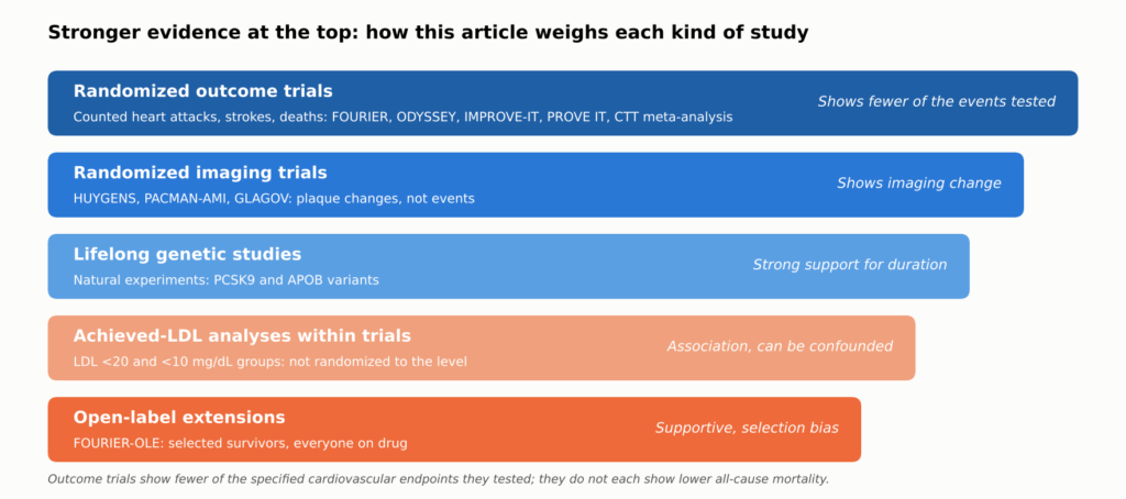
आकृति 16. इस लेख में साक्ष्यों को कैसे वज़न दिया गया है। रैंडमाइज्ड परिणाम परीक्षणों से यह स्थापित होता है कि एलडीएल-सी को कम करने से उन्होंने जिन घटनाओं का परीक्षण किया, वे रोकी जाती हैं; रैंडमाइज्ड इमेजिंग परीक्षण दिखाते हैं कि यह प्लाक के इमेजिंग मापदंडों को बदलता है; शेष श्रेणियाँ बहुत निम्न लक्ष्यों, दीर्घकालिक प्रभाव और आजीवन लाभ के दावों का समर्थन करती हैं, लेकिन स्वयं उन्हें साबित नहीं कर सकतीं। पाठ में प्रत्येक दावे को उसके पीछे मौजूद साक्ष्य के प्रकार के अनुसार लेबल किया गया है।.
The Numbers That Matter to Patients
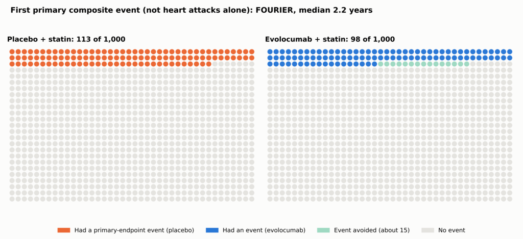
Relative reductions sound large; absolute reductions show what a person gains. In फूरियर, the primary composite (cardiovascular death, heart attack, stroke, hospitalization for अस्थिर एनजाइना, या कोरोनरी पुनर्संवहन) occurred in 9.8% with evolocumab versus 11.3% with placebo over a median 2.2 years: a 15% relative hazard reduction and an absolute difference of about 1.5 percentage points, or about 15 fewer people per 1,000 [2]. That composite is not the chance of a heart attack alone, and the original FOURIER परीक्षण did not establish a reduction in death.
See Figure 5 in Part 1.
| परीक्षण | What was tested | अनुवर्ती | Event rate, treatment vs control | Difference per 1,000 (people; फोरियर-ओले: events per 1,000 person-years) |
| MIRACL [43] | High-dose statin vs placebo, started in hospital | 16 weeks | 14.8% vs 17.4% | About 26 |
| PROVE IT–TIMI 22 [10] | Intensive vs moderate statin | दो साल | 22.4% vs 26.3% | About 39 |
| IMPROVE-IT [4] | Adding ezetimibe to a statin | 7 years | 32.7% vs 34.7% | About 20 |
| ओडिसी परिणाम [3] | Adding alirocumab after ACS | Median 2.8 years | 9.5% vs 11.1% | About 16 |
| फूरियर2] | Adding evolocumab, stable disease | Median 2.2 years | 9.8% vs 11.3% | About 15 |
| FOURIER-OLE [14] (open-label extension; not a randomized trial endpoint) | Earlier vs later evolocumab (open label) | Median 5 more years | CV death, MI, or stroke 2.05 vs 2.58 per 100 person-years | About 5.3 fewer events per 1,000 person-years (rate difference, not people) |
Each trial counted its own combination of events in its own population over its own follow-up, so the rows are not a ranking. FOURIER-OLE compares original randomized assignment in selected survivors, all on open-label evolocumab. A detailed version of this table, with relative effects, LDL-C levels, and mortality, is in the Technical Appendix.
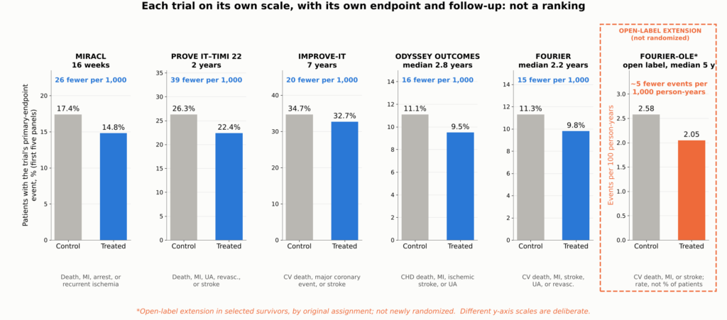
Figure 17. Absolute benefit in the major outcome trials, shown as small multiples: each panel gives one trial’s control and treated primary-endpoint event rates on its own y-axis scale, with the difference expressed per 1,000 patients treated. Each trial used a different कंपोजिट एंडपॉइंट, population, comparator, and follow-up period, so the panels cannot be compared with one another or read as a ranking; they show the scale of benefit within each trial. *FOURIER-OLE shows cardiovascular death, MI, or stroke during the open-label extension, comparing patients originally assigned evolocumab with those originally assigned placebo (2.58 vs 2.05 events per 100 person-years); because this is a rate, not a percentage of patients, its panel has its own y-axis label; it is an exploratory comparison in selected survivors, not a new randomized trial. Sources: [43, 10, 4, 3, 2, 14].
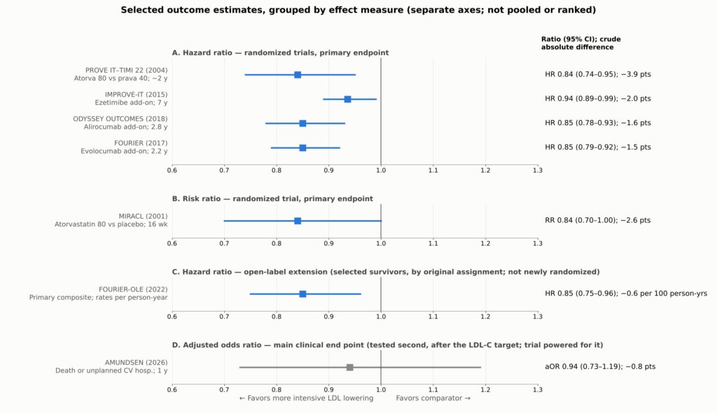
Figure 18. Selected outcome estimates from the trials discussed: primary endpoints for the randomized trials, the open-label extension comparison for FOURIER-OLE, and the main clinical end point for AMUNDSEN. Each trial used a different composite endpoint, population, and follow-up, and reported a different effect measure (risk ratio, Hazard ratio का हिंदी में अनुवाद है: **खतरा अनुपात** (या 'हेजर्ड रेशियो'), or adjusted odds ratio), so each effect measure is shown in its own panel with its own axis, for orientation, not pooled or ranked. Absolute differences are crude differences in each trial’s reported event rates over its own follow-up (for FOURIER-OLE, per 100 person-years). FOURIER-OLE is not a newly randomized outcome comparison: it compares selected surviving participants by original assignment during an open-label extension in which all received evolocumab. AMUNDSEN (gray) is shown for its main clinical end point, which was tested second in the trial’s prespecified hierarchy, after the LDL-C target endpoint; the trial was powered for it. PROVE IT–TIMI 22’s interval is derived from its reported 16% (5%–26%) relative reduction. MIRACL’s सापेक्ष जोखिम is shown as the trial reported it, estimated from a Cox proportional hazards model. Sources: [43, 10, 4, 3, 2, 14, 24].
Fewer Heart Attacks and Strokes; Death Is Harder to Move
The benefit is concentrated in nonfatal events. In FOURIER, heart attacks fell from 4.6% to 3.4%, strokes from 1.9% to 1.5%, and coronary procedures from 7.0% to 5.5% [2]. ODYSSEY OUTCOMES and IMPROVE-IT showed similar reductions in heart attack and ischemic stroke [3, 4]. These are secondary analyses that support the main findings.
Among the add-on trials (IMPROVE-IT, ODYSSEY OUTCOMES, and FOURIER), none showed a reduction in death that was statistically significant under its prespecified rules. ODYSSEY OUTCOMES’ lower all-cause death rate was nominal only. This does not prove that there is no mortality benefit; it means one should not be claimed as proven for these drugs individually.
Counting Every Event
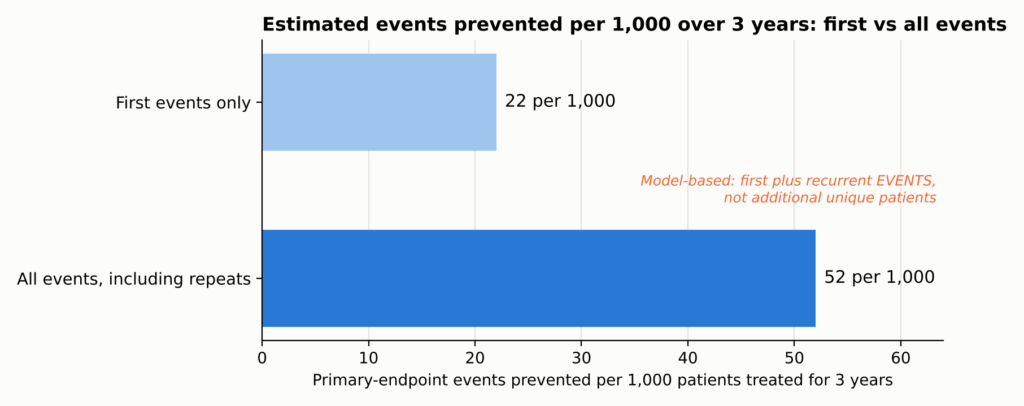
Trials usually count only each patient’s first event. But a patient who has one heart attack often has another, or a stroke, or a stent. When FOURIER counted every event, the estimated number of events prevented more than doubled, although the relative risk reduction did not: about 22 first events but 52 total events prevented per 1,000 patients treated for three years, counting first and repeat events rather than additional people helped (total events: rate ratio 0.82, 95% CI 0.75–0.90; first events: hazard ratio 0.85) [11].
See Figure 6 in Part 1.
The Big Picture
The Cholesterol Treatment Trialists pooled individual data from 26 statin trials and about 170,000 people. Each 38.7 mg/dL (1 mmol/L) reduction in LDL-C lowered major vascular events by about 22%, and all-cause death fell by about 10% per mmol/L, with no increase in cancer or other non-cardiovascular deaths [12]. A meta-regression of randomized trials found broadly similar proportional risk reductions per unit of LDL-C lowering across statins, ezetimibe, and PCSK9 inhibitors, which act through LDL-receptor pathways [13]. That supports a common LDL-mediated mechanism, though it does not mean every drug has identical clinical effects or safety profiles.
सरल शब्दों में।. The absolute LDL-C reduction and how long it lasts are major determinants of benefit, but therapies differ in evidence, timing, off-target effects, and tolerability.
Do the plaque changes explain this benefit? Plausibly, in part. The same drugs at similar LDL-C levels improve plaque in imaging trials and reduce events in outcome trials. But the imaging and outcome trials enrolled different people, and no trial has shown that the imaging changes account for the event reduction. The plaque findings make the benefit biologically coherent; the outcome trials establish the benefit for the endpoints they tested.
Time Is the Hidden Ingredient
Perhaps the most important lesson in the newer data is that benefit builds over time.
The Lag Effect
In FOURIER, the relative reduction in cardiovascular death, heart attack, or stroke was about 16% in the first year and about 25% after the first year [2]. Earlier statin trials showed the same pattern: benefit grows the longer treatment continues. That is consistent with, though it does not prove, plaque gradually becoming more stable.
The Legacy Effect
FOURIER did not demonstrate a significant cardiovascular mortality benefit during its median 2.2-year randomized phase. Afterward, 6,635 surviving participants who chose to continue entered an open-label extension, FOURIER-OLE, in which everyone received evolocumab; median extension follow-up was 5.0 years, and maximum evolocumab exposure across the parent trial and extension reached about 8.4 years; median LDL-C during the extension was about 30 mg/dL [14].
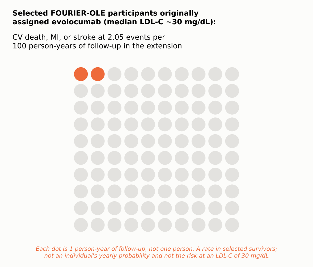
Even though both groups were now on the same drug at similar LDL-C, those originally assigned evolocumab, about two years earlier, had fewer subsequent events. Cardiovascular death, heart attack, or stroke occurred at 2.05 versus 2.58 events per 100 person-years (HR 0.80; 95% CI 0.68–0.93), and cardiovascular death at 0.68 versus 0.90 per 100 person-years (HR 0.77; 95% CI 0.60–0.99), as reported in the results table of the primary FOURIER-OLE publication, where events were counted during extension follow-up by original randomized assignment [14]. This is an exploratory comparison among selected survivors in open-label follow-up, not a new blinded randomized trial, so it cannot establish a mortality benefit by itself. It is compatible with a legacy effect from earlier, longer LDL lowering, but selection into the extension and open-label treatment mean it cannot show that early treatment caused the difference, or what carried it forward.
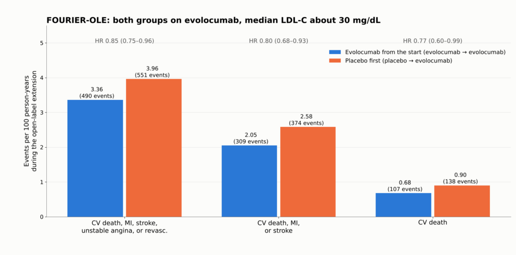
Figure 19. FOURIER-OLE: event rates per 100 person-years during the open-label extension, when all 6,635 participants received evolocumab and median LDL-C was about 30 mg/dL. Patients first randomized to evolocumab had 2.2 more years of low LDL-C exposure. Even in that group, cardiovascular death, MI, or stroke occurred at about 2 per 100 person-years. Extension participants were selected survivors and treatment was open label. Source: [14]. These are event rates in a selected-survivor, open-label extension; they cannot prove a randomized mortality benefit.
How Fast Things Change
- एलडीएल-सी falls within days with a PCSK9 antibody and within two to four weeks with a statin. In AMUNDSEN, median LDL-C was 16 mg/dL six weeks after evolocumab was started before stenting, versus 56 mg/dL with standard care [24].
- प्लाक changes in the randomized imaging trials were measured after roughly 9 to 24 months. YELLOW III documented within-person changes at 26 weeks, but it was uncontrolled, so it cannot establish the size of the drug’s effect or how early it began [37]. A trial that imaged only at the start and at one year cannot show that caps began thickening within days.
- Clinical events: MIRACL showed fewer recurrent ischemic events within 16 weeks [43]; FOURIER’s curves separated within about six months [2]; FOURIER-OLE’s exploratory cardiovascular-death difference emerged only during the open-label extension [14].
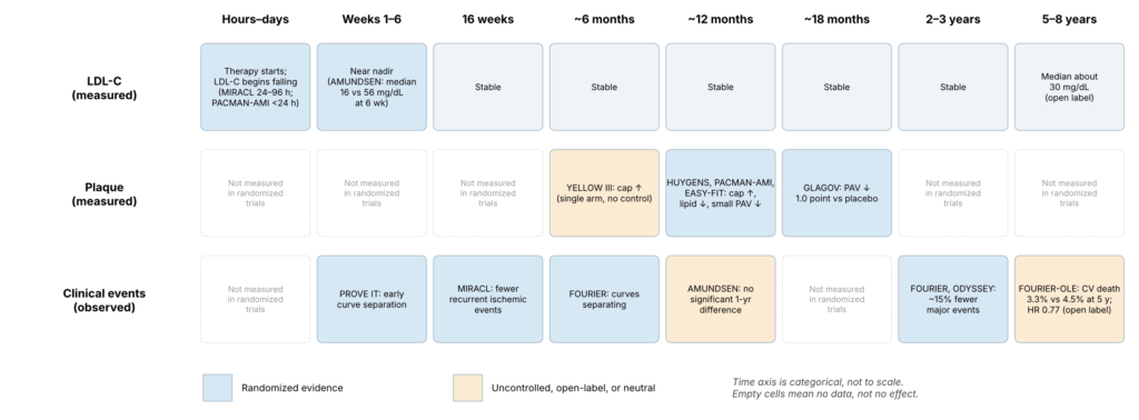
Figure 20. Timeline separating measured LDL-C changes, measured plaque changes, and observed clinical benefit. Each cell shows when a study made a measurement, not when a biological effect began; the figure cannot show when stabilization started, and no unmeasured time points are extrapolated. The time of a study measurement is not necessarily the time a biological benefit began; clinical benefit can begin before imaging is repeated, and no fixed biological lag is implied. A blank cell means not measured, not no effect.
Exposure Over a Lifetime
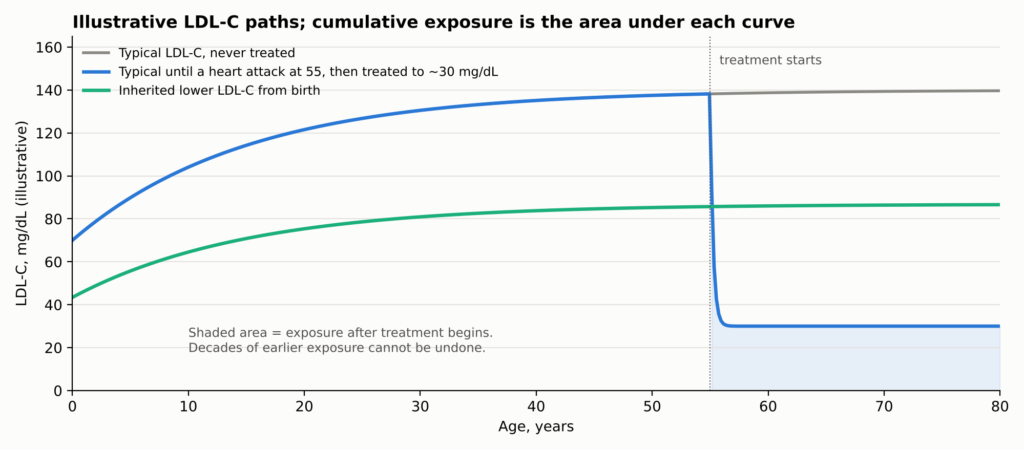
Why should time matter so much? Because धमनी काठिन्य reflects the total, accumulated exposure of the artery wall to ApoB particles over a lifetime. Genetic studies indicate that संचयी संपर्क drives risk [44]. A person treated after a heart attack lowers their future exposure but carries decades of past exposure and the plaque it built.
See Figure 7 in Part 1.
How much a patient gains depends on three things together: baseline risk, how much LDL-C falls, and how long it stays down. The same LDL reduction prevents far more events in a high-risk patient, and larger sustained reductions prevent more.
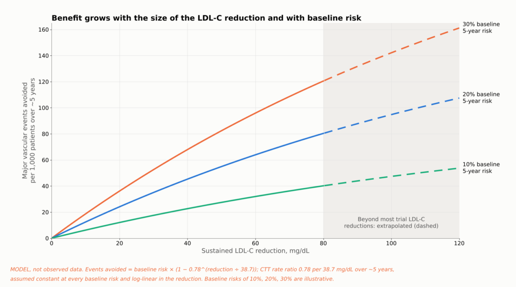
Figure 21. Evidence summary, not a patient-specific prediction. Lines apply the Cholesterol Treatment Trialists’ estimate of a 22% lower risk of major vascular events per 38.7 mg/dL (1 mmol/L) of LDL-C lowering over about five years [12] to three illustrative baseline five-year risks. The same LDL-C reduction prevents more events in higher-risk patients, and larger sustained reductions prevent more. Longer duration adds further benefit beyond what a five-year estimate shows (see the lifetime-exposure figure). Reductions above about 80 mg/dL extend beyond much of the trial data. Assumptions: the CTT rate ratio (a relative risk from a मेटा-विश्लेषण, not a trial hazard ratio) is applied uniformly at every baseline risk and scales log-linearly with LDL-C reduction. This is not a patient जोखिम कैलकुलेटर.
एलडीएल कितना कम होना चाहिए?
Patients often ask whether 40 is good enough, or whether 20 or 10 is better. The honest answer depends on what kind of evidence you accept, because the lower one goes, the more the evidence shifts from randomized comparisons to observational ones.
| LDL-C level | What the evidence looks like | निष्कर्ष |
| 70 mg/dL से कम | Randomized trials (PROVE IT, statin imaging trials) | Established for drug regimens that reached these levels (not randomized to the threshold) |
| 55 मिलीग्राम/डेसीलीटर से कम | Randomized trials (IMPROVE-IT, ODYSSEY, FOURIER) | Established for drug regimens that reached these levels (not randomized to the threshold) |
| Below 40 mg/dL | Randomized trial arms reached it (FOURIER about 30; GLAGOV 36.6) | मजबूती से समर्थन |
| Below 30 mg/dL | Imaging trials (HUYGENS 28, PACMAN-AMI 24); achieved-level analyses | Supported for plaque; outcomes observational |
| 20 mg/dL से कम | Thousands of patients in FOURIER and FOURIER-OLE reached it, but were not randomized to it | Lower risk, no clear safety signal; observational |
| 10 मिलीग्राम/डेली से कम | Small subsets of FOURIER, FOURIER-OLE, and ODYSSEY | Consistent but statistically fragile |
No trial cited here randomized post-heart-attack patients between specific LDL-C targets below 55 mg/dL. People in the drug trials were randomized to drugs, not to “20 versus 40.” That is the most important caveat in this area.
Within those limits, the data point one way. In FOURIER, 2,669 participants reached LDL-C below 20 mg/dL (0.5 mmol/L) by week 4; lower achieved LDL-C was associated with steadily lower cardiovascular risk across the range studied, and serious adverse events were not related to achieved LDL-C over the randomized period [17]. In FOURIER-OLE, about 1,604 participants maintained LDL-C below 20 mg/dL; lower levels remained associated with lower risk and no detected increase in serious adverse events over the extension’s follow-up [18]. In ODYSSEY OUTCOMES, patients who transiently reached below 15 mg/dL had fewer subsequent events in an adjusted post hoc comparison (HR 0.72; 95% CI 0.51–0.997), which does not establish that aiming below 15 mg/dL is better [45].
सरल शब्दों में।. Lower achieved LDL-C has generally been linked to lower event rates across the ranges examined, including very low levels. The studies reviewed have not identified a clear lower threshold; evidence becomes sparse at extreme LDL-C, and achieved-level analyses cannot identify the best target. No randomized trial has shown that aiming for 10 or 20 mg/dL beats aiming for 30 or 40 mg/dL.
How Low Can Risk Go?
There is a different question hiding inside “how low should LDL go.” Suppose LDL-C is pushed nearly to zero. How low can the rate of heart attack, stroke, and cardiovascular death fall? The answer differs sharply between people who already have coronary disease and people who have had low LDL-C since birth.
In People Who Already Have Atherosclerosis
This is not a randomized comparison of LDL-C targets: patients sorted themselves into achieved-LDL groups, so selection and confounding can only partly be adjusted for, and the analysis does not prove there is no biological floor. Within that limit, the peer-reviewed FOURIER-OLE achieved-LDL analysis found risk falling steadily down to the lowest levels studied, below 20 mg/dL [18].
Two points follow. Risk was lowest at the lowest achieved levels studied, an observational association. And the floor is well above zero: among selected participants entering FOURIER-OLE, cardiovascular death, heart attack, or stroke occurred at 2.05 events per 100 person-years of follow-up in those originally assigned evolocumab, the group treated longest; this is a rate by original randomized assignment, not the risk at an LDL-C of 30 mg/dL [14].
These are observational comparisons within trials; patients who reach the lowest levels may differ in ways that statistical adjustment cannot fully remove.
Why the Floor Exists
- Plaque already built. These patients carried decades of accumulated plaque, and IVUS shows plaque burden falling by only a few percentage points.
- Short exposure. Two to eight years of very low LDL-C cannot erase forty or more years of higher exposure.
- Other pathways. Lp(a), triglyceride-rich remnants, and residual inflammation persist. In कैंटोस, COLCOT, and LoDoCo2, anti-inflammatory therapy reduced events in patients already on lipid-lowering drugs [19, 20, 46].
- Other causes. Some strokes come from the heart or tiny vessels, and some heart attacks from erosion, artery tears, or clots carried from elsewhere. धूम्रपान, रक्तचाप, diabetes, and गुर्दे की बीमारी also continue to drive risk.
Nature’s Experiment: People Born With Low LDL
Some people inherit जीन वेरिएंट्स that lower LDL-C from birth. Because those variants are assigned at conception, largely independent of lifestyle, they act like natural randomized experiments. Their results are dramatic.
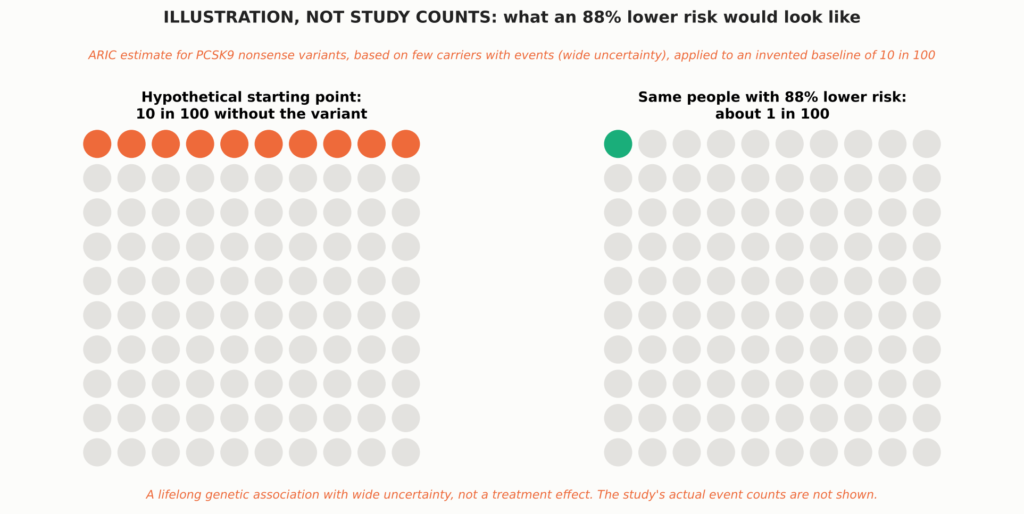
See Figure 8 in Part 1.
- PCSK9 nonsense variants. में एआरआईसी study, Black participants carrying inactivating PCSK9 variants had about 28% lower LDL-C and an 88% lower risk of कोरोनरी हृदय रोग over 15 years. White participants with a milder variant had about 15% lower LDL-C and 47% lower risk [5]. Few carriers had events, so the estimate is imprecise (hazard ratio 0.11; 95% CI 0.02–0.81).
- APOB truncating variants. Carriers had 43 mg/dL lower LDL-C and 72% lower odds of early coronary disease [47].
- PCSK9 R46L in large studies. This common variant lowers LDL-C only modestly, yet it was associated with less coronary or इस्केमिक हृदय रोग [48, 15] and lower cardiovascular mortality [16].
- Across nine variants in six genes (312,321 people), each 38.7 mg/dL of lifelong lower LDL-C was associated with 5% lower coronary risk, about three times the effect per unit of statins started later in life; this is an illustrative comparison across different study designs, not causal equivalence [6].
The genetic data also mark limits. The same R46L variant that lowered coronary risk was not associated with lower ischemic stroke [15], and lifelong lower LDL-C from PCSK9 variants was associated with lower cardiovascular but not सभी कारणों से होने वाली मृत्यु दर [16]. Across these datasets, reductions in ischemic stroke or all-cause mortality were not consistently demonstrated; estimates differ by variant, population, endpoint, and statistical precision. These are observational genetic associations with population-specific uncertainty. Effect sizes differ across variants, populations, and endpoints, and because they reflect lifelong exposure, their scale cannot be equated with a drug treatment effect started in adulthood; quantitative comparisons with trials are illustrative.
People who inherit two inactivating PCSK9 variants are extremely rare. The best-documented case is a 32-year-old woman with no detectable PCSK9 and LDL-C of 14 mg/dL who was healthy, fertile, and working as an aerobics instructor [49]. She was described at a single point in time; nobody knows her long-term cardiovascular outcome.
Putting the Two Together
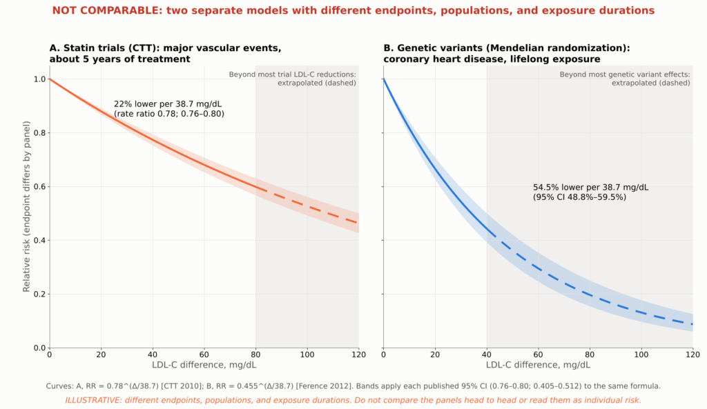
Figure 22. Illustrative model, not trial data or a patient risk calculator. Lines show two published log-linear associations with different endpoints, populations, and exposure durations: the Cholesterol Treatment Trialists’ rate ratio of 0.78 (95% CI 0.76–0.80) for major vascular events per 38.7 mg/dL over about five years of statin treatment [12], and the मेंडेलियन रैंडमाइजेशन estimate of 54.5% (95% CI 48.8%–59.5%) lower coronary heart disease risk per 38.7 mg/dL of lifelong exposure, from 312,321 participants and 9 variants in 6 genes [6]. Each curve is ratio^(LDL-C difference/38.7); shaded bands carry each published 95% विश्वास अंतराल through the same formula and are not observed trial confidence bands. Extrapolated segments are dashed: in panel B beyond about 40 mg/dL, past the range of most genetic variants, and in panel A beyond about 80 mg/dL, past most trial LDL-C reductions, and the model assumes the लॉग-रैखिक संबंध holds at extreme exposures, which has not been shown. The two estimates are shown in separate panels, without individual study or trial points, because their endpoints and populations differ; they must not be read as a head-to-head comparison of statin therapy with lifelong genetic LDL reduction or as a prediction of individual lifetime risk.
The figure compares two published estimates. Applied to a difference of about 75 mg/dL, the statin-trial estimate predicts roughly 38% fewer major vascular events over about five years, while the genetic estimate predicts roughly 78% fewer coronary events over a lifetime. Both are extrapolations, especially the genetic one. They illustrate why duration matters, not what any individual should expect.
The defensible conclusion. Early, sustained, very low exposure to ApoB particles probably drives atherosclerotic coronary events toward a low floor. But no human data show near-zero cardiovascular events, the genetic data suggest stroke and total mortality would remain above zero, and in people who already have coronary disease, events still occur even at extremely low LDL-C.
क्या बहुत कम एलडीएल सुरक्षित है?
A natural worry is that the body needs cholesterol. It does, but cells make their own, and the evidence so far is reassuring, with honest limits.
- Memory and thinking. In EBBINGHAUS, 1,204 FOURIER patients underwent formal cognitive testing over a median 19 months, with no difference between evolocumab and placebo and no link to achieved LDL-C [21]. Longer follow-up found no excess of reported cognitive events [18], though that relies on adverse-event reports.
- Bleeding strokes. उच्च खुराक अटोरवास्टेटिन after stroke modestly increased हेमरेजिक स्ट्रोक in SPARCL [50]. FOURIER, ODYSSEY OUTCOMES, and the very-low-LDL analyses showed no significant excess [2, 17, 18], though such strokes are uncommon.
- Statins modestly raise the risk of new diabetes, about 9% in relative terms, mostly in people already near the threshold [22]. PCSK9 inhibitors and very low LDL-C have not shown a comparable increase [2, 17].
- The adrenal glands and gonads can make their own cholesterol and also take it up from circulating लाइपोप्रोटीन, and large PCSK9 programs have not shown a clinically important hormonal signal.
Achieved-LDL analyses are observational within trials and subject to selection and शेष भ्रमितीकरण; any isolated nominal findings are hypothesis-generating, and very long-term safety at extreme levels remains incompletely characterized.
Drug side effects are a separate matter: PCSK9 antibodies cause slightly more injection-site reactions (2.1% vs 1.6% in FOURIER) [2], and statins can cause muscle symptoms, though true muscle injury is rare. Randomized safety data for PCSK9 antibodies cover about two to three years; open-label exposure reached about 8.4 years [14]. In selected FOURIER extension participants, very low achieved LDL-C was not associated with a detected increase in several prespecified safety outcomes over the parent trial and its open-label extension [18]. That is reassuring but limited by participant selection, observational comparisons, and the inability to exclude rare or lifelong effects. Unlimited lifelong safety at extremely low levels has not been, and cannot yet be, established.
What Guidelines Recommend
Guidelines turn many trials into practical goals. The 2026 U.S. multisociety डिस्लिपिडिमिया guideline reintroduced explicit LDL-C goals and divides people with atherosclerotic हृदय रोग (ASCVD) into two groups [23]. Very-high-risk ASCVD means multiple major ASCVD events, or one major event plus multiple high-risk conditions; a recent heart attack alone does not automatically qualify.
| Who | What the 2026 U.S. guideline recommends |
| ASCVD, not very high risk | High-intensity statin; LDL-C below 70 mg/dL and non-HDL-C below 100 mg/dL (recommended) |
| ASCVD, not very high risk | Add ezetimibe, a PCSK9 antibody, or बेंपेडोइक एसिड if needed to reach below 70 mg/dL |
| ASCVD, not very high risk | Going further, to below 55 mg/dL (non-HDL-C below 85) (reasonable) |
| Very-high-risk ASCVD | High-intensity statin; LDL-C below 55 mg/dL and non-HDL-C below 85 mg/dL (recommended) |
| Very-high-risk ASCVD | Add ezetimibe and/or a PCSK9 antibody if needed to reach those goals |
| Very-high-risk ASCVD | Bempedoic acid is a further add-on option |
“Recommended” and “reasonable” are shown only for the LDL-C and non-HDL-C goals, where they follow the guideline’s own wording for stronger and moderate recommendations. For add-on drugs, the guideline’s recommendation tables give the formal classes, evidence levels, and eligibility criteria, and separate recommendations apply in special situations such as गंभीर हाइपरकोलेस्ट्रॉलएमिया [23].
The 2025 U.S. acute coronary syndrome guideline recommends high-intensity statin therapy and timely addition of nonstatin therapy for those above recommended levels [51]. European guidance after a heart attack recommends at least a 50% reduction and LDL-C below 55 mg/dL [52], and allows a goal below 40 mg/dL after a second vascular event within two years [53]. None of these specific targets was directly tested against another in a randomized target trial; they are expert judgments built on the evidence above.
Should a Second Drug Start in the Hospital?
AMUNDSEN randomized 2,161 heart attack patients and demonstrated that a strategy of giving the first evolocumab injection before PCI substantially improved attainment of the combined LDL-C target at 12 months (82% versus 40%). However, the difference in the trial’s main clinical end point (tested second in the prespecified order, after the LDL-C target), death or unplanned cardiovascular hospitalization, was not statistically significant at one year (14.6% versus 15.4%; adjusted OR 0.94, 95% CI 0.73–1.19). The trial therefore supports faster, more reliable target attainment, but does not demonstrate an additional short-term clinical-event benefit from this initiation strategy [24]. Standard care permitted a PCSK9 inhibitor when indicated, but only 3.8% of the standard-care group received one. The trial was powered (80%) to detect an odds ratio of 0.68 for this end point, so it argues against a large early clinical effect of starting a PCSK9 inhibitor in the catheterization laboratory; it does not exclude a smaller benefit or one that emerges with longer treatment. A prespecified per-protocol analysis suggested fewer events with evolocumab (10.2% versus 14.3%), but the investigators caution that this is likely affected by selection bias. A one-year result does not refute the longer outcome trials. EVOLVE-MI, testing evolocumab within 10 days of a heart attack, had not reported by the search cutoff. For now, early combination therapy is साबित to reach lower LDL-C sooner and, in PACMAN-AMI, to improve one-year plaque changes; it is not yet proven to prevent more events than adding the same drug weeks later.
Where ApoB Fits
The guideline gives ApoB a role in assessing अवशिष्ट जोखिम. ApoB is most useful when it and LDL-C disagree, as in people with high triglycerides, diabetes, or obesity, where LDL-C can look “at goal” while particle number remains high. No randomized trial reviewed here has compared ApoB treatment targets with LDL-C targets, so ApoB is best used as an added measure of remaining कण भार rather than as a proven separate target.
निष्कर्ष
If someone has already had a heart attack, how much can intensive लिपिड कम करने वाला change the disease and lower the chance of another event?
The disease: Within about a year, lowering LDL-C to roughly 25 to 30 mg/dL produces about twice the cap thickening seen with a statin alone, reduces imaging markers of lipid in untreated plaques, and adds roughly a 1.2 to 1.7 percentage-point reduction in percent atheroma volume compared with control groups. The plaque does not disappear, calcium does not dissolve, and the artery is not restored to normal.
The risk: In randomized trials after an acute coronary syndrome, relative reductions in each trial’s main composite endpoint were roughly 15%, with absolute benefits that differ by trial and follow-up. Across the pooled statin trials, each 38.7 mg/dL of LDL-C lowering reduced major vascular events by about 22%. Heart attacks and ischemic strokes are clearly reduced; a reduction in death is proven for statins in pooled analyses, but for add-on therapies only in nominal or open-label analyses.
How low can risk go: The studies reviewed have not identified a clear lower threshold, evidence becomes sparse at extreme LDL-C, and achieved-level analyses cannot define the optimal target; but events still occur even at extremely low LDL-C. People with lifelong low LDL-C have far less coronary disease, which points to duration of exposure as a major lever, but reductions in ischemic stroke and total mortality were not consistently shown. No evidence shows that cardiovascular events can be driven to zero.
| Well established | संकेतात्मक | Still unresolved |
| Lowering LDL-C after a heart attack reduces repeat heart attack, ischemic stroke, and procedures, in proportion to the LDL reduction | Benefit and safety at sustained LDL-C below 20 and even below 10 mg/dL | Whether starting a PCSK9 inhibitor in the hospital prevents more events than starting it weeks later |
| The benefit continues below 70 and 55 mg/dL in randomized comparisons | A lasting legacy benefit from earlier treatment (open-label, selected survivors) | Whether any target below 55 mg/dL beats another in a randomized trial |
| Intensive LDL lowering improves plaque composition and modestly reduces plaque volume over about a year | Far greater protection from lifelong low LDL-C | The true floor of risk with lifelong near-zero LDL-C |
| For statins, ezetimibe, and PCSK9 antibodies, benefits generally outweigh recognized harms in the populations studied | A cardiovascular-mortality benefit from adding PCSK9 inhibition | Lifelong safety at extremely low LDL-C |
| That plaque changes explain part of the benefit; that ApoB and Lp(a) modify the plaque response | How to prevent heart attacks caused by erosion; whether imaging-guided therapy helps |
Practical Points to Discuss With Your Cardiologist
- Start or continue high-intensity statin therapy during the hospital stay after an acute coronary syndrome, unless it is contraindicated or not tolerated.
- Know your numbers and your risk category. The 2026 U.S. guideline recommends an LDL-C goal below 55 mg/dL for people with ASCVD classified as very high risk and below 70 mg/dL for those who are not, with a goal below 55 mg/dL reasonable in selected patients who are not at very high risk; European ACS guidance generally recommends at least a 50% reduction and LDL-C below 55 mg/dL after an acute coronary syndrome. Ask your clinician which category you are in and what your goal is. If you are not at goal on a statin, ask about adding ezetimibe or a PCSK9 inhibitor.
- Ask for a one-time Lp(a) test, and consider ApoB, especially if your triglycerides are high or you have diabetes.
- Do not read a rising calcium score as failure on its own. Discuss what it means in your case.
- Treat the whole picture. Stop smoking, control blood pressure and ब्लड शुगर, attend हृदय पुनर्वास, and take prescribed एंटीप्लेटलेट थेरेपी.
- Stay on therapy. LDL-C rises when treatment stops, and the benefit depends on how long it stays low.
चिकित्सा अस्वीकरण: This article is for educational purposes and summarizes published research. It is not medical advice and does not replace individualized care from your physician. Do not start, stop, or change any medication without talking to your doctor.
Technical Appendix
About This Review
यह वर्णनात्मक समीक्षा selectively summarizes landmark peer-reviewed studies available through October 2026 and is not a व्यवस्थित समीक्षा or meta-analysis. Where full text was not available, I used the peer-reviewed abstract only when it contained enough detail to support the specific statement. Press releases, registry pages, and news coverage were not used as evidence. Numbers not supported by the sources consulted were excluded. Every value plotted in a data figure was checked against the published abstract or primary report, as listed in the companion figure register; supplementary tables were not used. The bibliographic details of all 54 references (authors, title, journal, year, volume, and pages) were checked against the Crossref registry.
A1. HUYGENS Results
| HUYGENS measure (change over about one year) | एवोलोकुमाब + स्टैटिन | Placebo + statin | Difference* | P value |
| Achieved LDL-C, mg/dL | 28.1 | 87.2 | about −59 | <0.001 |
| Minimum fibrous cap thickness, μm (primary endpoint) | +42.7 | +21.5 | +21.2 | 0.015 |
| Maximum lipid arc, degrees (secondary) | −57.5 | −31.4 | −26.1 | 0.04 |
| Macrophage index, mm (exploratory) | −3.17 | −1.45 | −1.72 | 0.04 |
| Percent atheroma volume, percentage points (IVUS subset; exploratory) | −2.29 | −0.61 | −1.68 | 0.009 |
| Microchannels and calcium (exploratory) | कोई महत्वपूर्ण अंतर नहीं | एनएस |
*Values and P values are as reported in the published HUYGENS abstract; 135 treated patients had evaluable imaging at follow-up. The cap-thickness values are least-squares means from the trial’s analysis of covariance in the full analysis set of 161 treated patients, 119 with paired OCT (registry results: difference 21.2 μm, 95% CI 4.7–37.7); IVUS values come from a smaller subset. Other differences are simple arithmetic on the reported group means; the P values come from the trial’s own models. Source: [1].
The protocol prespecified the absolute change in minimum cap thickness as the primary endpoint; secondary endpoints were the percent change in minimum cap thickness, the change in average minimum cap thickness, and the change in maximum lipid arc; IVUS plaque burden, macrophages, and microchannels were exploratory. The trial planned about 150 patients so that 125 would complete paired imaging, with 90% power to detect a 51 μm benefit; the observed adjusted benefit, 21.2 μm, was less than half that assumption yet still statistically significant [9]. Imaging trials can only measure patients who survive, stay in the study, and undergo a second catheterization with usable images, so missing follow-up imaging can introduce selection bias.
A2. HUYGENS Subgroup Analyses
These small, post hoc findings are hypothesis-generating and do not establish separate treatment thresholds or independently causal mechanisms. They were based on subsets and not adjusted for multiple comparisons.
- लाइपोप्रोटीन(ए). In HUYGENS, the difference in cap thickening between evolocumab and placebo appeared larger in patients with high Lp(a) than in those with lower Lp(a), partly because the placebo-plus-statin group with high Lp(a) showed a smaller response [28]. Evolocumab lowered Lp(a) only modestly, and the analysis does not show that lowering Lp(a) caused the benefit. In PACMAN-AMI, baseline Lp(a) did not modify the plaque-volume or cap-thickness response to alirocumab, although patients with the highest Lp(a) had a smaller reduction in the plaque lipid signal [54].
- Patients who reached lower ApoB levels showed more favorable OCT findings than those who did not [29]. Achieved ApoB was not randomized, and the full adjustment model could not be examined, so this does not establish an optimal ApoB target or an independent causal effect.
- Achieved LDL-C. Pooling both arms, lower achieved LDL-C and larger LDL-C reductions were associated with greater cap thickening [1]; also non-randomized.
A3. Plaque-Imaging Trials
| परीक्षण | जनसंख्या | Treatment vs comparator | Achieved LDL-C, mg/dL (mmol/L) | Imaging, duration | Main plaque finding | Key limitations |
| HUYGENS (2022) [1] | 161 post-NSTEMI | Evolocumab vs placebo, both + statin | 28.1 vs 87.2 (0.73 vs 2.25) | OCT; IVUS subset; 50 wk | Cap +42.7 vs +21.5 μm; lipid arc −57.5° vs −31.4°; PAV −2.29 vs −0.61 | 135 with evaluable follow-up imaging; denominators vary by endpoint; selected plaque |
| PACMAN-AMI (2022) [7] | 300 acute MI, started <24 h after PCI | Alirocumab vs placebo, both + rosuvastatin 20 | 23.6 vs 74.4 (0.61 vs 1.92) | IVUS + NIRS + OCT; 52 wk | PAV diff −1.21; LCBI diff −41.24; cap diff +29.65 μm | 265 paired IVUS; 81% men |
| GLAGOV (2016) [30] | 968 stable CAD on statin | Evolocumab vs placebo | 36.6 vs 93.0 (0.95 vs 2.41) | IVUS; 76 wk treatment, week-78 IVUS | PAV −0.95 vs +0.05; regression 64% vs 47% | Not post-MI; composition substudy neutral |
| REVERSAL (2004) [32] | 654 stable CAD | Atorvastatin 80 vs प्रवास्टेटिन 40 | 79 vs 110 (2.0 vs 2.8) | IVUS; 18 mo | Progression halted vs continued | Moderate LDL levels |
| ASTEROID (2006) [33] | 507 CAD | Rosuvastatin 40, single arm | 60.8 (1.57) | IVUS; 24 mo | PAV −0.98 | No control; attrition |
| SATURN (2011) [34] | 1,385 CAD randomized; 1,039 with follow-up IVUS | Rosuvastatin 40 vs atorvastatin 80 | 62.6 vs 70.2 (1.62 vs 1.82) | IVUS; 104 wk | Median PAV −1.22 vs −0.99 (P=0.17) | No significant difference between the two statins; both regressed; no placebo arm |
| PRECISE-IVUS (2015) [35] | 246 after PCI, Japan | Atorvastatin + ezetimibe vs atorvastatin | 63.2 vs 73.3 (1.63 vs 1.90) | IVUS; 9–12 mo | PAV −1.4 vs −0.3; regression 78% vs 58% | Open label |
| EASY-FIT (2014) [36] | 70 unstable angina | Atorvastatin 20 vs 5 | 69 vs 78 (1.78 vs 2.02) | OCT; 12 mo | Cap +69% vs +17% | Small; low-dose comparator |
| ODYSSEY J-IVUS (2019) [39] | 206 recent ACS, Japan | Alirocumab vs standard care | Not shown | IVUS; 36 wk | No significant difference in primary plaque endpoint | Neutral; short duration |
| YELLOW III (2026) [37] | 110 stable CAD, lipid-rich plaque | Evolocumab + statin, single arm | Not reported in sources reviewed | OCT + NIRS-IVUS; 26 wk | Cap 70.9 → 97.7 μm; maxLCBI4mm 306.8 → 213.1; PAV −1.38 points | No control group |
| CCTA/PET study (2025) [38] | 47 stable, noncalcified plaque | Evolocumab, single arm | Not reported in sources reviewed | CCTA + PET; 18 mo | Noncalcified plaque fell; calcified plaque rose | No control group |
Values are not directly comparable across trials. PACMAN-AMI “diff” values are model-based between-group differences; other entries are within-arm changes as reported. PAV, percent atheroma volume; TAV, total atheroma volume; LCBI, लिपिड कोर burden index.
A4. Major Outcome Trials in Detail
| परीक्षण | Population, timing, follow-up | Treatment vs comparator | LDL-C, mg/dL (baseline; achieved) | Primary endpoint | परिणाम | Mortality |
| MIRACL (2001) [43] | 3,086 unstable angina or non-Q-wave MI; 24–96 h; 16 wk | Atorvastatin 80 vs placebo | ~124; 72 vs 135 at 16 wk | Death, MI, resuscitated arrest, or recurrent इस्क्रीमिया with rehospitalization | 14.8% vs 17.4%; RR 0.84 (0.70–1.00) | No significant reduction in death or MI; benefit driven mainly by fewer rehospitalizations for symptomatic ischemia |
| PROVE IT–TIMI 22 (2004) [10] | 4,162 ACS; within 10 days; mean 24 mo | Atorvastatin 80 vs pravastatin 40 | 106; 62 vs 95 | Death, MI, UA rehospitalization, revascularization, or stroke | 22.4% vs 26.3%; 16% reduction (5%–26%) | Death 2.2% vs 3.2%; P=0.07 |
| IMPROVE-IT (2015) [4] | 18,144 ACS; within 10 days; median ~6 y | Simvastatin + ezetimibe vs simvastatin | 93.8; 53.7 vs 69.5 | CV death, major coronary event, or nonfatal stroke | 32.7% vs 34.7% at 7 y; HR 0.936 (0.89–0.99) | No difference |
| ODYSSEY OUTCOMES (2018) [3] | 18,924 ACS 1–12 mo earlier; median 2.8 y | Alirocumab vs placebo + statin | ~92 (mean); ~40 vs ~93 at 4 mo | CHD death, MI, ischemic stroke, or UA hospitalization | 9.5% vs 11.1%; HR 0.85 (0.78–0.93) | All-cause death 3.5% vs 4.1%, nominal only |
| FOURIER (2017) [2] | 27,564 stable ASCVD; median 2.2 y | Evolocumab vs placebo + statin | 92 (median); 30 with evolocumab at 48 wk, 56 mg/dL lower than placebo | CV death, MI, stroke, UA hospitalization, or revascularization | 9.8% vs 11.3%; HR 0.85 (0.79–0.92) | CV death HR 1.05 (0.88–1.25) |
| FOURIER-OLE (2022) [14] | 6,635 FOURIER completers; median 5 more years | Original evolocumab vs original placebo, all on open-label drug | Median ~30 at extension week 12, on open-label evolocumab | Same as FOURIER | 3.36 vs 3.96 per 100 person-years; HR 0.85 (0.75–0.96) | CV death 0.68 vs 0.90 per 100 person-years, HR 0.77 (0.60–0.99); exploratory |
| AMUNDSEN (2026) [24] | 2,161 STEMI/NSTEMI (1,087 vs 1,074); open label; 12 mo | Evolocumab before PCI + standard care vs standard care (PCSK9 inhibitor permitted; used in 3.8%) | Median 16 vs 56 at 6 wk | LDL-C <55 and ≥50% reduction; main clinical end point tested second in a prespecified hierarchy (80% power for OR 0.68) | 792/970 (82%) vs 370/934 (40%); aOR 5.54 (4.50–6.82). Clinical composite 159/1,087 (14.6%) vs 165/1,074 (15.4%); aOR 0.94 (0.73–1.19) | Not significant |
Event rates are Kaplan–Meier estimates for PROVE IT–TIMI 22 and IMPROVE-IT and observed proportions for the others. Endpoints, populations, and follow-up differ, so these figures should not be compared across trials. AMUNDSEN was checked against the full primary JAMA article. CV, cardiovascular; CHD, coronary heart disease; UA, unstable angina.
| Endpoint (treatment vs control) | IMPROVE-IT (7 y) [4] | ODYSSEY OUTCOMES (median 2.8 y) [3] | FOURIER (median 2.2 y) [2] |
| मायोकार्डियल इन्फार्क्शन | 13.1% vs 14.8% | 6.6% vs 7.6% | 3.4% vs 4.6% (HR 0.73) |
| Stroke (trial-specific definition) | Ischemic stroke 3.4% vs 4.1% | Ischemic stroke 1.2% vs 1.6% | Any stroke 1.5% vs 1.9% (HR 0.79) |
| Coronary revascularization | 21.8% vs 23.4% (not significant) | Ischemia-driven, lower with alirocumab | 5.5% vs 7.0% (HR 0.78) |
| Cardiovascular death | कोई महत्वपूर्ण अंतर नहीं | Not significant (HR 0.88) | No difference (HR 1.05) |
| सभी कारणों से मृत्यु | कोई महत्वपूर्ण अंतर नहीं | 3.5% vs 4.1% (HR 0.85), nominal only | No difference |
These components are secondary or nominal analyses, not multiplicity-controlled results.
A5. Limitations of the Evidence
- Small imaging trials. HUYGENS randomized 161 patients, 135 of whom had evaluable follow-up imaging; PACMAN-AMI randomized 300, with 265 having paired IVUS. Both selected particular segments, had missing follow-up imaging, and enrolled mostly men. HUYGENS selected patients with already vulnerable-looking plaque.
- Imaging is not histology. OCT can mistake fibrous tissue for lipid, and macrophage signals are indirect.
- उद्योग वित्तपोषण. Amgen funded HUYGENS, GLAGOV, and FOURIER; Sanofi and Regeneron supported ODYSSEY OUTCOMES, and Sanofi, Regeneron, and Infraredx funded PACMAN-AMI; Merck funded IMPROVE-IT; AstraZeneca funded SATURN. The major outcome trials used blinded treatment and independent event न्यायनिर्णय, which limits but does not eliminate the concern.
- Different populations. The outcome trials differ in timing, background therapy, and endpoint definitions.
- Achieved-LDL and open-label analyses are vulnerable to selection and confounding.
- Genetic studies estimate lifelong effects of specific variants; their effect sizes cannot be transferred directly to drugs started in adulthood.
- Abstract-level sources. The following were reviewed at the abstract or published-summary level rather than in full text: the HUYGENS primary report (supplemented by its registry results) and ApoB analysis, Cohen 2006 (PCSK9 variants), Peloso 2019 (APOB variants), Benn 2010 and Benn 2019 (PCSK9 R46L), and Ference 2012 (Mendelian यादृच्छिकीकरण). These are also marked in the reference list.
- Sources of figure data. Each data figure plots values reported in the study cited in its caption; conceptual and model figures are labeled as illustrative.
- Review method. This narrative review is based on selected peer-reviewed studies and guidelines rather than a registered systematic search. Some findings were extracted from peer-reviewed abstracts or published summaries. Numbers not supported by the sources consulted were excluded. Figure values were checked against the published abstracts or primary reports listed in the companion figure register, and each statement in the text is supported by its cited source at the level indicated above (abstract or full text).
संदर्भ
- Nicholls SJ, Kataoka Y, Nissen SE, et al. Effect of evolocumab on coronary plaque phenotype and burden in statin-treated patients following myocardial infarction (HUYGENS). JACC Cardiovasc Imaging. 2022;15(7):1308-1321. doi:10.1016/j.jcmg.2022.03.002. PubMed: https://pubmed.ncbi.nlm.nih.gov/35431172/
- Sabatine MS, Giugliano RP, Keech AC, et al. Evolocumab and clinical outcomes in patients with cardiovascular disease (FOURIER). N Engl J Med. 2017;376:1713-1722. doi:10.1056/NEJMoa1615664. PubMed: https://pubmed.ncbi.nlm.nih.gov/28304224/
- Schwartz GG, Steg PG, Szarek M, et al. Alirocumab and cardiovascular outcomes after acute coronary syndrome (ODYSSEY OUTCOMES). N Engl J Med. 2018;379:2097-2107. doi:10.1056/NEJMoa1801174. PubMed: https://pubmed.ncbi.nlm.nih.gov/30403574/
- Cannon CP, Blazing MA, Giugliano RP, et al. Ezetimibe added to statin therapy after acute coronary syndromes (IMPROVE-IT). N Engl J Med. 2015;372:2387-2397. doi:10.1056/NEJMoa1410489. PubMed: https://pubmed.ncbi.nlm.nih.gov/26039521/
- Cohen JC, Boerwinkle E, Mosley TH Jr, Hobbs HH. Sequence variations in PCSK9, low LDL, and protection against coronary heart disease. N Engl J Med. 2006;354:1264-1272. doi:10.1056/NEJMoa054013 [abstract reviewed]
- Ference BA, Yoo W, Alesh I, et al. Effect of long-term exposure to lower low-density lipoprotein cholesterol beginning early in life on the risk of coronary heart disease: a Mendelian randomization analysis. J Am Coll Cardiol. 2012;60:2631-2639. doi:10.1016/j.jacc.2012.09.017 [abstract summary reviewed]
- Räber L, Ueki Y, Otsuka T, et al. Effect of alirocumab added to high-intensity statin therapy on coronary atherosclerosis in patients with acute myocardial infarction: the PACMAN-AMI randomized clinical trial. JAMA. 2022;327:1771-1781. doi:10.1001/jama.2022.5218
- Stone GW, Maehara A, Lansky AJ, et al. A prospective natural-history study of coronary atherosclerosis (PROSPECT). N Engl J Med. 2011;364:226-235. doi:10.1056/NEJMoa1002358
- Nicholls SJ, Nissen SE, Prati F, et al. Assessing the impact of PCSK9 inhibition on coronary plaque phenotype with optical coherence tomography: rationale and design of the randomized, placebo-controlled HUYGENS study. Cardiovasc Diagn Ther. 2021;11(1):120-129. doi:10.21037/cdt-20-684. PMCID: PMC7944215. PubMed: https://pubmed.ncbi.nlm.nih.gov/33708484/
- Cannon CP, Braunwald E, McCabe CH, et al. Intensive versus moderate lipid lowering with statins after acute coronary syndromes (PROVE IT–TIMI 22). N Engl J Med. 2004;350:1495-1504. doi:10.1056/NEJMoa040583. PubMed: https://pubmed.ncbi.nlm.nih.gov/15007110/
- Murphy SA, Pedersen TR, Gaciong ZA, et al. Effect of the PCSK9 inhibitor evolocumab on total cardiovascular events in patients with cardiovascular disease: a prespecified analysis from the FOURIER trial. JAMA Cardiol. 2019;4:613-619. doi:10.1001/jamacardio.2019.0886
- Cholesterol Treatment Trialists’ (CTT) Collaboration. Efficacy and safety of more intensive lowering of LDL cholesterol: a meta-analysis of data from 170,000 participants in 26 randomised trials. Lancet. 2010;376:1670-1681. doi:10.1016/S0140-6736(10)61350-5. PubMed: https://pubmed.ncbi.nlm.nih.gov/21067804/
- Silverman MG, Ference BA, Im K, et al. Association between lowering LDL-C and cardiovascular risk reduction among different therapeutic interventions: a systematic review and meta-analysis. JAMA. 2016;316:1289-1297. doi:10.1001/jama.2016.13985
- O’Donoghue ML, Giugliano RP, Wiviott SD, et al. Long-term evolocumab in patients with established atherosclerotic cardiovascular disease (FOURIER-OLE). Circulation. 2022;146:1109-1119. doi:10.1161/CIRCULATIONAHA.122.061620
- Hopewell JC, Malik R, Valdés-Márquez E, Worrall BB, Collins R; METASTROKE Collaboration of the ISGC. Differential effects of PCSK9 variants on risk of coronary disease and ischaemic stroke. Eur Heart J. 2018;39:354-359. doi:10.1093/eurheartj/ehx373
- Benn M, Tybjærg-Hansen A, Nordestgaard BG. Low LDL cholesterol by PCSK9 variation reduces cardiovascular mortality. J Am Coll Cardiol. 2019;73:3102-3114. doi:10.1016/j.jacc.2019.03.517 [abstract reviewed]
- Giugliano RP, Pedersen TR, Park JG, et al. Clinical efficacy and safety of achieving very low LDL-cholesterol concentrations with the PCSK9 inhibitor evolocumab: a prespecified secondary analysis of the FOURIER trial. Lancet. 2017;390:1962-1971. doi:10.1016/S0140-6736(17)32290-0
- Gaba P, O’Donoghue ML, Park JG, et al. Association between achieved low-density lipoprotein cholesterol levels and long-term cardiovascular and safety outcomes: an analysis of FOURIER-OLE. Circulation. 2023;147:1192-1203. doi:10.1161/CIRCULATIONAHA.122.063399
- Ridker PM, Everett BM, Thuren T, et al. Antiinflammatory therapy with canakinumab for atherosclerotic disease (CANTOS). N Engl J Med. 2017;377:1119-1131. doi:10.1056/NEJMoa1707914
- Tardif JC, Kouz S, Waters DD, et al. Efficacy and safety of low-dose colchicine after myocardial infarction (COLCOT). N Engl J Med. 2019;381:2497-2505. doi:10.1056/NEJMoa1912388
- Giugliano RP, Mach F, Zavitz K, et al. Cognitive function in a randomized trial of evolocumab (EBBINGHAUS). N Engl J Med. 2017;377:633-643. doi:10.1056/NEJMoa1701131. PubMed: https://pubmed.ncbi.nlm.nih.gov/28813214/
- Sattar N, Preiss D, Murray HM, et al. Statins and risk of incident diabetes: a collaborative meta-analysis of randomised statin trials. Lancet. 2010;375:735-742. doi:10.1016/S0140-6736(09)61965-6
- Blumenthal RS, et al. 2026 ACC/AHA/AACVPR/ABC/ACPM/ADA/AGS/APhA/ASPC/NLA/PCNA guideline on the management of dyslipidemia. Circulation. 2026;153(17). doi:10.1161/CIR.0000000000001423. Copublished in J Am Coll Cardiol. 2026;87(19):2624-2757. doi:10.1016/j.jacc.2025.11.016. PubMed: https://pubmed.ncbi.nlm.nih.gov/41824552/
- Montalescot G, Ferrari E, Souteyrand G, et al. LDL cholesterol lowering with evolocumab before percutaneous coronary intervention for acute myocardial infarction: the AMUNDSEN randomized clinical trial. JAMA. Published online August 29, 2026. doi:10.1001/jama.2026.17302. PubMed: https://pubmed.ncbi.nlm.nih.gov/42666092/
- Burke AP, Farb A, Malcom GT, et al. Coronary risk factors and plaque morphology in men with coronary disease who died suddenly. N Engl J Med. 1997;336:1276-1282. doi:10.1056/NEJM199705013361802
- Jia H, Abtahian F, Aguirre AD, et al. In vivo diagnosis of plaque erosion and calcified nodule in patients with acute coronary syndrome by intravascular optical coherence tomography. J Am Coll Cardiol. 2013;62:1748-1758. doi:10.1016/j.jacc.2013.05.071
- Fujino M, Nicholls SJ. PCSK9 inhibitors and coronary atherosclerosis: insights from recent intravascular imaging clinical trials. Cardiometab Syndr J. 2023;3(2):103-109. doi:10.51789/cmsj.2023.3.e14
- Di Giovanni G, et al. Impact of evolocumab on plaque phenotypic changes in patients with acute coronary syndrome and elevated lipoprotein(a) levels: a HUYGENS secondary analysis. Eur J Prev Cardiol. 2026;33(11):2047-2054. doi:10.1093/eurjpc/zwaf211
- Fujino M, et al. Achieved levels of apolipoprotein B and plaque composition after acute coronary syndromes: insights from HUYGENS. Atherosclerosis. 2025;403:119145. doi:10.1016/j.atherosclerosis.2025.119145 [abstract reviewed]
- Nicholls SJ, Puri R, Anderson T, et al. Effect of evolocumab on progression of coronary disease in statin-treated patients: the GLAGOV randomized clinical trial. JAMA. 2016;316:2373-2384. doi:10.1001/jama.2016.16951. PubMed: https://pubmed.ncbi.nlm.nih.gov/27846344/
- Nicholls SJ, et al. Effect of evolocumab on coronary plaque composition. J Am Coll Cardiol. 2018;72:2012-2021. doi:10.1016/j.jacc.2018.06.078
- Nissen SE, Tuzcu EM, Schoenhagen P, et al. Effect of intensive compared with moderate lipid-lowering therapy on progression of coronary atherosclerosis: a randomized controlled trial (REVERSAL). JAMA. 2004;291:1071-1080. doi:10.1001/jama.291.9.1071
- Nissen SE, Nicholls SJ, Sipahi I, et al. Effect of very high-intensity statin therapy on regression of coronary atherosclerosis: the ASTEROID trial. JAMA. 2006;295:1556-1565. doi:10.1001/jama.295.13.jpc60002
- Nicholls SJ, Ballantyne CM, Barter PJ, et al. Effect of two intensive statin regimens on progression of coronary disease (SATURN). N Engl J Med. 2011;365:2078-2087. doi:10.1056/NEJMoa1110874. PubMed: https://pubmed.ncbi.nlm.nih.gov/22085316/
- Tsujita K, Sugiyama S, Sumida H, et al. Impact of dual lipid-lowering strategy with ezetimibe and atorvastatin on coronary plaque regression in patients with percutaneous coronary intervention: the multicenter randomized controlled PRECISE-IVUS trial. J Am Coll Cardiol. 2015;66:495-507. doi:10.1016/j.jacc.2015.05.065. PubMed: https://pubmed.ncbi.nlm.nih.gov/26227186/
- Komukai K, Kubo T, Kitabata H, et al. Effect of atorvastatin therapy on fibrous cap thickness in coronary atherosclerotic plaque as assessed by optical coherence tomography: the EASY-FIT study. J Am Coll Cardiol. 2014;64:2207-2217. doi:10.1016/j.jacc.2014.08.045
- Kini AS, Vengrenyuk Y, Liu J, et al. Intracoronary imaging and transcriptomic characteristics before and after maximum lipid-lowering therapy: YELLOW-III study of evolocumab and maximum statin therapy in chronic coronary disease. JACC Cardiovasc Imaging. 2026;19(4):516-534. doi:10.1016/j.jcmg.2025.12.013. PubMed: https://pubmed.ncbi.nlm.nih.gov/41758107/
- Han D, et al. Effects of evolocumab on coronary plaque composition and microcalcification activity by coronary PET and CT angiography. JACC Cardiovasc Imaging. 2025;18:589-599. doi:10.1016/j.jcmg.2025.01.005. PubMed: https://pubmed.ncbi.nlm.nih.gov/40178463/
- Ako J, Hibi K, Tsujita K, et al. Effect of alirocumab on coronary atheroma volume in Japanese patients with acute coronary syndrome: the ODYSSEY J-IVUS trial. Circ J. 2019;83(10):2025-2033. doi:10.1253/circj.CJ-19-0412
- Prati F, Romagnoli E, Gatto L, et al. Relationship between coronary plaque morphology of the left anterior descending artery and 12 months clinical outcome: the CLIMA study. Eur Heart J. 2020;41:383-391. doi:10.1093/eurheartj/ehz520
- Puri R, Nicholls SJ, Shao M, et al. Impact of statins on serial coronary calcification during atheroma progression and regression. J Am Coll Cardiol. 2015;65:1273-1282. doi:10.1016/j.jacc.2015.01.036
- Lee SE, Chang HJ, Sung JM, et al. Effects of statins on coronary atherosclerotic plaques: the PARADIGM study. JACC Cardiovasc Imaging. 2018;11:1475-1484. doi:10.1016/j.jcmg.2018.04.015
- Schwartz GG, Olsson AG, Ezekowitz MD, et al. Effects of atorvastatin on early recurrent ischemic events in acute coronary syndromes: the MIRACL study. JAMA. 2001;285:1711-1718. doi:10.1001/jama.285.13.1711. PubMed: https://pubmed.ncbi.nlm.nih.gov/11277825/
- Ference BA, Ginsberg HN, Graham I, et al. Low-density lipoproteins cause atherosclerotic cardiovascular disease. 1. Evidence from genetic, epidemiologic, and clinical studies. A consensus statement from the European Atherosclerosis Society Consensus Panel. Eur Heart J. 2017;38:2459-2472. doi:10.1093/eurheartj/ehx144
- Schwartz GG, Szarek M, Bittner VA, et al. Transiently achieved very low LDL-cholesterol levels by statin and alirocumab after acute coronary syndrome are associated with cardiovascular risk reduction: the ODYSSEY OUTCOMES trial. Eur Heart J. 2023;44:1408-1417. doi:10.1093/eurheartj/ehad144. PubMed: https://pubmed.ncbi.nlm.nih.gov/36879424/
- Nidorf SM, Fiolet ATL, Mosterd A, et al. Colchicine in patients with chronic coronary disease (LoDoCo2). N Engl J Med. 2020;383:1838-1847. doi:10.1056/NEJMoa2021372
- Peloso GM, Nomura A, Khera AV, et al. Rare protein-truncating variants in APOB, lower low-density lipoprotein cholesterol, and protection against coronary heart disease. Circ Genom Precis Med. 2019;12:e002376. doi:10.1161/CIRCGEN.118.002376 [abstract reviewed]
- Benn M, Nordestgaard BG, Grande P, Schnohr P, Tybjaerg-Hansen A. PCSK9 R46L, low-density lipoprotein cholesterol levels, and risk of ischemic heart disease: 3 independent studies and meta-analyses. J Am Coll Cardiol. 2010;55:2833-2842. doi:10.1016/j.jacc.2010.02.044 [abstract reviewed]
- Zhao Z, Tuakli-Wosornu Y, Lagace TA, et al. Molecular characterization of loss-of-function mutations in PCSK9 and identification of a compound heterozygote. Am J Hum Genet. 2006;79(3):514-523. doi:10.1086/507488. PMCID: PMC1559532
- Amarenco P, Bogousslavsky J, Callahan A 3rd, et al. High-dose atorvastatin after stroke or transient ischemic attack (SPARCL). N Engl J Med. 2006;355:549-559. doi:10.1056/NEJMoa061894
- Rao SV, O’Donoghue ML, Ruel M, et al. 2025 ACC/AHA/ACEP/NAEMSP/SCAI guideline for the management of patients with acute coronary syndromes. J Am Coll Cardiol. 2025;85:2135-2237. doi:10.1016/j.jacc.2024.11.009. PubMed: https://pubmed.ncbi.nlm.nih.gov/40013746/
- Byrne RA, Rossello X, Coughlan JJ, et al. 2023 ESC guidelines for the management of acute coronary syndromes. Eur Heart J Acute Cardiovasc Care. 2024;13:55-161. doi:10.1093/ehjacc/zuad107. PubMed: https://pubmed.ncbi.nlm.nih.gov/37740496/
- Mach F, Baigent C, Catapano AL, et al. 2019 ESC/EAS guidelines for the management of dyslipidaemias: lipid modification to reduce cardiovascular risk. Atherosclerosis. 2019;290:140-205. doi:10.1016/j.atherosclerosis.2019.08.014. PubMed: https://pubmed.ncbi.nlm.nih.gov/31591002/
- Koskinas KC, Häner J, Ueki Y, et al. Association of lipoprotein(a) with changes in coronary atherosclerosis in patients treated with alirocumab. Circ Cardiovasc Imaging. 2024;17:e016683. doi:10.1161/CIRCIMAGING.124.016683. PubMed: https://pubmed.ncbi.nlm.nih.gov/39561225/