
El renacimiento tecnológico en la cardiología preventiva: inteligencia artificial, tomografía coronaria cuantitativa
Introducción
La medicina cardiovascular está experimentando un cambio de paradigma sustancial, pasando de un modelo predominantemente reactivo y guiado por los síntomas hacia un marco preventivo basado en la detección temprana y la evaluación de riesgos individualizada. En el centro de esta transición se encuentra la integración de la inteligencia artificial (IA) con la coronaria tomografía computarizada angiografía (CCTA), en particular mediante el desarrollo de ateroesclerosis tomografía computarizada cuantitativa basada en imágenes (AI-QCT). Este enfoque aborda una limitación de larga data en la cardiología: la incapacidad de las herramientas de diagnóstico tradicionales para detectar de manera confiable la enfermedad coronaria no obstructiva y rica en lípidos placa, a pesar de eso lesiones ser responsable de la mayoría de síndromes coronarios agudos y muerte súbita cardíaca eventos.
La relevancia clínica de esta limitación es sustancial. Los estudios epidemiológicos y patológicos demuestran consistentemente que la mayoría infartos de miocardio surgen de placas que no limitaban el flujo antes de la ruptura (2). Como enfermedad cardiovascular sigue siendo la principal causa de mortalidad en todo el mundo, y se proyecta que las muertes anuales superarán los 20 millones para 2030, por lo que cada vez son más necesarios métodos mejorados para identificar la aterosclerosis coronaria de alto riesgo antes de que ocurran eventos clínicos (3).

Evolución de la estratificación del riesgo cardíaco y la biología de la aterosclerosis
La estratificación tradicional del riesgo cardiovascular se basa en marcadores indirectos como los niveles de lípidos séricos, presión arterial, control glucémico, fumar estado y variables demográficas incorporadas en la población calculadoras de riesgo. Si bien estos modelos han demostrado ser útiles a nivel poblacional, a menudo carecen de precisión cuando se aplican a pacientes individuales, en particular a aquellos con resultados aparentemente “normales” factor de riesgo perfiles que, sin embargo, albergan una enfermedad subclínica significativa (4).
La aterosclerosis es una enfermedad inflamatoria crónica de la pared arterial en lugar de un trastorno definido únicamente por la obstrucción luminal. La placa se desarrolla dentro de la íntima y consiste en proporciones variables de lípidos, tejido fibroso, células inflamatorias y calcio. Las pruebas diagnósticas convencionales, que incluyen la prueba de esfuerzo, el electrocardiograma y la angiografía coronaria invasiva, detectan principalmente la enfermedad una vez que esta produce estenosis hemodinámicamente significativa, que por lo general se define como un estrechamiento luminal de 70% o más (5).
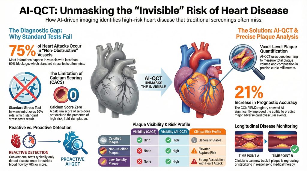
Perspicacia 1: Los estudios de autopsia y angiográficos demuestran que aproximadamente entre el 70 y el 75% de los infartos de miocardio se producen en vasos con menos del 50% estenosis previo al evento índice, lo que indica que la mayoría de los pacientes que experimentan síndromes coronarios agudos no habrían sido identificados como de alto riesgo únicamente mediante pruebas de esfuerzo estándar (2).
Perspicacia 2: Coronario arteria la puntuación de calcio (CACS) se desarrolló para mejorar la detección de la enfermedad subclínica mediante la identificación de placa calcificada. Sin embargo, CACS no detecta placa no calcificada, and a puntaje de calcio of zero does not exclude the presence of lipid-rich, rupture-prone lesions (6).
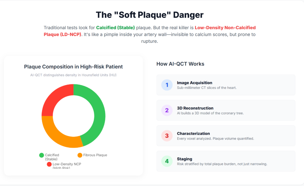
Insight 3: AI-QCT enables non-invasive quantification of total coronary carga de placa, incluido low-density non-calcified plaque, which has been shown to carry a higher association with future cardiovascular events than stenosis severity alone (7).
| Plaque Characteristic | Visibility (CACS) | Visibility (Standard CCTA) | Visibility (AI-QCT) | Clinical Risk Profile |
| Placa calcificada | Alto | Alto | Alto | Generally stable; marker of chronic disease |
| Placa no calcificada | Ninguno | Qualitative / limited | Quantitative / high | Elevated rupture risk |
| Low-density plaque | Ninguno | Variable | Alto | Strong association with ACS |
| Total volumen de placa | Ninguno | Indirect estimate | Precise (mm³) | Strong predictor of future events |
Technical Architecture of AI-Enabled Coronary Analysis
AI-QCT platforms perform voxel-level analysis of the coronary tree rather than relying on visual interpretation alone. These systems employ deep-learning architectures—including convolutional neural networks (CNNs), three-dimensional U-Net segmentation models, and VGG-derived classifiers—trained on large, curated datasets that link imaging features with invasive validation and long-term clinical outcomes (8).
Insight 4: AI-QCT begins with acquisition of high-resolution CCTA. Contemporary multi-detector CT systems, including 64- and 640-slice scanners, provide the temporal and resolución espacial necessary to minimize motion artifact and enable accurate segmentation of coronary anatomy (5).
Insight 5: Automated algorithms identify the coronary lumen and vessel wall and classify plaque components according to Hounsfield unit attenuation values, allowing differentiation of lipid-rich, fibrous, and calcified tissue (9).
Insight 6: AI-derived isquemia indices integrate plaque morphology, lesion length, vessel remodeling, and luminal geometry to estimate flow limitation, providing a non-invasive correlate of invasive reserva de flujo fraccional (FFR) (10).
Clinical Validation and Peer-Reviewed Evidence
AI-QCT has been evaluated in multiple prospective trials, registries, and comparative studies published in leading cardiovascular and imaging journals, including Journal of the American College of Cardiology, Circulation: Cardiovascular Imaging, y European Heart Journal – Cardiovascular Imaging (11).
Landmark Trials and Registries
Insight 7: El CREDENCE trial demonstrated close agreement between AI-assisted CCTA analysis and invasive angiografía coronaria cuantitativa and FFR, supporting the diagnostic accuracy of AI-QCT for identifying hemodynamically significant disease (12).
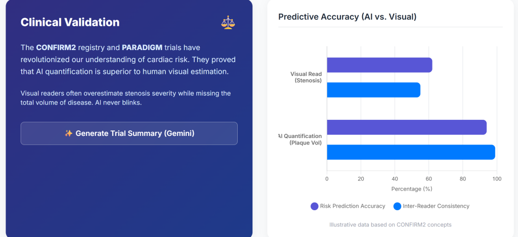
Insight 8: El Registro CONFIRM2, encompassing tens of thousands of patients across multiple countries, showed that AI-quantified plaque features identify a wide gradient of cardiovascular risk among patients with non-obstructive enfermedad de las arterias coronarias (7).
Insight 9: El Estudio CERTAIN demonstrated that AI-QCT significantly improved diagnostic certainty compared with standard interpretation, resulting in more appropriate medical therapy and fewer unnecessary downstream tests (1).
| Estudio | N | Hallazgo clave | P-value | Implicancia clínica |
| CONFIRM2 | 6,550 | AUC for MACE improved from 0.62 to 0.75 | <0.001 | Improved prognostic accuracy |
| CREDENCE | 513 | High concordance with invasive FFR | <0.01 | Viable non-invasive ischemia assessment |
| PACIFIC-1 | 208 | AI exceeded expert readers | <0.05 | Reduced inter-observer variability |
| PROMISE (subset) | 4,347 | 41% stenoses reclassified | — | Reduced false positives |
Preventive Cardiology Implementation and Longitudinal Disease Monitoring
Integration of AI-QCT into preventive cardiology workflows shifts the clinical focus from episodic evaluation toward longitudinal monitoring of atherosclerotic disease.
Insight 10: Quantification of total plaque burden enables objective assessment of disease progression or regression over time, facilitating a treat-to-target approach guided by serial imaging (7).
Insight 11: AI-based comparison tools permit evaluation of changes in plaque composition, including stabilization or regression of placa rica en lípidos in response to medical therapy (13).
Hardware Advances: Photon-Counting Computed Tomography
The performance of AI-QCT is dependent on the quality of input imaging data, which continues to improve with advances in CT hardware.
Insight 12: Tomografía computarizada de conteo de fotones (PCCT) systems directly convert X-ray photons into electrical signals, reducing electronic noise and calcium blooming artifacts compared with conventional energy-integrating detectors (14).
Insight 13: PCCT provides higher spatial resolution with lower radiation and contrast dose, expanding eligibility for coronary CT imaging to patients with high frecuencias cardíacas, elevated índice de masa corporal, or extensive coronary calcificación (15).
Economic and Health System Implications
The adoption of AI-enabled cardiac imaging aligns with broader healthcare trends toward value-based care and prevention.
Insight 14: Market analyses project rapid growth of AI applications in cardiology, driven by improved diagnostic efficiency and potential reductions in downstream costs associated with acute coronary events (16).
Insight 15: Early identification of high-risk plaque phenotypes may reduce myocardial infarction rates and associated healthcare expenditures by enabling earlier and more targeted intervention (17).
Conclusions and Future Directions
Preventive cardiology is increasingly transitioning toward a biologically informed model that emphasizes plaque burden and composition rather than stenosis severity alone. AI-QCT provides a reproducible, quantitative framework for assessing coronary atherosclerosis before clinical events occur.
Insight 16: Future developments are likely to include integration of agentic AI systems, longitudinal digital heart models, and multimodal data streams incorporating genomic and metabolic information (18).
Insight 17: As AI assumes a greater role in image interpretation and quantification, clinicians may increasingly focus on higher-level clinical decision-making, risk communication, and individualized therapy optimization (19).
Referencias
- Nurmohamed NS, Cole JH, Budoff MJ, et al. Impact of atherosclerosis imaging-quantitative computed tomography on diagnostic certainty, downstream testing, coronary revascularization, and medical therapy: the CERTAIN study. Eur Heart J Cardiovasc Imaging. 2024;25(6):857-866. doi:10.1093/ehjci/jeae029
- Falk E, Shah PK, Fuster V. Coronary plaque disruption. Circulation. 1995;92(3):657-671. doi:10.1161/01.cir.92.3.657
- Global Burden of Cardiovascular Diseases and Risks 2023 Collaborators. Global, Regional, and National Burden of Cardiovascular Diseases and Risk Factors in 204 Countries and Territories, 1990-2023. J Am Coll Cardiol. 2025;86(22):2167-2243. doi:10.1016/j.jacc.2025.08.015
- Lloyd-Jones DM. Cardiovascular risk prediction: basic concepts, current status, and future directions. Circulation. 2010;121(15):1768-1777. doi:10.1161/CIRCULATIONAHA.109.849166
- Douglas PS, Hoffmann U, Patel MR, et al. Outcomes of anatomical versus functional testing for coronary artery disease. N Engl J Med. 2015;372(14):1291-1300. doi:10.1056/NEJMoa1415516
- Greenland P, Blaha MJ, Budoff MJ, Erbel R, Watson KE. Coronary Calcium Score and Cardiovascular Risk. J Am Coll Cardiol. 2018;72(4):434-447. doi:10.1016/j.jacc.2018.05.027
- Feuchtner GM, Lacaita PG, Bax JJ, et al. AI-Quantitative CT Coronary Plaque Features Associate With a Higher Relative Risk in Women: CONFIRM2 Registry. Circ Cardiovasc Imaging. 2025;18(6):e018235. doi:10.1161/CIRCIMAGING.125.018235
- Oikonomou EK, Khera R. Machine learning in precision diabetes care and cardiovascular risk prediction. Cardiovasc Diabetol. 2023;22(1):259. Published 2023 Sep 25. doi:10.1186/s12933-023-01985-3
- Koo BK, Yang S, Jung JW, et al. Artificial Intelligence-Enabled Quantitative Coronary Plaque and Hemodynamic Analysis for Predicting Acute Coronary Syndrome. JACC Cardiovasc Imaging. 2024;17(9):1062-1076. doi:10.1016/j.jcmg.2024.03.015
- Nørgaard BL, Leipsic J, Gaur S, et al. Diagnostic performance of noninvasive fractional flow reserve derived from coronary computed tomography angiography in suspected coronary artery disease: the NXT trial (Analysis of Coronary Blood Flow Using CT Angiography: Next Steps). J Am Coll Cardiol. 2014;63(12):1145-1155. doi:10.1016/j.jacc.2013.11.043
- Beerkens FJ, Tang GHL, Kini AS, et al. Transcatheter Aortic Valve Replacement Beyond Severe Aortic Stenosis: JACC State-of-the-Art Review. J Am Coll Cardiol. 2025;85(9):944-964. doi:10.1016/j.jacc.2024.11.051
- Griffin WF, Choi AD, Riess JS, et al. AI Evaluation of Stenosis on Coronary CTA, Comparison With Quantitative Coronary Angiography and Fractional Flow Reserve: A CREDENCE Trial Substudy. JACC Cardiovasc Imaging. 2023;16(2):193-205. doi:10.1016/j.jcmg.2021.10.020
- Zaman S, Wasfy JH, Kapil V, et al. The Lancet Commission on rethinking coronary artery disease: moving from ischaemia to atheroma. Lancet. 2025;405(10486):1264-1312. doi:10.1016/S0140-6736(25)00055-8
- Willemink MJ, Persson M, Pourmorteza A, Pelc NJ, Fleischmann D. Photon-counting CT: Technical Principles and Clinical Prospects. Radiology. 2018;289(2):293-312. doi:10.1148/radiol.2018172656
- Schiebler ML, Jinzaki M, Yanagawa M, et al. Future Applications of Cardiothoracic CT. Radiology. 2025;315(3):e240085. doi:10.1148/radiol.240085
- Windecker S, Gilard M, Achenbach S, et al. Device innovation in cardiovascular medicine: a report from the European Society of Cardiology Cardiovascular Round Table. Eur Heart J. 2024;45(13):1104-1115. doi:10.1093/eurheartj/ehae069
- Undas A, Musiał J, Ząbczyk M. Antiphospholipid antibodies and atherosclerotic vascular disease: recent advances. Rheumatol Int. 2025;45(12):279. Published 2025 Nov 29. doi:10.1007/s00296-025-06050-8
- Topol EJ. High-performance medicine: the convergence of human and artificial intelligence. Nat Med. 2019;25(1):44-56. doi:10.1038/s41591-018-0300-7
- Shetty NS, Gaonkar M, Pampana A, et al. Association of Pathogenic/Likely Pathogenic Inherited Cardiomyopathy Variants With Heart Failure: A TOPMed Multiancestry Analysis. Mayo Clin Proc. 2025;100(11):1948-1955. doi:10.1016/j.mayocp.2025.01.021

