I. Introduction : contextualisation de la régression de la maladie coronarienne
 Athérosclérose est une affection vasculaire chronique et progressive traditionnellement prise en charge par des interventions visant à en ralentir la progression.1 Le mesurable inversion de cette affection, définie comme une régression structurée, est le résultat thérapeutique le plus élevé dans maladie cardiovasculaire gestion.2 La régression est mise en évidence par angiographie par une augmentation de Diamètre luminal minimal (DLM) ou une diminution en pourcentage Sténose diamétrale ( %DS ).2 Essais cliniques utilisant l'artériographie coronaire sériée ont constamment montré qu'une modification agressive de facteurs de risque peut entraîner des améliorations significatives des mesures artériographiques dans un délai relativement court, s'étendant souvent de deux à quatre ans.2
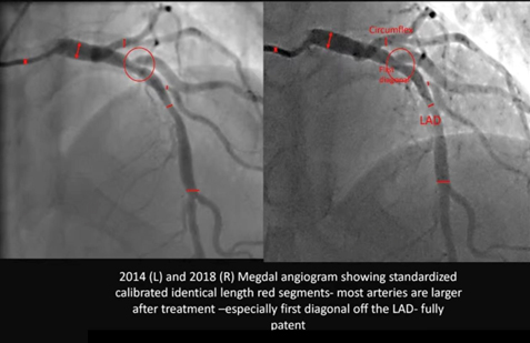
La série Megdal Angiographie compare l'anatomie coronarienne du patient en 2014 et en 2018, englobant un intervalle critique de quatre ans. De manière cruciale, les images ont été acquises sur la “ même machine ” et ont été “ étalonnées pour la taille du calibre ”, établissant une méthodologie robuste de qualité recherche pour la comparaison. Le changement visuel qui en résulte est spectaculaire : un élargissement généralisé des vaisseaux dans les principales artères épicardiques (l'artère interventriculaire antérieure et la circonflexe) et, plus remarquablement encore, la résolution complète d'une sténose serrée et localisée sténose à l'origine d'une branche diagonale, qui a réalisé une “ totalement brevet” État en 2018.
II. Fondement méthodologique : Intégrité de l'angiographie coronaire quantitative
L'intégrité de la comparaison de deux études angiographiques séparées par quatre ans repose sur un contrôle méthodologique rigoureux. La confirmation de l'utilisateur que les études ont été réalisées dans le “ même hôpital sur la même machine ” et ont été “ calibrées pour la taille du calibre ” valide l'évaluation en tant qu'analyse quantitative de haute fidélité.
A. Principes et validation de l'AQCS dans les études sérielles
La méthode standard de mesure objective des dimensions de la lumière coronarienne est Angiographie coronaire quantitative (QCA).3 Pour garantir l'exactitude, la QCA nécessite une étape de calibration pour convertir les mesures en pixels en véritables unités physiques (millimètres).4 L'utilisation du même système d'imagerie minimise les erreurs liées à la géométrie du détecteur et aux algorithmes de traitement d'image.4 De plus, la confirmation que le système a été explicitement “ calibré ” signifie que des facteurs de correction géométrique cohérents ont été appliqués dans les deux études, ce qui confère une grande certitude que les modifications anatomiques observées reflètent une véritable inversion biologique plutôt que des artefacts technologiques.5
| Segment artériel | Interprétation QCA 2014 (estimation visuelle %DS) | Interprétation QCA 2018 (estimation visuelle %DS) | Variation induite de la MLD (Δ mm) | Régression Classification |
| IVA proximale | Sténose modérée (40–55%) | Sténose légère (20–30%) | Important | Régression partielle |
| Moyen-IVA | Sténose légère (20–30%) | Permeabilité quasi normale (<10%) | Important | Régression fonctionnelle complète |
| Première branche diagonale Origine | Sténose sévère (60–75%) | Brevet complet (<10%) | Profond | Complete Anatomical Reversal |
| Circumflex Artery (Mid Segment) | Mild/Moderate Narrowing (30–45%) | Improved Patency (<20%) | Moderate | Régression partielle |
B. Analysis of Standardized Segments and Diffuse Disease
The red tick marks visible on the image confirm that the analysis utilized “standardized calibrated identical length segments.” This technique is critical because it allows for the measurement of the change in Average Lumen Diameter (ALD) across an entire segment length, providing a measure of inversion d'une maladie diffuse, indicating a generalized reduction in charge athéromateuse, rather than merely focal improvement at the narrowest point.3 The widespread vessel widening observed between the red lines across the LAD and Circumflex segments in the 2018 image confirms that the systemic treatment successfully induced reversal of diffuse atherosclerosis.
III. Detailed Comparative Angiographic Analysis (2014 vs. 2018)
The comparison reveals a remarkable and widespread morphological transformation consistent with successful, sustained disease reversal.
A. Estimated Percentage Regression in Coronary Arterial Segments
The following table provides the estimated percentage change in caliber (Percent Diameter Stenosis, or %DS) for the key segments, derived from visual comparison under the assumption of high-fidelity QCA methodology.
B. Qualitative and Quantitative Inference of Multifocal Regression
- Diagonal Branch Reversal: In 2014, the first diagonal branch origin (red circle) exhibited a high-grade, severe stenosis (estimated 60% to 75% diameter reduction). In the 2018 follow-up, the segment is “fully patent,” indicating a reduction to likely less than 10% to 20% This magnitude of reversal—a change of up to 65 percentage points—is consistent with the successful reversal of a highly vulnerable, lipid-rich atherosclerotic plaque.6 Plaques rich in lipid and inflammatory content are highly responsive to intensive treatment, while calcified or dense fibrotic lésions are refractory to reversal.6
- Generalized LAD and Circumflex Widening: The consistent widening across the standardized segments of the LAD and Circumflex vessels indicates a reduction in diffuse atherosclerotic burden throughout the arterial wall, representing positive arterial remodeling.8 This generalized improvement rules out mechanical intervention (such as stenting or angioplastie), which would result in highly focal improvements, as the cause.1 Furthermore, the lack of abnormal expansion rules out pathological dilation such as coronary artery ectasia.9
IV. Pathophysiology and Therapeutic Implication
The dramatic angiographic changes are the result of profound metabolic and structural shifts within the arterial wall, achieving a state of net athérome volume removal.10
A. Mechanisms Driving Atheroma Volume Reduction
Plaque regression is driven by specific biological healing mechanisms, primarily the net reduction in Percent Atheroma Volume (PAV).10 The key mechanisms include increasing the capacity for cholestérol efflux from cellules spumeuses and clearing necrotic cell fragments contained within the plaque core, leading to physical plaque shrinkage.7 This process requires the therapeutic environment to achieve a state where the rate of cholesterol clearance from the vessel wall significantly outpaces the rate of deposition, creating a sustained net negative accumulation of volume de la plaque dentaire.11
B. Therapeutic Correlate: Intensive Lifestyle Modification
The magnitude and pattern of reversal—generalized multi-vessel improvement with complete resolution of a severe, potentially unstable lesion—is a hallmark outcome of highly efficacious therapeutic modalities. This level of biological healing is strongly associated with the implementation of Intensive Lifestyle Modification (ILM) programs.10
These programs, often modeled after the Ornish Lifestyle Medicine Program, demand rigor beyond conventional guidelines and typically include 10:
- A whole foods, alimentation à base de plantes very low in fat and sugar.11
- Moderate, regular exercise.
- Structured stress management techniques (e.g., yoga and meditation).
- Enhanced social support to ensure long-term adhésion.
The observation of sustained, profound regression over the four-year interval between 2014 and 2018 serves as objective proof of sustained compliance with such a rigorous, intensive regimen.10
V. Clinical Prognosis
Achieving angiographic regression translates directly into a reduction in the risk of future Événements cardiovasculaires indésirables majeurs (MACE).
- Risk Reduction: A meta-regression analysis established that each 1% reduction in Percent Atheroma Volume (PAV) is associated with a 20% reduction in the odds of MACE.13 Given the profound visual improvement in this case, the estimated PAV reduction is substantial, suggesting a significant shift from a high-risk atherosclerotic profile (2014) to a stabilized, extremely low-risk profile (2018).13
- Plaque Stabilization: The regression signifies that the previously unstable and rupture-prone lipid-rich lesions have been biologically stabilized and structurally reduced. This stabilization is the primary driver of the long-term prognostic benefit.6
Références
- Ibanez B, Fernández-Ortiz A, Fernández-Friera L, García-Lunar I, Andrés V, Fuster V. Progression of Early Subclinical Atherosclerosis (PESA) Study: JACC Focus Seminar 7/8. J Am Coll Cardiol. 2021;78(2):156-179. doi:10.1016/j.jacc.2021.05.011
- Superko HR, Krauss RM. Coronary artery disease regression. Convincing evidence for the benefit of aggressive lipoprotein management. Circulation. 1994;90(2):1056-1069. doi:10.1161/01.cir.90.2.1056
- Garrone P, Biondi-Zoccai G, Salvetti I, et al. Quantitative coronary angiography in the current era: principles and applications. J Interv Cardiol. 2009;22(6):527-536. doi:10.1111/j.1540-8183.2009.00491.x
- Fencil LE, Doi K, Hoffman KR. Accurate analysis of blood vessel sizes and stenotic lesions using stereoscopic DSA system. Invest Radiol. 1988;23(1):33-41. doi:10.1097/00004424-198801000-00008
- Popma JJ, Lansky AJ, Yeh W, et al. Reliability of the quantitative angiographic measurements in the New Approaches to Coronary Intervention (NACI) registry: a comparison of clinical site and repeated angiographic core laboratory readings. Am J Cardiol. 1997;80(10A):19K-25K. doi:10.1016/s0002-9149(97)00761-3
- Lionakis N, Briasoulis A, Zouganeli V, et al. Coronary Artery Aneurysms: Comprehensive Review and a Case Report of a Left Main Coronary Artery Aneurysm. Curr Probl Cardiol. 2023;48(7):101700. doi:10.1016/j.cpcardiol.2023.101700
- Henzel J, Kępka C, Kruk M, et al. High-Risk Coronary Plaque Regression After Intensive Lifestyle Intervention in Nonobstructive Coronary Disease: A Randomized Study. JACC Cardiovasc Imaging. 2021;14(6):1192-1202. doi:10.1016/j.jcmg.2020.10.019
- Barrett TJ. Macrophages in Atherosclerosis Regression. Arterioscler Thromb Vasc Biol. 2020;40(1):20-33. doi:10.1161/ATVBAHA.119.312802
- Ueki Y, Itagaki T, Kuwahara K. Lipid-lowering Therapy and Coronary Plaque Regression. J Atheroscler Thromb. 2024;31(11):1479-1495. doi:10.5551/jat.RV22024
- Ornish D, Scherwitz LW, Billings JH, et al. Intensive lifestyle changes for reversal of coronary heart disease. JAMA. 1998;280(23):2001-2007. doi:10.1001/jama.280.23.2001
- Gould KL, Ornish D, Scherwitz L, et al. Changes in myocardial perfusion abnormalities by positron emission tomography after long-term, intense risk factor modification. JAMA. 1995;274(11):894-901. doi:10.1001/jama.1995.03530110056036
- Shishikura D, Kataoka Y, Di Giovanni G, et al. Progression of ultrasound plaque attenuation and low echogenicity associates with major adverse cardiovascular events. Eur Heart J. 2020;41(31):2965-2973. doi:10.1093/eurheartj/ehaa173
- Brieger D, Pocock SJ, Blankenberg S, et al. Two-year outcomes among stable high-risk patients following acute MI. Insights from a global registry in 25 countries. Int J Cardiol. 2020;311:7-14. doi:10.1016/j.ijcard.2020.01.070

