
I. Einleitung: Kontextualisierung der Regression der koronaren Herzkrankheit
 Arteriosklerose ist eine chronische, fortschreitende Gefäßerkrankung, die traditionell durch Maßnahmen behandelt wird, die darauf abzielen, ihr Fortschreiten zu verlangsamen.1 Das Messbare Umkehrung Dieses Krankheitsbild – definiert als strukturelle Regression – führt zum besten Therapieergebnis bei Herz-Kreislauf-Erkrankung Management.2 Eine Regression zeigt sich angiographisch durch eine Zunahme von Minimaler Lumen-Durchmesser (MLD) oder ein Rückgang in Prozent Durchmesser-Stenose ( %DS ).2 Klinische Studien Untersuchungen unter Einsatz der seriellen Koronarangiographie haben durchweg gezeigt, dass eine aggressive Modifikation von Risikofaktoren kann innerhalb eines relativ kurzen Zeitraums, der oft zwei bis vier Jahre umfasst, zu erheblichen Verbesserungen bei den arteriographischen Messungen führen.2
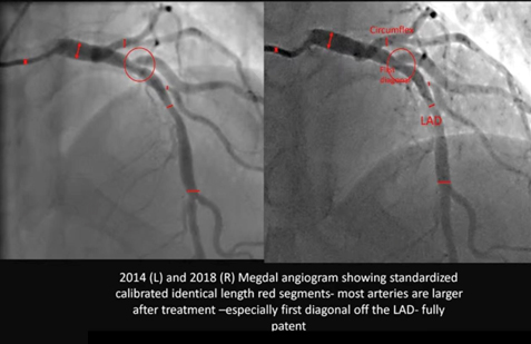
Die Serie „Megdal“ Angiogramm vergleicht die Koronaranatomie des Patienten in den Jahren 2014 und 2018 und deckt damit einen entscheidenden Zeitraum von vier Jahren ab. Entscheidend ist, dass die Bilder auf dem “selben Gerät” aufgenommen und “auf das Kaliber kalibriert” wurden, wodurch eine robuste, forschungsgerechte Methodik für den Vergleich geschaffen wurde. Die daraus resultierende visuelle Veränderung ist dramatisch: eine allgemeine Gefäßerweiterung in den großen epikardialen Arterien (linke vordere absteigende Arterie und Circumflex) und, am bemerkenswertesten, die vollständige Auflösung einer engen, lokalisierten Stenose am Anfang eines diagonalen Zweigs, der eine “vollständige“ Patent”Bundesstaat im Jahr 2018.“.
II. Methodologische Grundlagen: Integrität der quantitativen Koronarangiographie
Die Aussagekraft eines Vergleichs zweier angiographischer Untersuchungen, die im Abstand von vier Jahren durchgeführt wurden, beruht auf einer strengen methodischen Kontrolle. Die Bestätigung des Anwenders, dass die Untersuchungen im “selben Krankenhaus mit demselben Gerät” durchgeführt und “auf das Kaliber abgestimmt” wurden, bestätigt die Bewertung als hochpräzise quantitative Analyse.
A. Prinzipien und Validierung von QCA in seriellen Studien
Die Standardmethode zur objektiven Messung der Lumenmaße der Koronararterien ist Quantitative Koronarangiographie (QCA).3 Um die Genauigkeit sicherzustellen, erfordert die QCA einen Kalibrierungsschritt, um Pixelmessungen in echte physikalische Einheiten (Millimeter) umzuwandeln.4 Die Verwendung desselben bildgebenden Systems minimiert Fehler im Zusammenhang mit der Detektorgeometrie und den Bildverarbeitungsalgorithmen.4 Darüber hinaus bedeutet die Bestätigung, dass das System ausdrücklich “kalibriert” wurde, dass in beiden Studien einheitliche geometrische Korrekturfaktoren angewendet wurden, was ein hohes Maß an Sicherheit dafür bietet, dass die beobachteten anatomischen Veränderungen eine echte biologische Umkehrung widerspiegeln und keine technischen Artefakte sind.5
| Arterielles Segment | QCA-Auslegung 2014 (Schätzung für Visual %DS) | QCA-Auswertung 2018 (Schätzung „Visual %DS“) | Abgeleitete Änderung der MLD (Δ mm) | Regression Klassifikation |
| Proximale RIVA | Mäßige Stenose (40–55%) | Leichte Stenose (20–30%) | Bedeutend | Partielle Regression |
| mittlerer LAD | Leichte Stenose (20–30%) | Nahezu normale Durchgängigkeit (<10%) | Bedeutend | Vollständige Funktionsregression |
| Erster Diagonaler Zweig Herkunft | Schwere Stenose (60–75%) | Vollständiges Patent (<10%) | Tiefgründig | Vollständige anatomische Umkehrung |
| Arteria circumflexa (Mittlerer Abschnitt) | Leichte bis mäßige Verengung (30–45%) | Verbesserte Durchgängigkeit (<20%) | Mäßig | Partielle Regression |
B. Analyse standardisierter Segmente und diffuser Erkrankungen
Die auf dem Bild sichtbaren roten Markierungen bestätigen, dass bei der Analyse “standardisierte, kalibrierte Segmente gleicher Länge” verwendet wurden. Diese Technik ist von entscheidender Bedeutung, da sie die Messung der Veränderung von Durchschnittlicher Lumen-Durchmesser (ALD) über die gesamte Segmentlänge hinweg, was ein Maß für Rückbildung diffuser Erkrankungen, was auf eine allgemeine Verringerung von Plaquebelastung, anstatt sich lediglich auf eine punktuelle Verbesserung an der engsten Stelle zu beschränken.3 Die in dem Bild von 2018 zwischen den roten Linien über die Segmente des LAD und des Zirkumflex hinweg beobachtete weitverbreitete Gefäßerweiterung bestätigt, dass die systemische Behandlung erfolgreich eine Rückbildung der diffusen Atherosklerose bewirkt hat.
III. Detaillierte vergleichende angiographische Analyse (2014 vs. 2018)
Der Vergleich offenbart eine bemerkenswerte und weitverbreitete morphologische Transformation, die mit einer erfolgreichen, anhaltenden Rückbildung der Erkrankung vereinbar ist.
A. Geschätzter prozentualer Regressionsgrad in koronaren Arteriensegmenten
Die folgende Tabelle enthält die geschätzte prozentuale Veränderung des Kalibers (prozentuale Durchmesserstenose, kurz %DS) für die wichtigsten Segmente, die aus einem visuellen Vergleich unter der Annahme einer hochpräzisen QCA-Methodik abgeleitet wurden.
B. Qualitative und quantitative Inferenz der multifokalen Regression
- Diagonale Astumkehr Im Jahr 2014 wies der erste diagonale Ast (roter Kreis) eine hochgradige, schwere Stenose auf (geschätzte Durchmesserverringerung von 60% bis 75%). Bei der Nachuntersuchung im Jahr 2018 ist das Segment “vollständig patentiert,”, was auf eine Verringerung auf wahrscheinlich weniger als 10% bis 20% hindeutet. Das Ausmaß dieser Umkehr – eine Veränderung um bis zu 65 Prozentpunkte – steht im Einklang mit der erfolgreichen Umkehrung einer hochgradig anfälligen, lipidreichen atherosklerotischen Plaque.6 Plaques mit hohem Lipid- und Entzündungsanteil sprechen sehr gut auf eine intensive Behandlung an, während verkalkte oder stark fibrotische Läsionen lassen sich nicht rückgängig machen.6
- Generalisierte LAD- und Circumflex-Verbreiterung: Die gleichmäßige Erweiterung über die standardisierten Segmente des LAD- und des Zirkumflexgefäßes hinweg weist auf eine Reduktion der diffusen atherosklerotischen Last in der gesamten Arterienwand hin, was darstellt positiv arterielles Remodeling.8 Diese allgemeine Verbesserung schließt mechanische Eingriffe (wie beispielsweise die Implantation von Stents oder Angioplastie), was zu sehr punktuellen Verbesserungen führen würde, als Ursache.1 Darüber hinaus schließt das Fehlen einer abnormalen Ausdehnung eine pathologische Erweiterung aus, wie beispielsweise Koronararterienektasie.9
IV. Pathophysiologie und therapeutische Implikation
Die dramatischen angiographischen Veränderungen sind das Ergebnis tiefgreifender metabolischer und struktureller Veränderungen innerhalb der Arterienwand, wodurch ein Zustand der Netto- Atherom Entfernung des Volumens.10
A. Mechanismen der Reduktion des Atheromvolumens
Plaque-Rückbildung wird durch spezifische biologische Heilungsmechanismen gesteuert, in erster Linie durch die Nettoverringerung von Prozentuales Atheromvolumen (PAV).10 Zu den wichtigsten Mechanismen gehört der Ausbau der Kapazitäten für Cholesterin Abfluss aus Schaumzellen und die Entfernung nekrotischer Zellfragmente aus dem Plaquekern, was zu einer physikalischen Verkleinerung der Plaque führt.7 Dieser Prozess erfordert, dass das therapeutische Milieu einen Zustand erreicht, in dem die Rate der Cholesterinausscheidung aus der Gefäßwand signifikant überholt die Ablagerungsrate, wodurch eine anhaltende negative Nettoakkumulation von Plaquevolumen.11
B. Therapeutisches Korrelat: Intensive Lebensstiländerung
Das Ausmaß und Muster der Regression – eine generalisierte Verbesserung an mehreren Gefäßen mit vollständiger Rückbildung einer schweren, potenziell instabilen Läsion – ist ein charakteristisches Ergebnis hochwirksamer therapeutischer Modalitäten. Dieses Ausmaß der biologischen Heilung ist stark mit der Durchführung von Intensive Lebensstiländerung Programme.10
Diese Programme, die sich häufig am Ornish-Modell orientieren Lebensstilmedizin Das Programm stellt höhere Anforderungen als herkömmliche Richtlinien und umfasst in der Regel 10:
- Vollwertkost, pflanzliche Ernährung sehr fett- und zuckerarm.11
- Moderates, regelmäßiges Training.
- Strukturierte Stressbewältigungstechniken (z. B. Yoga und Meditation).
- Verstärkte soziale Unterstützung zur Sicherung einer langfristigen Einhaltung.
Die Beobachtung einer anhaltenden, tiefgreifenden Rückbildung im Vierjahreszeitraum zwischen 2014 und 2018 dient als objektiver Beleg für die kontinuierliche Einhaltung eines derartig rigiden, intensiven Regimes.10
V. Klinische Prognose
Erreichen angiographische Regression führt unmittelbar zu einer Verringerung des Risikos künftiger Schwerwiegende kardiovaskuläre Ereignisse (MACE).
- Risikoreduzierung: Eine Metaregressionsanalyse ergab, dass jede Verringerung des prozentualen Atheromvolumens (PAV) um 1% mit einer Verringerung der Wahrscheinlichkeit für MACE um 20% einhergeht.13 Angesichts der tiefgreifenden visuellen Verbesserung in diesem Fall ist die geschätzte PAV-Reduktion beträchtlich, was auf einen signifikanten Wandel von einem Hochrisiko-Atheroskleroseprofil (2014) zu einem stabilisierten, extrem risikoarmen Profil (2018) hindeutet.13
- Plaque-Stabilisierung: Die Regression bedeutet, dass die zuvor instabilen und rupturgefährdeten lipidreichen Läsionen biologisch stabilisiert und strukturell verkleinert wurden. Diese Stabilisierung ist der primäre Treiber des langfristigen prognostischen Nutzens.6
Referenzen
- Ibanez B, Fernández-Ortiz A, Fernández-Friera L, García-Lunar I, Andrés V, Fuster V. Progression of Early Subclinical Atherosclerosis (PESA) Study: JACC Focus Seminar 7/8. J Am Coll Cardiol. 2021;78(2):156-179. doi:10.1016/j.jacc.2021.05.011
- Superko HR, Krauss RM. Coronary artery disease regression. Convincing evidence for the benefit of aggressive lipoprotein management. Circulation. 1994;90(2):1056-1069. doi:10.1161/01.cir.90.2.1056
- Garrone P, Biondi-Zoccai G, Salvetti I, et al. Quantitative coronary angiography in the current era: principles and applications. J Interv Cardiol. 2009;22(6):527-536. doi:10.1111/j.1540-8183.2009.00491.x
- Fencil LE, Doi K, Hoffman KR. Accurate analysis of blood vessel sizes and stenotic lesions using stereoscopic DSA system. Invest Radiol. 1988;23(1):33-41. doi:10.1097/00004424-198801000-00008
- Popma JJ, Lansky AJ, Yeh W, et al. Reliability of the quantitative angiographic measurements in the New Approaches to Coronary Intervention (NACI) registry: a comparison of clinical site and repeated angiographic core laboratory readings. Am J Cardiol. 1997;80(10A):19K-25K. doi:10.1016/s0002-9149(97)00761-3
- Lionakis N, Briasoulis A, Zouganeli V, et al. Coronary Artery Aneurysms: Comprehensive Review and a Case Report of a Left Main Coronary Artery Aneurysm. Curr Probl Cardiol. 2023;48(7):101700. doi:10.1016/j.cpcardiol.2023.101700
- Henzel J, Kępka C, Kruk M, et al. High-Risk Coronary Plaque Regression After Intensive Lifestyle Intervention in Nonobstructive Coronary Disease: A Randomized Study. JACC Cardiovasc Imaging. 2021;14(6):1192-1202. doi:10.1016/j.jcmg.2020.10.019
- Barrett TJ. Macrophages in Atherosclerosis Regression. Arterioscler Thromb Vasc Biol. 2020;40(1):20-33. doi:10.1161/ATVBAHA.119.312802
- Ueki Y, Itagaki T, Kuwahara K. Lipid-lowering Therapy and Coronary Plaque Regression. J Atheroscler Thromb. 2024;31(11):1479-1495. doi:10.5551/jat.RV22024
- Ornish D, Scherwitz LW, Billings JH, et al. Intensive lifestyle changes for reversal of coronary heart disease. JAMA. 1998;280(23):2001-2007. doi:10.1001/jama.280.23.2001
- Gould KL, Ornish D, Scherwitz L, et al. Changes in myocardial perfusion abnormalities by positron emission tomography after long-term, intense risk factor modification. JAMA. 1995;274(11):894-901. doi:10.1001/jama.1995.03530110056036
- Shishikura D, Kataoka Y, Di Giovanni G, et al. Progression of ultrasound plaque attenuation and low echogenicity associates with major adverse cardiovascular events. Eur Heart J. 2020;41(31):2965-2973. doi:10.1093/eurheartj/ehaa173
- Brieger D, Pocock SJ, Blankenberg S, et al. Two-year outcomes among stable high-risk patients following acute MI. Insights from a global registry in 25 countries. Int J Cardiol. 2020;311:7-14. doi:10.1016/j.ijcard.2020.01.070

