I. Introducción: Contextualización de la regresión de la enfermedad arterial coronaria
 Ateroesclerosis is a chronic, progressive vascular condition traditionally managed by interventions aimed at slowing its advancement.1 The measurable reversión of this condition—defined as structural regression—is the highest therapeutic outcome in enfermedad cardiovascular gestión.2 Regression is evidenced angiographically by an increase in Minimal Lumen Diameter (MLD) or a decrease in Percent Diameter Stenosis ( %DS ).2 Ensayos clínicos utilizing serial coronary arteriography have consistently shown that aggressive modification of factores de riesgo can lead to significant improvements in arteriographic measurements within a relatively short period, often spanning two to four years.2
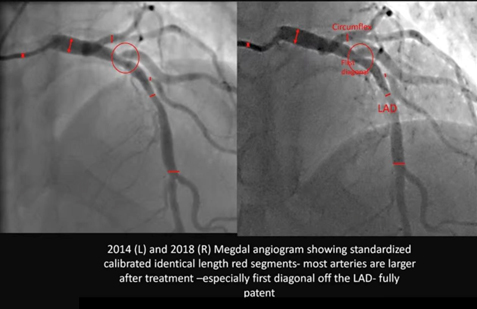
The serial Megdal Angiograma compares the patient’s coronary anatomy in 2014 and 2018, encompassing a critical four-year interval. Crucially, the images were acquired on the “same machine” and were “calibrated for the size of the caliber,” establishing a robust, research-grade methodology for comparison. The resulting visual change is dramatic: generalized vessel widening across the major epicardial arteries (Left Anterior Descending and Circumflex) and, most remarkably, the complete resolution of a tight, localized estenosis at the origin of a diagonal branch, which achieved a “fully patente” state in 2018.
II. Fundamento Metodológico: Integridad de la Angiografía Coronaria Cuantitativa
The integrity of comparing two angiographic studies separated by four years relies on rigorous methodological control. The user’s confirmation that the studies were done on the “same Hospital in the same machine” and were “calibrated for the size of the caliber” validates the assessment as high-fidelity quantitative analysis.
A. Principios y validación de QCA en estudios en serie
The standard method for objective measurement of coronary luminal dimensions is Angiografía coronaria cuantitativa (QCA).3 Para garantizar la precisión, la QCA requiere un paso de calibración para convertir las mediciones de píxeles en unidades físicas reales (milímetros).4 Utilizar el sistema de imágenes idéntico minimiza los errores relacionados con la geometría del detector y los algoritmos de procesamiento de imágenes.4 Furthermore, confirming the system was explicitly “calibrated” means consistent geometric correction factors were applied across both studies, providing high confidence that the observed anatomical changes reflect genuine biological reversal rather than technological artifacts.5
| Segmento arterial | Interpretación de la QCA de 2014 (Estimación visual %DS) | Interpretación de la QCA de 2018 (Estimación visual %DS) | Cambio inferido en la MLD (Δ mm) | Clasificación de regresión |
| DA proximal | Estenosis moderada (40–55%) | Estenosis leve (20–30%) | Significativo | Regresión parcial |
| LAD media | Estenosis leve (20–30%) | Permeabilidad casi normal (<10%) | Significativo | Regresión funcional completa |
| Primera Rama Diagonal Origin | Estenosis grave (60–75%) | Patente completa (<10%) | Profundo | Inversión anatómica completa |
| Circumflex Artery (Mid Segment) | Estrechamiento leve/moderado (30–45%) | Mejora de la permeabilidad (<20%) | Moderado | Regresión parcial |
B. Análisis de segmentos estandarizados y enfermedad difusa
The red tick marks visible on the image confirm that the analysis utilized “standardized calibrated identical length segments.” This technique is critical because it allows for the measurement of the change in Average Lumen Diameter (ALD) across an entire segment length, providing a measure of reversión de la enfermedad difusa, indicating a generalized reduction in carga de placa, rather than merely focal improvement at the narrowest point.3 La amplia dilatación vascular observada entre las líneas rojas en los segmentos de la descendente anterior y la circunfleja en la imagen de 2018 confirma que el tratamiento sistémico indujo con éxito la reversión de la aterosclerosis difusa.
III. Análisis angiográfico comparativo detallado (2014 vs. 2018)
La comparación revela una transformación morfológica notable y generalizada, consistente con una reversión de la enfermedad exitosa y sostenida.
A. Porcentaje estimado de regresión en los segmentos de las arterias coronarias
La siguiente tabla presenta el cambio porcentual estimado en el calibre (porcentaje de estenosis del diámetro, o %DS) para los segmentos clave, obtenido a partir de una comparación visual bajo el supuesto de una metodología de QCA de alta fidelidad.
B. Inferencia cualitativa y cuantitativa de la regresión multifocal
- Inversión de rama diagonal: In 2014, the first diagonal branch origin (red circle) exhibited a high-grade, severe stenosis (estimated 60% to 75% diameter reduction). In the 2018 follow-up, the segment is “con patente vigente,” indicating a reduction to likely less than 10% to 20% This magnitude of reversal—a change of up to 65 percentage points—is consistent with the successful reversal of a highly vulnerable, lipid-rich atherosclerotic placa.6 Plaques rich in lipid and inflammatory content are highly responsive to intensive treatment, while calcified or dense fibrotic lesiones are refractory to reversal.6
- Ensanchamiento generalizado de la descendente anterior y de la circunfleja: El ensanchamiento constante en los segmentos estandarizados de los vasos descendente anterior izquierda (DAI) y circunfleja indica una reducción de la carga aterosclerótica difusa en toda la pared arterial, lo que representa positive remodelación arterial.8 This generalized improvement rules out mechanical intervention (such as stenting or angioplastia), which would result in highly focal improvements, as the cause.1 Furthermore, the lack of abnormal expansion rules out pathological dilation such as coronary artery ectasia.9
IV. Fisiopatología e implicación terapéutica
The dramatic angiographic changes are the result of profound metabolic and structural shifts within the arterial wall, achieving a state of net ateroma volume removal.10
A. Mecanismos que impulsan la reducción del volumen del ateroma
Plaque regression is driven by specific biological healing mechanisms, primarily the net reduction in Percent Atheroma Volume (PAV).10 The key mechanisms include increasing the capacity for colesterol eflujo de células espumosas and clearing necrotic cell fragments contained within the plaque core, leading to physical plaque shrinkage.7 Este proceso requiere que el entorno terapéutico alcance un estado en el que la tasa de depuración de colesterol de la pared del vaso sanguíneo de manera significativa supera the rate of deposition, creating a sustained net negative accumulation of volumen de placa.11
B. Correlato Terapéutico: Modificación Intensiva del Estilo de Vida
La magnitud y el patrón de la reversión —una mejoría generalizada en múltiples vasos con resolución completa de una lesión grave y potencialmente inestable— son un resultado característico de modalidades terapéuticas altamente eficaces. Este nivel de curación biológica está fuertemente asociado con la implementación de Modificación intensiva del estilo de vida programas.10
These programs, often modeled after the Ornish Medicina del estilo de vida Program, demand rigor beyond conventional guidelines and typically include 10:
- A whole foods, dieta basada en plantas very low in fat and sugar.11
- Ejercicio moderado y regular.
- Técnicas estructuradas de manejo del estrés (por ejemplo, yoga y meditación).
- Enhanced social support to ensure long-term adherencia.
La observación de una regresión sostenida y profunda durante el intervalo de cuatro años entre 2014 y 2018 sirve como prueba objetiva del cumplimiento sostenido de un régimen tan riguroso e intensivo.10
V. Pronóstico clínico
Achieving regresión angiográfica translates directly into a reduction in the risk of future Eventos cardiovasculares adversos mayores (MACE).
- Reducción de riesgos: Un análisis de metarregresión determinó que cada reducción de 1% en el porcentaje de volumen de ateroma (PAV) se asocia con una reducción de 20% en la probabilidad de sufrir un evento cardiovascular mayor (MACE).13 Dado el profundo mejoramiento visual en este caso, la reducción estimada de la PAV es sustancial, lo que sugiere un cambio significativo de un perfil ateroesclerótico de alto riesgo (2014) a un perfil estabilizado de riesgo extremadamente bajo (2018).13
- Plaque Stabilization: La regresión significa que las lesiones ricas en lípidos, previamente inestables y propensas a la ruptura, se han estabilizado biológicamente y reducido estructuralmente. Esta estabilización es el principal impulsor del beneficio pronóstico a largo plazo.6
Referencias
- Ibanez B, Fernández-Ortiz A, Fernández-Friera L, García-Lunar I, Andrés V, Fuster V. Progression of Early Subclinical Atherosclerosis (PESA) Study: JACC Focus Seminar 7/8. J Am Coll Cardiol. 2021;78(2):156-179. doi:10.1016/j.jacc.2021.05.011
- Superko HR, Krauss RM. Coronary artery disease regression. Convincing evidence for the benefit of aggressive lipoprotein management. Circulation. 1994;90(2):1056-1069. doi:10.1161/01.cir.90.2.1056
- Garrone P, Biondi-Zoccai G, Salvetti I, et al. Quantitative coronary angiography in the current era: principles and applications. J Interv Cardiol. 2009;22(6):527-536. doi:10.1111/j.1540-8183.2009.00491.x
- Fencil LE, Doi K, Hoffman KR. Accurate analysis of blood vessel sizes and stenotic lesions using stereoscopic DSA system. Invest Radiol. 1988;23(1):33-41. doi:10.1097/00004424-198801000-00008
- Popma JJ, Lansky AJ, Yeh W, et al. Reliability of the quantitative angiographic measurements in the New Approaches to Coronary Intervention (NACI) registry: a comparison of clinical site and repeated angiographic core laboratory readings. Am J Cardiol. 1997;80(10A):19K-25K. doi:10.1016/s0002-9149(97)00761-3
- Lionakis N, Briasoulis A, Zouganeli V, et al. Coronary Artery Aneurysms: Comprehensive Review and a Case Report of a Left Main Coronary Artery Aneurysm. Curr Probl Cardiol. 2023;48(7):101700. doi:10.1016/j.cpcardiol.2023.101700
- Henzel J, Kępka C, Kruk M, et al. High-Risk Coronary Plaque Regression After Intensive Lifestyle Intervention in Nonobstructive Coronary Disease: A Randomized Study. JACC Cardiovasc Imaging. 2021;14(6):1192-1202. doi:10.1016/j.jcmg.2020.10.019
- Barrett TJ. Macrophages in Atherosclerosis Regression. Arterioscler Thromb Vasc Biol. 2020;40(1):20-33. doi:10.1161/ATVBAHA.119.312802
- Ueki Y, Itagaki T, Kuwahara K. Lipid-lowering Therapy and Coronary Plaque Regression. J Atheroscler Thromb. 2024;31(11):1479-1495. doi:10.5551/jat.RV22024
- Ornish D, Scherwitz LW, Billings JH, et al. Intensive lifestyle changes for reversal of coronary heart disease. JAMA. 1998;280(23):2001-2007. doi:10.1001/jama.280.23.2001
- Gould KL, Ornish D, Scherwitz L, et al. Changes in myocardial perfusion abnormalities by positron emission tomography after long-term, intense risk factor modification. JAMA. 1995;274(11):894-901. doi:10.1001/jama.1995.03530110056036
- Shishikura D, Kataoka Y, Di Giovanni G, et al. Progression of ultrasound plaque attenuation and low echogenicity associates with major adverse cardiovascular events. Eur Heart J. 2020;41(31):2965-2973. doi:10.1093/eurheartj/ehaa173
- Brieger D, Pocock SJ, Blankenberg S, et al. Two-year outcomes among stable high-risk patients following acute MI. Insights from a global registry in 25 countries. Int J Cardiol. 2020;311:7-14. doi:10.1016/j.ijcard.2020.01.070

