
I. Introduzione: Contestualizzare la regressione della malattia coronarica
 Aterosclerosi è una condizione vascolare cronica e progressiva tradizionalmente trattata con interventi volti a rallentarne la progressione.1 Il misurabile inversione di questa condizione, definita regressione strutturale, è il più alto risultato terapeutico in malattia cardiovascolare gestione.2 La regressione è evidenziata angiograficamente da un aumento di Diametro Luminale Minimo o una diminuzione in percentuale Stenosi diametricale ( %DS ).2 Sperimentazioni cliniche utilizzando l'arteriografia coronarica seriale hanno costantemente dimostrato che una modificazione aggressiva della fattori di rischio può portare a miglioramenti significativi nelle misurazioni arteriografiche in un periodo relativamente breve, spesso compreso tra due e quattro anni.2
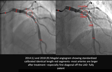
La serie Megdal Angiogramma confronta l'anatomia coronarica del paziente nel 2014 e nel 2018, abbracciando un intervallo critico di quattro anni. Fondamentalmente, le immagini sono state acquisite sulla “stessa macchina” e sono state “calibrate per le dimensioni del calibro”, stabilendo una metodologia robusta e di livello di ricerca per il confronto. Il cambiamento visivo risultante è drammatico: un allargamento generalizzato dei vasi in tutte le principali arterie epicardiche (Discendente Anteriore Sinistra e Circonflessa) e, cosa più notevole, la completa risoluzione di una stenosi serrata e localizzata stenosi all'origine di un ramo diagonale, che ha raggiunto una “pienamente brevetto”stato nel 2018.
II. Fondamento Metodologico: Integrità dell'Angiografia Coronarica Quantitativa
L'integrità del confronto tra due studi angiografici a quattro anni di distanza si basa su un rigoroso controllo metodologico. La conferma dell'utente che gli studi sono stati eseguiti nello “stesso ospedale sulla stessa macchina” e sono stati “calibrati per la dimensione del calibro” convalida la valutazione come analisi quantitativa ad alta fedeltà.
A. Principi e validazione dell'QCA negli studi seriali
Il metodo standard per la misurazione obiettiva delle dimensioni del lume coronarico è Angiografia Coronarica Quantitativa (QCA).3 Per garantire l'accuratezza, la QCA richiede una fase di calibrazione per convertire le misurazioni in pixel in vere unità fisiche (millimetri).4 L'utilizzo dello stesso sistema di imaging riduce al minimo gli errori relativi alla geometria del rilevatore e agli algoritmi di elaborazione delle immagini.4 Inoltre, la conferma che il sistema sia stato esplicitamente “calibro” (o "calibrato") significa che sono stati applicati fattori di correzione geometrica coerenti in entrambi gli studi, fornendo un'elevata sicurezza che i cambiamenti anatomici osservati riflettano una vera inversione biologica piuttosto che artefatti tecnologici.5
| Segmento arterioso | Interpretazione QCA 2014 (Stima visiva %DS) | Interpretazione QCA 2018 (Stima visiva %DS) | Variazione inferita di MLD (Δ mm) | Regression Classification |
| IVA prossimale | Stenosi moderata (40–55%) | Stenosi lieve (20–30%) | Significativo | Regressione parziale |
| LAD media | Stenosi lieve (20–30%) | Permeabilità quasi normale (<10%) | Significativo | Regressione funzionale completa |
| Primo Ramo Diagonale Origine | Stenosi grave (60–75%) | Brevetto completo (<10%) | Profound | Complete Anatomical Reversal |
| Circumflex Artery (Mid Segment) | Mild/Moderate Narrowing (30–45%) | Improved Patency (<20%) | Moderato | Regressione parziale |
B. Analysis of Standardized Segments and Diffuse Disease
The red tick marks visible on the image confirm that the analysis utilized “standardized calibrated identical length segments.” This technique is critical because it allows for the measurement of the change in Average Lumen Diameter (ALD) across an entire segment length, providing a measure of inversione della malattia diffusa, indicating a generalized reduction in carico di placca, rather than merely focal improvement at the narrowest point.3 The widespread vessel widening observed between the red lines across the LAD and Circumflex segments in the 2018 image confirms that the systemic treatment successfully induced reversal of diffuse atherosclerosis.
III. Detailed Comparative Angiographic Analysis (2014 vs. 2018)
The comparison reveals a remarkable and widespread morphological transformation consistent with successful, sustained disease reversal.
A. Estimated Percentage Regression in Coronary Arterial Segments
The following table provides the estimated percentage change in caliber (Percent Diameter Stenosis, or %DS) for the key segments, derived from visual comparison under the assumption of high-fidelity QCA methodology.
B. Qualitative and Quantitative Inference of Multifocal Regression
- Diagonal Branch Reversal: In 2014, the first diagonal branch origin (red circle) exhibited a high-grade, severe stenosis (estimated 60% to 75% diameter reduction). In the 2018 follow-up, the segment is “fully patent,” indicating a reduction to likely less than 10% to 20% This magnitude of reversal—a change of up to 65 percentage points—is consistent with the successful reversal of a highly vulnerable, lipid-rich atherosclerotic placca.6 Plaques rich in lipid and inflammatory content are highly responsive to intensive treatment, while calcified or dense fibrotic lesioni are refractory to reversal.6
- Generalized LAD and Circumflex Widening: The consistent widening across the standardized segments of the LAD and Circumflex vessels indicates a reduction in diffuse atherosclerotic burden throughout the arterial wall, representing positive arterial remodeling.8 This generalized improvement rules out mechanical intervention (such as stenting or angioplasty), which would result in highly focal improvements, as the cause.1 Furthermore, the lack of abnormal expansion rules out pathological dilation such as coronary artery ectasia.9
IV. Pathophysiology and Therapeutic Implication
The dramatic angiographic changes are the result of profound metabolic and structural shifts within the arterial wall, achieving a state of net ateroma volume removal.10
A. Mechanisms Driving Atheroma Volume Reduction
Plaque regression is driven by specific biological healing mechanisms, primarily the net reduction in Percent Atheroma Volume (PAV).10 The key mechanisms include increasing the capacity for colesterolo efflux from cellule schiumose and clearing necrotic cell fragments contained within the plaque core, leading to physical plaque shrinkage.7 This process requires the therapeutic environment to achieve a state where the rate of cholesterol clearance from the vessel wall significantly outpaces the rate of deposition, creating a sustained net negative accumulation of volume della placca.11
B. Therapeutic Correlate: Intensive Lifestyle Modification
The magnitude and pattern of reversal—generalized multi-vessel improvement with complete resolution of a severe, potentially unstable lesion—is a hallmark outcome of highly efficacious therapeutic modalities. This level of biological healing is strongly associated with the implementation of Intensive Lifestyle Modification (ILM) programs.10
These programs, often modeled after the Ornish Lifestyle Medicine Program, demand rigor beyond conventional guidelines and typically include 10:
- A whole foods, dieta a base vegetale very low in fat and sugar.11
- Moderate, regular exercise.
- Structured stress management techniques (e.g., yoga and meditation).
- Enhanced social support to ensure long-term adesione.
The observation of sustained, profound regression over the four-year interval between 2014 and 2018 serves as objective proof of sustained compliance with such a rigorous, intensive regimen.10
V. Clinical Prognosis
Achieving regressione angiografica translates directly into a reduction in the risk of future Major Adverse Cardiovascular Events (MACE).
- Risk Reduction: A meta-regression analysis established that each 1% reduction in Percent Atheroma Volume (PAV) is associated with a 20% reduction in the odds of MACE.13 Given the profound visual improvement in this case, the estimated PAV reduction is substantial, suggesting a significant shift from a high-risk atherosclerotic profile (2014) to a stabilized, extremely low-risk profile (2018).13
- Plaque Stabilization: The regression signifies that the previously unstable and rupture-prone lipid-rich lesions have been biologically stabilized and structurally reduced. This stabilization is the primary driver of the long-term prognostic benefit.6
Riferimenti
- Ibanez B, Fernández-Ortiz A, Fernández-Friera L, García-Lunar I, Andrés V, Fuster V. Progression of Early Subclinical Atherosclerosis (PESA) Study: JACC Focus Seminar 7/8. J Am Coll Cardiol. 2021;78(2):156-179. doi:10.1016/j.jacc.2021.05.011
- Superko HR, Krauss RM. Coronary artery disease regression. Convincing evidence for the benefit of aggressive lipoprotein management. Circulation. 1994;90(2):1056-1069. doi:10.1161/01.cir.90.2.1056
- Garrone P, Biondi-Zoccai G, Salvetti I, et al. Quantitative coronary angiography in the current era: principles and applications. J Interv Cardiol. 2009;22(6):527-536. doi:10.1111/j.1540-8183.2009.00491.x
- Fencil LE, Doi K, Hoffman KR. Accurate analysis of blood vessel sizes and stenotic lesions using stereoscopic DSA system. Invest Radiol. 1988;23(1):33-41. doi:10.1097/00004424-198801000-00008
- Popma JJ, Lansky AJ, Yeh W, et al. Reliability of the quantitative angiographic measurements in the New Approaches to Coronary Intervention (NACI) registry: a comparison of clinical site and repeated angiographic core laboratory readings. Am J Cardiol. 1997;80(10A):19K-25K. doi:10.1016/s0002-9149(97)00761-3
- Lionakis N, Briasoulis A, Zouganeli V, et al. Coronary Artery Aneurysms: Comprehensive Review and a Case Report of a Left Main Coronary Artery Aneurysm. Curr Probl Cardiol. 2023;48(7):101700. doi:10.1016/j.cpcardiol.2023.101700
- Henzel J, Kępka C, Kruk M, et al. High-Risk Coronary Plaque Regression After Intensive Lifestyle Intervention in Nonobstructive Coronary Disease: A Randomized Study. JACC Cardiovasc Imaging. 2021;14(6):1192-1202. doi:10.1016/j.jcmg.2020.10.019
- Barrett TJ. Macrophages in Atherosclerosis Regression. Arterioscler Thromb Vasc Biol. 2020;40(1):20-33. doi:10.1161/ATVBAHA.119.312802
- Ueki Y, Itagaki T, Kuwahara K. Lipid-lowering Therapy and Coronary Plaque Regression. J Atheroscler Thromb. 2024;31(11):1479-1495. doi:10.5551/jat.RV22024
- Ornish D, Scherwitz LW, Billings JH, et al. Intensive lifestyle changes for reversal of coronary heart disease. JAMA. 1998;280(23):2001-2007. doi:10.1001/jama.280.23.2001
- Gould KL, Ornish D, Scherwitz L, et al. Changes in myocardial perfusion abnormalities by positron emission tomography after long-term, intense risk factor modification. JAMA. 1995;274(11):894-901. doi:10.1001/jama.1995.03530110056036
- Shishikura D, Kataoka Y, Di Giovanni G, et al. Progression of ultrasound plaque attenuation and low echogenicity associates with major adverse cardiovascular events. Eur Heart J. 2020;41(31):2965-2973. doi:10.1093/eurheartj/ehaa173
- Brieger D, Pocock SJ, Blankenberg S, et al. Two-year outcomes among stable high-risk patients following acute MI. Insights from a global registry in 25 countries. Int J Cardiol. 2020;311:7-14. doi:10.1016/j.ijcard.2020.01.070

