آلية القلب والأوعية الدموية لتراجع الأداء الطولي لدى الرياضيين المتقدمين في السن:
A مراجعة سردية للشيخوخة،, مرض الشريان التاجي, احشاء عضلة القلب الصامت نقص الدم, ، وغير معترف به احتشاء عضلة القلب
ملخص
تدمج هذه المراجعة السردية أدلة مختارة ومحكّمة من الزملاء حول الآليات القلبية الوعائية والفسيولوجية الكامنة وراء التراجع في الأداء على المدى الطويل لدى الرياضيين المخضرمين الذين تتراوح أعمارهم بين 50 و80 عاماً. وتشمل المواضيع التي يتم تناولها (1) المسار الطبيعي للقدرة البدنية من دراسة الأتراب السكانية السويدية للنشاط البدني واللياقة البدنية (SPAF)[1]; (2) التناقض بين الرياضي المحترف والخامل بدنياً V̇O2أقصى انخفاض2][3][4]; (3) الآليات القلبية الوعائية المركزية والمحيطية لـ V̇O2التآكل الأقصى؛ (4) مفارقة قصور الشريان التاجي تحت السريري تصلب الشرايين لدى الرياضيين الذين يمارسون رياضات التحمل طوال حياتهم؛ (5) نقص تروية عضلة القلب الصامت واحتشاء عضلة القلب غير المعترف به — بما في ذلك علم الوبائيات, ، والآليات، والمآل، والنهج التشخيصي؛ (6) الشريان التاجي اختلال وظيفة الأوعية الدقيقة كمساهم محتمل في عدم تحمل التمرين لدى رياضيين كبار السن مختارين؛ و (7) اعتبارات فحص أمراض القلب الرياضية المعاصرة المرتكزة على إرشادات الجمعية الأوروبية لأمراض القلب لعام 2020 بشأن أمراض القلب الرياضية. هذا التركيب تفسيري وليس شاملاً؛ وتتم وصف استراتيجية البحث المحددة ونطاقه في قسم تحديد الأدبيات.
1. مقدمة
عملية الشيخوخة لدى البشر مرتبطة حتماً بتراجع تدريجي في القدرة الفسيولوجية، ومع ذلك فإن معدل ومسار هذا التراجع يتأثران بشكل كبير بالنشاط البدني الاعتيادي. بين الأفراد الذين يحافظون على مستويات عالية من التدريب على التحمل حتى عقدهم السادس والسابع والثامن - والمعروفين عموماً بالرياضيين المخضرمين - فإن التراجع في الحد الأقصى للقدرة الهوائية (استهلاك الأكسجين الأقصى V̇O2الحد الأقصى) وأداء التحمل يوفر نموذجاً بيولوجياً فريداً لدراسة حدود الشيخوخة البشرية[2]. في حين أن الرياضيين الأساتذة يظهرون باستمرار تفوقاً اللياقة القلبية التنفسية (اللياقة القلبية التنفسية)، مقارنة بنظرائهم الخاملين، ليسوا في مأمن من العمليات البيولوجية الأساسية التي تآكل وظيفة القلب والأوعية الدموية بمرور الوقت3][4].
الانحدار في الأداء على المدى الطويل بين سن الخمسين والثمانين يتحكم فيه تفاعل معقد بين القيود القلبية المركزية، والتدهور الوعائي والميتوكوندري المحيطي، والظهور المتناقض لأمراض القلب والأوعية الدموية تحت السريرية. وتشمل هذه الشرايين التاجية شريان المرض لدى رياضيي التحمل مدى الحياة13][14][15] وفي مجتمع المسنين الأوسع،, قصور عضلة القلب الصامت, احشاء عضلة القلب غير المُشخّص, ، و اختلال وظائف الأوعية الدموية الدقيقة التاجية[21][22]. لفهم هذه الآليات في سياقها، من المفيد أولاً وضع خط أساس على مستوى السكان من مجموعة SPAF الطولية [1ثم مقارنة خط الأساس هذا بالتكيفات الفسيولوجية المحددة - والمخاطر المرضية - التي لوحظت لدى الرياضيين الذين يمارسون رياضات التحمل طوال حياتهم.
2. تحديد الأدبيات
هذه المقالة عبارة عن مراجعة سردية. تم تحديد الدراسات الأدبية بشكل أساسي من خلال عمليات البحث في بوبمد وإمبيز (1990–2026) ومن خلال مراجعة المراجع الخلفية للدراسات الأولية ذات الصلة، والتحليلات التلوية، ووثائق إرشادات الجمعيات الكبرى التي تتناول الرياضيين المخضرمين، والانهيار المرتبط بالعمر في السعة الهوائية، وتصلب الشرايين التاجية لدى رياضي التحمل، ونقص تروية عضلة القلب الصامت، واحتشاء عضلة القلب غير المُشخّص، وخلل الأوعية الدقيقة التاجية، وفحص طب القلب الرياضي. تم إعطاء الأولوية لدراسات الأبحاث الأصلية، والتجارب العشوائية،, المراجعات المنهجية, ، والتحليلات التلوية، وبيانات الإرشادات الرسمية. ونظراً لكون هذه المراجعة المنهجية غير مدعومة ببروتوكول، فإن اختيار الدراسات كان نوعياً، وينبغي تفسير التوليف على أنه تفسيري وليس استقصائياً. لم يتم إجراء تجميع تحليلي تلوي رسمي؛ وتم نقل القيم الكمية من المصادر الأولية المذكورة دون إعادة تحليل.
3. خط الأساس لعامة السكان: رؤى مستمدة من دراسة وسترستوهل SPAF
تقدم دراسة SPAF الطولية التي استمرت 47 عاماً والتي أبلغ عنها ويسترهول وزملاؤه واحدة من أكثر التقييمات شمولاً لكيفية تغير القدرة البدنية من مرحلة المراهقة وحتى الشيخوخة المبكرة في مجموعة سكانية عامة وممثلة1تتبعت الدراسة، التي شملت 427 فرداً ولدوا في عام 1958، تقييمات موضوعية متكررة للقدرة الهوائية، والتحمل العضلي، والقوة الانفجارية من سن 16 إلى سن 63.1تُعتبر مجموعة البيانات هذه مفيدة لأنها تحدد المعدل الطبيعي للتراجع في مجموعة سكانية لا يخضع فيها النشاط البدني للمعايير، مما يسلط الضوء على الطبيعة المتسارعة للشيخوخة البيولوجية بغض النظر عن حالة الرياضة النخبوية.
عموماً، يتم الوصول إلى ذروة الأداء البدني بين سن 26 و 36 عاماً لكل من القدرة الهوائية المطلقة والتحمل العضلي1على وجه التحديد، بلغ الرجال ذروة قدرتهم الهوائية المطلقة في سن 35 (3.26 لتر·دقيقة-١)، بينما بلغت النساء ذروتهن عند سن 36 (2.61 لتر·دقيقة-١)[1]. وبعد بلوغ هذه المستويات القصوى، بدأ الانخفاض بمعدل يتراوح بين 0.3 و0.61 تي بي 9 تي سنويًّا تقريبًا، ثم تسارع ليصل إلى ما بين 2.0 و2.51 تي بي 9 تي سنويًّا تقريبًا مع اقتراب المشاركين من سن الستين[1]. بحلول سن 63 عامًا، بلغ الانخفاض التراكمي في السعة الهوائية المطلقة مقارنةً بالذروة حوالي 33% لدى الرجال و30% لدى النساء[1].
الجدول 1. مسارات القدرة البدنية من دراسة الأتراب الطولية لـ SPAF لمدة 47 عاماً.
| معامل | الرجال (ذروة العمر 35–36) | النساء (ذروة العمر 36) | إجمالي الانخفاض بمقدار 63 |
| القدرة الهوائية المطلقة (لتر·دقيقة⁻¹) | 3.26 | 2.61 | 30–33% |
| القدرة الهوائية النسبية (مل·كجم⁻¹·دقيقة⁻¹) | 42.2 في سن 26 | 39.7 في عمر 31 عاماً | 37–40% |
| الوثب العمودي (سم) | ٤٥.٥ في عمر ٢٧ | 33.0 في سن 19 | 41–48% |
| التحمل العضلي (ضغط العضلة الصدرية، التكرارات) | 52.7 في عمر 36 | ٣٩.٧ في عمر ٣٤ عامًا | 32–35% |
ويسترستوهل إم، وآخرون. مجلة الدُّعف ضمور العضلات عضلة. 2025[1]. القيم هي متوسطات الأتراب في العمر المشار إليه. SPAF = الأتراب السكاني السويدي للنشاط البدني واللياقة البدنية.
أكدت بيانات دراسة SPAF أيضاً دور عوامل نمط الحياة في تعديل هذه المسارات. فقد ارتبط ارتفاع النشاط البدني في أوقات الفراغ في سن 16 عاماً ونمط الحياة النشط خلال مرحلة البلوغ بأداء متفوق عبر جميع المقاييس طوال الحياة.1.[ ارتباط التحصيل التعليمي الأعلى بارتباط وثيق بسعة هوائية مطلقة وتحمل عضلي أعلى، وربما يُعزى ذلك إلى السلوكيات الباحثة عن الصحة. ومع ذلك، فحتى مع وجود هذه العوامل الوقائية، كان تسارع الانخفاض بعد سن الأربعين نتيجة ثابتة، مما يعزز مفهوم أن الشيخوخة البيولوجية تفرض قيداً فسيولوجياً تدريجياً على القدرة الوظيفية يبدأ في العمل قبل وقت طويل من أن يصبح ذا أهمية سريرية.
4. التراجع المقارن: الرياضيون الأكبر سنّاً مقابل عامة السكان
عند مقارنة خط الأساس لـ SPAF مع البيانات الطولية للرياضيين المخضرمين، يظهر تباين واضح في ارتفاع البداية وميل استهلاك الأكسجين (V̇O₂)2أقصى انخفاض. يمثل الرياضيون المحترفون في التحمل مجموعة أظهرت الحد الأقصى من القدرة التكيفية وقد تحمل V̇O2الحد الأقصى في سن الستين الذي يتجاوز المتوسط لشخص يبلغ من العمر 40 عاماً ويتبع نمط حياة خاملاً2]. وعلى الرغم من هذه الميزة، فإن الانخفاض الحتمي في معدل استهلاك الأكسجين (V̇O2يظل الحد الأقصى لاستهلاك الأكسجين الآلية الفيزيولوجية الأساسية المرتبطة بانخفاض الأداء التحملي لدى الرياضيين المتقدمين في السن3][4].
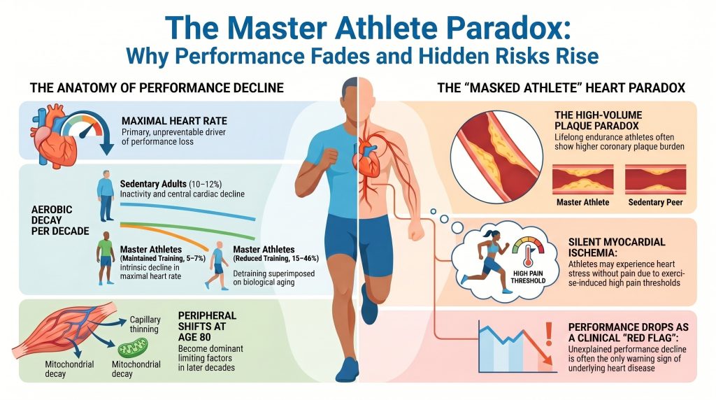
تشير التقييمات طولية المدى إلى أن الرياضيين المخضرمين الذين يحافظون على تدريبات عالية الكثافة والحجم يقللون من معدل استهلاك الأكسجين (V̇O2انخفاض أقصى إلى ما يقارب 5–71 تي بي 9 تي لكل عقد، مقارنة بـ 10–121 تي بي 9 تي لكل عقد لدى الأفراد الذين يتبعون نمط حياة خامل من نفس الفئة العمرية[2][3][4مع ذلك، فإن هذا الانخفاض الملطف بالتدريب هش؛ وتُشير تحليلات الانحدار إلى أن نسبة كبيرة من التباين في استهلاك الأكسجين2يعزى الانخفاض الأقصى في أداء الرياضيين الكبار بشكل مباشر إلى انخفاض حجم التدريب وشدته، حيث لوحظت أكبر معدلات الانخفاض (15–46% لكل عقد) لدى الرياضيين الذين خفضوا حجم تدريبهم بشكل كبير[3][4].
الجدول 2. المعدلات العقدية لاستهلاك الأكسجين الأقصى ($\dot{V}O_2$ max)2أقصى انخفاض حسب المجموعة.
| مجموعة | انخفاض عقدي | السائق الرئيسي |
| البالغون الخاملون | 10–12% | ضمور العضلات، وقلة الحركة، والقصور القلبي المركزي |
| الرياضيون الأساتذة (ذوو التدريب المستمر) | 5–7% | الإنحدار الذاتي في الحد الأقصى معدل ضربات القلب |
| رياضيو الماسترز (بتدريب مخفض) | 15–46% | فقدان التدريب متراكماً على الشيخوخة |
5. الآليات القلبية الوعائية لتآكل السعة الهوائية
تآكل الحجم الاستهلاكي للأكسجين2يتم وصف الحد الأقصى رياضياً بواسطة مبدأ فيك: V̇O2الحد الأقصى = Q̇ × (a–v)O2 الفرق، حيث Q̇ هي ناتج القلب و (a–v)O2 هو الفرق في محتوى الأكسجين الشرياني الوريدي. وفي الرياضيين المخضرمين، تبلغ المساهمة النسبية لعوامل القلب المركزية (معدل ضربات القلب و حجم القذفمقابل العوامل المحيطية (استخلاص الأكسجين) تتغير مع التقدم في السن8][9].
5.1 الآليات المركزية: مضخة الشيخوخة
أكثر محرك ثابت وغير قابل للتعديل إلى حد كبير لاستهلاك الأكسجين الأقصى2الانخفاض الأقصى هو التراجع المرتبط بالتقدم في العمر في معدل ضربات القلب الأقصىالحد الأقصى). واسع الانتشار معادلة تاناكا — الموارد البشريةالحد الأقصى ≈ 208 − 0.7 × العمر — يشير إلى انخفاض متوسط يقارب 0.7 نبضة في الدقيقة-١ سنوياً7]. ويرجع هذا أساساً إلى تغيرات في الفيزيولوجيا الكهربية الذاتية لـ العقدة الجيبية الأذينية وبالتخفيض استجابة بيتا الأدرينالية مع التقدم في العمر. وفي حين أن التدريب على التحمل يمكن أن يخفض معدل ضربات القلب أثناء الراحة ويعزز التوتر نظير الودي، إلا أن قدرته محدودة على الحفاظ على الحد الأقصى لمعدل ضربات القلب أثناء بذل أقصى جهدي.
سكتة دماغية يقدم الحجم (SV) قصة أكثر دقة. لدى الأفراد الخاملين، ينخفض الحجم (SV) بسبب زيادة صلابة عضلة القلب، وانخفاض مطاوعة البطين، واختلال الامتلاء انبساطي10في الرياضيين المخضرمين، يؤدي التدريب على التحمل المزمن إلى إعادة تشكيل البطين اللامركزي الذي يتميز بأبعاد حجرة أكبر ومرونة عضلة القلب المحسنة.10][11يتيح هذا التكيف للرياضيين الحفاظ على حجم ضربة أعلى من خلال آلية فرانك-ستارلينغ. مع ذلك، يمكن للرياضيين المتقدمين في السن أن يشهدوا انخفاضاً في حجم الضربة (SV) عند ذروة التمرين بسبب التقصير غير الطبيعي لوقت الامتلاء الانبساطي عند معدلات ضربات القلب العالية جداً، حيث تكون الفترة الانبساطية أقصر من أن تسمح بالامتلاء البطيني الكامل.
5.2 الآليات الطرفية: استخراج الأكسجين والاضطراب الميتوكوندري
بينما تسيطر العوامل المركزية على المراحل المبكرة من التدهور، تصبح العوامل الطرفية مقيدة بشكل متزايد مع اقتراب الرياضيين من سن 809]. وتعتبر ثلاث عمليات محيطية هي الأكثر تأثيرًا:
- شريان وريدي أو2 الحد الأقصى (a–v)O2 يتراجع الفارق بشكل طفيف مع التقدم في العمر لدى الرياضيين المتميزين، مدفوعاً إلى حد كبير بـ تخلخل الشعيرات الدموية - ترقق شبكات الشعيرات الدموية المغذية للعضلة الفردية ألياف — مما يزيد من مسافة الانتشار للأكسجين9].
- وظيفة الميتوكوندريا. يرتبط التقدم في العمر بانخفاض في الكثافة الميتوكوندريا وفي نشاط الإنزيمات التأكسدية الرئيسية مثل سيترات سينثاز وسكسينات ديهيدروجيناز2الحفاظ على التدريب عالي الشدة لدى الرياضيين المتقدمين في السن يحافظ على جودة الميتوكوندريا، إلا أن تقليل حجم التدريب ينتج عنه انخفاض سريع وشبه خطي في القدرة التأكسدية.
- ضمور انتقائي لألياف النوع الثاني (سريعة الانقباض) وفقدان إجمالي لكتلة العضلات يقللان من الطلب الأيضي ومخرجات التمارين، وهو تأثير موثق في مجموعة SPAF وفي أماكن أخرى[1].
تصلب الشرايين التاجية لدى الرياضيين النخبة: مفارقة التكلس
إحدى أكثر النتائج تحدياً في أمراض قلب الرياضيين الحديثة هي المفارقة الظاهرة المتمثلة في الارتفاع كالسيوم الشريان التاجي النتائج لدى الرياضيين الذين يمارسون رياضات التحمل طوال حياتهم. أظهرت البيانات المستعرضة، بما في ذلك دراسة (Master@Heart)، أن رياضيي التحمل على مدار الحياة (الغالبية العظمى منهم من الذكور) غالباً ما يحملون معدلاً انتشار أعلى للشريان التاجي لوحات وتهق درجات أعلى في الكالسيوم التاجي (CAC) مقارنة بالأقران الخاملين ذوي الملف الشخصي المماثل للخطر13][14][15][16][17].
شكل اللويحات وتوزيعها
الـ دراسة ماستر@هارت بواسطة دي بوسشر وزمائه، ضم 191 رياضياً طوال حياتهم، و191 رياضياً متأخري البداية، و176 شخصاً سليماً غير رياضي (جميعهم من الذكور) وأظهروا أن الرياضيين طوال حياتهم لديهم أعلى نسبة شرايين تاجية عبء اللويحات[15]. ولم تقتصر الأعباء على لويحات متكلسة — تُعتبر عموماً مستقرة — ولكنها أيضاً لويحات غير متكلسة ومختلطة في القطع الدانية للشرايين التاجية[15].
الجدول 3. نتائج اللويحات التاجية من مجموعة Master@Heart.
| مجموعة | لويحة إكليلية واحدة على الأقل (نسبة الرجحان) | ≥1 لويحة قريبة (أو) | ≥50% لومينال تضيق |
| غير الرياضيين (مرجع) | 1.00 | 1.00 | مرجع |
| رياضيو الظهور المتأخر | متوسط | متوسط | منخفض |
| رياضيو التحمل مدى الحياة | 1.86 | 1.96 | غير شایع، ولکنه مرتفع مقارنةً بالمجموعات الضابطة |
De Bosscher R, et al. Eur Heart J. 2023[15]. OR = نسبة الاحتمالات مقارنةً بمجموعة مرجعية من غير الرياضيين؛ القيم هي تقديرات معدلة. كان جميع المشاركين من الذكور؛ ومتوسط العمر ≈ 60 سنة. كان التضيق الانسدادي (≥50%) نادرًا من الناحية المطلقة في جميع المجموعات؛ وينبغي التحقق من معدل الانتشار الدقيق المُبلغ عنه في مجموعة الرياضيين مدى الحياة بمقارنته بالمنشور الأصلي قبل التنضيد النهائي.
في دراسة Master@Heart، كان الرياضيون مدى الحياة أكثر عرضة بشكل غير متناسب لـ لويحات غير متكلسة في القطع القريبة مقارنة وغير الرياضيين [15الموقع القريب ذو صلة سريرية لأن هذه اللويحات تتحكم في تدفق الدم إلى مناطق عضلة القلب الكبيرة. وفي حين أن لياقة القلب والأوعية التنفسية العالية ترتبط بتكوين اللويحات المستقرة في بعض التحليلات، إلا أن الوجود المطلق لهذه آفات يقترح أن التدريب التحملي عالي الحجم قد يتفاعل مع التقليدي عوامل الخطر لتسريع تطور المرض تحت السريري الموجود مسبقاً16][17].
6.2 العوامل الميكانيكية المقترحة
لا يزال تطور مرض الشريان التاجي لدى الرياضيين المتقدمين في السن غير مفسر بشكل كامل. وتثير الأدبيات الرصدية الحالية عدة الآليات المقترحة, التي يجب تفسيرها على أنها فرضيات محتملة وليست مسارات سببية مثبتة:
- إجهاد القص الديناميكي الدموي. يؤدي التمرين الشاق إلى تدفق تاجي مفرط الديناميكية. وفي مواقع التدفق المضطرب أو المتدفق، قد تساهم القوى الميكانيكية في اختلال وظيفة بطانة الأوعية الدموية[13][17].
- الإجهاد التأكسدي. قد تؤدي المتطلبات الأيضية الشديدة للتدريب فائق التحمل إلى توليد مؤقت أنواع الأكسجين التفاعلية أكثر من مضاد للأكسدة القدرة، مع وجود تأثيرات محتملة لاحقة على الوظيفة البطانية و بروتين دهني احتفاظ17].
- الناجم عن التمرين ارتفاع ضغط الدم. في مجموعة حديثة من الرياضيين الذكور المخضرمين في التحمل، كان ارتفاع ضغط الدم الخفي في الراحة والناتج عن التمرين منتشراً والمرتبط بعبء أعلى لويحات الشريان التاجي18]. تمرين مبالغ فيه ضغط الدم الانقباضي يُعترف بالاستجابات بشكل متزايد باعتبارها مظهراً سريرياً قابلاً للتدخل.
- التفاعلات مع عوامل الخطر التقليدية. حتى لدى الرياضيين، عوامل الخطر التقليدية - الكوليسترول الضار (LDL)،, أبروليپوبروتين ب, بروتين دهني (أ), ، ارتفاع ضغط الدم،, التاريخ العائلي تظل المحركات الأساسية لتصلب الشرايين49][50].
الإِقْرَاضُ القَلْبِيُّ الصَّامِتُ وَاحْتِسَاءُ عَضَلَةِ القَلْبِ غَيْرُ المُعْتَرَفِ بِهِ
7.1 التعريفات والتمييز الرئيسي
نقص تروية عضلة القلب الصامت يشير إلى دليل موضوعي على نقص التروية القابل للحث العابر — عادةً ما يكون هناك انخفاض في قطعة ST على تخطيط القلب المتنقل أو أثناء التمرين، أو تشوه قابل للحث في التروية على تصوير الإجهاد — وذلك في غياب أعراض الذبحة الصدرية تماماً21][22]. احشاء عضلة القلب غير المُشخص يشير إلى احتشاء مكتمل لم يتم تشخيصه سريرياً وقت حدوثه28][29][30][31]. تعزيز الجادولينium المتأخر (إل جي) على التصوير بالرنين المغناطيسي للقلب يتميز سابق إصابة عضلة القلب - ندبة، أو تليف، أو احتشاء مكتمل - وبالتالي فهي طريقة التصوير المعيارية المرجعية لاحتشاء عضلة القلب الصامت بدلاً من كونها اختباراً أساسياً لنقص الإسكيميا النشط30][36يتم الحفاظ على هذا التمييز طوال ما تبقى من هذا القسم.
٧.٢ وبائيات نقص الدم الموضعي الصامت واحتشاء العضلة القلبية غير المعروف
تشكل احتشاءات العضل القلبية الصامتة أو غير المعترف بها نسبة كبيرة من جميع احتشاءات العضل القلبية في الأتراب الوبائية. الأصلية دراسة قلب فرامينغهام أفادت الدراسة أن ما يقارب ربع حالات النوبات القلبية لم يتم التعرف عليها عند اكتشافها لأول مرة خلال الفحص الروتيني الذي يُجرى كل سنتين[28]. دراسة مخاطر تصلب الشرايين في المجتمعات (اريك) أظهرت الدراسة أن نسبة حالات احتشاء عضلة القلب الصامتة التي لم يتم الكشف عنها إلا عن طريق تخطيط القلب بلغت حوالي 22%، مع وجود اختلافات كبيرة في معدل الكشف حسب الجنس والعرق[29]. في «دراسة صحة القلب والأوعية الدموية» التي شملت كبار السن المقيمين في المجتمع المحلي، ارتفعت نسبة انتشار «UMI» مع تقدم العمر[31].
ظهور التصوير بالرنين المغناطيسي للقلب وقد أدى استخدام تقنية LGE إلى رفع تقديرات معدل انتشار UMI بشكل كبير. وقد وجدت دراسة ICELAND-MI التي أجراها شيلبرت وزملاؤه أن حالات UMI المكتشفة بواسطة التصوير بالرنين المغناطيسي القلبي باستخدام LGE كانت أكثر شيوعًا بمقدار الضعف مقارنةً بحالات UMI المكتشفة بواسطة تخطيط القلب الكهربائي (ECG) في مجموعة مجتمعية من كبار السن، وأن حالات UMI المكتشفة بواسطة تخطيط القلب الكهربائي لم تمثل سوى جزء ضئيل من إجمالي الحالات المكتشفة بواسطة التصوير بالرنين المغناطيسي القلبي[30]. في ميسا المجموعة، المكتشفة بواسطة التصوير بالرنين المغناطيسي القلبي (CMR) ندبة عضلة القلب تم الكشف عن الإصابة لدى ما يقارب 7.9% من البالغين غير المصابين بأعراض، الذين تتراوح أعمارهم بين 45 و84 عامًا، والذين ليس لديهم تاريخ سريري للإصابة بنوبة قلبية[33].
وبتطبيق ذلك على علم الأوبئة في الولايات المتحدة، فمن بين ما يقرب من 805,000 حالة احتشاء عضلة القلب التي تحدث سنويًّا في الولايات المتحدة، تشير المراقبة المجتمعية القائمة على تخطيط القلب إلى أن حوالي 170,000 (≈21%) منها تكون صامتة سريريًّا أو غير مُدرَكة عند حدوثها — وهو رقم يتوافق مع النسب المسجلة في دراستي ARIC و Framingham[28][29][31]. نظرًا لأن الكشف القائم على التصوير بالرنين المغناطيسي القلبي (CMR) قد حدد عددًا من حالات عدم تحديد الموقع (UMI) يزيد بأكثر من الضعف عن عدد الحالات التي حددها تخطيط القلب الكهربائي (ECG) في مجموعات السكان المسنين في المجتمع المحلي[30]، فإن التقدير المستمد من تخطيط القلب (ECG) البالغ 170,000 يُفسر على أفضل وجه على أنه الحد الأدنى حول العبء السنوي الحقيقي لحالات احتشاء عضلة القلب غير المشخصة سريريًّا في الولايات المتحدة.
7.3 لماذا تكون بعض حالات نقص التروية القلبية واحتشاء عضلة القلب صامتة: الآليات المقترحة
يعود عدم الشعور بالألم أثناء نقص التروية أو النوبة القلبية إلى عوامل متعددة. وقد تم اقتراح ثماني آليات عامة في الأدبيات العلمية:
- الاعتلال العصبي اللاإرادي. يمكن أن تتضرر الألياف الوديّة والمبهمية الواردة التي تنقل الإشارات المؤلمة القلبية بسبب مرض السكري داء السكري، أو الاعتلال العصبي اللاإرادي المرتبط بالتقدم في العمر، أو الخضوع لجراحة قلبية سابقة، مما يخفف من الإحساس بالألم الإقفاري أو يزيله تمامًا[21].
- عتبة ألم أعلى. تختلف عتبات الألم الجسدي الأساسية بشكل كبير من شخص لآخر، وقد ثبت أن مجموعة فرعية من المرضى الذين يعانون من نقص التروية الموثق لديهم عتبات مرتفعة بشكل واضح تجاه محفزات الألم المختبرية[34].
- الأفيونيات الذاتية و أنظمة الإندوكانابينويد. ترتفع مستويات الـβ-إندورفين والأفيونيات ذات الصلة في الدم لدى بعض المرضى المصابين بـ قصور إفقاري صامت وقد تم ربطها بالتنظيم المركزي لإدراك الألم القلبي[34].
- تخفيف الألم الناجم عن ممارسة الرياضة (EIH). يؤثر التدريب المنتظم على القدرة على التحمل على نظامي الأفيونيات الداخلية والكانابينويدات الداخلية، ويرتبط باستمرار بارتفاع عتبات الألم وقدرة الجسم على تحمله في الاختبارات المعملية[25][26]. فيما يتعلق بالرياضيين من فئة «ماستر»، يُرجح أن EIH يسهم في عدم ظهور أعراض تحذيرية أثناء نقص التروية الناجم عن الجهد.
- حجم نوبة قلبية صغيرة ومدى انتشار محدود عبر جدار القلب. قد لا تولد النوبات القلبية الصغيرة تحت الشغافية أو غير العابرة للجدار إشارات واردة كافية لتجاوز العتبة اللازمة للإدراك الواعي[32].
- التوزيع التشريحي. تكون حالات نقص التروية في الجدار السفلي والجدار الخلفي صامتة في أغلب الأحيان أكثر من حالات الجدار الأمامي، وربما يرجع ذلك إلى الاختلافات في كثافة التوصيل العصبي الوارد[21].
- تداول الأوراق المالية. يساعد ممارسة التمارين الرياضية بانتظام على تنمية الأوعية التفرعية التاجية، والتي يمكنها الحفاظ على تدفق الدم في الأجزاء البعيدة عن منطقة التضيق أثناء الراحة وأثناء بذل مجهود خفيف إلى متوسط، مما يمنع حدوث نقص التروية والألم إلى أن يتجاوز الطلب الأقصى قدرة الأوعية التفرعية[27].
- التكييف المسبق الإقفاري. قد تؤدي النوبات القصيرة المتكررة من نقص التروية دون الأعراض إلى حماية عضلة القلب للحماية من الإهانات اللاحقة، وقد تقلل أيضًا من حدة الإشارات المؤدية للألم[35].
7.4 الأعراض — وغياب الأعراض
بحكم التعريف، الإجهاد الكلاسيكي ذبحة صدرية يغيب في حالات الإقفار الصامت والنوبة القلبية غير المشخصة. ومع ذلك، فإن مصطلح صامت يُعتبر أحيانًا مصطلحًا غير دقيق. وعند الاستجواب الرجعي الدقيق، تصف نسبة من المرضى الذين تعرضوا لأحداث تبدو صامتة أعراضًا خفية في ذلك الوقت — وأكثرها شيوعًا ضيق التنفس غير المعتاد، والتعب عند بذل الجهد بشكل لا يتناسب مع حجم العمل، وعدم الراحة غير النمطية في الصدر التي تُعزى إلى عسر الهضم أو أسباب عضلية هيكلية، والإغماء أو شبه الإغماء، أو انخفاض غير مبرر في القدرة على تحمل التمارين[21][22][32]. ما يُسمى بـ الأعراض المماثلة لنوبة الذبحة الصدرية وتكتسب هذه الأعراض أهمية خاصة لدى الرياضيين من فئة كبار السن، حيث قد يكون تراجع الأداء — وليس ألم الصدر — هو العلامة التحذيرية السريرية الوحيدة. وعندما لا يتم الإبلاغ عن أي أعراض بأي شكل من الأشكال، فإن الحالة تكون خالية من الأعراض بالفعل ولا يتم اكتشافها إلا من خلال الفحوصات الوقائية، أو التصوير التشخيصي العرضي، أو كجزء من الفحوصات اللاحقة.
7.5 التوقعات
إن الإقفار الصامت والنوبة القلبية غير المشخصة ليسا حالة حميدة. ففي المتابعة طويلة الأمد لمجموعة ICELAND-MI، ارتبطت النوبة القلبية غير المشخصة (UMI) التي تم الكشف عنها بواسطة التصوير بالرنين المغناطيسي القلبي (CMR) بمعدل وفيات مماثل لمعدل الوفيات المرتبط بالنوبة القلبية المشخصة سريريًّا[30]. عبر عدة مجموعات، يتعرض الأفراد المصابون بـ UMI لمخاطر مرتفعة للإصابة بـ فشل القلب, ، والنوبات القلبية المتكررة، و الموت القلبي المفاجئ[29][30][31]. وفيما يتعلق بالرياضيين الذين يمارسون رياضات التحمل على وجه التحديد، فقد ارتبط وجود التباين في التبييض (LGE) ذي النمط الإقفاري بارتفاع ملحوظ في خطر حدوث مضاعفات خطيرة لاحقًا أحداث قلبية بما في ذلك الموت القلبي المفاجئ[36].
7.6 التشخيص
تختلف الأساليب التشخيصية بين الاضطراب العقلي الشديد (SMI) والاضطراب العقلي المعتدل (UMI):
- أما بالنسبة لنقص التروية الصامت،, وتتمثل الطرق الأساسية في تخطيط القلب المتنقل (هولتر)، وتخطيط القلب أثناء ممارسة التمارين، والتصوير تحت الإجهاد (تخطيط صدى القلب تحت الإجهاد، والتصوير المقطعي بالإصدار أحادي الفوتون،, تصوير المقطعي بالإصدار البوزيتروني, ، أو تقييم التروية في التصوير بالرنين المغناطيسي القلبي تحت الضغط)[21][22].
- في حالة النوبة القلبية غير المُشخَّصة،, الراحة تخطيط القلب الكهربائي بـ 12 اتجاهاً تتميز بحساسية محدودة، حيث لا تكتشف سوى جزء ضئيل من الحالات التي تم تأكيدها عبر التصوير بالرنين المغناطيسي القلبي[30]. يُعد التصوير بالرنين المغناطيسي القلبي (CMR) مع التباين المتأخر (LGE) هو الأكثر حساسية اختبار غير جراحي وهي المعيار المرجعي للكشف عن الندبات غير الحادة[30][36]. الاضطرابات الإقليمية في حركة جدران القلب الملحوظة في تخطيط صدى القلب وارتفاع مستويات البروتين عالي الحساسية تروبونين في السياقات غير الحادة، قد يثير ذلك الشكوك أيضًا.
7.7 العبء الخاص بالرياضيين الكبار في السن: ظاهرة “الرياضي المقنع”
في دراسة رائدة دراسة مستعرضة, ، وجد كاتزل وزملاؤه أن ما يقارب 16% من الرياضيين الذكور من فئة «ماستر» (متوسط أعمارهم 60 عامًا) يعانون من نقص تروية صامت خلال اختبار الجهد التدريجي المحدود بالأعراض، وهو معدل انتشار يمكن مقارنته إحصائيًا بالمعدل الملحوظ في المجموعة الضابطة من الأشخاص غير النشطين بديناميكية من نفس الفئة العمرية[23]. لم يؤدِ النشاط الرياضي إلى انخفاض معدل انتشار العبء الإقفاري؛ بل غيَّر من تصور هذا العبء. وبالاقتران مع الأدبيات المتعلقة بـ EIH[25][26]، تدعم هذه النتائج مفهوم رياضي مقنع — شخص تتميز لياقته القلبية التنفسية العالية وعتباته المرتفعة لتحمل الألم بقدرته على تأخير إدراكه للإقفار حتى تقترب متطلبات الجهد البدني من حدود الإمداد التاجي.
تباينت التقارير حول معدل انتشار CMR-LGE لدى رياضيي التحمل من فئة المحترفين، حيث تراوح بين حوالي 11% لدى عدائي الماراثون الأصغر سنًّا وأرقام أعلى بكثير لدى مجموعات رياضيي التحمل الأكبر سنًّا[37][38]. إن التباين في التلوين باللون الأزرق (LGE) ذي النمط الإقفاري (تباين تحت البطانية بتوزيع شبيه بالشرايين التاجية) والتباين في التلوين باللون الأزرق (LGE) غير الإقفاري (تباين بؤري متقطع عند نقاط مفصل البطين الأيمن أو في منتصف عضلة القلب) لهما دلالات آلية مختلفة وأهمية تنبؤية مختلفة[36]. انخفاض مفاجئ أو غير مبرر في الأداء أو في معدل استهلاك الأكسجين (V̇O2لذلك، ينبغي اعتبار ارتفاع مستوى ماكس لدى رياضي محترف كان حالته مستقرة في السابق بمثابة علامة تحذير سريرية تستدعي النظر في إجراء فحوصات تصويرية متقدمة.
8. الخلل الوظيفي في الأوعية الدموية الدقيقة التاجية
يُعد الخلل الوظيفي في الأوعية الدموية التاجية الدقيقة (CMD) أحد العوامل المحتملة المساهمة في عدم تحمل التمرين لدى بعض الرياضيين المسنين، لكن قاعدة الأدلة ذات الصلة في مجموعات الرياضيين المخضرمين محدودة، وينبغي توخي الحذر عند وصف النتائج[39][40]. في مجموعة مختارة من الرياضيين الذين أظهرت اختبارات الجهد لديهم نتائج غير طبيعية، ولكن لم تظهر لديهم أي إصابة بمرض الشريان التاجي الانسدادي فوق الشغافي في تصوير الأوعية المرجلة بالشرايين التاجية بالأشعة المقطعية, ، أفاد فولكس وزملاؤه بأن النسبة كانت أقل بشكل ملحوظ احتياطي التدفق التاجي (3.3 مقابل 4.2 في المجموعة الضابطة) وارتفاع مستويات الإندوثيلين-1[39]. وقد طُرحت عدة آليات تتعلق بالرياضيين على وجه التحديد، بما في ذلك فرضية احتمال وجود “عدم توافق بين الشعيرات الدموية والخلايا العضلية”، حيث يتفوق التضخم الناتج عن التدريب على توسع الأوعية الدموية الدقيقة. وأفضل تفسير لهذه النتائج هو تمهيدي ومولد للفرضيات بدلاً من اعتباره دليلاً على أن اضطراب الحركة المركزي (CMD) يفسر بشكل عام تراجع الأداء لدى فئة الرياضيين المحترفين.
قد تظهر أعراض مرض CMD بسمات غير نمطية، بما في ذلك انخفاض القدرة على التمرين, ، وارتفاع غير معتاد في معدل ضربات القلب أثناء بذل جهد دون الحد الأقصى، وضيق التنفس الناتج عن المجهود بدلاً من الألم الصدري النموذجي[40]. يتطلب التشخيص عادةً إجراء فحوصات متخصصة (قياس احتياطي تدفق الدم التاجي باستخدام التصوير المقطعي بالإصدار البوزيتروني، أو تقييم وظائف القلب التاجية بطريقة غازية باستخدام التخفيف الحراري بالجرعة المفاجئة، أو التصوير بالرنين المغناطيسي للقلب تحت الإجهاد) وهي متوفرة في المراكز ذات الخبرة.
9. الفحص وتصنيف المخاطر لدى الرياضيين الكبار في السن
نظرًا لارتفاع معدل انتشار مرض الشريان التاجي غير السريري ونقص التروية الصامت لدى الرياضيين المسنين، فإن أدوات الفحص التقليدية تنطوي على قيود مهمة. ورغم أن تخطيط القلب الكهربائي ذي الـ 12 قطبًا أثناء الراحة يُعد أداة قيّمة للكشف عن الاضطرابات الكهربائية، إلا أنه يتميز بحساسية منخفضة تجاه تصلب الشرايين تحت السريري[45]. اختبار الجهد على جهاز المشي في الرياضيين الذين لا تظهر عليهم أعراض يُحدّ من ذلك ارتفاع معدلات النتائج الإيجابية الكاذبة[44]. ومن المهم أن يتم اختيار الاختبارات الفردية وفقًا للسؤال السريري المطروح — سواء كان يتعلق بالتشريح، أو النقص الترويضي القابل للتحريض، أو الندبة السابقة، أو وظيفة الأوعية الدموية الدقيقة — بدلاً من الاعتماد على مؤشرات الأداء المجمعة عبر هذه المجالات المتميزة.
الجدول 4. طرق التشخيص القلبي لدى الرياضيين الكبار في سن المتقدمين، مصنفة حسب السؤال السريري الذي يجيب عليه كل اختبار.
| الطريقة | تمت الإجابة على السؤال الرئيسي | نقطة القوة الرئيسية | القيد الرئيسي |
| تخطيط القلب الكهربائي ذو الـ12 قطبًا أثناء الراحة | مرض كهربائي؛ نوبة قلبية سابقة مصحوبة بموجة Q | اختبار أساسي متاح على نطاق واسع | حساسية منخفضة في حالات مرض الشريان التاجي الصامت أو حالات النوبة القلبية السابقة التي لا تظهر فيها موجة Q |
| تخطيط القلب أثناء ممارسة التمارين على جهاز المشي | الاضطرابات في مخطط كهربية القلب الناتجة عن ممارسة التمارين الرياضية؛ الارتباط بالأعراض | الاستفزاز الوظيفي؛ يكشف عن استجابة ضغط الدم أثناء ممارسة التمارين الرياضية | تعد كل من النتائج الإيجابية الكاذبة والنتائج السلبية الكاذبة شائعة لدى الرياضيين المخضرمين: أما السبب وراء الأخيرة فهو أن الاحتياطي القلبي التنفسي العالي والتدفق الجانبي قد يسمحان لبعض الرياضيين بالحفاظ على حجم العمل على الرغم من الإصابة بنقص التروية القابل للتحفيز، حتى الوصول إلى مستويات أعلى من شدة التمرين |
| التصوير المقطعي المحوسب للأوعية التاجية (CCTA) | تشريح الشرايين التاجية؛ حجم اللويحات والتضيق | تحديد تشريحي واضح للوحة الترسبات/التضيق | لا يثبت بشكل مباشر حدوث نقص التروية القابل للتحفيز |
| تصوير القلب بالموجات فوق الصوتية تحت الإجهاد / تصوير التروية تحت الإجهاد (SPECT، PET، التصوير بالرنين المغناطيسي للقلب تحت الإجهاد) | نقص التروية القابل للتحفيز | التقييم الوظيفي لعبء الإقفار | يعتمد الأداء على البروتوكول وجودة الصورة |
| CMR مع LGE | نمط احتشاء سابق / ندبة / تليف (UMI) | قيمة عالية في توصيف الأنسجة؛ المعيار الذهبي لتقييم الندبات غير الحادة | ليس فحصًا أوليًّا لتشخيص مرض الشريان التاجي الانسدادي |
| التصوير المقطعي بالإصدار البوزيتروني (PET) مع قياس احتياطي تدفق الدم التاجي | نقص التروية القابل للتحفيز بالإضافة إلى وظيفة الأوعية الدموية الدقيقة | مفيد في تقييم CMD في المراكز المتخصصة | توافر محدود؛ درجة أعلى من التعقيد |
المصادر: refs [40][44][45]. CCTA = تصوير الأوعية التاجية بالأشعة المقطعية؛ CMR = التصوير بالرنين المغناطيسي للقلب؛ LGE = التباين المتأخر بالغادولينيوم؛ UMI = احتشاء عضلة القلب غير المشخص؛ CMD = الخلل الوظيفي في الأوعية الدموية التاجية الدقيقة؛ SPECT = التصوير بالإصدار الفوتوني الأحادي التصوير المقطعي المحوسب; ؛ PET = التصوير المقطعي بالإصدار البوزيتروني. وقد تم تنظيم هذا الجدول عمدًا حسب السؤال السريري بدلاً من تجميع قيم الحساسية/النوعية، لأن الاختبارات التي تجيب عن أسئلة مختلفة تتعلق بالمعايير المرجعية لا يمكن مقارنتها بشكل مباشر.
الـ تجربة ISCHEMIA لا ينبغي تلخيص هذه النتائج على أنها تثبت أن القدرة على ممارسة النشاط البدني تفوق أهمية التشريح التاجي أو شدة نقص التروية. ومن الاستنتاجات الأكثر قابلية للدفاع عنها المستمدة من برنامج ISCHEMIA أن الاستراتيجية الغازية الأولية، في حالات مرض الشريان التاجي المزمن المستقر، لم تؤدِ إلى انخفاض إجمالي في الأحداث القلبية الوعائية الكبرى مقارنةً بالاستراتيجية المحافظة الأولية، وتباينت التوقعات السريرية وفقًا لعبء المرض في التحليلات المحددة مسبقًا[41][42]. إن الفكرة الأوسع نطاقاً التي مفادها أن اللياقة القلبية التنفسية تُعد مؤشراً قوياً على معدل الوفيات، تدعمها بقوة الدراسات المتعلقة باختبارات الجهد القلبي الرئوي خارج نطاق برنامج «إيشيميا»[43]. وبالنسبة للرياضيين المخضرمين، فإن المعنى العملي لذلك هو أن أي انخفاض مفاجئ أو غير مبرر في الأداء أو في معدل استهلاك الأكسجين (V̇O2ينبغي التعامل مع «ماكس» باعتباره مؤشراً سريرياً يستدعي إجراء تقييم منظم[47].
10. الفروق بين الجنسين والرياضيات المحترفات
يستمد جزء كبير من الأدبيات المتعلقة بأمراض الشريان التاجي لدى الرياضيين من فئة كبار السن من دراسات أجريت على مجموعات من الذكور، بما في ذلك دراسة «Master@Heart»، التي شارك فيها رجال فقط[15]. فيما يتعلق بالرياضيات من فئة «ماستر»، يكون معدل انتشار تصلب الشرايين التاجية (CAC) أقل عمومًا منه لدى نظرائهن من الرجال في نفس الفئة العمرية وبنفس مستوى التعرض للتدريب، كما أن النمط الظاهري التاجي المرتبط بالتدريب على التحمل طوال الحياة لم يتم توصيفه بشكل كافٍ[48]. تشدد «إرشادات ESC لعام 2020 بشأن أمراض القلب والرياضة» على أهمية تقييم المخاطر بشكل فردي لدى الرياضيات من فئة «الماستر»، مع إيلاء الاهتمام لعوامل الخطر التقليدية، وحالة انقطاع الطمث، وممارسة التمارين الرياضية ضغط الدم الرد[47]. لا يُعد تعميم نتائج الدراسات التي أُجريت على مجموعات من الذكور على الرياضيات المحترفات أمراً ملائماً إلى أن تتوفر بيانات موازية.
11. المبادئ السريرية الخاصة بالرياضيين المحترفين المتقدمين في السن
تشير الأدلة الحالية إلى أن التدريب المخصص لكل فرد وإدارة مخاطر أمراض القلب والأوعية الدموية بدلاً من اتباع صيغة تدريبية واحدة مثبتة الفعالية للوقاية من تراكم اللويحات التاجية لدى الرياضيين المخضرمين. وتشمل المبادئ العملية التي تدعمها الدراسات الأولية وإرشادات الجمعيات الطبية الكبرى ما يلي:
- حافظ على ممارسة أنشطة التحمل بانتظام بدرجات شدة وحجم مناسبين لكل فرد، مع الأخذ في الاعتبار أن توقف التدريب يؤدي إلى تسريع معدل استهلاك الأكسجين (V̇O)2أقصى انخفاض يتجاوز ما ينتج عن الشيخوخة البيولوجية وحدها[2][3].
- دمج تدريب المقاومة للمساعدة في الحفاظ على الكتلة العضلية ووظائف العضلات مع تقدم العمر.
- إدارة عوامل الخطر التقليدية لأمراض القلب والأوعية الدموية بشكل صارم — مثل الكوليسترول من النوع LDL أو البروتين الدهني B، والبروتين الدهني (أ) حيثما كان متاحًا، وضغط الدم، ومستوى السكر في الدم، والتعرض للتبغ — وفقًا للإرشادات الوقائية الحالية[45][49][50].
- تقييم الاستجابات المبالغ فيها لضغط الدم أثناء ممارسة التمارين الرياضية، والتي ترتبط بشكل مستقل بزيادة حجم اللويحات التاجية لدى الرياضيين من فئة كبار السن[18].
- يجب التعامل مع انخفاض الأداء غير المبرر باعتباره مؤشراً سريرياً يستدعي إجراء تقييم منظم، نظراً لظاهرة “الرياضي المخفي”[23][25].
- استخدم تقنيات التصوير المتقدمة (التصوير المقطعي المحوسب بالأشعة السينية، والتصوير تحت الإجهاد، والتصوير بالرنين المغناطيسي للقلب مع التباين المتأخر) بشكل انتقائي، استرشادًا بالسياق السريري ونتائج اختبار الجهد وملف المخاطر التقليدي[47].
قد تكون توزيعات التدريب المُستقطبة (التي تُوصف عادةً بأنها تتوزع بنسبة 80/20 تقريبًا بين التدريب منخفض الشدة وعالي الشدة) مفيدة تحسين الأداء الأطر ولكن لديها ليس وقد ثبت في دراسات مستقبلية أنها تمنع تراكم الكلس في الشرايين التاجية أو تخفف ما يُسمى بـ مفارقة الرياضي, ، ولا ينبغي تقديمها على أنها وصفات وقائية[51][52].
12. التوليف والمناقشة
12.1 التآكل الحتمي مقابل التآكل القابل للتعديل
تؤكد دراسة SPAF أن صعود وقدر القدرات البدنية وانهيارها يمثلان سمة أساسية من سمات حياة الإنسان[1]. يبدو أن سن ذروة الأداء وتسارع التدهور بعد سن الأربعين ثابتان إلى حد كبير عبر مختلف المجموعات السكانية. ومع ذلك، يُظهر الرياضيون المحترفون أن المستوى المطلق للأداء ومعدل V̇O لكل عقد من الزمن2يمكن تعديل الخسارة القصوى بشكل كبير من خلال الحفاظ على حجم التدريب وشدته[2][3][4].
12.2 هيمنة العوامل المركزية
في الفئة العمرية ما بين 50 و80 عامًا، يكمن السبب الرئيسي لتراجع الأداء لدى الرياضيين المخضرمين في عامل أساسي، ألا وهو: التراجع المستمر في معدل ضربات القلبالحد الأقصى يقلل من إجمالي النتاج القلبي أثناء ذروة الجهد البدني، وهو قيد لا يمكن حتى للتدريب المكثف أن يعكسه[7]. وعلى الرغم من أن الرياضيين يحافظون على حجم الضربة القلبية بشكل أفضل من أقرانهم الذين يمارسون نمط حياة خامل، فإن تصلب عضلة القلب المرتبط بالتقدم في العمر يؤدي في نهاية المطاف إلى تقييد الملء الانبساطي عند ارتفاع معدل ضربات القلب[10][11].
12.3 المفارقة المرضية
يبدو أن التدريب على التحمل على مدى الحياة يرتبط بكل من التكيف الإيجابي للجهاز القلبي الوعائي وزيادة معدل انتشار تصلب الشرايين التاجية غير السريري في بعض الدراسات الاستقصائية القائمة على الملاحظة[15][16][17]. إن عبء تصلب الشرايين — مقترناً بالتأثيرات المسكنة الناجمة عن انخفاض الحساسية للألم الناتج عن ممارسة الرياضة — يجعل من الإقفار الصامت والنوبة القلبية غير المشخصة مخاطر ذات أهمية سريرية تستدعي إجراء فحوصات وعلاجاً مخصصين لكل حالة على حدة[23][25][26].
12.4 الآليات الطرفية عند حدود الشيخوخة البشرية
مع اقتراب الرياضيين من سن الثمانين، تزداد أهمية الاستخراج المحيطي ليصبح عاملاً محدداً لـ V̇O يضاهي العوامل المركزية2يؤدي ندرة الأوعية الشعرية والانحلال الميتوكوندري إلى تقليل قدرة العضلات على الاستفادة من الأكسجين الذي يصل إليها — وهو تأثير يتسارع عند تقليل حجم التدريب[2][9].
أهم النقاط السريرية المستخلصة
| • تنخفض القدرة الهوائية وفقًا لمسار منحني يبدأ في منتصف الثلاثينيات ويتسارع في الستينيات. يقلل الرياضيون الكبار الذين يواصلون التدريب من معدل التدهور إلى ما يقارب نصف معدل نظرائهم الذين لا يمارسون أي نشاط بدني؛ في حين أن التوقف عن التدريب يُسرّع من التدهور عدة أضعاف.
• لا يمنح التدريب على التحمل على مدى الحياة مناعة ضد تصلب الشرايين التاجية. تُظهر دراسة «Master@Heart» والدراسات المماثلة ارتفاعًا في مستويات الكالسيوم التاجي وحجم اللويحات القريبة من الشريان التاجي لدى الرياضيين الذين مارسوا الرياضة طوال حياتهم مقارنةً بأقرانهم الذين يتبعون نمط حياة خاملًا؛ وقد تكون شكل اللويحات أكثر إيجابية، لكن ذلك لا يلغي المخاطر. • لا يُعدّ نقص التروية الصامت واحتشاء عضلة القلب غير المُكتشف أمرًا نادرًا لدى الرياضيين المسنين. يعاني حوالي 1 من كل 6 رياضيين كبار من الذكور من نقص تروية قابل للتحريض عند إجراء اختبار الجهد المتدرج؛ وتفوق الندبات المكتشفة بواسطة التصوير بالرنين المغناطيسي القلبي (CMR) في المجموعات السكانية الندبات المكتشفة بواسطة تخطيط القلب (ECG) بنسبة تزيد عن 2:1. • قد تكون الأعراض غائبة، أو غير نمطية، أو تتجلى في شكل انخفاض في الأداء. إن أي انخفاض غير مبرر في أوقات السباق، أو ضيق التنفس عند بذل المجهود، أو انخفاض القدرة على تحمل التمارين لدى رياضي مسن، يجب أن يدفع إلى إجراء تقييم قلبٍ منظم — وليس مجرد طمأنة. • اختر الفحص التشخيصي الذي يتناسب مع السؤال السريري. تُجيب كل من التشريح (التصوير المقطعي المحوسب بالأشعة المقطعية)، والنقص الترويقي القابل للتحفيز (التصوير تحت الإجهاد)، والندبة السابقة (التصوير بالرنين المغناطيسي القلبي مع التباين المتأخر)، ووظيفة الأوعية الدموية الدقيقة (التصوير بالإصدار البوزيتروني مع معدل تدفق الدم) على أسئلة مختلفة، ولا يمكن استبدال أي منها بآخر. • تظل الإدارة القياسية لعوامل الخطر القلبية الوعائية هي الأساس. إدارة الكوليسترول الضار (LDL-C) بشكل مكثف/أبوبروتين ب, ، والبروتين الدهني (أ) حيثما كان متاحًا، وضغط الدم (بما في ذلك الاستجابات المفرطة للتمارين الرياضية)، ومستوى السكر في الدم، والتعرض للتبغ. ولم يثبت حتى الآن، من خلال أي دراسة استباقية، أن أي نمط من أنماط التدريب يمنع تراكم الترسبات الكلسية في الشرايين التاجية. • لا تزال الفوائد الصحية الشاملة لممارسة الرياضة على مدار الحياة كبيرة. وتتمثل المهمة السريرية في ضبط الشدة، والمراقبة، والسيطرة على عوامل الخطر — وليس تجنب التدريب. |
13. الاستنتاجات
بالنسبة للرياضيين الكبار الذين تتراوح أعمارهم بين 50 و80 عامًا، يتطلب الحفاظ على أداء القدرة على التحمل تحقيق توازن دقيق بين التدريب المستمر (للحفاظ على V̇O2(القدرة القصوى والقدرة التأكسدية للعضلات الهيكلية) وإدارة مخاطر أمراض القلب والأوعية الدموية بشكل فردي. يؤدي الانتقال من نمط الحياة النشط إلى نمط الحياة الخامل إلى حدوث تغير في معدل استهلاك الأكسجين (V̇O2الانخفاض الأقصى أكثر حدة بكثير من التقدم في العمر وحده[2][3].
ومع ذلك، فإن رياضي مقنع هذا المفهوم يستدعي توخي الحذر بدلاً من التهاون. فقد يعاني رياضيو التحمل الأكبر سنًا من انخفاض في القدرة على الأداء، أو ضيق التنفس عند بذل المجهود، أو أعراض غير نمطية أخرى بدلاً من الذبحة الصدرية الكلاسيكية، وينبغي أن يدفع التدهور غير المبرر في الأداء إلى إجراء إعادة تقييم سريري منهجي[23][25][47].
تشير الدراسات المتوفرة إلى أن ممارسة التمارين الرياضية على المدى الطويل تنطوي على فوائد صحية عامة كبيرة[43][53]. تشير بعض الأدلة إلى أن نمط ظاهري للويحة يتحول توزيع الآفات لدى الرياضيين الذين يمارسون الرياضة طوال حياتهم نحو المزيد من الآفات المتكلسة وقلة الآفات المختلطة/غير المتكلسة عند المقارنة على أساس الحمل الإجمالي للوحة الترسبية، وهو ما قد يمنحهم استقرارًا نسبيًا مقارنةً بالمرضى الذين لا يمارسون نشاطًا بدنيًا والذين لديهم درجة CAC مماثلة[16]. ومع ذلك، حتى لو قبلنا هذه الملاحظة، فإن الأدلة المتوفرة حالياً لا تبرر الادعاء بأن الشريان التاجي تکلس على الرغم من أن الحالة لدى الرياضيين الكبار تكون حميدة بشكل عام، أو أن مزايا انخفاض معدل الوفيات تستمر بغض النظر عن النمط الظاهري للوحة التصلب الشرياني — إلا أن البيانات المتعلقة بمعدل الوفيات المصنفة حسب مؤشر الكلس الشرياني (CAC) الخاصة بالرياضيين الكبار لا تزال محدودة. والاستنتاج الأكثر اتساقًا مع الأدلة هو أن يمكن أن يتعايش ممارسة الرياضة مدى الحياة مع مرض الشريان التاجي غير السريري, ، وأن شكل اللويحات قد يكون له أهمية بقدر كمية اللويحات، وأن التقييم الفردي لمخاطر الإصابة أمر ضروري لدى الرياضيين كبار السن.
14. القيود
هذه مراجعة سردية. لم يتم تنفيذ أي بروتوكول بحث منهجي أو تجميع رسمي للتحليل التلوي. تتألف مجموعة «Master@Heart» والعديد من الدراسات الرئيسية الأخرى المتعلقة بتصوير الشرايين التاجية لدى الرياضيين ذوي القدرة على التحمل مدى الحياة من ذكور فقط، مما يحد من قابلية تعميم النتائج على الرياضيات من فئة «ماستر». ولا تزال بيانات الوفيات المصنفة حسب تكلس الشرايين التاجية (CAC) الخاصة بالرياضيين من فئة «الماستر» نادرة، كما أن الأهمية التنبؤية للويحات القريبة غير المتكلسة التي تم تحديدها عرضياً في التصوير المقطعي المحوسب للأوعية التاجية (CCTA) لدى الرياضيين الذين لا يعانون من أعراض لم تتأكد بعد. وتستمد الأدبيات ذات الصلة بمرض الشريان التاجي (CMD) لدى الرياضيين من فئة «الماستر» من مجموعات صغيرة مختارة تم إحالتها للعلاج، ولا ينبغي تعميمها.
الإفصاحات والملاحق
تضارب المصالح
يُفيد المؤلف بعدم وجود أي تضارب مالي في المصالح يتعلق بموضوع هذه المقالة. ويُشغّل المؤلف موقع CuringHeartDisease.com، وهو منصة مخصصة للأطباء والمعلمين الطبيين تقدم محتوى عن صحة القلب والأوعية الدموية خاضعًا لمراجعة الأقران، دون تسويق للمكملات الغذائية أو رعاية تجارية.
التمويل
لم يسهم أي تمويل خارجي أو رعاية أو دعم مؤسسي في وضع فكرة هذه المخطوطة أو صياغتها أو نشرها.
توافر البيانات
لم يتم إنشاء أي مجموعات بيانات جديدة لهذه المراجعة السردية. وترد جميع الأدلة المذكورة في الأدبيات المنشورة ويمكن الاطلاع عليها من خلال المراجع المذكورة.
الأخلاق
لم تكن هناك حاجة إلى موافقة لجنة الأخلاقيات أو الحصول على موافقة مستنيرة، لأن هذه المقالة عبارة عن مراجعة لأبحاث منشورة سابقًا ولا تتضمن أي أبحاث جديدة أجريت على البشر.
النشر المسبق
لم يُنشر أي نسخة من هذه المخطوطة سابقًا أو تُتاح للجمهور في أي منصة، بما في ذلك خوادم النشر المسبق والمنصة التعليمية للمؤلف. ويُعد موقع CuringHeartDisease.com هو المنصة المخصصة لنشر هذه المخطوطة.
مساهمات المؤلفين
قام «PM» بصياغة فكرة هذه الورقة البحثية، وأجرى عملية تحديد المراجع، وكتب المسودة الأولى للنص وقام بمراجعته، ووافق على النسخة النهائية للنشر.
شكر وتقدير
يتقدم المؤلف بالشكر للمراجعين الخارجيين الذين ساهمت تعليقاتهم في إثراء هذه النسخة المنقحة.
المراجع
يتم سرد المراجع حسب ترتيب أول ذكر لها في النص، وفقًا لاتفاقية NLM/فانكوفر. وجميع المراجع عبارة عن دراسات أولية أو تحليلات تلوية أو مراجعات منهجية أو إرشادات صادرة عن الجمعيات الطبية الكبرى، وهي مفهرسة في قاعدة بيانات PubMed. أما قوائم المؤلفين التي تضم سبعة مؤلفين أو أكثر، فيتم اختصارها بعد ذكر أول ستة مؤلفين، تليهم عبارة “وآخرون”.”
- Westerståhl M, Jörnåker G, Jansson E, et al. Rise and Fall of Physical Capacity in a General Population: A 47-Year Longitudinal Study. J Cachexia Sarcopenia Muscle. 2025;16(6):e70134. doi:10.1002/jcsm.70134
- Trappe S. Master athletes. Int J Sport Nutr Exerc Metab. 2001;11 Suppl:S196-S207. doi:10.1123/ijsnem.11.s1.s196
- Pollock ML, Mengelkoch LJ, Graves JE, et al. Twenty-year follow-up of aerobic power and body composition of older track athletes. J Appl Physiol (1985). 1997;82(5):1508-1516. doi:10.1152/jappl.1997.82.5.1508
- Tanaka H, Seals DR. Endurance exercise performance in Masters athletes: age-associated changes and underlying physiological mechanisms. J Physiol. 2008;586(1):55-63. doi:10.1113/jphysiol.2007.141879
- Hawkins S, Wiswell R. Rate and mechanism of maximal oxygen consumption decline with aging: implications for exercise training. Sports Med. 2003;33(12):877-888. doi:10.2165/00007256-200333120-00002
- Joyner MJ, Coyle EF. Endurance exercise performance: the physiology of champions. J Physiol. 2008;586(1):35-44. doi:10.1113/jphysiol.2007.143834
- Tanaka H, Monahan KD, Seals DR. Age-predicted maximal heart rate revisited. J Am Coll Cardiol. 2001;37(1):153-156. doi:10.1016/s0735-1097(00)01054-8
- Levine BD. .VO2max: what do we know, and what do we still need to know?. J Physiol. 2008;586(1):25-34. doi:10.1113/jphysiol.2007.147629
- Proctor DN, Joyner MJ. Skeletal muscle mass and the reduction of VO2max in trained older subjects. J Appl Physiol (1985). 1997;82(5):1411-1415. doi:10.1152/jappl.1997.82.5.1411
- Pelliccia A, Caselli S, Sharma S, et al. European Association of Preventive Cardiology (EAPC) and European Association of Cardiovascular Imaging (EACVI) joint position statement: recommendations for the indication and interpretation of cardiovascular imaging in the evaluation of the athlete’s heart. Eur Heart J. 2018;39(21):1949-1969. doi:10.1093/eurheartj/ehx532
- La Gerche A, Burns AT, Mooney DJ, et al. Exercise-induced right ventricular dysfunction and structural remodelling in endurance athletes. Eur Heart J. 2012;33(8):998-1006. doi:10.1093/eurheartj/ehr397
- Levine BD. Can intensive exercise harm the heart? The benefits of competitive endurance training for cardiovascular structure and function. Circulation. 2014;130(12):987-991. doi:10.1161/CIRCULATIONAHA.114.008142
- Aengevaeren VL, Mosterd A, Braber TL, et al. Relationship Between Lifelong Exercise Volume and Coronary Atherosclerosis in Athletes. Circulation. 2017;136(2):138-148. doi:10.1161/CIRCULATIONAHA.117.027834
- Möhlenkamp S, Lehmann N, Breuckmann F, et al. Running: the risk of coronary events : Prevalence and prognostic relevance of coronary atherosclerosis in marathon runners. Eur Heart J. 2008;29(15):1903-1910. doi:10.1093/eurheartj/ehn163
- De Bosscher R, Dausin C, Claus P, et al. Lifelong endurance exercise and its relation with coronary atherosclerosis. Eur Heart J. 2023;44(26):2388-2399. doi:10.1093/eurheartj/ehad152
- Aengevaeren VL, Mosterd A, Sharma S, et al. Exercise and Coronary Atherosclerosis: Observations, Explanations, Relevance, and Clinical Management. Circulation. 2020;141(16):1338-1350. doi:10.1161/CIRCULATIONAHA.119.044467
- Merghani A, Maestrini V, Rosmini S, et al. Prevalence of Subclinical Coronary Artery Disease in Masters Endurance Athletes With a Low Atherosclerotic Risk Profile. Circulation. 2017;136(2):126-137. doi:10.1161/CIRCULATIONAHA.116.026964
- O’Driscoll J, Parry-Williams G, Moser J, et al. Resting and exercise-induced occult hypertension and coronary atherosclerosis in male masters endurance athletes. Br J Sports Med. Published online May 14, 2026. doi:10.1136/bjsports-2025-111347
- Heidbuchel H, Adami PE, Antz M, et al. Recommendations for participation in leisure-time physical activity and competitive sports in patients with arrhythmias and potentially arrhythmogenic conditions: Part 1: Supraventricular arrhythmias. A position statement of the Section of Sports Cardiology and Exercise from the European Association of Preventive Cardiology (EAPC) and the European Heart Rhythm Association (EHRA), both associations of the European Society of Cardiology. Eur J Prev Cardiol. 2021;28(14):1539-1551. doi:10.1177/2047487320925635
- Sharma S, Drezner JA, Baggish A, et al. International recommendations for electrocardiographic interpretation in athletes. Eur Heart J. 2018;39(16):1466-1480. doi:10.1093/eurheartj/ehw631
- Cohn PF, Fox KM, Daly C. Silent myocardial ischemia. Circulation. 2003;108(10):1263-1277. doi:10.1161/01.CIR.0000088001.59265.EE
- Deedwania PC, Carbajal EV. Silent ischemia during daily life is an independent predictor of mortality in stable angina. Circulation. 1990;81(3):748-756. doi:10.1161/01.cir.81.3.748
- Katzel LI, Fleg JL, Busby-Whitehead MJ, et al. Exercise-induced silent myocardial ischemia in master athletes. Am J Cardiol. 1998;81(3):261-265. doi:10.1016/s0002-9149(97)00898-9
- Sheps DS, Maixner W, Hinderliter AL. Mechanisms of pain perception in patients with silent myocardial ischemia. Am Heart J. 1990;119(4):983-987. doi:10.1016/s0002-8703(05)80351-5
- Koltyn KF. Analgesia following exercise: a review. Sports Med. 2000;29(2):85-98. doi:10.2165/00007256-200029020-00002
- Naugle KM, Fillingim RB, Riley JL 3rd. A meta-analytic review of the hypoalgesic effects of exercise. J Pain. 2012;13(12):1139-1150. doi:10.1016/j.jpain.2012.09.006
- Heaps CL, Parker JL. Effects of exercise training on coronary collateralization and control of collateral resistance. J Appl Physiol (1985). 2011;111(2):587-598. doi:10.1152/japplphysiol.00338.2011
- Kannel WB, Abbott RD. Incidence and prognosis of unrecognized myocardial infarction. An update on the Framingham study. N Engl J Med. 1984;311(18):1144-1147. doi:10.1056/NEJM198411013111802
- Zhang ZM, Rautaharju PM, Prineas RJ, et al. Race and Sex Differences in the Incidence and Prognostic Significance of Silent Myocardial Infarction in the Atherosclerosis Risk in Communities (ARIC) Study. Circulation. 2016;133(22):2141-2148. doi:10.1161/CIRCULATIONAHA.115.021177
- Schelbert EB, Cao JJ, Sigurdsson S, et al. Prevalence and prognosis of unrecognized myocardial infarction determined by cardiac magnetic resonance in older adults. JAMA. 2012;308(9):890-896. doi:10.1001/2012.jama.11089
- Sheifer SE, Manolio TA, Gersh BJ. Unrecognized myocardial infarction. Ann Intern Med. 2001;135(9):801-811. doi:10.7326/0003-4819-135-9-200111060-00010
- Acharya T, Aspelund T, Jonasson TF, et al. Association of Unrecognized Myocardial Infarction With Long-term Outcomes in Community-Dwelling Older Adults: The ICELAND MI Study. JAMA Cardiol. 2018;3(11):1101-1106. doi:10.1001/jamacardio.2018.3285
- Turkbey EB, Nacif MS, Guo M, et al. Prevalence and Correlates of Myocardial Scar in a US Cohort. JAMA. 2015;314(18):1945-1954. doi:10.1001/jama.2015.14849
- Sheps DS, Adams KF, Hinderliter A, et al. Endorphins are related to pain perception in coronary artery disease. Am J Cardiol. 1987;59(6):523-527. doi:10.1016/0002-9149(87)91161-1
- Murry CE, Jennings RB, Reimer KA. Preconditioning with ischemia: a delay of lethal cell injury in ischemic myocardium. Circulation. 1986;74(5):1124-1136. doi:10.1161/01.cir.74.5.1124
- Małek ŁA, Miłosz-Wieczorek B, Marczak M. Diagnostic Yield of Cardiac Magnetic Resonance in Athletes with and without Features of the Athlete’s Heart and Suspected Structural Heart Disease. Int J Environ Res Public Health. 2022;19(8):4829. Published 2022 Apr 15. doi:10.3390/ijerph19084829
- Möhlenkamp S, Leineweber K, Lehmann N, et al. Coronary atherosclerosis burden, but not transient troponin elevation, predicts long-term outcome in recreational marathon runners. Basic Res Cardiol. 2014;109(1):391. doi:10.1007/s00395-013-0391-8
- Breuckmann F, Möhlenkamp S, Nassenstein K, et al. Myocardial late gadolinium enhancement: prevalence, pattern, and prognostic relevance in marathon runners. Radiology. 2009;251(1):50-57. doi:10.1148/radiol.2511081118
- van de Sande DAJP, Barneveld PC, Hoogsteen J, Doevendans PA, Kemps HMC. Coronary microvascular function in athletes with abnormal exercise test results. Neth Heart J. 2019;27(12):621-628. doi:10.1007/s12471-019-01336-6
- Camici PG, Crea F. Coronary microvascular dysfunction. N Engl J Med. 2007;356(8):830-840. doi:10.1056/NEJMra061889
- Maron DJ, Hochman JS, Reynolds HR, et al. Initial Invasive or Conservative Strategy for Stable Coronary Disease. N Engl J Med. 2020;382(15):1395-1407. doi:10.1056/NEJMoa1915922
- Reynolds HR, Shaw LJ, Min JK, et al. Outcomes in the ISCHEMIA Trial Based on Coronary Artery Disease and Ischemia Severity. Circulation. 2021;144(13):1024-1038. doi:10.1161/CIRCULATIONAHA.120.049755
- Mandsager K, Harb S, Cremer P, Phelan D, Nissen SE, Jaber W. Association of Cardiorespiratory Fitness With Long-term Mortality Among Adults Undergoing Exercise Treadmill Testing. JAMA Netw Open. 2018;1(6):e183605. Published 2018 Oct 5. doi:10.1001/jamanetworkopen.2018.3605
- Knuuti J, Ballo H, Juarez-Orozco LE, et al. The performance of non-invasive tests to rule-in and rule-out significant coronary artery stenosis in patients with stable angina: a meta-analysis focused on post-test disease probability. Eur Heart J. 2018;39(35):3322-3330. doi:10.1093/eurheartj/ehy267
- Knuuti J, Wijns W, Saraste A, et al. 2019 ESC Guidelines for the diagnosis and management of chronic coronary syndromes. Eur Heart J. 2020;41(3):407-477. doi:10.1093/eurheartj/ehz425
- Maron BJ, Zipes DP, Kovacs RJ. Eligibility and Disqualification Recommendations for Competitive Athletes With Cardiovascular Abnormalities: Preamble, Principles, and General Considerations: A Scientific Statement From the American Heart Association and American College of Cardiology. J Am Coll Cardiol. 2015;66(21):2343-2349. doi:10.1016/j.jacc.2015.09.032
- Pelliccia A, Sharma S, Gati S, et al. 2020 ESC Guidelines on sports cardiology and exercise in patients with cardiovascular disease. Eur Heart J. 2021;42(1):17-96. doi:10.1093/eurheartj/ehaa605
- Parry-Williams G, Sharma S. The effects of endurance exercise on the heart: panacea or poison?. Nat Rev Cardiol. 2020;17(7):402-412. doi:10.1038/s41569-020-0354-3
- Tsimikas S. A Test in Context: Lipoprotein(a): Diagnosis, Prognosis, Controversies, and Emerging Therapies. J Am Coll Cardiol. 2017;69(6):692-711. doi:10.1016/j.jacc.2016.11.042
- Sniderman AD, Thanassoulis G, Glavinovic T, et al. Apolipoprotein B Particles and Cardiovascular Disease: A Narrative Review. JAMA Cardiol. 2019;4(12):1287-1295. doi:10.1001/jamacardio.2019.3780
- Seiler S. What is best practice for training intensity and duration distribution in endurance athletes?. Int J Sports Physiol Perform. 2010;5(3):276-291. doi:10.1123/ijspp.5.3.276
- Stöggl T, Sperlich B. Polarized training has greater impact on key endurance variables than threshold, high intensity, or high volume training. Front Physiol. 2014;5:33. Published 2014 Feb 4. doi:10.3389/fphys.2014.00033
- Eijsvogels TMH, Thompson PD, Franklin BA. The “Extreme Exercise Hypothesis”: Recent Findings and Cardiovascular Health Implications. Curr Treat Options Cardiovasc Med. 2018;20(10):84. Published 2018 Aug 28. doi:10.1007/s11936-018-0674-3
- Eijsvogels TMH, Thompson PD, Franklin BA. The “Extreme Exercise Hypothesis”: Recent Findings and Cardiovascular Health Implications. Curr Treat Options Cardiovasc Med. 2018;20(10):84. Published 2018 Aug 28. doi:10.1007/s11936-018-0674-3

