Cardiovasculaire mechanismen van langetermijndeficiëntie in prestaties bij mastersporters:
A Verhalend overzicht van Veroudering, Coronaire hartziekte, Stille hartinfarct Ischemie, en Onbekend Myocardinfarct
Abstract
Dit narratieve overzicht synthetiseert geselecteerd door vakgenoten getoetst bewijs over de cardiovasculaire en fysiologische mechanismen die ten grondslag liggen aan de longitudinale prestatievermindering bij mastersporters tussen 50 en 80 jaar oud. Behandelde onderwerpen omvatten (1) het natuurlijke traject van de fysieke capaciteit op basis van de Swedish Population Cohort for Physical Activity and Fitness (SPAF)-studie[1]; (2) het contrast tussen meesteratleet en sedentaire V̇O2maximale daling2][3][4]; (3) centrale en perifere cardiovasculaire mechanismen van V̇O2maximale erosie; (4) de paradox van subklinische coronaire aderverkalking bij duursporters met een levenslange trainingsachtergrond; (5) stille myocardischemie en niet-erkend myocardinfarct — inclusief epidemiologie, mechanismen, prognose en diagnostische benadering; (6) coronaire microvasculaire disfunctie als een kandidaat-bijdrager aan inspanningsintolerantie bij geselecteerde oudere sporters; en (7) hedendaagse overwegingen voor sportcardiologische screening op basis van de 2020 ESC-richtlijn voor sportcardiologie. De synthese is interpretatief in plaats van uitputtend; een specifieke zoekstrategie en reikwijdte worden beschreven in de sectie Literatuuridentificatie.
1. Inleiding
Het verouderingsproces bij mensen is onlosmakelijk verbonden met een progressieve afname van de fysiologische capaciteit, hoewel de snelheid en het verloop van deze afname aanzienlijk worden beïnvloed door regelmatige fysieke activiteit. Bij individuen die tot in hun zesde, zevende en achtste decennium een hoog niveau van duiertraining handhaven — algemeen bekend als master-atleten — is de afname van de maximale aërobe capaciteit (V̇O2max) en het uithoudingsvermogen biedt een uniek biologisch model voor het bestuderen van de grenzen van de menselijke veroudering[2]. Terwijl mastersporters consistent superieure prestaties leveren cardiorespiratoire conditie (CRF) in vergelijking met hun sedentaire leeftijdsgenoten zijn ze niet immuun voor de fundamentele biologische processen die de cardiovasculaire functie in de loop van de tijd aantasten[3][4].
Longitudinale prestatieachteruitgang tussen de leeftijd van 50 en 80 jaar wordt gestuurd door een complex samenspel van centrale cardiale beperkingen, perifeer vasculair en mitochondrieel verval, en het paradoxale ontstaan van subklinische cardiovasculaire aandoeningen. Deze omvatten coronaire slagader ziekte bij levenslange duursporters13][14][15] en, binnen de bredere ouder wordende bevolking, stille myocardischemie, niet-herkend myocardinfarct, en coronaire microvasculaire dysfunctie (CMD)[21][22Om deze mechanismen in hun context te begrijpen, is het nuttig om eerst een populatiebrede baseline vast te stellen aan de hand van het longitudinale SPAF-cohort1] en vervolgens die nulmeting te contrasteren met de specifieke fysiologische aanpassingen — en pathologische risico's — die worden waargenomen bij duursporters op de lange termijn.
2. Identificatie van literatuur
Dit artikel is een verhalend overzicht. Literatuur werd primair geïdentificeerd via zoekopdrachten in PubMed en Embase (1990–2026) en door middel van een achterwaartse literatuurlijstanalyse van relevante primaire studies, meta-analyses en richtlijnen van belangrijke verenigingen met betrekking tot mastersporters, leeftijdsgebonden achteruitgang van de aërobe capaciteit, coronaire atherosclerose bij duursporters, stille myocardischemie, niet-erkend myocardinfarct, coronaire microvasculaire disfunctie en sportcardiologische screening. Prioriteit werd gegeven aan originele cohortstudies, gerandomiseerde trials, systematische literatuuronderzoeken, meta-analyses en officiële richtlijnen. Omdat dit geen op een protocol gebaseerde systematische review was, was de selectie van studies kwalitatief en moet de synthese worden geïnterpreteerd als interpreterend in plaats van uitputtend. Er werd geen formele meta-analytische samenvoeging uitgevoerd; kwantitatieve waarden zijn overgenomen uit de geciteerde primaire bronnen zonder heranalyse.
3. De algemene populatiebasislijn: Inzichten uit de Westerståhl SPAF-studie
De 47 jaar durende longitudinale SPAF-studie, gerapporteerd door Westerståhl en collega's, biedt een van de meest uitgebreide beoordelingen van hoe de fysieke capaciteit verandert van de adolescentie tot de vroege ouderdom in een representatieve algemene-populatiecohort1]. Door 427 personen te volgen die in 1958 zijn geboren, maakte de studie gebruik van herhaalde objectieve beoordelingen van het uithoudingsvermogen, spieruithoudingsvermogen en explosieve kracht van de leeftijd van 16 tot 63 jaar[1]. De dataset is informatief omdat deze het natuurlijke vervaltempo identificeert in een populatie waar fysieke activiteit niet is gestandaardiseerd, wat het versnellende karakter van biologische veroudering benadrukt, ongeacht de status van elite-atleet.
Piekende fysieke prestaties werden over het algemeen bereikt tussen de leeftijd van 26 en 36 jaar voor zowel het absolute aërobe uithoudingsvermogen als het spieruithoudingsvermogen1]. In het bijzonder bereikten mannen hun maximale absolute aërobe capaciteit op 35-jarige leeftijd (3,26 L·min−1), terwijl vrouwen hun piek bereikten op 36-jarige leeftijd (2,61 L·min−1)[1]. Na deze pieken zette de daling in met ongeveer 0,3–0,61 TP9T per jaar en versnelde deze tot ongeveer 2,0–2,51 TP9T per jaar naarmate de deelnemers de leeftijd van 60 jaar naderden[1]. Op de leeftijd van 63 jaar bedroeg de cumulatieve afname van de absolute aërobe capaciteit ten opzichte van het hoogtepunt ongeveer 33% bij mannen en 30% bij vrouwen[1].
Tabel 1. Trajectorieën van fysieke capaciteit uit de 47-jarige longitudinale SPAF-cohort.
| Parameter | Mannen (piekleeftijd 35–36) | Vrouwen (piekleeftijd 36) | Totale afname met 63 |
| Absolute aërobe capaciteit (L·min⁻¹) | 3.26 | 2.61 | 30–33% |
| Relatieve aërobe capaciteit (mL·kg⁻¹·min⁻¹) | 42,2 op 26-jarige leeftijd | 39,7 op 31-jarige leeftijd | 37–40% |
| Verticale sprong (cm) | 45,5 op 27-jarige leeftijd | 33,0 op 19-jarige leeftijd | 41–48% |
| Spieruithoudingsvermogen (bankdrukken, herhalingen) | 52,7 op 36-jarige leeftijd | 39,7 op 34-jarige leeftijd | 32–35% |
Westerståhl M, et al. J Cachexia Sarcopenie Muscle. 2025[1]. Waarden zijn cohortgemiddelden op de aangegeven leeftijd. SPAF = Zweeds populatiecohort voor fysieke activiteit en fitheid.
De SPAF-gegevens onderstreepten bovendien de rol van leefstijlafactoren bij het beïnvloeden van deze trajecten. Een hogere fysieke activiteit in de vrije tijd op 16-jarige leeftijd en een actieve levensstijl tijdens de volwassen leeftijd waren geassocieerd met betere prestaties op alle meetwaarden gedurende het hele leven1]. Een hoger opleidingsniveau was geassocieerd met een hoger absoluut aëroob vermogen en een groter spieruithoudingsvermogen, vermoedelijk via gezondheidsbevorderend gedrag. Zelfs met deze beschermende factoren was de versnelling van de achteruitgang na de leeftijd van 40 jaar echter een consistente bevinding, wat het concept versterkt dat biologische veroudering een progressieve fysiologische beperking oplegt aan de functionele capaciteit die al begint te werken ruim voordat deze klinisch significant wordt.
4. Relatieve achteruitgang: Master-atleten versus de algemene bevolking
Bij het vergelijken van de SPAF-baseline met longitudinale gegevens van mastersporters, ontstaat er een duidelijk onderscheid in de startaltitude en de helling van de V̇O2maximale afname. Duuratleten van wereldklasse vertegenwoordigen een cohort dat het adaptieve potentieel heeft gemaximaliseerd en mogelijk een V̇O2maximum op 60-jarige leeftijd dat hoger is dan de mediaan voor een sedentaire 40-jarige2]. Ondanks dit voordeel zorgt de onvermijdelijke afname van het V̇O2max blijft het belangrijkste fysiologische mechanisme dat geassocieerd wordt met verminderde uithoudingsprestaties bij verouderende atletes[3][4].
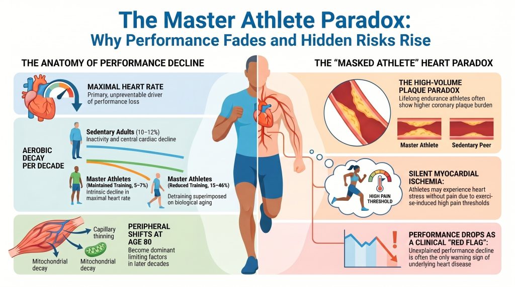
Longitudinale beoordelingen geven aan dat masters-atletes die trainingen met een hoge intensiteit en een hoog volume handhaven, de mate van V̇O verminderen2maximale afname tot ongeveer 5–71 TP9T per decennium, vergeleken met 10–121 TP9T per decennium bij personen van dezelfde leeftijd die een zittend leven leiden[2][3][4]. Deze door training afgeremde afname is echter kwetsbaar; uit regressieanalyses blijkt dat een aanzienlijk deel van de variantie in V̇O2De maximale afname bij masteratleten is rechtstreeks toe te schrijven aan een afname van het trainingsvolume en de trainingsintensiteit, waarbij de sterkste dalingen (15–46% per decennium) worden waargenomen bij atleten die hun training aanzienlijk verminderen[3][4].
Tabel 2. V̇O-waarden per decennium2maximale daling per groep.
| Groep | Afname over een periode van tien jaar | Hoofdbestuurder |
| Volwassenen met een zittend leven | 10–12% | Sarcopenie, gebrek aan lichaamsbeweging, afname van de hartfunctie |
| Masteratleten (die hun training hebben voortgezet) | 5–7% | Intrinsieke achteruitgang in maximaal hartslag |
| Masteratleten (minder training) | 15–46% | De effecten van het stoppen met sporten in combinatie met veroudering |
5. Cardiovasculaire mechanismen van de afname van het aërobe uithoudingsvermogen
De afname van V̇O2max wordt wiskundig beschreven door de Het Fick-principe: V̇O2max = Q̇ × (a–v)O2 verschil, waarbij Q̇ het hartminuutvolume is en (a–v)O2 is het verschil in zuurstofgehalte tussen arteriële en veneuze bloedvaten. Bij mastersatleten is de relatieve bijdrage van centrale hartfactoren (hartslag en slagvolume) ten opzichte van perifere factoren (zuurstofopname) verandert met de leeftijd[8][9].
5.1 Centrale mechanismen: de verouderingspomp
De meest consistente en grotendeels onveranderlijke factor die V̇O beïnvloedt2De maximale afname is de leeftijdsgebonden daling van de maximale hartslag (HRmax). Het veel geciteerde Tanaka-vergelijking — HRmax ≈ 208 − 0,7 × leeftijd — dit betekent een gemiddelde afname van ongeveer 0,7 slagen·min−1 per jaar[7]. Dit wordt voornamelijk veroorzaakt door veranderingen in de intrinsieke elektrofysiologie van de sinusnodus en door vermindering van β-adrenerge responsiviteit met het ouder worden. Hoewel duurtraining de hartslag in rust kan verlagen en de parasympathische tonus kan versterken, heeft deze training slechts een beperkt vermogen om de maximale hartslag tijdens maximale inspanning op peil te houden.
Beroerte Het slagvolume (SV) geeft een genuanceerder beeld. Bij mensen met een zittende levensstijl neemt het SV af als gevolg van een toegenomen stijfheid van het hartspierweefsel, een verminderde ventrikelcompliantie en een verminderde diastolische vulling[10]. Bij masteratleten leidt chronische duurtraining tot excentrische ventrikelremodellering, die wordt gekenmerkt door grotere kamerafmetingen en een verhoogde myocardiale compliantie[10][11]. Dankzij deze aanpassing kunnen sporters een hogere slagvolume behouden tijdens de het Frank-Starling-mechanisme. Niettemin kunnen masters-sporters een verminderd slagvolume ervaren bij maximale inspanning als gevolg van een abnormale verkorting van de diastolische vullingstijd bij zeer hoge hartfrequenties, waarbij het diastolische interval te kort is voor een volledige ventriculaire vulling.
5.2 Perifere mechanismen: zuurstofextractie en mitochondriale achteruitgang
Terwijl centrale beperkingen de vroege fases van achteruitgang domineren, worden perifeer gerelateerde factoren in toenemende mate beperkend naarmate atleten de leeftijd van 80 benaderen9]. Drie periferieprocessen zijn het belangrijkst:
- Arterioveneuze O2 Maximaal (a–v)O2 verschil neemt bescheiden af met de leeftijd bij mastersporters, grotendeels gedreven door capillaire zeldzaamheid — verdunning van de capillaire netwerken die individuele spieren van bloed voorzien vezels — wat de diffusieafstand voor zuurstof vergroot9].
- Mitochondriale functie. Veroudering is geassocieerd met verminderingen in mitochondriale dichtheid en bij de activiteit van belangrijke oxidatieve enzymen zoals citraatsynthase en succinaat dehydrogenase2Het onderhouden van intensieve training bij masteratleten behoudt de mitochondriale kwaliteit, maar verminderingen in trainingsvolume veroorzaken een snelle, nagenoeg lineaire daling van de oxidatieve capaciteit.
- Selectieve atrofie van type II (snelle) vezels en algeheel verlies van spierassa verminderen de metabole vraag en de inspanningscapaciteit, een effect dat is gedocumenteerd in het SPAF-cohort en elders[1].
6. Coronaire Atherosclerose bij Topsporters: De Verkalkingsparadox
Een van de meest uitdagende bevindingen in de moderne sportcardiologie is de schijnbare paradox van hoge kransslagaderkalk (CAC) scores bij duursporters voor het leven. Cross-sectionele gegevens, waaronder het Master@Heart-cohort, hebben aangetoond dat duursporters voor het leven (voornamelijk mannelijk) vaak een hogere prevalentie van coronaire plaques en hogere CAC-scores dan sedentaire leeftijdsgenoten met een vergelijkbaar risicoprofiel13][14][15][16][17].
6.1 Plaque morfologie en distributie
De Master@Heart-studie door De Bosscher en collega's namen 191 levenslange sporters, 191 laat-gestarte sporters en 176 gezonde niet-sporters (allen mannelijk) deel en toonden aan dat levenslange sporters de hoogste coronaire plaquebelasting[15]. De last omvatte niet alleen verkalkte plaques — over het algemeen als stabiel beschouwd — maar ook niet-verkalkte en gemengde plaques in proximale coronairsegmenten[15].
Tabel 3. Bevindingen over coronaire plaque uit het Master@Heart-cohort.
| Groep | ≥1 coronair plaque (OR) | ≥1 proximale plaque (OR) | ≥50% luminaal stenose |
| Niet-sporters (referentie) | 1.00 | 1.00 | ≈ referentie |
| Sporters die op latere leeftijd beginnen | gemiddeld | gemiddeld | laag |
| Duursporters voor het leven | 1.86 | 1.96 | ongewoon, maar verhoogd ten opzichte van controles |
De Bosscher R, et al. Eur Heart J. 2023[15]. OR = oddsratio ten opzichte van de referentiegroep van niet-sporters; de waarden zijn gecorrigeerde schattingen. Alle deelnemers waren mannen; gemiddelde leeftijd ≈ 60 jaar. Obstructieve (≥50%) stenose kwam in absolute termen in alle groepen zelden voor; de exacte gerapporteerde prevalentie in de groep van levenslange sporters moet vóór de definitieve opmaak worden gecontroleerd aan de hand van de oorspronkelijke publicatie.
In Master@Heart waren levenslange sporters ook onredelijk vaak voorzien van niet-verkalkte plaques in proximale segmenten vergeleken met niet-sporters15]. Een proximale lokalisatie is klinisch relevant omdat dergelijke plaques de doorstroming naar grote myocardiale gebieden controleren. Hoewel een hogere cardiorespiratoire conditie in sommige analyses geassocieerd is met een stabiele plaquemorfologie, is de absolute aanwezigheid van deze laesies suggereert dat duurtraining met een hoog volume een wisselwerking kan aangaan met conventionele risicofactoren om de progressie van reeds bestaande subklinische ziekten te versnellen16][17].
6.2 Voorgestelde mechanistische drijfveren
De ontwikkeling van CAD bij mastersporters blijft onvolledig verklaard. De huidige observationele literatuur roept verschillende voorgestelde mechanismen, die moeten worden geïnterpreteerd als plausibele hypothesen in plaats van als vaststaande causale verbanden:
- Hemodynamische afschuifspanning. Heftige inspanning produceert een hyperdynamische coronaire doorbloeding. Op plaatsen met verstoorde of turbulente stroming kunnen mechanische krachten bijdragen aan endotheeldisfunctie[13][17].
- Oxidatieve stress. Extreme metabole eisen van ultraduurtraining kunnen tijdelijk genereren reactieve zuurstofcomponenten meer dan antioxidant capacity, with possible downstream effects on endotheelfunctie en lipoproteïne retention[17].
- Exercise-induced hypertensie. In a recent cohort of male master endurance athletes, occult resting and exercise-induced hypertension was prevalent and associated with higher coronary plaque burden[18]. Exaggerated exercise systolische bloeddruk responses are increasingly recognized as a clinically actionable phenotype.
- Interactions with conventional risk factors. Even in athletes, conventional risk factors — LDL-cholesterol, apolipoproteïne B, lipoproteïne(a), hypertension, familiegeschiedenis — remain the dominant drivers of atherosclerosis[49][50].
7. Silent Myocardial Ischemia and Unrecognized Myocardial Infarction
7.1 Definitions and key distinction
Silent myocardial ischemia (SMI) refers to objective evidence of transient inducible ischemia — typically ST-segment depression on ambulatory or exercise ECG, or inducible perfusion abnormality on stress imaging — in the complete absence of anginal symptoms[21][22]. Unrecognized myocardial infarction (UMI) refers to a completed infarction that was not clinically diagnosed at the time it occurred[28][29][30][31]. Late gadolinium enhancement (LGE) on hart-MRI characterizes prior myocardial injury — scar, fibrosis, or completed infarction — and is therefore the reference standard imaging modality for UMI rather than a primary test for active ischemia[30][36]. This distinction is preserved throughout the remainder of this section.
7.2 Epidemiology of silent ischemia and unrecognized MI
Silent or unrecognized MIs account for a substantial fraction of all myocardial infarctions in epidemiological cohorts. The original Framingham-hartonderzoek reported that approximately one-quarter of MIs were unrecognized when first detected during routine biennial examination[28]. The Atherosclerosis Risk in Communities (ARIC) study found that the proportion of MIs that were silent and detected only by ECG was approximately 22%, with substantial sex and race differences in detection[29]. In the Cardiovascular Health Study of older community-dwelling adults, the prevalence of UMI rose with age[31].
The advent of MRI-scan van het hart with LGE has substantially raised estimates of UMI prevalence. The ICELAND-MI study by Schelbert and colleagues found that UMI detected by CMR-LGE was more than twice as common as UMI detected by ECG in an older community cohort, and that ECG-detected UMI represented only a fraction of all CMR-detected events[30]. In de MESA cohort, CMR-detected myocardial scar was identified in approximately 7.9% of asymptomatic adults aged 45–84 with no clinical history of MI[33].
Translated to US epidemiology, of the approximately 805,000 myocardial infarctions occurring annually in the United States, ECG-based community surveillance suggests that roughly 170,000 (≈21%) are clinically silent or unrecognized at the time of occurrence — a figure consistent with ARIC and Framingham proportions[28][29][31]. Because CMR-based detection has identified more than twice as many UMIs as ECG in older community cohorts[30], the ECG-based extrapolation of 170,000 is best interpreted as a lower bound on the true annual burden of clinically unrecognized myocardial infarction in the United States.
7.3 Why some myocardial ischemia and infarction is silent: proposed mechanisms
The absence of pain during ischemia or infarction is multifactorial. Eight broad mechanisms have been proposed in the literature:
- Autonomic neuropathy. Afferent sympathetic and vagal fibers that transmit cardiac nociceptive signals can be damaged by diabetes mellitus, age-related autonomic neuropathy, or prior cardiac surgery, attenuating or eliminating the perception of ischemic pain[21].
- Higher pain threshold. Individuals vary substantially in baseline somatic pain thresholds, and a subset of patients with documented ischemia have demonstrably elevated thresholds for laboratory pain stimuli[34].
- Endogenous opioid and endocannabinoid systems. Circulating β-endorphin and related opioids are elevated in some patients with stille ischemie and have been implicated in central modulation of cardiac pain perception[34].
- Exercise-induced hypoalgesia (EIH). Regular endurance training engages the endogenous opioid and endocannabinoid systems and is consistently associated with elevated pain thresholds and tolerances in laboratory testing[25][26]. In master athletes, EIH plausibly contributes to the absence of warning symptoms during demand-induced ischemia.
- Small infarct size and limited transmural extent. Small subendocardial or non-transmural infarcts may not generate sufficient afferent signaling to cross the threshold for conscious perception[32].
- Anatomic distribution. Inferior-wall ischemia and posterior-wall ischemia are more frequently silent than anterior-wall events, possibly due to differences in afferent innervation density[21].
- Collateral circulation. Chronic exercise promotes the development of coronary collateral vessels, which can maintain blood flow distal to a stenosis at rest and during low-to-moderate exertion, thereby preventing ischemia and pain until peak demand exceeds collateral capacity[27].
- Ischemic preconditioning. Repeated brief episodes of subclinical ischemia may protect the myocardium against subsequent insults and may also blunt afferent pain signaling[35].
7.4 Symptoms — and the absence of symptoms
By definition, classic exertional angina is absent in silent ischemia and unrecognized MI. However, the term silent is sometimes a misnomer. On careful retrospective questioning, a proportion of patients with apparently silent events describe subtle symptoms at the time — most commonly unaccustomed dyspnea, exertional fatigue out of proportion to workload, atypical chest discomfort attributed to indigestion or musculoskeletal causes, syncope or near-syncope, or unexplained reduction in exercise tolerance[21][22][32]. These so-called anginal equivalents are particularly important in master athletes, in whom dropping performance — rather than chest pain — may be the only clinical warning. When no symptoms are reported in any form, the event is genuinely asymptomatic and is detected only through screening, incidental imaging, or as part of a subsequent workup.
7.5 Prognosis
Silent ischemia and unrecognized MI are not benign. In long-term follow-up of the ICELAND-MI cohort, UMI detected by CMR was associated with mortality comparable to that of clinically recognized MI[30]. Across multiple cohorts, individuals with UMI carry elevated risks of hartfalen, recurrent MI, and plotselinge hartdood[29][30][31]. In endurance athletes specifically, the presence of ischemic-pattern LGE has been associated with markedly elevated risk of subsequent major adverse cardiaque voorvallen including sudden cardiac death[36].
7.6 Diagnosis
Diagnostic approaches differ for SMI versus UMI:
- For silent ischemia, the foundational modalities are ambulatory (Holter) ECG, exercise ECG, and stress imaging (stress echocardiography, single-photon emission CT, positron-emissietomografie, or stress CMR perfusion)[21][22].
- For unrecognized MI, the resting 12-afgeleide-ecg has limited sensitivity, detecting only a fraction of CMR-confirmed events[30]. CMR with LGE is the most sensitive niet-invasieve test and is the reference standard for non-acute scar detection[30][36]. Echocardiographic regional wall-motion abnormalities and elevated high-sensitivity troponine in non-acute contexts may also raise suspicion.
7.7 Master-athlete-specific burden: the “masked athlete” phenomenon
In a seminal cross-sectional study, Katzel and colleagues found that approximately 16% of male master athletes (mean age 60 years) had silent ischemia on a symptom-limited graded exercise test, a prevalence statistically comparable to that observed in sedentary controls of similar age[23]. Athletic status did not reduce the prevalence of ischemic burden; rather, it changed the perception of that burden. Combined with the EIH literature[25][26], these findings support the concept of the gemaskerde atleet — an individual whose high cardiorespiratory fitness and elevated pain thresholds can defer the perception of ischemia until exertional demand approaches the limits of coronary supply.
CMR-LGE prevalence in master endurance athletes has been variably reported, ranging from approximately 11% in younger marathon runners to considerably higher figures in older endurance-athlete cohorts[37][38]. Ischemic-pattern LGE (subendocardial enhancement in a coronary distribution) and non-ischemic LGE (focal patchy enhancement at the right-ventricular hinge points or mid-myocardium) carry different mechanistic implications and different prognostic weights[36]. A sudden or unexplained decline in performance or in V̇O2max in a previously stable master athlete should therefore be treated as a clinical red flag warranting consideration of advanced imaging.
8. Coronary Microvascular Dysfunction
Coronary microvascular dysfunction (CMD) is a plausible contributor to exercise intolerance in some older athletes, but the relevant evidence base in master-athlete cohorts is limited and findings should be described cautiously[39][40]. In a selected cohort of athletes with abnormal exercise tests but no obstructive epicardial CAD on coronaire CT-angiografie, Foulkes and colleagues reported significantly lower coronaire flowreserve (3.3 vs 4.2 in controls) and elevated endothelin-1 levels[39]. Several mechanisms have been proposed in athletes specifically, including the hypothesis of a possible “capillary-to-myocyte mismatch” in which training-induced hypertrophy outpaces microvascular expansion. These findings are best interpreted as preliminary and hypothesis-generating rather than as evidence that CMD broadly explains performance decline across the master-athlete population.
CMD may present with atypical features, including reduced inspanningscapaciteit, unusually elevated heart rates during submaximal effort, and exertional dyspnea rather than classic chest pain[40]. Diagnosis typically requires specialized testing (PET-based coronary flow reserve, invasive coronary physiology with bolus thermodilution, or stress CMR) available at experienced centers.
9. Screening and Risk Stratification in the Master Athlete
Given the high prevalence of subclinical CAD and silent ischemia in older athletes, traditional screening tools have important limitations. The resting 12-lead ECG, while valuable for detecting electrical disorders, has low sensitivity for subklinische atherosclerose[45]. Exercise treadmill testing in asymptomatic athletes is constrained by high false-positive rates[44]. Importantly, individual tests should be selected according to the clinical question being asked — anatomy, inducible ischemia, prior scar, or microvascular function — rather than by pooled performance metrics across these distinct domains.
Tabel 4. Cardiac diagnostic modalities in older master athletes, organized by the clinical question each test answers.
| Modaliteit | Primary question answered | Main strength | Main limitation |
| Resting 12-lead ECG | Electrical disease; prior Q-wave MI | Widely available baseline test | Low sensitivity for silent CAD or non-Q-wave prior MI |
| Exercise treadmill ECG | Exercise-provoked ECG abnormalities; symptom correlation | Functional provocation; reveals exercise BP response | Both false positives and false negatives are common in master athletes: the latter because high cardiorespiratory reserve and collateral flow may allow some athletes to maintain workload despite inducible ischemia until higher exercise intensities |
| Coronary CT angiography (CCTA) | Coronary anatomy; plaque burden and stenosis | Strong anatomic definition of plaque/stenosis | Does not directly establish inducible ischemia |
| Stress echocardiography / stress perfusion imaging (SPECT, PET, stress CMR) | Inducible ischemia | Functional assessment of ischemic burden | Performance depends on protocol and image quality |
| CMR with LGE | Prior infarction / scar / fibrosis pattern (UMI) | High tissue characterization value; gold standard for non-acute scar | Not a primary test for obstructive CAD |
| PET with coronary flow reserve | Inducible ischemia plus microvascular function | Useful for CMD assessment in specialized centers | Limited availability; higher complexity |
Bronnen: refs [40][44][45]. CCTA = coronary CT angiography; CMR = cardiac magnetic resonance; LGE = late gadolinium enhancement; UMI = unrecognized myocardial infarction; CMD = coronary microvascular dysfunction; SPECT = single-photon emission computertomografie; PET = positron emission tomography. The table is intentionally structured by clinical question rather than by pooled sensitivity/specificity, because tests answering different reference-standard questions are not directly comparable.
De ISCHEMIA-studie should not be summarized as proving that exercise capacity outweighs coronary anatomy or ischemia severity. A more defensible conclusion from the ISCHEMIA program is that, in stable chronic coronary disease, an initial invasive strategy did not produce an overall reduction in major cardiovascular events compared with an initial conservative strategy, and prognosis varied according to disease burden in pre-specified analyses[41][42]. The broader point that cardiorespiratory fitness is a strong predictor of mortality is robustly supported by cardiopulmonary exercise testing literature outside the ISCHEMIA program[43]. For master athletes, the practical implication is that a sudden or unexplained decline in performance or in V̇O2max should be treated as a clinical signal warranting structured evaluation[47].
10. Sex Differences and the Female Master Athlete
A substantial portion of the master-athlete coronary literature is derived from male cohorts, including Master@Heart, which enrolled only men[15]. In female master athletes, CAC prevalence is generally lower than in male counterparts at the same age and training exposure, and the coronary phenotype associated with lifelong endurance training is less well characterized[48]. The 2020 ESC Sports Cardiology Guideline emphasizes individualized risk assessment in female master athletes, with attention to traditional risk factors, menopausal status, and exercise bloeddruk response[47]. Generalizing male-cohort findings to female master athletes is inappropriate until parallel data are available.
11. Clinical Principles for the Aging Master Athlete
Current evidence supports individualized training and cardiovascular risk management rather than a single proven training formula for preventing coronary plaque in master athletes. Practical principles supported by primary literature and major-society guidance include:
- Maintain regular endurance activity at intensities and volumes appropriate to the individual, recognizing that detraining accelerates V̇O2max decline beyond what biological aging alone produces[2][3].
- Incorporate krachttraining to help preserve muscle mass and function across the aging trajectory.
- Aggressively manage conventional cardiovascular risk factors — LDL-cholesterol or apolipoprotein B, lipoprotein(a) where available, blood pressure, glycemia, and tobacco exposure — in accordance with current prevention guidelines[45][49][50].
- Evaluate exaggerated exercise blood pressure responses, which are independently associated with higher coronary plaque burden in master athletes[18].
- Treat unexplained performance decline as a clinical signal warranting structured assessment, given the “masked athlete” phenomenon[23][25].
- Apply advanced imaging (CCTA, stress imaging, CMR with LGE) selectively, guided by clinical context, exercise findings, and conventional risk profile[47].
Polarized training distributions (commonly described as approximately 80/20 low- to high-intensity) may be useful performance optimization frameworks but have niet been prospectively shown to prevent CAC accrual or to mitigate the so-called atletenparadox, and should not be presented as preventive prescriptions[51][52].
12. Synthesis and Discussion
12.1 Inevitable versus modifiable decay
The SPAF study confirms that the rise and fall of physical capacity is a constitutive feature of human life[1]. The age of peak performance and the acceleration of decline after age 40 appear largely invariant across populations. However, master athletes demonstrate that the absolute level of performance and the decadal rate of V̇O2max loss are highly modifiable through maintenance of training volume and intensity[2][3][4].
12.2 Dominance of central factors
Between ages 50 and 80, the primary driver of performance decline in master athletes is central: a relentless decline in HRmax reduces total cardiac output during peak exertion, a limitation that even aggressive training cannot reverse[7]. Although athletes preserve stroke volume better than sedentary peers, age-related myocardial stiffening eventually restricts diastolic filling at high heart rates[10][11].
12.3 The pathological paradox
Lifelong endurance training appears associated with both favorable cardiovascular adaptation and an increased prevalence of subclinical coronary atherosclerosis in some observational cohorts[15][16][17]. The atherosclerotic burden — combined with the analgesic effects of exercise-induced hypoalgesia — makes silent ischemia and unrecognized MI clinically important risks that warrant individualized screening and management[23][25][26].
12.4 Peripheral mechanisms at the limit of human aging
As athletes approach age 80, peripheral extraction increasingly catches up to central factors as a determinant of V̇O2max. Capillary rarefaction and mitochondrial decay reduce the muscle’s capacity to utilize delivered oxygen — an effect that is accelerated when training volume is reduced[2][9].
Key Clinical Takeaways
| • Aerobic capacity declines along a curvilinear trajectory beginning in the mid-30s and accelerating in the 60s. Master athletes who maintain training reduce the rate of decline to roughly half that of sedentary peers; detraining accelerates decline several-fold.
• Lifelong endurance training does not confer immunity to coronary atherosclerosis. Master@Heart and related cohorts show higher coronary calcium and proximal plaque burden in lifelong athletes than in sedentary peers; plaque morphology may be more favorable but does not abolish risk. • Silent ischemia and unrecognized myocardial infarction are not uncommon in older athletes. Approximately 1 in 6 male master athletes has inducible ischemia on graded exercise testing; CMR-detected scar in community cohorts exceeds ECG-detected scar by more than 2:1. • Symptoms may be absent, atypical, or expressed as performance loss. An unexplained decline in race times, exertional dyspnea, or reduced exercise tolerance in an older athlete should prompt structured cardiac evaluation — not reassurance. • Choose the diagnostic test to match the clinical question. Anatomy (CCTA), inducible ischemia (stress imaging), prior scar (CMR with LGE), and microvascular function (PET with CFR) answer different questions and are not interchangeable. • Standard cardiovascular risk-factor management remains the foundation. Aggressively manage LDL-C/ApoB, lipoprotein(a) where available, blood pressure (including exaggerated exercise responses), glycemia, and tobacco exposure. No training distribution has been prospectively shown to prevent CAC accrual. • Overall health benefits of lifelong exercise remain substantial. The clinical task is calibration of intensity, surveillance, and risk-factor control — not avoidance of training. |
13. Conclusions
For master athletes between the ages of 50 and 80, maintenance of endurance performance requires a careful balance between sustained training (to preserve V̇O2max and skeletal-muscle oxidative capacity) and individualized cardiovascular risk management. Transitioning from active to sedentary status produces a V̇O2max decline substantially steeper than aging alone[2][3].
However, the gemaskerde atleet concept warrants vigilance rather than complacency. Older endurance athletes may present with reduced performance capacity, exertional dyspnea, or other atypical symptoms rather than classic angina, and unexplained decline in performance should prompt structured clinical reassessment[23][25][47].
The available literature supports substantial overall health benefits of long-term exercise[43][53]. Some evidence suggests that the plaquefenotype of lifelong athletes is shifted toward more calcified and fewer mixed/non-calcified lesions when matched for total plaque burden, which may confer relative stability compared with sedentary patients of equivalent CAC score[16]. Even granting that observation, however, the present evidence base does not justify claiming that coronary verkalking in master athletes is uniformly benign, or that mortality advantages persist regardless of plaque phenotype — CAC-stratified mortality data specific to master athletes remain limited. A more evidence-consistent conclusion is that lifelong exercise and subclinical coronary disease can coexist, that plaque morphology likely matters as well as plaque quantity, and that individualized risk assessment is essential in older athletes.
14. Limitations
This is a narrative review. No systematic search protocol or formal meta-analytic pooling was performed. The Master@Heart cohort and several other key coronary-imaging studies in lifelong endurance athletes are all-male, limiting generalizability to female master athletes. CAC-stratified mortality data specific to master athletes remain sparse, and the prognostic significance of non-calcified proximal plaques identified incidentally on CCTA in asymptomatic athletes is not yet established. The relevant CMD literature in master athletes is derived from small selected cohorts on referral and should not be generalized.
Disclosures and End-Matter
Conflicts of interest
The author reports no financial conflicts of interest related to the subject matter of this manuscript. The author is the operator of CuringHeartDisease.com, a clinician-educator platform that provides peer-reviewed cardiovascular health content without supplement marketing or commercial sponsorship.
Funding
No external funding, sponsorship, or institutional support contributed to the conception, drafting, or publication of this manuscript.
Data availability
No new datasets were generated for this narrative review. All cited evidence is contained in the published literature and is accessible through the cited references.
Ethics
Ethics approval and informed consent were not required because this manuscript is a review of previously published literature and does not report new human-subject research.
Prior dissemination
No version of this manuscript has been previously published or made publicly available in any venue, including preprint servers and the author’s educational platform. CuringHeartDisease.com is the intended publication venue.
Author contributions
PM conceived the manuscript, conducted the literature identification, drafted and revised the text, and approved the final version for publication.
Acknowledgments
The author thanks the external reviewers whose comments informed the present revision.
Referenties
References are listed in order of first mention in the text, in accordance with NLM/Vancouver convention. All entries are PubMed-indexed primary studies, meta-analyses, systematic reviews, or major-society guidelines. Author lists with seven or more authors are truncated after the first six followed by “et al.”
- Westerståhl M, Jörnåker G, Jansson E, et al. Rise and Fall of Physical Capacity in a General Population: A 47-Year Longitudinal Study. J Cachexia Sarcopenia Muscle. 2025;16(6):e70134. doi:10.1002/jcsm.70134
- Trappe S. Master athletes. Int J Sport Nutr Exerc Metab. 2001;11 Suppl:S196-S207. doi:10.1123/ijsnem.11.s1.s196
- Pollock ML, Mengelkoch LJ, Graves JE, et al. Twenty-year follow-up of aerobic power and body composition of older track athletes. J Appl Physiol (1985). 1997;82(5):1508-1516. doi:10.1152/jappl.1997.82.5.1508
- Tanaka H, Seals DR. Endurance exercise performance in Masters athletes: age-associated changes and underlying physiological mechanisms. J Physiol. 2008;586(1):55-63. doi:10.1113/jphysiol.2007.141879
- Hawkins S, Wiswell R. Rate and mechanism of maximal oxygen consumption decline with aging: implications for exercise training. Sports Med. 2003;33(12):877-888. doi:10.2165/00007256-200333120-00002
- Joyner MJ, Coyle EF. Endurance exercise performance: the physiology of champions. J Physiol. 2008;586(1):35-44. doi:10.1113/jphysiol.2007.143834
- Tanaka H, Monahan KD, Seals DR. Age-predicted maximal heart rate revisited. J Am Coll Cardiol. 2001;37(1):153-156. doi:10.1016/s0735-1097(00)01054-8
- Levine BD. .VO2max: what do we know, and what do we still need to know?. J Physiol. 2008;586(1):25-34. doi:10.1113/jphysiol.2007.147629
- Proctor DN, Joyner MJ. Skeletal muscle mass and the reduction of VO2max in trained older subjects. J Appl Physiol (1985). 1997;82(5):1411-1415. doi:10.1152/jappl.1997.82.5.1411
- Pelliccia A, Caselli S, Sharma S, et al. European Association of Preventive Cardiology (EAPC) and European Association of Cardiovascular Imaging (EACVI) joint position statement: recommendations for the indication and interpretation of cardiovascular imaging in the evaluation of the athlete’s heart. Eur Heart J. 2018;39(21):1949-1969. doi:10.1093/eurheartj/ehx532
- La Gerche A, Burns AT, Mooney DJ, et al. Exercise-induced right ventricular dysfunction and structural remodelling in endurance athletes. Eur Heart J. 2012;33(8):998-1006. doi:10.1093/eurheartj/ehr397
- Levine BD. Can intensive exercise harm the heart? The benefits of competitive endurance training for cardiovascular structure and function. Circulation. 2014;130(12):987-991. doi:10.1161/CIRCULATIONAHA.114.008142
- Aengevaeren VL, Mosterd A, Braber TL, et al. Relationship Between Lifelong Exercise Volume and Coronary Atherosclerosis in Athletes. Circulation. 2017;136(2):138-148. doi:10.1161/CIRCULATIONAHA.117.027834
- Möhlenkamp S, Lehmann N, Breuckmann F, et al. Running: the risk of coronary events : Prevalence and prognostic relevance of coronary atherosclerosis in marathon runners. Eur Heart J. 2008;29(15):1903-1910. doi:10.1093/eurheartj/ehn163
- De Bosscher R, Dausin C, Claus P, et al. Lifelong endurance exercise and its relation with coronary atherosclerosis. Eur Heart J. 2023;44(26):2388-2399. doi:10.1093/eurheartj/ehad152
- Aengevaeren VL, Mosterd A, Sharma S, et al. Exercise and Coronary Atherosclerosis: Observations, Explanations, Relevance, and Clinical Management. Circulation. 2020;141(16):1338-1350. doi:10.1161/CIRCULATIONAHA.119.044467
- Merghani A, Maestrini V, Rosmini S, et al. Prevalence of Subclinical Coronary Artery Disease in Masters Endurance Athletes With a Low Atherosclerotic Risk Profile. Circulation. 2017;136(2):126-137. doi:10.1161/CIRCULATIONAHA.116.026964
- O’Driscoll J, Parry-Williams G, Moser J, et al. Resting and exercise-induced occult hypertension and coronary atherosclerosis in male masters endurance athletes. Br J Sports Med. Published online May 14, 2026. doi:10.1136/bjsports-2025-111347
- Heidbuchel H, Adami PE, Antz M, et al. Recommendations for participation in leisure-time physical activity and competitive sports in patients with arrhythmias and potentially arrhythmogenic conditions: Part 1: Supraventricular arrhythmias. A position statement of the Section of Sports Cardiology and Exercise from the European Association of Preventive Cardiology (EAPC) and the European Heart Rhythm Association (EHRA), both associations of the European Society of Cardiology. Eur J Prev Cardiol. 2021;28(14):1539-1551. doi:10.1177/2047487320925635
- Sharma S, Drezner JA, Baggish A, et al. International recommendations for electrocardiographic interpretation in athletes. Eur Heart J. 2018;39(16):1466-1480. doi:10.1093/eurheartj/ehw631
- Cohn PF, Fox KM, Daly C. Silent myocardial ischemia. Circulation. 2003;108(10):1263-1277. doi:10.1161/01.CIR.0000088001.59265.EE
- Deedwania PC, Carbajal EV. Silent ischemia during daily life is an independent predictor of mortality in stable angina. Circulation. 1990;81(3):748-756. doi:10.1161/01.cir.81.3.748
- Katzel LI, Fleg JL, Busby-Whitehead MJ, et al. Exercise-induced silent myocardial ischemia in master athletes. Am J Cardiol. 1998;81(3):261-265. doi:10.1016/s0002-9149(97)00898-9
- Sheps DS, Maixner W, Hinderliter AL. Mechanisms of pain perception in patients with silent myocardial ischemia. Am Heart J. 1990;119(4):983-987. doi:10.1016/s0002-8703(05)80351-5
- Koltyn KF. Analgesia following exercise: a review. Sports Med. 2000;29(2):85-98. doi:10.2165/00007256-200029020-00002
- Naugle KM, Fillingim RB, Riley JL 3rd. A meta-analytic review of the hypoalgesic effects of exercise. J Pain. 2012;13(12):1139-1150. doi:10.1016/j.jpain.2012.09.006
- Heaps CL, Parker JL. Effects of exercise training on coronary collateralization and control of collateral resistance. J Appl Physiol (1985). 2011;111(2):587-598. doi:10.1152/japplphysiol.00338.2011
- Kannel WB, Abbott RD. Incidence and prognosis of unrecognized myocardial infarction. An update on the Framingham study. N Engl J Med. 1984;311(18):1144-1147. doi:10.1056/NEJM198411013111802
- Zhang ZM, Rautaharju PM, Prineas RJ, et al. Race and Sex Differences in the Incidence and Prognostic Significance of Silent Myocardial Infarction in the Atherosclerosis Risk in Communities (ARIC) Study. Circulation. 2016;133(22):2141-2148. doi:10.1161/CIRCULATIONAHA.115.021177
- Schelbert EB, Cao JJ, Sigurdsson S, et al. Prevalence and prognosis of unrecognized myocardial infarction determined by cardiac magnetic resonance in older adults. JAMA. 2012;308(9):890-896. doi:10.1001/2012.jama.11089
- Sheifer SE, Manolio TA, Gersh BJ. Unrecognized myocardial infarction. Ann Intern Med. 2001;135(9):801-811. doi:10.7326/0003-4819-135-9-200111060-00010
- Acharya T, Aspelund T, Jonasson TF, et al. Association of Unrecognized Myocardial Infarction With Long-term Outcomes in Community-Dwelling Older Adults: The ICELAND MI Study. JAMA Cardiol. 2018;3(11):1101-1106. doi:10.1001/jamacardio.2018.3285
- Turkbey EB, Nacif MS, Guo M, et al. Prevalence and Correlates of Myocardial Scar in a US Cohort. JAMA. 2015;314(18):1945-1954. doi:10.1001/jama.2015.14849
- Sheps DS, Adams KF, Hinderliter A, et al. Endorphins are related to pain perception in coronary artery disease. Am J Cardiol. 1987;59(6):523-527. doi:10.1016/0002-9149(87)91161-1
- Murry CE, Jennings RB, Reimer KA. Preconditioning with ischemia: a delay of lethal cell injury in ischemic myocardium. Circulation. 1986;74(5):1124-1136. doi:10.1161/01.cir.74.5.1124
- Małek ŁA, Miłosz-Wieczorek B, Marczak M. Diagnostic Yield of Cardiac Magnetic Resonance in Athletes with and without Features of the Athlete’s Heart and Suspected Structural Heart Disease. Int J Environ Res Public Health. 2022;19(8):4829. Published 2022 Apr 15. doi:10.3390/ijerph19084829
- Möhlenkamp S, Leineweber K, Lehmann N, et al. Coronary atherosclerosis burden, but not transient troponin elevation, predicts long-term outcome in recreational marathon runners. Basic Res Cardiol. 2014;109(1):391. doi:10.1007/s00395-013-0391-8
- Breuckmann F, Möhlenkamp S, Nassenstein K, et al. Myocardial late gadolinium enhancement: prevalence, pattern, and prognostic relevance in marathon runners. Radiology. 2009;251(1):50-57. doi:10.1148/radiol.2511081118
- van de Sande DAJP, Barneveld PC, Hoogsteen J, Doevendans PA, Kemps HMC. Coronary microvascular function in athletes with abnormal exercise test results. Neth Heart J. 2019;27(12):621-628. doi:10.1007/s12471-019-01336-6
- Camici PG, Crea F. Coronary microvascular dysfunction. N Engl J Med. 2007;356(8):830-840. doi:10.1056/NEJMra061889
- Maron DJ, Hochman JS, Reynolds HR, et al. Initial Invasive or Conservative Strategy for Stable Coronary Disease. N Engl J Med. 2020;382(15):1395-1407. doi:10.1056/NEJMoa1915922
- Reynolds HR, Shaw LJ, Min JK, et al. Outcomes in the ISCHEMIA Trial Based on Coronary Artery Disease and Ischemia Severity. Circulation. 2021;144(13):1024-1038. doi:10.1161/CIRCULATIONAHA.120.049755
- Mandsager K, Harb S, Cremer P, Phelan D, Nissen SE, Jaber W. Association of Cardiorespiratory Fitness With Long-term Mortality Among Adults Undergoing Exercise Treadmill Testing. JAMA Netw Open. 2018;1(6):e183605. Published 2018 Oct 5. doi:10.1001/jamanetworkopen.2018.3605
- Knuuti J, Ballo H, Juarez-Orozco LE, et al. The performance of non-invasive tests to rule-in and rule-out significant coronary artery stenosis in patients with stable angina: a meta-analysis focused on post-test disease probability. Eur Heart J. 2018;39(35):3322-3330. doi:10.1093/eurheartj/ehy267
- Knuuti J, Wijns W, Saraste A, et al. 2019 ESC Guidelines for the diagnosis and management of chronic coronary syndromes. Eur Heart J. 2020;41(3):407-477. doi:10.1093/eurheartj/ehz425
- Maron BJ, Zipes DP, Kovacs RJ. Eligibility and Disqualification Recommendations for Competitive Athletes With Cardiovascular Abnormalities: Preamble, Principles, and General Considerations: A Scientific Statement From the American Heart Association and American College of Cardiology. J Am Coll Cardiol. 2015;66(21):2343-2349. doi:10.1016/j.jacc.2015.09.032
- Pelliccia A, Sharma S, Gati S, et al. 2020 ESC Guidelines on sports cardiology and exercise in patients with cardiovascular disease. Eur Heart J. 2021;42(1):17-96. doi:10.1093/eurheartj/ehaa605
- Parry-Williams G, Sharma S. The effects of endurance exercise on the heart: panacea or poison?. Nat Rev Cardiol. 2020;17(7):402-412. doi:10.1038/s41569-020-0354-3
- Tsimikas S. A Test in Context: Lipoprotein(a): Diagnosis, Prognosis, Controversies, and Emerging Therapies. J Am Coll Cardiol. 2017;69(6):692-711. doi:10.1016/j.jacc.2016.11.042
- Sniderman AD, Thanassoulis G, Glavinovic T, et al. Apolipoprotein B Particles and Cardiovascular Disease: A Narrative Review. JAMA Cardiol. 2019;4(12):1287-1295. doi:10.1001/jamacardio.2019.3780
- Seiler S. What is best practice for training intensity and duration distribution in endurance athletes?. Int J Sports Physiol Perform. 2010;5(3):276-291. doi:10.1123/ijspp.5.3.276
- Stöggl T, Sperlich B. Polarized training has greater impact on key endurance variables than threshold, high intensity, or high volume training. Front Physiol. 2014;5:33. Published 2014 Feb 4. doi:10.3389/fphys.2014.00033
- Eijsvogels TMH, Thompson PD, Franklin BA. The “Extreme Exercise Hypothesis”: Recent Findings and Cardiovascular Health Implications. Curr Treat Options Cardiovasc Med. 2018;20(10):84. Published 2018 Aug 28. doi:10.1007/s11936-018-0674-3
- Eijsvogels TMH, Thompson PD, Franklin BA. The “Extreme Exercise Hypothesis”: Recent Findings and Cardiovascular Health Implications. Curr Treat Options Cardiovasc Med. 2018;20(10):84. Published 2018 Aug 28. doi:10.1007/s11936-018-0674-3

