Mecanismos cardiovasculares do declínio longitudinal do desempenho em atletas master:
A Revisão Narrativa do Envelhecimento, Doença arterial coronariana, Infarto do miocárdio silencioso Isquemia, e Não reconhecido Infarto do miocárdio
Resumo
Esta revisão narrativa sintetiza evidências selecionadas e revisadas por pares sobre os mecanismos cardiovasculares e fisiológicos subjacentes ao declínio longitudinal do desempenho em atletas master com idades entre 50 e 80 anos. Os tópicos abordados incluem (1) a trajetória natural da capacidade física a partir do estudo da Coorte Populacional Sueca para Atividade Física e Condição Física (SPAF)[1]; (2) o contraste entre o V̇O dos atletas veteranos e o dos sedentários2declínio máximo[2][3][4]; (3) mecanismos cardiovasculares centrais e periféricos do V̇O2erosão máxima; (4) o paradoxo da doença coronariana subclínica aterosclerose em atletas de resistência ao longo da vida; (5) isquemia miocárdica assintomática e infarto do miocárdio não diagnosticado — incluindo epidemiologia, mecanismos, prognóstico e abordagem diagnóstica; (6) coronariana disfunção microvascular como um fator que pode contribuir para a intolerância ao exercício em determinados atletas idosos; e (7) considerações atuais sobre exames de triagem em cardiologia esportiva, baseadas no Diretriz da ESC de 2020 sobre Cardiologia do Esporte. A síntese tem caráter interpretativo, e não exaustivo; a estratégia específica de pesquisa e o escopo estão descritos na seção “Identificação da literatura”.
1. Introdução
O processo de envelhecimento em seres humanos está inexoravelmente ligado a um declínio progressivo da capacidade fisiológica; no entanto, a taxa e a trajetória desse declínio são significativamente influenciadas pela atividade física habitual. Entre os indivíduos que mantêm altos níveis de treinamento de resistência ao longo da sexta, sétima e oitava décadas de vida — comumente chamados de atletas veteranos —, o declínio na capacidade aeróbica máxima (V̇O2(máx.) e o desempenho de resistência oferecem um modelo biológico único para o estudo dos limites do envelhecimento humano[2]. Embora os atletas master demonstrem consistentemente um desempenho superior aptidão cardiorrespiratória (CRF) — mesmo em comparação com seus pares sedentários —, eles não estão imunes aos processos biológicos fundamentais que prejudicam a função cardiovascular ao longo do tempo[3][4].
O declínio longitudinal do desempenho entre os 50 e os 80 anos é determinado por uma interação complexa entre limitações cardíacas centrais, deterioração vascular periférica e mitocondrial e o surgimento paradoxal de patologias cardiovasculares subclínicas. Entre elas estão as doenças coronárias artéria doença em atletas de resistência de longa duração[13][14][15] e, no contexto mais amplo do envelhecimento da população, isquemia miocárdica silenciosa (SMI), infarto do miocárdio não diagnosticado (UMI), e disfunção microvascular coronária (CMD)[21][22]. Para compreender esses mecanismos no contexto, é útil, em primeiro lugar, estabelecer uma linha de base em nível populacional a partir da coorte longitudinal do SPAF[1] e, em seguida, comparar essa linha de base com as adaptações fisiológicas específicas — e os riscos patológicos — observados em atletas de resistência de longa data.
2. Identificação da literatura
Este artigo é uma revisão narrativa. A literatura foi identificada principalmente por meio de pesquisas no PubMed e no Embase (1990–2026) e pela análise retrospectiva das referências de estudos primários relevantes, meta-análises e documentos de orientação das principais sociedades médicas que abordam atletas master, declínio da capacidade aeróbica relacionado à idade, aterosclerose coronariana em atletas de resistência, isquemia miocárdica silenciosa, infarto do miocárdio não reconhecido, disfunção microvascular coronariana e triagem em cardiologia esportiva. Foi dada prioridade a estudos de coorte originais, ensaios clínicos randomizados, revisões sistemáticas, metanálises e diretrizes oficiais. Como esta não foi uma revisão sistemática baseada em protocolo, a seleção dos estudos foi qualitativa e a síntese deve ser interpretada como interpretativo, em vez de exaustivo. Não foi realizada nenhuma combinação formal por meio de meta-análise; os valores quantitativos foram reproduzidos a partir das fontes primárias citadas, sem nova análise.
3. A linha de base da população geral: conclusões do estudo SPAF de Westerståhl
O estudo longitudinal SPAF, com duração de 47 anos, relatado por Westerståhl e colegas, oferece uma das avaliações mais abrangentes sobre como a capacidade física se altera desde a adolescência até o início da terceira idade em uma coorte representativa da população em geral[1]. Acompanhando 427 indivíduos nascidos em 1958, o estudo utilizou avaliações objetivas repetidas da capacidade aeróbica, resistência muscular e potência explosiva, dos 16 aos 63 anos[1]. O conjunto de dados é informativo porque identifica a taxa natural de declínio em uma população na qual a atividade física não é padronizada, destacando a natureza acelerada do envelhecimento biológico, independentemente do status de atleta de elite.
O pico de desempenho físico foi, em geral, alcançado entre os 26 e os 36 anos, tanto no que diz respeito à capacidade aeróbica absoluta quanto à resistência muscular[1]. Especificamente, os homens atingiram seu pico de capacidade aeróbica absoluta aos 35 anos (3,26 L·min−1), enquanto as mulheres atingiram o pico aos 36 anos (2,61 L·min−1)[1]. Após esses picos, o declínio começou a uma taxa de aproximadamente 0,3–0,61 TP9T por ano e se acelerou para cerca de 2,0–2,51 TP9T por ano à medida que os participantes se aproximavam dos 60 anos[1]. Aos 63 anos, o declínio cumulativo da capacidade aeróbica absoluta em relação ao pico foi de aproximadamente 33% nos homens e 30% nas mulheres[1].
Tabela 1. Trajetórias da capacidade física da coorte longitudinal SPAF de 47 anos.
| Parâmetro | Homens (idade de pico: 35–36 anos) | Mulheres (idade máxima: 36 anos) | Queda total de 63 |
| Capacidade aeróbica absoluta (L·min⁻¹) | 3.26 | 2.61 | 30–33% |
| Capacidade aeróbica relativa (mL·kg⁻¹·min⁻¹) | 42,2 aos 26 anos | 39,7 aos 31 anos | 37–40% |
| Salto vertical (cm) | 45,5 aos 27 anos | 33,0 aos 19 anos | 41–48% |
| Resistência muscular (supino, repetições) | 52,7 aos 36 anos | 39,7 aos 34 anos | 32–35% |
Fonte: Westerståhl M, et al. J Cachexia Sarcopenia Muscle. 2025[1]. Os valores correspondem às médias da coorte na idade indicada. SPAF = Coorte da População Sueca para Atividade Física e Condição Física.
Os dados do SPAF também destacaram o papel dos fatores relacionados ao estilo de vida na modificação dessas trajetórias. Níveis mais elevados de atividade física no tempo livre aos 16 anos e um estilo de vida ativo durante a idade adulta foram associados a um desempenho superior em todos os indicadores ao longo da vida[1]. Um nível de escolaridade mais elevado foi associado a maior capacidade aeróbica absoluta e resistência muscular, provavelmente por meio de comportamentos voltados para a saúde. Mesmo com esses fatores protetores, no entanto, a aceleração do declínio após os 40 anos foi um achado consistente, reforçando o conceito de que o envelhecimento biológico impõe uma restrição fisiológica progressiva à capacidade funcional, que começa a se manifestar bem antes de se tornar clinicamente significativa.
4. Declínio comparativo: atletas master x população em geral
Ao comparar a linha de base do SPAF com dados longitudinais de atletas master, surge uma distinção clara na altitude inicial e na inclinação da V̇O2declínio máximo. Os atletas de resistência de alto nível representam um grupo que maximizou seu potencial de adaptação e pode apresentar um V̇O2valor máximo aos 60 anos que exceda a mediana de uma pessoa sedentária de 40 anos[2]. Apesar dessa vantagem, o inevitável declínio no V̇O2O max continua sendo o principal mecanismo fisiológico associado à redução do desempenho em provas de resistência em atletas mais velhos[3][4].
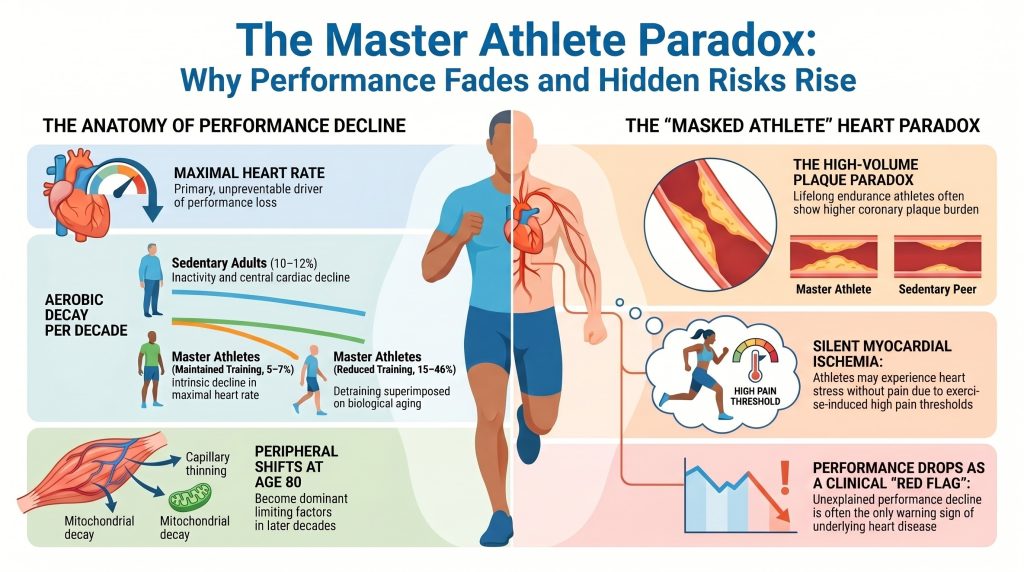
Avaliações longitudinais indicam que atletas da terceira idade que mantêm um treinamento de alta intensidade e alto volume reduzem a taxa de V̇O2redução máxima para aproximadamente 5–71 TP9T por década, em comparação com 10–121 TP9T por década em indivíduos sedentários da mesma faixa etária[2][3][4]. Esse declínio atenuado pelo treinamento é, no entanto, frágil; análises de regressão indicam que uma proporção substancial da variância no V̇O2A queda máxima observada nos atletas master é diretamente atribuível à redução do volume e da intensidade do treinamento, sendo que as quedas mais acentuadas (15–46% por década) foram observadas em atletas que reduziram substancialmente o treinamento[3][4].
Tabela 2. Taxas decenais de V̇O2declínio máximo por grupo.
| Grupo | Declínio ao longo de uma década | Motivador principal |
| Adultos sedentários | 10–12% | Sarcopenia, sedentarismo, declínio cardíaco central |
| Atletas master (treinamento contínuo) | 5–7% | Declínio intrínseco na capacidade máxima frequência cardíaca |
| Atletas master (treinamento reduzido) | 15–46% | Perda de condicionamento físico associada ao envelhecimento |
5. Mecanismos cardiovasculares da perda da capacidade aeróbica
A erosão do V̇O2O valor máximo é descrito matematicamente pela Princípio de Fick: V̇O2max = Q̇ × (a–v)O2 diferença, em que Q̇ é o débito cardíaco e (a–v)O2 é a diferença no teor de oxigênio entre as artérias e as veias. Em atletas veteranos, a contribuição relativa dos fatores cardíacos centrais (frequência cardíaca e volume sistólico) em relação aos fatores periféricos (extração de oxigênio) sofre alterações com a idade[8][9].
5.1 Mecanismos centrais: a bomba do envelhecimento
O fator mais consistente e, em grande parte, imutável que influencia o V̇O2O declínio máximo é a redução da frequência cardíaca máxima (FC) relacionada à idademáximo). O amplamente citado Equação de Tanaka — RHmáximo ≈ 208 − 0,7 × idade — o que implica uma redução média de aproximadamente 0,7 batimentos·min−1 por ano[7]. Isso se deve principalmente a alterações na eletrofisiologia intrínseca do nó sinoatrial e pela redução de resposta β-adrenérgica com a idade. Embora o treinamento de resistência possa reduzir a frequência cardíaca em repouso e aumentar o tônus parassimpático, ele tem capacidade limitada de preservar a frequência cardíaca máxima durante esforços intensos.
Acidente vascular cerebral O volume sistólico (VS) oferece um quadro mais matizado. Em indivíduos sedentários, o VS diminui devido ao aumento da rigidez miocárdica, à redução da complacência ventricular e ao comprometimento do enchimento diastólico[10]. Em atletas da categoria master, o treinamento crônico de resistência induz uma remodelação ventricular excêntrica, caracterizada por dimensões maiores das câmaras e maior complacência miocárdica[10][11]. Essa adaptação permite que os atletas mantenham um SV mais elevado durante o Mecanismo de Frank-Starling. No entanto, atletas master podem apresentar um volume sistólico (VS) reduzido no pico do exercício devido ao encurtamento anormal do tempo de enchimento diastólico em frequências cardíacas muito elevadas, nas quais o intervalo diastólico é curto demais para permitir o enchimento ventricular completo.
5.2 Mecanismos periféricos: extração de oxigênio e deterioração mitocondrial
Embora as limitações centrais predominem nas fases iniciais do declínio, os fatores periféricos tornam-se cada vez mais limitantes à medida que os atletas se aproximam dos 80 anos[9]. Três processos periféricos são os mais importantes:
- Arteriovenoso O2 (a–v)O máximo2 A diferença diminui modestamente com a idade nos atletas veteranos, em grande parte devido a rarefação capilar — redução das redes capilares que irrigam cada músculo fibras — o que aumenta a distância de difusão do oxigênio[9].
- Função mitocondrial. O envelhecimento está associado a reduções em densidade mitocondrial e na atividade de enzimas oxidativas fundamentais, como a citrato sintase e a succinato desidrogenase[2]. A manutenção do treinamento de alta intensidade em atletas master preserva a qualidade mitocondrial, mas reduções no volume de treinamento produzem uma queda rápida e quase linear na capacidade oxidativa.
- A atrofia seletiva das fibras do tipo II (de contração rápida) e a perda geral de massa muscular reduzem a demanda metabólica e o rendimento físico, um efeito documentado na coorte SPAF e em outros estudos[1].
6. Aterosclerose coronariana em atletas da terceira idade: o paradoxo da calcificação
Uma das descobertas mais desafiadoras da cardiologia esportiva moderna é o aparente paradoxo da alta escore de cálcio coronariano (ECC) resultados em atletas de resistência ao longo da vida. Dados transversais, incluindo a coorte Master@Heart, demonstraram que atletas de resistência ao longo da vida (predominantemente do sexo masculino) costumam apresentar uma prevalência mais elevada de doença coronariana placas e pontuações no CAC mais elevadas do que as de seus pares sedentários com perfil de risco semelhante[13][14][15][16][17].
6.1 Morfologia e distribuição da placa
O Estudo Master@Heart O estudo de De Bosscher e colegas envolveu 191 atletas de longa data, 191 atletas de início tardio e 176 indivíduos saudáveis não atletas (todos do sexo masculino) e demonstrou que os atletas de longa data apresentavam o maior risco coronariano carga de placa[15]. Esse fardo incluía não apenas placas calcificadas — geralmente consideradas estáveis —, mas também placas não calcificadas e mistas em segmentos coronários proximais[15].
Tabela 3. Achados relativos à placa coronariana na coorte Master@Heart.
| Grupo | ≥1 placa coronariana (OR) | ≥1 placa proximal (OR) | ≥50% luminal estenose |
| Pessoas que não praticam esportes (referência) | 1.00 | 1.00 | ≈ referência |
| Atletas que começaram a praticar esportes mais tarde | intermediário | intermediário | baixo |
| Atletas de resistência ao longo da vida | 1.86 | 1.96 | pouco comum, mas elevado em comparação com o grupo de controle |
Fonte: De Bosscher R, et al. Eur Heart J. 2023[15]. OR = razão de chances em relação à referência de não atletas; os valores são estimativas ajustadas. Todos os participantes eram do sexo masculino; idade média ≈ 60 anos. A estenose obstrutiva (≥50%) foi incomum em termos absolutos em todos os grupos; a prevalência exata relatada no grupo de atletas de longa data deve ser verificada em relação à publicação original antes da composição final do texto.
No estudo Master@Heart, os atletas de longa data também apresentavam uma probabilidade desproporcionalmente maior de ter placas não calcificadas nos segmentos proximais, em comparação com os não atletas[15]. A localização proximal é clinicamente relevante, pois essas placas controlam o fluxo sanguíneo para grandes territórios miocárdicos. Embora uma melhor aptidão cardiorrespiratória esteja associada a uma morfologia estável das placas em algumas análises, a presença absoluta dessas lesões sugere que o treinamento de resistência de alto volume pode interagir com o convencional fatores de risco para acelerar a progressão de uma doença subclínica pré-existente[16][17].
6.2 Fatores mecânicos propostos
O desenvolvimento da CAD em atletas da terceira idade ainda não foi totalmente explicado. A literatura observacional atual levanta várias mecanismos propostos, que devem ser interpretadas como hipóteses plausíveis, e não como relações causais comprovadas:
- Esforço de cisalhamento hemodinâmico. O exercício físico intenso produz um fluxo coronariano hiperdinâmico. Em locais onde o fluxo é perturbado ou turbulento, as forças mecânicas podem contribuir para disfunção endotelial[13][17].
- Estresse oxidativo. As exigências metabólicas extremas do treinamento de ultra-resistência podem gerar, de forma transitória, espécies reativas de oxigênio superior a antioxidante capacidade, com possíveis efeitos em cadeia sobre função endotelial e lipoproteína retenção[17].
- Induzido pelo exercício hipertensão. Em uma coorte recente de atletas masculinos de resistência de nível master, a hipertensão oculta em repouso e induzida pelo exercício foi prevalente e associada a uma maior carga de placa coronariana[18]. Exercício físico excessivo pressão arterial sistólica As respostas são cada vez mais reconhecidas como um fenótipo clinicamente relevante.
- Interações com fatores de risco convencionais. Mesmo entre os atletas, os fatores de risco convencionais — colesterol LDL, apolipoproteína B, lipoproteína(a), hipertensão, histórico familiar — continuam sendo os principais fatores que levam à aterosclerose[49][50].
7. Isquemia miocárdica silenciosa e infarto do miocárdio não diagnosticado
7.1 Definições e distinção fundamental
Isquemia miocárdica silenciosa (SMI) refere-se a evidências objetivas de isquemia induzível transitória — normalmente, depressão do segmento ST no ECG ambulatorial ou de esforço, ou anormalidade de perfusão induzível em exames de imagem sob estresse — na ausência total de sintomas anginosos[21][22]. Infarto do miocárdio não diagnosticado (UMI) refere-se a um infarto consumado que não foi clinicamente diagnosticado no momento em que ocorreu[28][29][30][31]. Realce tardio com gadolínio (LGE) em ressonância magnética cardíaca (RMC) caracteriza anterior lesão miocárdica — cicatriz, fibrose ou infarto consumado — e, portanto, é a modalidade de imagem padrão-ouro para a UMI, e não um exame primário para isquemia ativa[30][36]. Essa distinção é mantida ao longo do restante desta seção.
7.2 Epidemiologia da isquemia silenciosa e do infarto do miocárdio não diagnosticado
Os infartos do miocárdio silenciosos ou não reconhecidos representam uma parcela substancial de todos os infartos do miocárdio em coortes epidemiológicas. O estudo original Estudo do Coração de Framingham relatou que aproximadamente um quarto dos infartos do miocárdio não foram reconhecidos quando detectados pela primeira vez durante o exame bienal de rotina[28]. O Estudo sobre o Risco de Aterosclerose nas Comunidades (ARIC) O estudo constatou que a proporção de infartos do miocárdio silenciosos, detectados apenas pelo ECG, era de aproximadamente 22%, com diferenças significativas na detecção em função do sexo e da raça[29]. No Estudo de Saúde Cardiovascular com idosos que vivem em comunidade, a prevalência de UMI aumentou com a idade[31].
O advento de ressonância magnética cardíaca com LGE aumentou substancialmente as estimativas da prevalência de UMI. O estudo ICELAND-MI, conduzido por Schelbert e colegas, constatou que a UMI detectada por CMR-LGE era mais de duas vezes mais comum do que a UMI detectada por ECG em uma coorte comunitária de idosos, e que a UMI detectada por ECG representava apenas uma fração de todos os eventos detectados por CMR[30]. No MESA coorte, detectada por RM cardíaca cicatriz miocárdica foi identificado em aproximadamente 7,9% de adultos assintomáticos com idades entre 45 e 84 anos, sem histórico clínico de infarto do miocárdio[33].
Aplicando esses dados à epidemiologia dos EUA, dos cerca de 805.000 infartos do miocárdio que ocorrem anualmente nos Estados Unidos, a vigilância comunitária baseada no ECG sugere que cerca de 170.000 (≈21%) são clinicamente silenciosos ou não reconhecidos no momento da ocorrência — um número consistente com as proporções dos estudos ARIC e Framingham[28][29][31]. Como a detecção baseada na RM cardíaca identificou mais do que o dobro de UMIs em comparação com o ECG em coortes de idosos da comunidade[30], a extrapolação com base no ECG de 170.000 deve ser interpretada, em primeiro lugar, como uma limite inferior sobre o verdadeiro impacto anual do infarto do miocárdio não diagnosticado clinicamente nos Estados Unidos.
7.3 Por que alguns casos de isquemia miocárdica e infarto são assintomáticos: mecanismos propostos
A ausência de dor durante a isquemia ou o infarto é multifatorial. Oito mecanismos gerais foram propostos na literatura:
- Neuropatia autonômica. As fibras aferentes simpáticas e vagais que transmitem sinais nociceptivos cardíacos podem ser danificadas por diabetes diabetes mellitus, neuropatia autonômica relacionada à idade ou cirurgia cardíaca prévia, atenuando ou eliminando a percepção da dor isquêmica[21].
- Maior limiar de dor. Os indivíduos apresentam variações substanciais nos limiares basais de dor somática, e um subgrupo de pacientes com isquemia comprovada apresenta limiares comprovadamente elevados para estímulos dolorosos em ambiente de laboratório[34].
- Opioides endógenos e sistemas endocanabinoides. Os níveis de β-endorfina circulante e de opioides relacionados estão elevados em alguns pacientes com isquemia silenciosa e têm sido associados à modulação central da percepção da dor cardíaca[34].
- Hipualgesia induzida pelo exercício (EIH). O treinamento regular de resistência ativa os sistemas opióide e endocanabinoide endógenos e está consistentemente associado a limiares e tolerâncias à dor mais elevados em testes laboratoriais[25][26]. Em atletas master, é plausível que a EIH contribua para a ausência de sintomas de alerta durante a isquemia induzida por esforço.
- Infarto de pequeno tamanho e extensão transmural limitada. Pequenos infartos subendocárdicos ou não transmurais podem não gerar sinais aferentes suficientes para ultrapassar o limiar da percepção consciente[32].
- Distribuição anatômica. A isquemia da parede inferior e a isquemia da parede posterior costumam ser mais frequentemente assintomáticas do que os eventos na parede anterior, possivelmente devido a diferenças na densidade da inervação aferente[21].
- Circulação colateral. A prática regular de exercícios promove o desenvolvimento de vasos colaterais coronários, que podem manter o fluxo sanguíneo distal à estenose, tanto em repouso quanto durante esforços de intensidade baixa a moderada, prevenindo assim a isquemia e a dor até que a demanda máxima exceda a capacidade colateral[27].
- Pré-condicionamento isquêmico. Episódios breves e repetidos de isquemia subclínica podem proteger o miocárdio contra insultos subsequentes e também pode atenuar a transmissão aferente da dor[35].
7.4 Sintomas — e a ausência de sintomas
Por definição, a prova de esforço clássica angina está ausente na isquemia silenciosa e no infarto do miocárdio não diagnosticado. No entanto, o termo silencioso às vezes é um termo impróprio. Ao ser submetido a um questionamento retrospectivo minucioso, uma parte dos pacientes com eventos aparentemente assintomáticos descreve sintomas sutis na ocasião — mais comumente dispneia incomum, fadiga à esforço desproporcional à carga de trabalho, desconforto torácico atípico atribuído a indigestão ou causas musculoesqueléticas, síncope ou quase-síncope, ou redução inexplicável da tolerância ao exercício[21][22][32]. Esses chamados equivalentes anginosos são particularmente importantes em atletas veteranos, nos quais a queda no desempenho — e não a dor no peito — pode ser o único sinal clínico de alerta. Quando não há relato de nenhum tipo de sintoma, o evento é genuinamente assintomático e só é detectado por meio de exames de triagem, exames de imagem incidentais ou como parte de uma investigação médica posterior.
7.5 Prognóstico
A isquemia silenciosa e o infarto do miocárdio não diagnosticado não são inofensivos. No acompanhamento de longo prazo da coorte ICELAND-MI, o infarto do miocárdio não diagnosticado (UMI) detectado pela ressonância magnética cardíaca (CMR) foi associado a uma mortalidade comparável à do infarto do miocárdio clinicamente diagnosticado[30]. Em várias coortes, indivíduos com UMI apresentam riscos elevados de insuficiência cardíaca, infarto do miocárdio recorrente e morte súbita cardíaca[29][30][31]. Especificamente em atletas de resistência, a presença de LGE com padrão isquêmico tem sido associada a um risco significativamente elevado de eventos adversos graves subsequentes eventos cardíacos incluindo morte cardíaca súbita[36].
7.6 Diagnóstico
As abordagens diagnósticas diferem entre o SMI e o UMI:
- No caso da isquemia silenciosa, as modalidades básicas são o ECG ambulatorial (Holter), o ECG de esforço e os exames de imagem sob estresse (ecocardiografia sob estresse, tomografia computadorizada por emissão de fóton único, tomografia por emissão de pósitrons, ou perfusão por RM cardíaca sob estresse)[21][22].
- No caso de infarto do miocárdio não diagnosticado, o repouso ECG de 12 derivações tem sensibilidade limitada, detectando apenas uma fração dos eventos confirmados por RM cardíaca[30]. A RM cardíaca com LGE é a mais sensível exame não invasivo e é o padrão de referência para a detecção de cicatrizes não agudas[30][36]. Anomalias regionais no movimento da parede detectadas por ecocardiografia e níveis elevados de biomarcadores de alta sensibilidade troponina em contextos não agudos também podem levantar suspeitas.
7.7 Carga específica do atleta veterano: o fenômeno do “atleta mascarado”
Em um artigo seminal estudo transversal, Katzel e colegas constataram que aproximadamente 16% de atletas master do sexo masculino (idade média de 60 anos) apresentavam isquemia assintomática em um teste de esforço gradual limitado pelos sintomas, uma prevalência estatisticamente comparável à observada em controles sedentários de idade semelhante[23]. O status atlético não reduziu a prevalência da carga isquêmica; ao contrário, alterou a percepção dessa carga. Em conjunto com a literatura sobre EIH[25][26], esses resultados corroboram o conceito de que o atleta mascarado — um indivíduo cuja elevada capacidade cardiorrespiratória e limiares de dor elevados podem adiar a percepção da isquemia até que a demanda de esforço se aproxime dos limites do suprimento coronariano.
A prevalência de CMR-LGE em atletas de resistência de alto nível tem sido relatada de forma variável, variando de aproximadamente 11% em maratonistas mais jovens a números consideravelmente mais elevados em coortes de atletas de resistência mais velhos[37][38]. O LGE de padrão isquêmico (realce subendocárdico com distribuição coronariana) e o LGE não isquêmico (realce focal e irregular nos pontos de articulação do ventrículo direito ou no miocárdio médio) apresentam implicações mecânicas diferentes e pesos prognósticos distintos[36]. Uma queda repentina ou inexplicável no desempenho ou no V̇O2A presença de um valor máximo em um atleta veterano anteriormente estável deve, portanto, ser considerada um sinal de alerta clínico que justifica a realização de exames de imagem avançados.
8. Disfunção microvascular coronariana
A disfunção microvascular coronariana (CMD) é um fator que pode contribuir para a intolerância ao exercício em alguns atletas idosos, mas a base de evidências relevante em coortes de atletas master é limitada, e os resultados devem ser descritos com cautela[39][40]. Em uma coorte selecionada de atletas com resultados anormais em testes de esforço, mas sem doença arterial coronariana (DAC) epicárdica obstrutiva em angiotomografia coronariana, Foulkes e colegas relataram valores significativamente mais baixos reserva de fluxo coronariano (3,3 contra 4,2 no grupo de controle) e níveis elevados de endotelina-1[39]. Vários mecanismos foram propostos especificamente para atletas, incluindo a hipótese de um possível “desequilíbrio entre capilares e miócitos”, no qual a hipertrofia induzida pelo treinamento supera a expansão microvascular. Esses achados são melhor interpretados como preliminar e gerador de hipóteses em vez de ser considerada uma evidência de que a CMD explique, de maneira geral, o declínio no desempenho em toda a população de atletas veteranos.
A CMD pode apresentar características atípicas, incluindo redução de capacidade de exercício, frequências cardíacas excepcionalmente elevadas durante esforço submáximo e dispneia de esforço, em vez da dor torácica clássica[40]. O diagnóstico geralmente requer exames especializados (reserva de fluxo coronariano por PET, fisiologia coronariana invasiva com termodiluição por bolus ou RMC sob estresse), disponíveis em centros especializados.
9. Triagem e estratificação de risco em atletas veteranos
Dada a alta prevalência de doença arterial coronariana (DAC) subclínica e isquemia silenciosa em atletas idosos, os métodos tradicionais de triagem apresentam limitações importantes. O ECG de 12 derivações em repouso, embora seja valioso para detectar distúrbios elétricos, apresenta baixa sensibilidade para aterosclerose subclínica[45]. Teste de esforço em esteira em atletas assintomáticos é limitada pelas altas taxas de falsos-positivos[44]. É importante ressaltar que os exames individuais devem ser selecionados de acordo com a questão clínica em questão — anatomia, isquemia induzível, cicatriz prévia ou função microvascular — e não com base em métricas de desempenho agregadas que abranjam esses domínios distintos.
Tabela 4. Modalidades de diagnóstico cardíaco em atletas master idosos, organizadas de acordo com a questão clínica à qual cada exame responde.
| Modalidade | Pergunta principal respondida | Principal ponto forte | Principal limitação |
| ECG de 12 derivações em repouso | Doença elétrica; infarto do miocárdio anterior com onda Q | Teste de referência amplamente disponível | Baixa sensibilidade para DAC silenciosa ou infarto do miocárdio prévio sem onda Q |
| ECG em esteira de exercício | Anomalias no ECG provocadas pelo exercício; correlação com os sintomas | Provocação funcional; revela a resposta da pressão arterial ao exercício | Tanto os falsos positivos quanto os falsos negativos são comuns em atletas veteranos: estes últimos porque a elevada reserva cardiorrespiratória e o fluxo colateral podem permitir que alguns atletas mantenham a carga de trabalho, apesar da isquemia induzível, até intensidades de exercício mais elevadas |
| Angiografia coronária por tomografia computadorizada (CCTA) | Anatomia coronariana; carga de placa e estenose | Definição anatômica bem definida da placa/estenose | Não estabelece diretamente a isquemia induzível |
| Ecocardiografia sob estresse / imagem de perfusão sob estresse (SPECT, PET, RMC sob estresse) | Isquemia induzível | Avaliação funcional da carga isquêmica | O desempenho depende do protocolo e da qualidade da imagem |
| RMC com LGE | Padrão de infarto prévio / cicatriz / fibrose (UMI) | Alto valor de caracterização tecidual; padrão-ouro para cicatrizes não agudas | Não é um exame de primeira linha para a doença arterial coronariana obstrutiva |
| PET com reserva de fluxo coronariano | Isquemia induzível e função microvascular | Útil para a avaliação da DCM em centros especializados | Disponibilidade limitada; maior complexidade |
Fontes: refs [40][44][45]. CCTA = angiografia coronária por tomografia computadorizada; CMR = ressonância magnética cardíaca; LGE = realce tardio com gadolínio; UMI = infarto do miocárdio não reconhecido; CMD = disfunção microvascular coronária; SPECT = emissão de fóton único tomografia computadorizada; PET = tomografia por emissão de pósitrons. A tabela foi estruturada intencionalmente por questão clínica, e não por sensibilidade/especificidade agregadas, pois os exames que respondem a diferentes questões relativas ao padrão-referência não são diretamente comparáveis.
O Estudo ISCHEMIA não deve ser interpretado como prova de que a capacidade de exercício supera a anatomia coronariana ou a gravidade da isquemia. Uma conclusão mais defensável do programa ISCHEMIA é que, na doença coronariana crônica estável, uma estratégia invasiva inicial não produziu uma redução geral nos eventos cardiovasculares graves em comparação com uma estratégia conservadora inicial, e o prognóstico variou de acordo com a gravidade da doença em análises pré-especificadas[41][42]. A ideia mais ampla de que a aptidão cardiorrespiratória é um forte indicador de mortalidade é amplamente corroborada pela literatura sobre testes de esforço cardiopulmonar fora do programa ISCHEMIA[43]. Para os atletas veteranos, a implicação prática é que uma queda repentina ou inexplicável no desempenho ou no V̇O2O valor máximo deve ser considerado um sinal clínico que justifica uma avaliação estruturada[47].
10. Diferenças entre os sexos e a atleta de alto rendimento do sexo feminino
Uma parte significativa da literatura sobre doenças coronárias em atletas master provém de coortes de homens, incluindo o estudo Master@Heart, que incluiu apenas homens[15]. Em atletas master do sexo feminino, a prevalência de CAC é geralmente menor do que em atletas do sexo masculino da mesma faixa etária e com o mesmo tempo de treinamento, e o fenótipo coronariano associado ao treinamento de resistência ao longo da vida é menos bem caracterizado[48]. A Diretriz de Cardiologia Esportiva da ESC de 2020 enfatiza a avaliação individualizada de risco em atletas master do sexo feminino, com atenção aos fatores de risco tradicionais, ao estado menopáusico e à prática de exercícios físicos pressão arterial resposta[47]. Não é adequado generalizar os resultados de coortes masculinas para atletas master do sexo feminino até que haja dados paralelos disponíveis.
11. Princípios clínicos para o atleta veterano
As evidências atuais indicam que treinamento personalizado e gestão de riscos cardiovasculares em vez de uma única fórmula comprovada de treinamento para prevenir a placa coronariana em atletas veteranos. Os princípios práticos, respaldados pela literatura científica primária e pelas diretrizes das principais sociedades médicas, incluem:
- Mantenha a prática regular de atividades de resistência em intensidades e volumes adequados a cada pessoa, tendo em conta que a perda de condicionamento físico acelera o V̇O2queda máxima além daquela causada apenas pelo envelhecimento biológico[2][3].
- Incorporar treinamento de resistência para ajudar a preservar a massa muscular e a função muscular ao longo do processo de envelhecimento.
- Gerenciar de forma agressiva os fatores de risco cardiovasculares convencionais — colesterol LDL ou apolipoproteína B, lipoproteína (a), quando disponível, pressão arterial, glicemia e exposição ao tabaco — de acordo com as diretrizes atuais de prevenção[45][49][50].
- Avaliar as respostas exageradas da pressão arterial ao exercício, que estão associadas de forma independente a uma maior carga de placa coronariana em atletas da categoria master[18].
- Trate a queda inexplicável no desempenho como um sinal clínico que justifica uma avaliação estruturada, tendo em vista o fenômeno do “atleta mascarado”[23][25].
- Aplique técnicas avançadas de imagem (CCTA, exames sob estresse, RM cardíaca com LGE) de forma seletiva, com base no contexto clínico, nos resultados dos exames de esforço e no perfil de risco convencional[47].
Distribuições polarizadas de treinamento (geralmente descritas como uma proporção de aproximadamente 80/20 entre baixa e alta intensidade) podem ser úteis otimização de desempenho frameworks, mas têm não foi demonstrado prospectivamente que previne o acúmulo de CAC ou atenua o chamado paradoxo do atleta, e não devem ser apresentadas como prescrições preventivas[51][52].
12. Síntese e Discussão
12.1 Decadência inevitável versus decadência modificável
O estudo do SPAF confirma que o aumento e a diminuição da capacidade física são características constitutivas da vida humana[1]. A idade em que se atinge o pico de desempenho e a aceleração do declínio após os 40 anos parecem ser, em grande parte, invariáveis entre as populações. No entanto, os atletas veteranos demonstram que o nível absoluto de desempenho e a taxa decenal de V̇O2A perda máxima é altamente modificável por meio da manutenção do volume e da intensidade do treinamento[2][3][4].
12.2 Predominância dos fatores centrais
Entre os 50 e os 80 anos, o principal fator responsável pelo declínio no desempenho dos atletas master é de natureza central: uma redução constante da frequência cardíacamáximo reduz o débito cardíaco total durante o pico do esforço, uma limitação que nem mesmo um treinamento intensivo consegue reverter[7]. Embora os atletas mantenham o volume sistólico melhor do que seus pares sedentários, o endurecimento miocárdico relacionado à idade acaba por restringir o enchimento diastólico em frequências cardíacas elevadas[10][11].
12.3 O paradoxo patológico
O treinamento de resistência ao longo da vida parece estar associado tanto a uma adaptação cardiovascular favorável quanto a uma maior prevalência de aterosclerose coronariana subclínica em algumas coortes observacionais[15][16][17]. A carga aterosclerótica — aliada aos efeitos analgésicos da hipoalgesia induzida pelo exercício — torna a isquemia silenciosa e o infarto do miocárdio não diagnosticado riscos clinicamente importantes que justificam triagem e manejo individualizados[23][25][26].
12.4 Mecanismos periféricos no limite do envelhecimento humano
À medida que os atletas se aproximam dos 80 anos, a extração periférica passa a ter um peso cada vez maior, equiparando-se aos fatores centrais como determinante do V̇O2A rarefação capilar máxima e a deterioração mitocondrial reduzem a capacidade do músculo de utilizar o oxigênio fornecido — um efeito que se acelera quando o volume de treinamento é reduzido[2][9].
Principais conclusões clínicas
| • A capacidade aeróbica diminui seguindo uma trajetória curvilínea, que começa por volta dos 35 anos e se acelera a partir dos 60. Os atletas da terceira idade que mantêm a prática de exercícios reduzem a taxa de declínio para cerca de metade da observada em pessoas sedentárias da mesma faixa etária; a interrupção do treinamento acelera o declínio em várias vezes.
• O treinamento de resistência ao longo da vida não confere imunidade à aterosclerose coronariana. O estudo Master@Heart e coortes relacionadas mostram níveis mais elevados de cálcio coronariano e maior carga de placa proximal em atletas de longa data do que em indivíduos sedentários da mesma faixa etária; a morfologia da placa pode ser mais favorável, mas não elimina o risco. • A isquemia silenciosa e o infarto do miocárdio não diagnosticado não são incomuns em atletas idosos. Aproximadamente 1 em cada 6 atletas master do sexo masculino apresenta isquemia induzível em testes de esforço graduados; a área de cicatriz detectada pela RMC em coortes da comunidade supera a área de cicatriz detectada pelo ECG em uma proporção superior a 2:1. • Os sintomas podem estar ausentes, ser atípicos ou se manifestar como perda de desempenho. Uma queda inexplicável nos tempos de corrida, dispneia de esforço ou redução da tolerância ao exercício em um atleta idoso deve levar a uma avaliação cardíaca estruturada — e não a palavras de tranquilização. • Escolha o exame diagnóstico adequado à questão clínica. A anatomia (CCTA), a isquemia induzível (imagem sob estresse), a cicatriz prévia (CMR com LGE) e a função microvascular (PET com CFR) respondem a questões diferentes e não são intercambiáveis. • O controle padrão dos fatores de risco cardiovasculares continua sendo a base. Gerenciar de forma agressiva o LDL-C/ApoB, lipoproteína (a), quando disponível, pressão arterial (incluindo respostas exageradas ao exercício), glicemia e exposição ao tabaco. Nenhuma modalidade de treinamento demonstrou prospectivamente ser capaz de prevenir o acúmulo de CAC. • Os benefícios gerais para a saúde decorrentes da prática de exercícios físicos ao longo da vida continuam sendo substanciais. A tarefa clínica consiste na calibração da intensidade, no acompanhamento e no controle dos fatores de risco — e não em evitar o treinamento. |
13. Conclusões
Para atletas master com idades entre 50 e 80 anos, a manutenção do desempenho de resistência exige um equilíbrio cuidadoso entre o treinamento contínuo (para preservar o V̇O2max e a capacidade oxidativa do músculo esquelético) e o gerenciamento individualizado do risco cardiovascular. A transição de um estilo de vida ativo para um sedentário produz um V̇O2a queda máxima é substancialmente mais acentuada do que a observada apenas com o envelhecimento[2][3].
No entanto, o atleta mascarado Esse conceito exige vigilância, e não complacência. Atletas de resistência mais idosos podem apresentar redução da capacidade de desempenho, dispneia de esforço ou outros sintomas atípicos, em vez da angina clássica, e um declínio inexplicável no desempenho deve levar a uma reavaliação clínica estruturada[23][25][47].
A literatura disponível corrobora os benefícios substanciais para a saúde em geral decorrentes da prática de exercícios físicos a longo prazo[43][53]. Algumas evidências sugerem que o fenótipo da placa nos atletas ao longo da vida, o perfil das lesões tende a apresentar mais lesões calcificadas e menos lesões mistas/não calcificadas quando comparados quanto à carga total de placa, o que pode conferir relativa estabilidade em comparação com pacientes sedentários com pontuação equivalente no CAC[16]. Mesmo admitindo essa observação, no entanto, as evidências disponíveis atualmente não justificam afirmar que a doença coronariana calcificação em atletas master seja uniformemente benigna, ou que as vantagens em termos de mortalidade persistam independentemente do fenótipo da placa — os dados de mortalidade estratificados por CAC específicos para atletas master continuam sendo limitados. Uma conclusão mais consistente com as evidências é que A prática de exercícios físicos ao longo da vida e a doença coronariana subclínica podem coexistir, que a morfologia da placa provavelmente é tão importante quanto a quantidade da placa, e que a avaliação individualizada do risco é essencial em atletas idosos.
14. Limitações
Esta é uma revisão narrativa. Não foi realizado nenhum protocolo de busca sistemática nem agrupamento meta-analítico formal. A coorte Master@Heart e vários outros estudos importantes de imagem coronariana em atletas de resistência de longa data são compostos exclusivamente por homens, o que limita a generalização para atletas master do sexo feminino. Os dados de mortalidade estratificados por CAC específicos para atletas master continuam escassos, e o significado prognóstico das placas proximais não calcificadas identificadas acidentalmente na TC coronariana (CCTA) em atletas assintomáticos ainda não foi estabelecido. A literatura relevante sobre doença coronariana (CMD) em atletas master provém de pequenas coortes selecionadas por indicação médica e não deve ser generalizada.
Divulgações e Apêndices
Conflitos de interesse
O autor declara não ter conflitos de interesse financeiros relacionados ao tema deste manuscrito. O autor é o administrador do site CuringHeartDisease.com, uma plataforma para médicos e educadores que oferece conteúdo sobre saúde cardiovascular revisado por pares, sem marketing de suplementos nem patrocínio comercial.
Financiamento
Nenhum financiamento externo, patrocínio ou apoio institucional contribuiu para a concepção, redação ou publicação deste manuscrito.
Disponibilidade de dados
Não foram gerados novos conjuntos de dados para esta revisão narrativa. Todas as evidências citadas estão contidas na literatura publicada e podem ser acessadas por meio das referências citadas.
Ética
Não foi necessária aprovação ética nem consentimento informado, pois este artigo é uma revisão da literatura já publicada e não relata novas pesquisas envolvendo seres humanos.
Divulgação prévia
Nenhuma versão deste manuscrito foi publicada anteriormente ou disponibilizada publicamente em qualquer meio, incluindo servidores de pré-impressão e a plataforma educacional do autor. O CuringHeartDisease.com é o meio de publicação pretendido.
Contribuições dos autores
PM concebeu o manuscrito, conduziu a identificação da literatura, redigiu e revisou o texto e aprovou a versão final para publicação.
Agradecimentos
O autor agradece aos revisores externos, cujos comentários contribuíram para a presente revisão.
Referências
As referências são listadas na ordem em que são mencionadas pela primeira vez no texto, de acordo com a convenção NLM/Vancouver. Todas as entradas são estudos primários indexados no PubMed, metanálises, revisões sistemáticas ou diretrizes de sociedades médicas de renome. As listas de autores com sete ou mais autores são abreviadas, indicando-se apenas os seis primeiros, seguidos de “et al.”
- Westerståhl M, Jörnåker G, Jansson E, et al. Rise and Fall of Physical Capacity in a General Population: A 47-Year Longitudinal Study. J Cachexia Sarcopenia Muscle. 2025;16(6):e70134. doi:10.1002/jcsm.70134
- Trappe S. Master athletes. Int J Sport Nutr Exerc Metab. 2001;11 Suppl:S196-S207. doi:10.1123/ijsnem.11.s1.s196
- Pollock ML, Mengelkoch LJ, Graves JE, et al. Twenty-year follow-up of aerobic power and body composition of older track athletes. J Appl Physiol (1985). 1997;82(5):1508-1516. doi:10.1152/jappl.1997.82.5.1508
- Tanaka H, Seals DR. Endurance exercise performance in Masters athletes: age-associated changes and underlying physiological mechanisms. J Physiol. 2008;586(1):55-63. doi:10.1113/jphysiol.2007.141879
- Hawkins S, Wiswell R. Rate and mechanism of maximal oxygen consumption decline with aging: implications for exercise training. Sports Med. 2003;33(12):877-888. doi:10.2165/00007256-200333120-00002
- Joyner MJ, Coyle EF. Endurance exercise performance: the physiology of champions. J Physiol. 2008;586(1):35-44. doi:10.1113/jphysiol.2007.143834
- Tanaka H, Monahan KD, Seals DR. Age-predicted maximal heart rate revisited. J Am Coll Cardiol. 2001;37(1):153-156. doi:10.1016/s0735-1097(00)01054-8
- Levine BD. .VO2max: what do we know, and what do we still need to know?. J Physiol. 2008;586(1):25-34. doi:10.1113/jphysiol.2007.147629
- Proctor DN, Joyner MJ. Skeletal muscle mass and the reduction of VO2max in trained older subjects. J Appl Physiol (1985). 1997;82(5):1411-1415. doi:10.1152/jappl.1997.82.5.1411
- Pelliccia A, Caselli S, Sharma S, et al. European Association of Preventive Cardiology (EAPC) and European Association of Cardiovascular Imaging (EACVI) joint position statement: recommendations for the indication and interpretation of cardiovascular imaging in the evaluation of the athlete’s heart. Eur Heart J. 2018;39(21):1949-1969. doi:10.1093/eurheartj/ehx532
- La Gerche A, Burns AT, Mooney DJ, et al. Exercise-induced right ventricular dysfunction and structural remodelling in endurance athletes. Eur Heart J. 2012;33(8):998-1006. doi:10.1093/eurheartj/ehr397
- Levine BD. Can intensive exercise harm the heart? The benefits of competitive endurance training for cardiovascular structure and function. Circulation. 2014;130(12):987-991. doi:10.1161/CIRCULATIONAHA.114.008142
- Aengevaeren VL, Mosterd A, Braber TL, et al. Relationship Between Lifelong Exercise Volume and Coronary Atherosclerosis in Athletes. Circulation. 2017;136(2):138-148. doi:10.1161/CIRCULATIONAHA.117.027834
- Möhlenkamp S, Lehmann N, Breuckmann F, et al. Running: the risk of coronary events : Prevalence and prognostic relevance of coronary atherosclerosis in marathon runners. Eur Heart J. 2008;29(15):1903-1910. doi:10.1093/eurheartj/ehn163
- De Bosscher R, Dausin C, Claus P, et al. Lifelong endurance exercise and its relation with coronary atherosclerosis. Eur Heart J. 2023;44(26):2388-2399. doi:10.1093/eurheartj/ehad152
- Aengevaeren VL, Mosterd A, Sharma S, et al. Exercise and Coronary Atherosclerosis: Observations, Explanations, Relevance, and Clinical Management. Circulation. 2020;141(16):1338-1350. doi:10.1161/CIRCULATIONAHA.119.044467
- Merghani A, Maestrini V, Rosmini S, et al. Prevalence of Subclinical Coronary Artery Disease in Masters Endurance Athletes With a Low Atherosclerotic Risk Profile. Circulation. 2017;136(2):126-137. doi:10.1161/CIRCULATIONAHA.116.026964
- O’Driscoll J, Parry-Williams G, Moser J, et al. Resting and exercise-induced occult hypertension and coronary atherosclerosis in male masters endurance athletes. Br J Sports Med. Published online May 14, 2026. doi:10.1136/bjsports-2025-111347
- Heidbuchel H, Adami PE, Antz M, et al. Recommendations for participation in leisure-time physical activity and competitive sports in patients with arrhythmias and potentially arrhythmogenic conditions: Part 1: Supraventricular arrhythmias. A position statement of the Section of Sports Cardiology and Exercise from the European Association of Preventive Cardiology (EAPC) and the European Heart Rhythm Association (EHRA), both associations of the European Society of Cardiology. Eur J Prev Cardiol. 2021;28(14):1539-1551. doi:10.1177/2047487320925635
- Sharma S, Drezner JA, Baggish A, et al. International recommendations for electrocardiographic interpretation in athletes. Eur Heart J. 2018;39(16):1466-1480. doi:10.1093/eurheartj/ehw631
- Cohn PF, Fox KM, Daly C. Silent myocardial ischemia. Circulation. 2003;108(10):1263-1277. doi:10.1161/01.CIR.0000088001.59265.EE
- Deedwania PC, Carbajal EV. Silent ischemia during daily life is an independent predictor of mortality in stable angina. Circulation. 1990;81(3):748-756. doi:10.1161/01.cir.81.3.748
- Katzel LI, Fleg JL, Busby-Whitehead MJ, et al. Exercise-induced silent myocardial ischemia in master athletes. Am J Cardiol. 1998;81(3):261-265. doi:10.1016/s0002-9149(97)00898-9
- Sheps DS, Maixner W, Hinderliter AL. Mechanisms of pain perception in patients with silent myocardial ischemia. Am Heart J. 1990;119(4):983-987. doi:10.1016/s0002-8703(05)80351-5
- Koltyn KF. Analgesia following exercise: a review. Sports Med. 2000;29(2):85-98. doi:10.2165/00007256-200029020-00002
- Naugle KM, Fillingim RB, Riley JL 3rd. A meta-analytic review of the hypoalgesic effects of exercise. J Pain. 2012;13(12):1139-1150. doi:10.1016/j.jpain.2012.09.006
- Heaps CL, Parker JL. Effects of exercise training on coronary collateralization and control of collateral resistance. J Appl Physiol (1985). 2011;111(2):587-598. doi:10.1152/japplphysiol.00338.2011
- Kannel WB, Abbott RD. Incidence and prognosis of unrecognized myocardial infarction. An update on the Framingham study. N Engl J Med. 1984;311(18):1144-1147. doi:10.1056/NEJM198411013111802
- Zhang ZM, Rautaharju PM, Prineas RJ, et al. Race and Sex Differences in the Incidence and Prognostic Significance of Silent Myocardial Infarction in the Atherosclerosis Risk in Communities (ARIC) Study. Circulation. 2016;133(22):2141-2148. doi:10.1161/CIRCULATIONAHA.115.021177
- Schelbert EB, Cao JJ, Sigurdsson S, et al. Prevalence and prognosis of unrecognized myocardial infarction determined by cardiac magnetic resonance in older adults. JAMA. 2012;308(9):890-896. doi:10.1001/2012.jama.11089
- Sheifer SE, Manolio TA, Gersh BJ. Unrecognized myocardial infarction. Ann Intern Med. 2001;135(9):801-811. doi:10.7326/0003-4819-135-9-200111060-00010
- Acharya T, Aspelund T, Jonasson TF, et al. Association of Unrecognized Myocardial Infarction With Long-term Outcomes in Community-Dwelling Older Adults: The ICELAND MI Study. JAMA Cardiol. 2018;3(11):1101-1106. doi:10.1001/jamacardio.2018.3285
- Turkbey EB, Nacif MS, Guo M, et al. Prevalence and Correlates of Myocardial Scar in a US Cohort. JAMA. 2015;314(18):1945-1954. doi:10.1001/jama.2015.14849
- Sheps DS, Adams KF, Hinderliter A, et al. Endorphins are related to pain perception in coronary artery disease. Am J Cardiol. 1987;59(6):523-527. doi:10.1016/0002-9149(87)91161-1
- Murry CE, Jennings RB, Reimer KA. Preconditioning with ischemia: a delay of lethal cell injury in ischemic myocardium. Circulation. 1986;74(5):1124-1136. doi:10.1161/01.cir.74.5.1124
- Małek ŁA, Miłosz-Wieczorek B, Marczak M. Diagnostic Yield of Cardiac Magnetic Resonance in Athletes with and without Features of the Athlete’s Heart and Suspected Structural Heart Disease. Int J Environ Res Public Health. 2022;19(8):4829. Published 2022 Apr 15. doi:10.3390/ijerph19084829
- Möhlenkamp S, Leineweber K, Lehmann N, et al. Coronary atherosclerosis burden, but not transient troponin elevation, predicts long-term outcome in recreational marathon runners. Basic Res Cardiol. 2014;109(1):391. doi:10.1007/s00395-013-0391-8
- Breuckmann F, Möhlenkamp S, Nassenstein K, et al. Myocardial late gadolinium enhancement: prevalence, pattern, and prognostic relevance in marathon runners. Radiology. 2009;251(1):50-57. doi:10.1148/radiol.2511081118
- van de Sande DAJP, Barneveld PC, Hoogsteen J, Doevendans PA, Kemps HMC. Coronary microvascular function in athletes with abnormal exercise test results. Neth Heart J. 2019;27(12):621-628. doi:10.1007/s12471-019-01336-6
- Camici PG, Crea F. Coronary microvascular dysfunction. N Engl J Med. 2007;356(8):830-840. doi:10.1056/NEJMra061889
- Maron DJ, Hochman JS, Reynolds HR, et al. Initial Invasive or Conservative Strategy for Stable Coronary Disease. N Engl J Med. 2020;382(15):1395-1407. doi:10.1056/NEJMoa1915922
- Reynolds HR, Shaw LJ, Min JK, et al. Outcomes in the ISCHEMIA Trial Based on Coronary Artery Disease and Ischemia Severity. Circulation. 2021;144(13):1024-1038. doi:10.1161/CIRCULATIONAHA.120.049755
- Mandsager K, Harb S, Cremer P, Phelan D, Nissen SE, Jaber W. Association of Cardiorespiratory Fitness With Long-term Mortality Among Adults Undergoing Exercise Treadmill Testing. JAMA Netw Open. 2018;1(6):e183605. Published 2018 Oct 5. doi:10.1001/jamanetworkopen.2018.3605
- Knuuti J, Ballo H, Juarez-Orozco LE, et al. The performance of non-invasive tests to rule-in and rule-out significant coronary artery stenosis in patients with stable angina: a meta-analysis focused on post-test disease probability. Eur Heart J. 2018;39(35):3322-3330. doi:10.1093/eurheartj/ehy267
- Knuuti J, Wijns W, Saraste A, et al. 2019 ESC Guidelines for the diagnosis and management of chronic coronary syndromes. Eur Heart J. 2020;41(3):407-477. doi:10.1093/eurheartj/ehz425
- Maron BJ, Zipes DP, Kovacs RJ. Eligibility and Disqualification Recommendations for Competitive Athletes With Cardiovascular Abnormalities: Preamble, Principles, and General Considerations: A Scientific Statement From the American Heart Association and American College of Cardiology. J Am Coll Cardiol. 2015;66(21):2343-2349. doi:10.1016/j.jacc.2015.09.032
- Pelliccia A, Sharma S, Gati S, et al. 2020 ESC Guidelines on sports cardiology and exercise in patients with cardiovascular disease. Eur Heart J. 2021;42(1):17-96. doi:10.1093/eurheartj/ehaa605
- Parry-Williams G, Sharma S. The effects of endurance exercise on the heart: panacea or poison?. Nat Rev Cardiol. 2020;17(7):402-412. doi:10.1038/s41569-020-0354-3
- Tsimikas S. A Test in Context: Lipoprotein(a): Diagnosis, Prognosis, Controversies, and Emerging Therapies. J Am Coll Cardiol. 2017;69(6):692-711. doi:10.1016/j.jacc.2016.11.042
- Sniderman AD, Thanassoulis G, Glavinovic T, et al. Apolipoprotein B Particles and Cardiovascular Disease: A Narrative Review. JAMA Cardiol. 2019;4(12):1287-1295. doi:10.1001/jamacardio.2019.3780
- Seiler S. What is best practice for training intensity and duration distribution in endurance athletes?. Int J Sports Physiol Perform. 2010;5(3):276-291. doi:10.1123/ijspp.5.3.276
- Stöggl T, Sperlich B. Polarized training has greater impact on key endurance variables than threshold, high intensity, or high volume training. Front Physiol. 2014;5:33. Published 2014 Feb 4. doi:10.3389/fphys.2014.00033
- Eijsvogels TMH, Thompson PD, Franklin BA. The “Extreme Exercise Hypothesis”: Recent Findings and Cardiovascular Health Implications. Curr Treat Options Cardiovasc Med. 2018;20(10):84. Published 2018 Aug 28. doi:10.1007/s11936-018-0674-3
- Eijsvogels TMH, Thompson PD, Franklin BA. The “Extreme Exercise Hypothesis”: Recent Findings and Cardiovascular Health Implications. Curr Treat Options Cardiovasc Med. 2018;20(10):84. Published 2018 Aug 28. doi:10.1007/s11936-018-0674-3

