
L'interazione tra apolipoproteina B e infiammazione sistemica nella malattia cardiovascolare aterosclerotica: causalità, meccanismi e paradigmi clinici
Aterosclerotico malattia cardiovascolare (ASCVD) rimane la principale causa di morbilità e mortalità in tutto il mondo. Per decenni, il campo ha discusso la natura fisiopatologica fondamentale di aterogenesi. Le prime formulazioni dell'ipotesi lipidica consideravano aterosclerosi principalmente come una malattia di colesterolo accumulo all'interno della parete arteriosa, mentre le successive osservazioni patologiche evidenziano macrofagi, cellule T e altre cellule infiammatorie all'interno targhe ha favorito la visione alternativa secondo cui l'ASCVD fosse principalmente un disturbo infiammatorio cronico. I progressi nella biologia vascolare, genetica, e studio clinico la scienza dimostra ora che queste non sono spiegazioni mutualmente esclusive, ma componenti interconnesse dello stesso processo patologico.1,12,13,22]
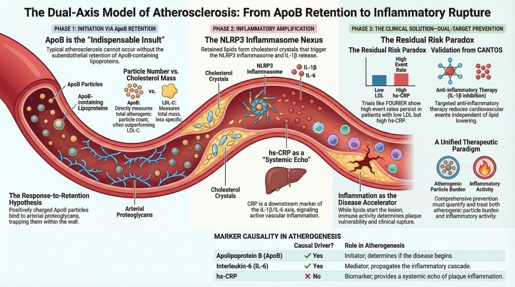
L'attuale consenso scientifico e clinico caratterizza l'aterosclerosi come una malattia infiammatoria guidata dai lipidi. All'interno di questo modello, apolipoproteina B (ApoBcontenente lipoproteine fornisci l'insulto iniziale indispensabile, mentre sistemico e locale infiammazione amplifica la crescita della placca, la vulnerabilità e il rischio di rottura. In altre parole, il lesione inizia con la ritenzione di particelle aterogeniche di ApoB e diventa pericolosa attraverso una risposta immunitaria maladattativa al lipide trattenuto e modificato.2-5,12,13,21,22]
Questa rassegna esamina la relazione tra ApoB, infiammazione e progressione dell'ASCVD. Tracciando la sequenza causale dell'inizio della placca, esaminando il percorso molecolare dalla ritenzione lipidica all'attivazione delle citochine e integrando la genetica umana con i principali studi sugli esiti cardiovascolari, viene sviluppato un modello unificato di aterogenesi. La tesi centrale è che il carico di ApoB determina se la biologia della malattia ha inizio, mentre la biologia infiammatoria influenza fortemente il ritmo della progressione e la probabilità di infarto miocardico, ictus, o morte cardiovascolare.2-5,6-9,12-18,21]
La Sequenza Causale: L'Ipotesi Risposta-Ritenzione
Per comprendere la relazione tra ApoB e infiammazione, è necessario stabilire la temporalità del primo evento aterogenico. La questione meccanicistica fondamentale è se le lipoproteine elevate contenenti ApoB diano inizio all'aterosclerosi prima che si sviluppi un'infiammazione localizzata, o se l'infiammazione prepari inizialmente la parete arteriosa e permetta successivamente il deposito di lipidi. Le prove attuali favoriscono fortemente una sequenza in cui la malattia inizia con la ritenzione arteriosa delle particelle di ApoB, con l'infiammazione che sorge come risposta biologica a tali particelle ritenute e che successivamente alimenta il processo peggiorando la malattia.2-5]
Evidenze istopatologiche e sperimentali supportano ipotesi della risposta alla ritenzione della precoce aterogenesi. Nella classica formulazione di Williams e Tabas, l'afflusso e la ritenzione di particelle ApoB ricche di colesterolo all'interno della parete arteriosa intima sono i primi passaggi necessari nella formazione della lesione. Il successivo aggiornamento di Circulation sulla ritenzione di lipoproteine subendoteliali afferma direttamente che il processo di innesco nell'aterogenesi è la ritenzione di lipoproteine contenenti ApoB al di sotto della endotelio, il che attiva poi la risposta infiammatoria dominata da macrofagi e cellule T che guida lo sviluppo della lesione.2,3]
Questo modello rimane affascinante perché integra la biologia arteriosa, matrice extracellulare legame e osservazione clinica. La particella di ApoB non è semplicemente un trasportatore passivo di lipidi. particelle contenenti ApoB si legano ai proteoglicani della parete arteriosa, specialmente nelle regioni vascolari predisposte alla formazione di lesioni. Una volta intrappolate, esse sono esposte a modificazioni ossidative, enzimatiche e aggregative, che le convertono in potenti stimoli infiammatori. Pertanto, l'evento più precoce non è l'infiammazione generalizzata o il danno endoteliale aspecifico, ma la ritenzione focale di lipoproteine aterogeniche in un microambiente arterioso suscettibile.2-5]
Confutazione del paradigma “Prima l'infiammazione”
Modelli alternativi, in particolare l'ipotesi della risposta al danno, hanno proposto che il danno endoteliale o un'infiammazione preesistente debbano precedere il deposito di lipidico. Questa visione ha preso piede perché le cellule infiammatorie sono abbondanti nelle placche stabili e perché le condizioni infiammatorie sistemiche accelerano l'ASCVD. Tuttavia, l'accelerazione infiammatoria non prova l'innesco infiammatorio. La questione decisiva è se l'infiammazione, in assenza di un'esposizione sufficiente ad ApoB, possa generare una tipica lesione ateromatosa ricca di colesterolo. Il peso delle prove suggerisce che non possa farlo.1-3,12,21,22]
I dati sperimentali e patologici indicano che disfunzione endoteliale, lo stress da taglio alterato e l'attivazione infiammatoria creano un ambiente permissivo per la formazione delle lesioni, ma il carico di ApoB determina se le lesioni si formano effettivamente e quanto estensivamente progrediscono. Il flusso alterato aiuta a spiegare la localizzazione delle lesioni; le lipoproteine contenenti ApoB ritenute spiegano l'inizio e l'espansione delle lesioni.2-5,12,22]
L'infiammazione sistemica agisce nondimeno come un potente amplificatore della malattia. Gli stati infiammatori cronici aumentano l'attivazione endoteliale, alterano la segnalazione vascolare e accelerano la progressione della placca. Tuttavia, in un ambiente povero di lipidi, non generano la classica lesione carica di colesterolo che definisce l'aterosclerosi. Questa distinzione è clinicamente importante perché spiega perché la terapia antinfiammatoria non elimina la necessità di una riduzione intensiva dell'ApoB.1,12,13,21,22]
Apolipoproteina B: La Variabile Iniziatrice
L'ApoB esiste in 2 principali isoforme aterogeniche: ApoB48 e ApoB100. L'ApoB48 viene sintetizzata nell'intestino ed è presente nei chilomicroni e i loro residui. L'ApoB100 viene sintetizzata nel fegato ed è presente nelle lipoproteine a densità molto bassa, nelle lipoproteine a densità intermedia, nelle lipoproteine a bassa densità e lipoproteina(a) [Lp(a)]. Poiché ciascuna particella aterogenica contiene esattamente 1 molecola di ApoB, la concentrazione plasmatica di ApoB riflette il numero totale di particelle aterogeniche circolanti. Ciò conferisce all'ApoB importanti vantaggi concettuali e clinici rispetto all'LDL-C, che riflette la massa di colesterolo anziché il numero di particelle.4,15,23,25,28]
L'importanza clinica dell'ApoB risiede nel fatto che l'aterosclerosi è guidata dal numero di particelle in grado di entrare ed essere trattenute nella parete arteriosa, e non semplicemente dalla quantità di colesterolo che trasportano. Il contenuto di colesterolo per particella varia notevolmente, specialmente in insulino-resistenza, sindrome metabolica, di tipo 2 diabete, e ipertrigliceridemia. In tali condizioni, l'LDL-C può sottostimare il vero carico di particelle aterogene, mentre l'ApoB cattura tale carico in modo più diretto.4,23,25,26,28]
L'ingresso di queste particelle nella parete arteriosa è guidato dal gradiente di concentrazione dal plasma all'intima. Una volta all'interno spazio sottendoteliale, i domini strutturali sull'ApoB facilitano la ritenzione da parte dei proteoglicani arteriosi. La ritenzione prolunga il tempo di residenza delle particelle e crea il substrato per l'ossidazione, l'aggregazione e l'attivazione immunitaria. Senza ritenzione, la cascata infiammatoria a valle non si sviluppa nella sua forma canonica.2-5]
La Lp(a) merita una menzione speciale all'interno della famiglia delle ApoB perché combina una particella simile alle LDL contenente ApoB con l'apolipoproteina(a) e sembra contribuire a effetti sia proaterogeni che proinfiammatori. Le prove genetiche e cliniche ora supportano la Lp(a) come causa di ASCVD fattore di rischio.[15]
Percorsi Meccanistici: Dalla Ritenzione Arteriosa all'Infiammazione Sistemica
Segue ritenzione subendoteliale di particelle contenenti ApoB, una serie coordinata di eventi biologici trasforma l'accumulo di lipidi focali in una lesione infiammatoria. Questo percorso collega l'iperlipidemia all'attivazione immunitaria innata e spiega come un quadro clinicamente silente stria lipidica può evolversi in una pericolosa placca.2,5,10-14,21,22]
Modificazione delle lipoproteine
Una volta trattenute nella matrice extracellulare intimale, le lipoproteine contenenti ApoB sono esposte a modificazioni ossidative ed enzimatiche. Questi processi generano LDL ossidata, fosfolipidi ossidati e particelle aggregate con proprietà biologiche distinte dalle lipoproteine native. Tali particelle modificate agiscono come segnali associati al pericolo all'interno della parete vascolare e alterano il comportamento delle cellule endoteliali e mieloidi.2,5,10-14]
Questi lipidi modificati non sono un carico passivo. Diventano ligandi infiammatori che attirano le cellule vascolari e immunitarie e sostengono l'evoluzione della lesione. La Lp(a), a causa del suo carico di fosfolipidi ossidati, può funzionare come un veicolo infiammatorio particolarmente potente dopo la ritenzione.5,13,15]
Disfunzione endoteliale e reclutamento dei monociti
I lipidi modificati interagiscono con le vie di riconoscimento dei pattern e attivano programmi trascrizionali infiammatori in cellule endoteliali e macrofagi. Questo sopprime la segnalazione endoteliale ateroprotettiva e promuove l'espressione di adesione leucocitaria molecole e chemochine, segnando la transizione dalla ritenzione silente di lipidi all'infiammazione vascolare attiva.12-14,21,22]
Il risultato è il reclutamento di monociti circolanti e linfociti T nello spazio subendoteliale. I monociti aderiscono, migrano attraverso l'endotelio attivato ed entrano in un microambiente ricco di lipidi che favorisce la differenziazione in macrofagi. Questo è il passaggio fondamentale da una lesione biochimica guidata dalla ritenzione di lipoproteine a una lesione cellulare dominata da effettori immunitari.12-14,21,22]
Differenziamento dei macrofagi, formazione di cellule schiumose e genesi dei cristalli
All'interno dell'intima, i monociti si differenziano in macrofagi e internalizzano lipoproteine modificate attraverso i recettori spazzini. Poiché queste vie di captazione non sono adeguatamente regolate inを下 (down-regulated) dal carico di colesterolo intracellulare, i macrofagi continuano a inglobare lipidi e diventano cellule schiumose, il segno distintivo della stria lipidica precoce.10-14]
Poiché l'assorbimento dei lipidi supera l'efflusso di colesterolo, il colesterolo libero intracellulare si accumula e può precipitare in cristalli di colesterolo. Questi cristalli sono oggi riconosciuti non come detriti inerti, ma come inneschi infiammatori che attivano le vie immunitarie innate centrali per la progressione della placca.10,11,14]
L'inflammasoma NLRP3 e la piroptosi
La formazione intracellulare e la fagocitosi dei cristalli di colesterolo rappresentano un legame meccanicistico chiave tra il lipide ritenuto e l'attivazione dell'immunità innata. Quando i macrofagi tentano di elaborare i cristalli di colesterolo, la rottura lisosomiale e lo stress cellulare associato attivano il inflammasoma NLRP3, un complesso multiproteico di rilevamento del pericolo implicato nell'aterogenesi.10,11,14]
Una volta assemblato, NLRP3 attiva la caspasi-1, che scinde la pro-IL-1β e la pro-IL-18 nelle loro forme mature e promuove vie di morte cellulare infiammatoria. Ciò porta al rilascio di citochine, proteasi e contenuti lipidici nello spazio extracellulare, contribuendo a centro necrotico espansione e destabilizzazione della placca. Questo è il punto in cui le lesioni croniche da accumulo di lipidi diventano lesioni infiammatorie attivamente destabilizzanti.10,11,14]
Questo modello incentrato sull'inflammasoma è terapeuticamente importante perché collega il lipide trattenuto alla produzione di IL-1β e fornisce una base meccanicistica per interventi quali canakinumab e colchicina.[6,10,11,14,29-32]
La sintesi epatica della proteina C-reattiva
Il rilascio di IL-1β all'interno della placca amplifica l'infiammazione sia localmente che a livello sistemico. L'IL-1β stimola la produzione di interleuchina-6 (IL-6) e altri mediatori a valle, e IL-6 a sua volta induce la sintesi epatica di Proteina C-reattiva. Un livello elevato di hs-CRP è quindi meglio compreso come un marcatore sistemico di segnalazione infiammatoria derivante, almeno in parte, da lesioni aterosclerotiche infiammate.6,12,14,17,18,21,29]
Poiché la PCR si trova a valle dell'asse IL-1β/IL-6, è clinicamente utile come biomarcatore dell'attività infiammatoria, ma potrebbe non essere di per sé il motore causale della malattia.17,18,21]
Il ruolo della PCR: biomarcatore contro mediatore causale
Data la forte associazione epidemiologica tra livelli elevati di hs-CRP e ASCVD incidenti, i ricercatori si sono chiesti se la CRP rifletta semplicemente l'infiammazione vascolare o contribuisca direttamente all'aterogenesi. Se la CRP fosse causale, essa stessa rappresenterebbe un interessante bersaglio terapeutico. Se fosse solo un marcatore, agire sulla CRP senza influenzare i pathway a monte difficilmente ridurrebbe gli eventi.17,18,21]
Prove della randomizzazione mendeliana
Randomizzazione mendeliana è stato fondamentale per la risoluzione di questo problema. Gli studi genetici sulle differenze a lungo termine della PCR hanno generalmente mostrato associazioni nulle o quasi nulle con la cardiopatia coronarica, mentre gli studi di mendeliana randomizzazione le analisi della segnalazione dell'IL-6 supportano la causalità del percorso. Questo schema suggerisce fortemente che la PCR sia un biomarcatore a valle piuttosto che il principale mediatore causale.17,18]
L'analisi di randomizzazione mendeliana di IL6R è particolarmente informativa. La sottoregolazione genetica della segnalazione del recettore dell'IL-6 è associata a una minore attività infiammatoria, a una minore proteina C-reattiva (PCR) a valle e a un minor rischio coronarico, a supporto della causalità del pathway dell'IL-6. Questa interpretazione si allinea con i successivi dati dei trial clinici che mostrano benefici dall'inibizione dell'IL-1β e con la più ampia prospettiva secondo cui i circuiti infiammatori a monte, e non la PCR in sé, rappresentano il bersaglio terapeutico rilevante.6,17,18,21]
Le prove provenienti da modelli animali sugli effetti diretti della proteina C-reattiva sono state contrastanti, soprattutto se confrontate con i dati genetici e clinici più coerenti che coinvolgono l'IL-1β e l'IL-6. Nel complesso, la proteina C-reattiva è da considerarsi preferibilmente un biomarcatore clinico di grande valore piuttosto che un bersaglio terapeutico centrale.17,18,21]
Interazione tra ApoB e infiammazione: prove cliniche fondamentali
Il riconoscimento che l'aterosclerosi dipende sia dall'accumulo di ApoB sia dall'amplificazione infiammatoria ha portato al moderno concetto di duplice rischio residuo percorsi. Alcuni pazienti rimangono ad alto rischio perché la loro aterogenica carico di particelle rimane inadeguatamente controllato. Altri ottengono importanti ipolipemizzante ma mantengono un rischio infiammatorio sostanziale, riflesso da livelli elevati di hs-CRP o da un'attivazione persistente delle citochine. Studi cardine come Giove, Canti, e Fourier illustra questi percorsi complementari.6-9,29,33]
GIOVE: infiammazione nella prevenzione primaria
Lo studio JUPITER ha arruolato individui apparentemente sani con livelli di LDL-C inferiori a 130 mg/dL e hs-CRP di almeno 2,0 mg/L, selezionando in tal modo una popolazione con livelli lipidici tradizionali modesti ma con un rischio infiammatorio accresciuto. Rosuvastatina ridotto sia il colesterolo LDL (LDL-C) che la proteina C-reattiva ad alta sensibilità (hs-CRP) e ridotto significativamente i principali eventi cardiovascolari. Lo studio ha dimostrato che può esistere un rischio cardiovascolare clinicamente rilevante nonostante livelli “normali” di LDL-C quando il carico infiammatorio è elevato, e che una terapia con effetti sia ipolipemizzanti che antinfiammatori può ridurre sostanzialmente il rischio.7]
CANTOS: isolare l'ipotesi infiammatoria
CANTOS ha testato direttamente se la riduzione dell'infiammazione, indipendente dalla riduzione dei lipidi, migliora gli esiti. Nei pazienti con precedente infarto miocardico e persistente elevazione della hs-CRP nonostante la terapia standard, il canakinumab ha ridotto sostanzialmente l'IL-6 e la hs-CRP senza abbassare i livelli di LDL-C, HDL-C o ApoB, e ha ridotto gli eventi cardiovascolari ricorrenti. Le analisi secondarie hanno suggerito un beneficio maggiore in coloro che hanno ottenuto una riduzione più profonda della hs-CRP. CANTOS ha quindi fornito la prova di principio che una terapia anti-infiammatoria mirata può ridurre gli eventi ASCVD anche quando i livelli lipidici rimangono invariati.6,29]
FOURIER: rischio infiammatorio persistente nonostante livelli di lipidi ultra-bassi
FOURIER ha testato la riduzione intensiva dei lipidi con evolocumab nei pazienti con ASCVD conclamata. L'evolocumab ha ridotto drasticamente l'LDL-C, mentre la hs-CRP è rimasta sostanzialmente invariata. Lo studio ha confermato il beneficio di una riduzione più marcata dei lipidi, ma le analisi successive hanno dimostrato che la hs-CRP e l'LDL-C sono rimasti predittori indipendenti dei risultati. Anche a livelli di LDL-C molto bassi, valori più elevati di hs-CRP identificavano un rischio residuo maggiore. Lo studio FOURIER ha così dimostrato che rischio infiammatorio residuo persiste anche quando il rischio lipidico viene ridotto a livelli molto bassi.8,9,33]
Insieme, questi studi clinici convalidano un modello a doppio asse. JUPITER ha evidenziato il rischio infiammatorio in prevenzione primaria, CANTOS ha dimostrato che la terapia antinfiammatoria selettiva riduce gli eventi senza abbassare i lipidi, e FOURIER ha dimostrato che l'intenso abbassamento dei lipidi lascia un pool misurabile di rischio infiammatorio residuo. L'implicazione non è quella di scegliere tra lipidi e infiammazione, ma di trattarli entrambi.6-9,29,33]
Sinergia Epidemiologica: Approfondimenti dalla UK Biobank e dalle Coorti Contemporanee
Le analisi su vaste popolazioni completano i dati degli studi clinici mostrando come il carico di ApoB e il rischio infiammatorio interagiscano su un'ampia distribuzione del rischio. Gli studi di coorte contemporanei e le analisi basate sulla UK Biobank indicano che il carico di ApoB e i marker infiammatori forniscono spesso informazioni complementari, con i tassi di eventi più elevati che si osservano frequentemente quando entrambi sono elevati. Particolarmente importanti sono discordanza analisi che dimostrano che l'ApoB spesso prevede il rischio meglio dell'LDL-C quando le due misure sono in disaccordo.4,23,25,28]
Nella sindrome metabolica e nell'ipertrigliceridemia, l'LDL-C può apparire ingannevolmente basso poiché ogni particella trasporta meno colesterolo, mentre l'ApoB riflette ancora il numero reale di particelle aterogene. Recenti studi di coorte suggeriscono inoltre che gli approcci basati sull'ApoB possono migliorare la discriminazione del rischio rispetto alle metriche basate sul colesterolo in condizioni di discordanza.4,23,25,26,28]
Queste osservazioni epidemiologiche rafforzano il quadro meccanicistico. Un valore elevato di ApoB indica una maggiore opportunità di ingresso e ritenzione arteriosa, mentre un valore elevato di hs-CRP segnala un'amplificazione infiammatoria attiva. Quando sono presenti entrambi, il rischio aumenta sensibilmente. Ciò non pone la PCR sullo stesso livello causale dell'ApoB, ma supporta la misurazione di entrambi per perfezionare la valutazione del rischio clinico.4,17,18,23,25,28]
Implicazioni cliniche e prospettive delle linee guida 2025
La lipidologia moderna riconosce sempre più che, poiché la massa di colesterolo per LDL la particella varia, la dipendenza da colesterolo totale o il solo C-LDL calcolato può classificare erroneamente il rischio. L'ApoB inventaria direttamente le particelle aterogeniche circolanti capaci di penetrare nella parete arteriosa. La rassegna del 2024 di Circulation sull'ApoB sostiene un uso clinico più ampio dell'ApoB a causa del suo maggiore allineamento biologico con la causalità delle malattie cardiovascolari aterosclerotiche (ASCVD) e della sua utilità negli stati discordanti.4,23,25,28]
Questo cambiamento si riflette nelle linee guida contemporanee. L'AACE del 2025 dislipidemia la linea guida enfatizza la gestione farmacologica moderna basata sul rischio, mentre l'aggiornamento mirato ESC/EAS del 2025 supporta un uso più precoce e ampio della terapia di combinazione, inclusi statine, ezetimibe, acido bempedoico, e PCSK9 inibizione, per ridurre il carico aterogenico. Sebbene le soglie specifiche delle diverse società differiscano, la direzione generale è coerente: l'ApoB sta acquisendo importanza come marcatore modificabile, soprattutto nei pazienti ad alto rischio e discordanti.19,20]
L'infiammazione è passata allo stesso modo da concetto accademico a concetto clinicamente applicabile. La dichiarazione scientifica dell'ACC del 2025 sottolinea la rilevanza clinica dell'infiammazione lungo l'intero continuum dell'ASCVD. La hs-CRP rimane utile per la stima del rischio in determinati contesti di prevenzione primaria e per l'identificazione del rischio infiammatorio residuo in prevenzione secondaria. La colchicina a basso dosaggio è emersa come un'opzione antinfiammatoria pratica in pazienti selezionati con ASCVD conclamata, e il lavoro in corso sulla modulazione del percorso dell'IL-6 e sulle strategie di riduzione della Lp(a) potrebbe perfezionare ulteriormente il trattamento.20,27,30-32,35]
Dibattiti scientifici irrisolti: Definire la malattia
Nonostante una vasta convergenza, permane un dibattito semantico sul fatto che l'aterosclerosi sia fondamentalmente guidata dai lipidi, infiammatoria o un'infiammazione indotta dai lipidi. La visione incentrata sui lipidi è corretta in un senso cruciale: senza lipoproteine contenenti ApoB, l'aterosclerosi tipica non si sviluppa. La genetica umana, la patologia e gli interventi di riduzione dei lipidi supportano fortemente questo punto.2-5,15,16,23]
Tuttavia, un modello puramente incentrato sui lipidi non spiega pienamente perché alcuni pazienti con un eccellente controllo lipidico continuino a manifestare rottura di placca e ricorrenti eventi. Per contro, la visione incentrata sull'infiammazione sottolinea giustamente che i meccanismi immunitari dominano la destabilizzazione della placca, la degradazione del cappuccio e la rottura, ma l'infiammazione in un ambiente povero di lipidi non produce il classico quadro ricco di colesterolo ateroma. La sintesi più accurata è quindi che l'aterosclerosi è una malattia infiammatoria indotta dai lipidi: iniziata dalla ritenzione di ApoB, poi amplificata e resa clinicamente pericolosa da risposte immunitarie innate e adattive maladattative al lipide ritenuto.1-3,10-14,21,22,27,29-35]
Conclusione
La fisiopatologia dell'ASCVD richiede la convergenza di due assi biologici strettamente correlati. La malattia inizia con l'afflusso e la ritenzione subendoteliale di lipoproteine contenenti ApoB all'interno della parete arteriosa. Questo costituisce la lesione iniziale. La progressione da una stria lipidica clinicamente silente a una placca vulnerabile, necrotica e soggetta a rottura dipende dall'amplificazione infiammatoria guidata da lipidi modificati, biologia delle cellule schiumose, cristalli di colesterolo, attivazione dell'inflammasoma NLRP3 e segnalazione citochinica tramite IL-1β e IL-6. La hs-CRP emerge da questa cascata come un marcatore sistemico informativo piuttosto che come l'agente causale primario.2-5,10-14,17,18,21]
Gli studi clinici fondamentali degli ultimi due decenni hanno dimostrato che il trattamento del fattore determinante ApoB mediante statine e inibizione di PCSK9 e il trattamento del fattore di amplificazione infiammatoria mediante inibizione di IL-1β o colchicina possono fornire un beneficio indipendente e complementare. Per ridurre rischio cardiovascolare residuo In modo significativo, la pratica contemporanea deve andare oltre la sola massa del colesterolo e valutare sia il carico di particelle aterogene sia l'attività infiammatoria residua. Questo quadro a duplice rischio è la più chiara implicazione pratica della ricerca moderna sull'aterosclerosi.4,6-9,19,20,27,29-35]
Riferimenti
- Libby P. Inflammation in atherosclerosis. Nature. 2002;420(6917):868-874. doi:10.1038/nature01323
- Tabas I, Williams KJ, Borén J. Subendothelial lipoprotein retention as the initiating process in atherosclerosis: update and therapeutic implications.
- Williams KJ, Tabas I. The response-to-retention hypothesis of early atherogenesis.
- De Oliveira-Gomes D, Joshi PH, Peterson ED, Rohatgi A, Khera A, Navar AM. Apolipoprotein B: Bridging the Gap Between Evidence and Clinical Practice. Circulation. 2024;150(1):62-79. doi:10.1161/CIRCULATIONAHA.124.068885
- Borén J, Williams KJ. The central role of arterial retention of cholesterol-rich apolipoprotein-B-containing lipoproteins in the pathogenesis of atherosclerosis: a triumph of simplicity. Curr Opin Lipidol. 2016;27(5):473-483. doi:10.1097/MOL.0000000000000330
- Ridker PM, Everett BM, Thuren T, et al. Antiinflammatory Therapy with Canakinumab for Atherosclerotic Disease. N Engl J Med. 2017;377(12):1119-1131. doi:10.1056/NEJMoa1707914
- Ridker PM, Danielson E, Fonseca FA, et al. Rosuvastatin to prevent vascular events in men and women with elevated C-reactive protein. N Engl J Med. 2008;359(21):2195-2207. doi:10.1056/NEJMoa0807646
- Sabatine MS, Giugliano RP, Keech AC, et al. Evolocumab and Clinical Outcomes in Patients with Cardiovascular Disease. N Engl J Med. 2017;376(18):1713-1722. doi:10.1056/NEJMoa1615664
- Pradhan AD, Aday AW, Rose LM, Ridker PM. Residual Inflammatory Risk on Treatment With PCSK9 Inhibition and Statin Therapy. Circulation. 2018;138(2):141-149. doi:10.1161/CIRCULATIONAHA.118.034645
- Duewell P, Kono H, Rayner KJ, et al. NLRP3 inflammasomes are required for atherogenesis and activated by cholesterol crystals. Nature. 2010;464(7293):1357-1361. doi:10.1038/nature08938
- Grebe A, Latz E. Cholesterol crystals and inflammation. Curr Rheumatol Rep. 2013;15(3):313. doi:10.1007/s11926-012-0313-z
- Libby P, Ridker PM, Hansson GK; Leducq Transatlantic Network on Atherothrombosis. Inflammation in atherosclerosis: from pathophysiology to practice. J Am Coll Cardiol. 2009;54(23):2129-2138. doi:10.1016/j.jacc.2009.09.009
- Tall AR, Yvan-Charvet L. Cholesterol, inflammation and innate immunity. Nat Rev Immunol. 2015;15(2):104-116. doi:10.1038/nri3793
- Grebe A, Hoss F, Latz E. NLRP3 Inflammasome and the IL-1 Pathway in Atherosclerosis. Circ Res. 2018;122(12):1722-1740. doi:10.1161/CIRCRESAHA.118.311362
- Tsimikas S, Stroes ESG. The dedicated “Lp(a) clinic”: A concept whose time has arrived?. Atherosclerosis. 2020;300:1-9. doi:10.1016/j.atherosclerosis.2020.03.003
- Ference BA, Kastelein JJP, Ray KK, et al. Association of Triglyceride-Lowering LPL Variants and LDL-C-Lowering LDLR Variants With Risk of Coronary Heart Disease. JAMA. 2019;321(4):364-373. doi:10.1001/jama.2018.20045
- Zacho J, Tybjaerg-Hansen A, Jensen JS, Grande P, Sillesen H, Nordestgaard BG. Genetically elevated C-reactive protein and ischemic vascular disease. N Engl J Med. 2008;359(18):1897-1908. doi:10.1056/NEJMoa0707402
- Interleukin-6 Receptor Mendelian Randomisation Analysis (IL6R MR) Consortium, Swerdlow DI, Holmes MV, et al. The interleukin-6 receptor as a target for prevention of coronary heart disease: a mendelian randomisation analysis. Lancet. 2012;379(9822):1214-1224. doi:10.1016/S0140-6736(12)60110-X
- Mach F, Koskinas KC, Roeters van Lennep JE, et al. 2025 Focused Update of the 2019 ESC/EAS Guidelines for the management of dyslipidaemias. Eur Heart J. 2025;46(42):4359-4378. doi:10.1093/eurheartj/ehaf190
- Patel SB, Wyne KL, Afreen S, et al. American Association of Clinical Endocrinology Clinical Practice Guideline on Pharmacologic Management of Adults With Dyslipidemia. Endocr Pract. 2025;31(2):236-262. doi:10.1016/j.eprac.2024.09.016
- Ridker PM. From C-Reactive Protein to Interleukin-6 to Interleukin-1: Moving Upstream To Identify Novel Targets for Atheroprotection. Circ Res. 2016;118(1):145-156. doi:10.1161/CIRCRESAHA.115.306656
- Hansson GK, Hermansson A. The immune system in atherosclerosis. Nat Immunol. 2011;12(3):204-212. doi:10.1038/ni.2001
- Sniderman AD, Thanassoulis G, Glavinovic T, et al. Apolipoprotein B Particles and Cardiovascular Disease: A Narrative Review. JAMA Cardiol. 2019;4(12):1287-1295. doi:10.1001/jamacardio.2019.3780
- Aday AW, Ridker PM. Targeting Residual Inflammatory Risk: A Shifting Paradigm for Atherosclerotic Disease. Front Cardiovasc Med. 2019;6:16. Published 2019 Feb 28. doi:10.3389/fcvm.2019.00016
- Mora S, Buring JE, Ridker PM. Discordance of low-density lipoprotein (LDL) cholesterol with alternative LDL-related measures and future coronary events. Circulation. 2014;129(5):553-561. doi:10.1161/CIRCULATIONAHA.113.005873
- Arsenault BJ, Després JP, Stroes ES, et al. Lipid assessment, metabolic syndrome and coronary heart disease risk. Eur J Clin Invest. 2010;40(12):1081-1093. doi:10.1111/j.1365-2362.2010.02357.x
- Mensah GA, Arnold N, Prabhu SD, Ridker PM, Welty FK. Inflammation and Cardiovascular Disease: 2025 ACC Scientific Statement: A Report of the American College of Cardiology. J Am Coll Cardiol. Published online September 29, 2025. doi:10.1016/j.jacc.2025.08.047
- Sniderman AD, Dufresne L, Pencina KM, Bilgic S, Thanassoulis G, Pencina MJ. Discordance among apoB, non-high-density lipoprotein cholesterol, and triglycerides: implications for cardiovascular prevention. Eur Heart J. 2024;45(27):2410-2418. doi:10.1093/eurheartj/ehae258
- Ridker PM, MacFadyen JG, Everett BM, et al. Relationship of C-reactive protein reduction to cardiovascular event reduction following treatment with canakinumab: a secondary analysis from the CANTOS randomised controlled trial. Lancet. 2018;391(10118):319-328. doi:10.1016/S0140-6736(17)32814-3
- Nidorf SM, Fiolet ATL, Mosterd A, et al. Colchicine in Patients with Chronic Coronary Disease. N Engl J Med. 2020;383(19):1838-1847. doi:10.1056/NEJMoa2021372
- Nidorf SM, Eikelboom JW, Budgeon CA, Thompson PL. Low-dose colchicine for secondary prevention of cardiovascular disease. J Am Coll Cardiol. 2013;61(4):404-410. doi:10.1016/j.jacc.2012.10.027
- Tardif JC, Kouz S, Waters DD, et al. Efficacy and Safety of Low-Dose Colchicine after Myocardial Infarction. N Engl J Med. 2019;381(26):2497-2505. doi:10.1056/NEJMoa1912388
- O’Donoghue ML, Fazio S, Giugliano RP, et al. Lipoprotein(a), PCSK9 Inhibition, and Cardiovascular Risk. Circulation. 2019;139(12):1483-1492. doi:10.1161/CIRCULATIONAHA.118.037184
- Ference BA, Kastelein JJP, Ginsberg HN, et al. Association of Genetic Variants Related to CETP Inhibitors and Statins With Lipoprotein Levels and Cardiovascular Risk. JAMA. 2017;318(10):947-956. doi:10.1001/jama.2017.11467
- Du Z, Li F, Jiang L, et al. Metabolic systems approaches update molecular insights of clinical phenotypes and cardiovascular risk in patients with homozygous familial hypercholesterolemia. BMC Med. 2023;21(1):275. Published 2023 Jul 27. doi:10.1186/s12916-023-02967-8

