心血管疾患 (CVD)は依然として現代における最も手強い公衆衛生上の課題であり、世界的死亡の主要な原因として君臨し、世界中の全死亡のおよそ3分の1を占めている。1の正統な理解 動脈硬化線維脂肪性病変による動脈血管の進行性かつ潜行性の狭窄および硬化 歯垢 供述調書:発症機序を、十分に確立された、主に修正可能な一連の要因に帰している 危険因子. これらの古典的な決定要因には、全身性の 高血圧, 脂質異常症 (具体的には高値の低密度 リポタンパク質 コレステロール)、2型 糖尿病 糖尿病, 肥満, 、運動不足、高齢化、およびタバコの消費。1しかし、根強く、かつ深く 交絡 疫学上のパラドックスは、この伝統的なパラダイムの絶対的な網羅性に対して長年にわたり疑問を投げかけてきた。すなわち、致命的な症例を含む心血管疾患の初回発症の約15%が、 心筋梗塞 さらに、これらの既確立された危険因子が完全にまったく存在しない患者においても、脳血管障害が発生する。1]
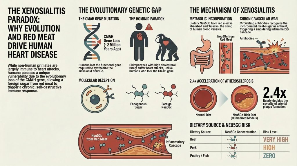
この深遠な臨床的異常は、種間比較心臓病学の分野によって、さらに複雑化されると同時に解明される。より広い哺乳類の王国を観察すると、アテローム性動脈硬化に起因する自然発生的な冠動脈血栓症や心筋梗塞は、 プラーク破裂 人間以外の種では事実上存在しない。1この抵抗性は、私たちの最も近い進化上の親戚である非ヒト霊長類においても、驚くほどしっかりと保たれている。飼育下のチンパンジーは、人間のような座りがちなライフスタイルを共有することが多く、重度の高脂血症、高齢化、全身性高血圧など、驚くほど類似した生理学的障害を示す。1] それにもかかわらず、人間のようなCVD(心血管疾患)リスクの高い血液を所有しているにもかかわらず、 脂質プロファイル そして軽度の初期アテローム性動脈硬化の証拠はあるものの、これらの霊長類が終末期のアテローム性動脈硬化性CVDイベントに苦しむことはめったにない。2] 壊滅的なとき 心臓発作 チンパンジーの個体群でも発生するが、厳密な死後分析により、それらの 心臓発作 まだ説明されていない未解明の要素によって圧倒的に引き起こされている 間質性心筋線維症心筋のびまん性瘢痕化であり、典型的なアテローム性プラークの破裂とその後の 血栓症 ヒト患者で観察される。2さらに、一方で重い 赤身肉 摂取はヒト集団におけるCVDリスクの普遍的に認められている疫学的増幅因子であるが、この正確な食習慣は誘発しない アテローム発生 他の肉食性または雑食性の哺乳類において。2]
この深い乖離は、ヒトにおけるアテローム性動脈硬化症への種特異的かつ内在的な遺伝的素因を容赦なく指し示している。1この特異な脆弱性の核心は、脂質代謝、血行動態のずり応力、あるいは行動変数の単独にあるのではなく、細胞表面の基本的な生物学に関わる太古の進化上の遺伝的異常にある。 糖鎖. 具体的には、ヒトの心血管系の感受性は、生合成能力の進化的な喪失と不可分に関連している。 N-グリコリルノラミン酸 (Neu5Gc), 、哺乳類のどこにでもあるシアル酸。1この重大な遺伝的欠損にもかかわらず、Neu5Gcが豊富な赤身肉の人間による摂取は、この非ヒト糖鎖の代謝的取り込みをもたらし、それは直接人間の組織、特には血管系に組み込まれる。 内皮.[3,4] その後、取り込まれたこの糖の免疫学的認識は、「“異種自己抗原”は、次のような持続的で燻るような炎症の連鎖を引き起こします。“異物唾液腺炎.”[5]
異種シアリル炎仮説の根底にある、非常に複雑な進化、生化学、免疫学的メカニズムを解明するこの網羅的な調査レポートは、ゲノム欠失の探索を通じて CMAH遺伝子, 、食事の複雑なマイクロバイオームを介した消化 Neu5Gc, ヒトの細胞内糖鎖付加経路の分子乗っ取り、およびその結果生じるポリクローナル抗体媒介性の内皮破壊に着目し、本分析は、なぜヒトが赤肉関連のアムロスクレローシス(動脈硬化症)に特異的に罹患しやすいのか、そしてこのメカニズムがどのように生涯にわたって心血管リスクを定量的に増大させるのかについての包括的な理解を提供する。.
進化のパラダイム:CMAH遺伝子の変異と人類の起源
シアル酸の基礎生物学
ヒト遺伝的欠損の大きさとその重大性を正確に理解するには、まず以下の生理学的および構造的役割を明らかにしなければならない。 シアル酸 脊椎動物の生物学において。すべての脊椎動物細胞の表面は、主に膜貫通タンパク質に結合した、高密度で複雑かつ高度に制御された糖鎖の配列によって緻密に飾られている。 タンパク質 そして表面脂質が形成する グリコカリックス.[6,7ヒトの血中を循環する可溶性分泌タンパク質のほとんども、同様にこのような特殊な糖鎖で修飾されている。6,7シアル酸は、9炭素骨格を基盤とする高度に修飾された単糖の多様なファミリーを構成しており、通常、これらグリカン鎖の最も外側の末端をキャップするように存在している。6,7]
糖タンパク質および糖脂質における末端に位置すること、ならびに生理学的pHにおいて強い負電荷を帯びていることが相まって、シアル酸は、細胞間接着の媒介、受容体とリガンドの相互作用の調節、および安定化において、不可欠かつ多面的な役割を果たしている 内皮グリコカリックス バリアとして機能し、特定のシアル酸結合性レクチン(Siglec)を介して免疫学的自己認識を調節する。6,8]
大半のほ乳類種、そして実際にはほとんどの陸生脊椎動物において、シア酸の2つの主要な形態は、N-アセチルノイラミン酸(Neu5Ac)とその直接の水酸化誘導体であるN-グリコイルノイラミン酸(Neu5Gc)である。6,7これら2つの分子は構造的にほぼ同一であり、わずか1つの酸素原子の有無のみが異なっている。5前駆体Neu5Acから誘導体Neu5Gcへの生化学的変換は、単一の特異性の高い酵素であるシチジン一リン酸-N-アセチルノイラミン酸ヒドロキシラーゼ(CMAH)によってのみ触媒される。1,3この crucial な酵素は、細胞質基質内においてヌクレオチド糖ドナーであるCMP-Neu5Acに作用し、水酸基を付加してCMP-Neu5Gcを生成するモノオキシゲナーゼ反応を促進する。6,7CMP-Neu5AcおよびCMP-Neu5Gcの両方は、その後、細胞質から ルーメン ゴルジ体に送られ、そこで新しく合成された物質への糖付加における主要なヌクレオチド糖供与体として機能する 糖複合体.[6,7その結果、通常の哺乳類組織は、その細胞表面にNeu5AcとNeu5Gcの両方が豊富に混ざり合った、高度に機能的なモザイク状の構造を示している。6,7]
| 特徴 | N-アセチルノラミン酸(Neu5Ac) | N-グリコリルノラミン酸 (Neu5Gc) |
| 構造的基盤 | 炭素数9の単糖 | 炭素数9の単糖 |
| 化学的違い | N-アセチル基を有する | N-グリコリル基を持つ(1つの酸素原子の付加) |
| 生合成的由来 | すべての脊椎動物においてデノボ合成される | CMAH酵素を介してNeu5Acから合成される |
| 人間におけるステータス | 主要な内因性のヒトシアル酸 | 遺伝的に欠損しており、「異物」とみなされる“ 異種抗原 |
| 哺乳類の分布 | 哺乳類の種に広く見られる | ヒト以外の哺乳類には広く見られるが、ヒトには存在しない |
アルミニウムを介した疑似遺伝子化事象
しかし、ヒトは哺乳類の系統において、顕著かつ重大な異常を示している。正常で健康なヒトの組織は、遺伝的かつ根本的にNeu5Gcを合成することができない。[5] この生化学的な切断は、調節による抑制ではなく、CMAH遺伝子そのものにおける種特異的で不可逆的な変異の直接的な結果である。[1,3]
古ゲノム推定および比較系統学的解析によると、およそ200万年から300万年前――これは属の出現に相当する時代――に ホモ ヒト科の祖先から――重要なゲノム上の出来事が起こった。[1,9,12] ある Alu可動要素, 、霊長類のゲノムに多く見られる「跳躍遺伝子」の一種が、CMAH遺伝子の遺伝子座に挿入された。この挿入により、CMAH遺伝子内の重要な92塩基対からなるエクソンにおいて壊滅的なフレームシフト欠失が生じ、その結果、早期終止コドンが出現し、完全な 擬原化 (その遺伝子座の完全な不活性化)。[9,12] CMP-Neu5AcからCMP-Neu5Gcへの酵素反応は厳密に一方向であり、完全にCMAHに依存しているため、この単一の酵素が失われたことで、生合成経路は永久に断たれてしまった。[1,3] その直接的な結果として、現代人は前駆体であるNeu5Acのみを生成し、細胞表面からは内因性のNeu5Gcが完全に失われ、ヒトの生体における生化学的状況が劇的に変化している。[1,3]
進化的選択圧:病原体と生殖的隔離
細胞表面に広く存在する主要な分子の合成に関与する遺伝子における機能喪失変異の固定は、極めて特異な進化的出来事である。 宿主のグリコカリックスにおけるこのような劇的かつ突然の変化は、極めて激しい進化的選択圧によって引き起こされたに違いない。なぜなら、このような根本的な生化学的変化が個体群全体にわたって完全に固定化されたことを、確率的な遺伝的浮動のみによって説明することは極めて考えにくいからである。[12] 現在の進化生物学では、祖先の人類集団においてCMAHノックアウト対立遺伝子が急速に固定されたことについて、多因子モデルが提唱されている。.
最も有力であり、かつ最も精力的に研究が進められている仮説は、宿主と病原体との間の熾烈な進化上の軍拡競争に関するもので、具体的にはマラリアの原因となる マラリア原虫 寄生虫。[9,12] 哺乳類細胞表面のシアル酸は、さまざまなウイルス、細菌、寄生虫などの病原体にとって、主要な結合受容体および認識モチーフとして機能している。[6,7] 選択のゲノムシグネチャに基づくと、祖先の人類は、Neu5Gcを豊富に含む赤血球に優先的に結合して細胞内へ侵入する、極めて毒性の強い「非ヒトヒト科動物(NHH)」マラリア株によって、その個体数が激減した可能性が極めて高い。[9,12] AluによるCMAH遺伝子の喪失は、他の点では有害である可能性があったものの、この変異を持つごく少数の個体には、命を救うような突然の生存上の優位性をもたらした。 病原体が好む細胞表面受容体を宿主から事実上剥奪することで、CMAH欠損のヒト科生物は、当時蔓延していたマラリアの脅威から逃れることができ、その結果、生存した集団全体にこのヌル変異が固定される大規模な選択的掃討が生じた。[9,12] しかし、この進化上の勝利は、深遠な時間のスケールで見れば一時的なものに過ぎなかった。. 熱帯熱マラリア原虫—現代の人間に悪性マラリアを引き起こす、極めて致死性の高い病原体は、やがて、新たに遍在するようになったヒト特有のNeu5Acを豊富に含む赤血球に優先的に結合する能力を進化させ、宿主と病原体の相互作用の動態を再構築した。[6,7,12]
二次的なものとはいえ、同様に説得力があり、前者の仮説を補完するもう一つの仮説として、生殖的隔離と、雌の免疫系を介した「破局的な選択」が挙げられる。[12] 哺乳類の精子は高度にシアル酸修飾されており、複雑な表面糖鎖を利用して、精子の生存率を高め、免疫原性のあるタンパク質を隠蔽し、雌の生殖管を通って卵管内の受精部位に至る危険な旅路において、その機能を最適化している。[12] 希少なCMAH-null変異を持つ女性ヒト属は、出生後に環境や食事からNeu5Gcにさらされると、免疫系がその分子を非自己と認識するため、当然ながら血中を循環する抗Neu5Gc抗体が産生されることになる。[10,12,18] もしこの変異体雌がその後、野生型(CMAH陽性)の雄と交尾した場合、その血中抗体はNeu5Gcを豊富に含む父方の精子を積極的に標的とし、中和してしまうため、雄の繁殖成功率を著しく低下させ、事実上、そのペアの存続可能性を損なうことになる。[12] 数千世代にわたって、父系由来のNeu5Gcに対するこの雌による免疫媒介的選択は、変異型集団と野生型集団の間に強烈かつ揺るぎない生殖的障壁を生み出し、CMAH欠損遺伝子型の急速な固定を促進するとともに、最終的にはヒトの祖先を他のヒト科動物から隔離することになった種分化の過程に直接寄与した可能性がある。[12]
食事のミスマッチ:外因性シアル酸の消化と微生物による代謝
CMAH遺伝子の進化上の喪失により、Neu5Gcは生化学的に現代人の生体にとって完全に異物となっている。[5] しかし、このような深刻な遺伝的欠乏があるにもかかわらず、現代人の食事――特に西洋化された社会における食事――には、この分子が大量に含まれている。Neu5Gcは、哺乳類由来の赤身肉、具体的には牛肉、豚肉、羊肉に高濃度で含まれており、これらは依然として世界中で主要な食糧となっている。[3,8,22] さらに、これらの哺乳類由来の乳製品にも、人間以外のシアル酸が、量は少ないものの、検出可能なレベルで含まれている。[8]
厳密な生化学的分析および高速液体クロマトグラフィーにより、Neu5Gcへの曝露に関する食事上の実態が明確に解明された。研究によると、牛肉にはこの分子が最も多く含まれており、Neu5Gcの含有量は約30.1 µg/gである。これに続いて、豚肉が25.5 µg/gと僅差で続いている。[7,8] 一方、鳥類(家禽など)や多様な魚の系統は、CMAH遺伝子が進化するはるか以前に哺乳類の系統樹から分岐したか、あるいは独立してその遺伝子を失ったため、生来、Neu5Gcを全く有していない。[7,8] この厳格な生化学的二分法により、食事によるNeu5Gcの摂取は、哺乳類の赤身肉の摂取に極めて特異的なものとなっている。[7,8]
| 食事からの摂取源 | Neu5Gc含有量の分類 | Neu5Gcの特定濃度 |
| 牛肉 | 極めて高い | ~30.1 µg/g |
| 豚肉 | 高 | ~25.5 µg/g |
| 子羊/ウサギ | 高 | 牛肉・豚肉と同等 |
| 乳製品 | 中程度 | 変異性があり、乳糖タンパク質に含まれる |
| 家禽(鶏、七面鳥) | ゼロ(生化学的に検出されない) | 0.0 µg/g |
| 魚・魚介類 | ゼロ(生化学的に検出されない) | 0.0 µg/g |
腸内微生物叢の重要な役割
食事由来のNeu5Gcが、胃腔内に留まっている摂取食品の状態から、吸収され全身的に作用する生化学的物質へと移行する過程は、宿主の胃腸マイクロバイオームによって厳密に制御されている。 食事由来のNeu5Gcは、一般的に遊離単糖として摂取されるのではなく、主にグリコシド結合した形態で摂取され、哺乳類の組織(ブタの顎下腺ムチンなど)を構成する複雑な糖タンパク質、ムチン、および糖脂質に深く組み込まれている。[8,17] 結合型と遊離型のシアル酸とのこの区別は、生理学的に極めて重要です。遊離状態で結合していないNeu5Gcは、技術的には腸管バリアを通過して吸収される可能性がありますが、体内では、小さく、溶解性が高く、利用できない分子として認識されます。 その結果、遊離のNeu5Gcは腎系によって迅速に除去され、尿中に効率的に排泄されるため、組織への長期的な蓄積や病変を引き起こすことは一切ない。[11,16] 複雑な高分子配列を形成して提示される、構造的に結合したNeu5Gcのみが、代謝による取り込みという危険な過程を経る。[11,16,17]
ヒトおよびマウスのメタゲノムシーケンシングにおける最近の進展 腸内マイクロバイオーム Neu5Gcを豊富に含む食事に対して、常在微生物叢が動的かつ迅速に反応することが明らかになった。[17] 具体的には、以下の目に属する個体群 バクテロイダ目 そして クロストリジアル目 赤身肉に関連する要因にさらされると、著しい表現型の拡大が見られる 炭水化物.[17] これらの特定の細菌系統は、宿主の粘膜ムチン、そしてとりわけ食物由来の糖タンパク質から末端のシアル酸を切断するように特別に設計された、高度に専門化された細菌性シアル酸分解酵素(ノイラミニダーゼ)を発現するように進化してきた。[17,20,21]
従来、ヒトの腸管内で最も豊富に存在する基質であることから、細菌のシアリダーゼは宿主由来のNeu5Acのみを特異的に好むと想定されていた。 しかし、最先端のX線結晶構造解析を用いた詳細な構造研究により、特定の細菌性シアリダーゼ内に、これまで観察されたことのない特有のアミノ酸配列が同定され、これがNeu5Gcを含む糖鎖に対して異常に高い親和性を示すことが明らかになった。[17] これらの特殊な腸内微生物は、マイクロバイオーム内で複雑な相互栄養の動態を調整しており、その過程でNan遺伝子経路を利用して、切断されたシアル酸を積極的に代謝し、エネルギーや炭素源として利用している。[20,21] 興味深いことに、Neu5Acの下流の分解過程では酢酸が副産物として生成されるのに対し、Neu5Gcの細菌による代謝ではグリコール酸が生成される。[20,21] こうした代謝産物の違いは、結腸上皮の局所的なpHや代謝環境を微妙に変化させ、より広範な炎症環境の形成に寄与している可能性がある。[20,21]
これらの微生物由来のシアリダーゼが、大量の肉由来糖タンパク質からNeu5Gcを酵素的に遊離させ、より小さな特定のオリゴ糖断片(GalNGcなど)へと分解する過程が、このヒト以外の糖のその後の腸管吸収および全身への分布に必要な第一段階である可能性が極めて高いと見られている。[8,17]
細胞内ハイジャック:代謝的取り込みのメカニズム
食事由来のグリコシド結合型Neu5Gc(またはそのマイクロバイオームによって遊離されたオリゴ糖中間体)が、複雑な腸上皮を通過すると、全身循環に入り込む。 赤身肉を摂取した後、これらの異種グリカンがヒトの循環系に現れ、数時間にわたって定常状態のレベルが維持されることが実証されており、これにより組織との相互作用が十分に起こり得る。[11,15,16] この全く異質な食事由来の分子が、ヒトの組織に恒久的に組み込まれるという現象は、ヒトの生化学における興味深く、かつ極めて危険な脆弱性を示している。 根本的な問題は、分子レベルの欺瞞にある。すなわち、我々の高度に保存された細胞内糖鎖形成機構は、内因性の前駆体(Neu5Ac)と外因性の非ヒト由来の類似体(Neu5Gc)とを区別することができないのである。[3,5,11] たった1つの小さな酸素原子だけが異なるこれらの分子は、細胞の構築を担う酵素と全く同じように見える。[3,5]
エンドサイトーシスおよびリソソーム経路
ヒトの細胞には、細胞外空間から遊離のシアル酸を取り込むために特化して設計された専用の細胞膜トランスポーターが完全に欠如しているため、循環しているNeu5Gc-グリココンジュゲートの細胞内取り込みは、別の経路を通じて行われる必要がある。[11,13,16] この取り込みは、主に液相エンドサイトーシスとマクロピノサイトーシスによって行われる。これらは、細胞が周囲の細胞外液とその中に溶解した成分を、いわば「飲み込む」ようなプロセスである。[11,12,16]
取り込まれると、これらの外因性分子はエンドソーム内に封入され、リソソームの極めて酸性度が高く、分解が活発な環境へと慎重に輸送される。[11,16] リソソーム内では、宿主由来のリソソーム性シアリダーゼが、複雑な食物由来の糖複合体を酵素的に加水分解し、最終的に細胞の深部で遊離したNeu5Gc単糖を放出する。[11,16]
この分子がその後取り込まれる上で、極めて重要かつかけがえのない入り口となるのが、リソソームのシアル酸輸送体であり、これはシアリンとして知られるタンパク質である(コードされるのは SLC17A5 遺伝子)。[11,13,16] シアリンは高度に保存された膜貫通タンパク質であり、その正常な生理的役割は、リソソーム内で分解された糖タンパク質から宿主由来のNeu5Acを回収・再利用し、それらを細胞質へ送り戻して再利用できるようにすることである。[11,16] しかし、シアリンは完全な選択性を備えているわけではない。赤身肉由来の遊離Neu5Gc分子を容易に取り込み、それらを細胞質空間へと能動輸送するため、事実上、細胞の最終的な内部バリアを突破してしまう。[11,12,16]
デ・ノボ合成と内皮細胞における発現
細胞質という比較的安全な環境に入ると、外因性のNeu5Gcは宿主細胞のデ・ノボシアル酸合成経路に入り込み、生化学的乗っ取りの最終段階を開始する。 この分子は、シチジン一リン酸(CMP)-シアル酸合成酵素の作用を受ける。この酵素は、高エネルギーのCMPヌクレオチドを付加することで単糖を活性化し、ドナー複合体であるCMP-Neu5Gcを形成する。[6,7] この活性化された糖ヌクレオチド供与体は、その後、細胞の選別および修飾の中枢であるゴルジ体の管腔へと能動輸送される。[6,7]
ゴルジ体内で、ヒトのグリコシルトランスフェラーゼ――新しく合成されたタンパク質や脂質上に、複雑で分岐した糖鎖構造を構築する役割を担う酵素――は、豊富に存在するCMP-Neu5Gcドナーを、あたかもそれが正しい内因性のCMP-Neu5Acであるかのように、無差別に利用している。[3,5,11] その結果、Neu5Gcは新しく合成された糖タンパク質や糖脂質に共有結合によって恒久的に結合する。こうして新たに組み立てられた、異種シアル化された分子は、その後、分泌小胞を介して細胞表面へと輸送され、そこで細胞膜に組み込まれる。[3,5]
この複雑な生化学的経路を通じて、ヒトの細胞は実質的に食事由来の化合物を乗っ取り、知らず知らずのうちに自身の細胞表面を武器化してしまう。 Neu5Gcの蓄積は、中空器官の内壁を覆う上皮を含むさまざまなヒト組織で観察されている(これが、さまざまな癌との強い疫学的関連を説明している)が、Neu5Gcは、すべての血管の最も内側を覆う繊細な層である血管内皮に対して、極めて選択的な親和性を示している。[5,22] 特異性の高い単特異性ニワトリ由来抗Neu5Gc抗体および親和精製ヒト血清の両方を用いた、ヒト剖検検体に対する厳密な免疫組織化学的調査により、正常に見えるヒト組織においてNeu5Gcが強く発現していることが明確に実証された。 大動脈, 、結腸や胎盤に見られる緻密な微小血管網、そしてとりわけ重要なのは、活動性のアテローム性動脈硬化プラークの直上に位置する内皮である。[5]
免疫学的起源:共生菌によるプライミングと異種自己抗体
ヒトの組織に共有結合で結合した非ヒト由来の分子の存在は、極めて危険な免疫学的パラドックスをもたらすものであり、文献上では正式に「異種自己抗原」として分類されている。[5] ニュー5Gcは、私たちの進化の歴史に起因して生物学的に異物(「ゼノ」)であるため、たとえそれが内皮細胞表面において宿主自身の自己関連分子パターンと密接に結びついた状態にあるとしても、ヒトの免疫系はこれを異物として認識する。[5,6]
免疫応答の個体発生
広範な免疫学的サーベイランスと疫学的検査により、驚くべき事実が明らかになった。すなわち、基本的にすべての正常で健康な成人には、血中を循環する、極めて活性の高い多クローン性抗Neu5Gc抗体が存在するのである。[5,6,10] これらの極めて多様な抗体レパートリーは、免疫グロブリンG(IgG)、IgA、およびIgMのアイソタイプから構成されており、ヒトの細胞に一般的に見られる多種多様なNeu5Gc含有エピトープを標的としている。 内皮細胞.[5,6,10] 重要な点として、これらの抗体は、生殖細胞系列にコード化された「天然」の抗体(特定の曝露とは無関係に存在する、ABO式血液型を決定する抗体など)ではない。[5,10,18] むしろ、これらの抗体は、出生後の特定の抗原刺激を受け、典型的な親和性成熟を経て、高い結合能力を獲得するものである。[10,18]
乳児の臍帯血を詳細に分析した結果、出生時点では抗Neu5Gc抗体(特にIgM)が完全に欠如していることが明らかになった。[10,18] 生後数ヶ月間は抗体価が検出限界以下にとどまっており、長期的な免疫の主な源が胎盤を通じた移行であるという説を強く否定する根拠となっている。[10,18] しかし、これらの抗体価は生後6ヶ月頃になると、急激かつ持続的に上昇し始める。[10,18] この正確な時期の推移は、牛乳ベースの粉ミルクや乳製品の導入、および固形食への一般的な離乳過程と完全に一致しており、乳児が食事を通じてNeu5Gcに初めて全身的にさらされる時期を示している。[8,10,18] さらに、より広範な環境曝露を調査した疫学研究によると、稼働中の農場で育った子どもたちは、動物由来の抗原に周囲環境中で著しく高いレベルで曝露されていると推測されるが、こうした子どもたちは、農業に従事していない都市部の子どもたちに比べ、血中抗Neu5Gc IgGの濃度が統計的に有意に高いことが示されている。[8,18]
細菌によるプライミングは、以下の経路を通じて インフルエンザ菌
ここで、重要なメカニズムに関する疑問が生じる。すなわち、単純な食事性糖類に対して、人間の免疫系はどのようにしてこれほど強力で生涯にわたる、親和性が成熟した抗体応答を引き起こすのか。特に、遊離のNeu5Gcは単体では免疫原性が極めて弱いことを考えると、なおさらである。
その答えは、微生物の分子模倣や細菌のステルス化という、巧妙な現象にある。生命の系統樹全体を見渡しても、細菌、ウイルス、真菌のいずれにおいても、内因性Neu5Gcの遺伝的合成が報告されたことは一度もないが、人間に共生する病原体の中には、高度に適応したものが存在し、これらは効率的な分子スカベンジャーとして機能している。[14,21] 型指定不可 インフルエンザ菌 (NTHi)は、至る所に存在するヒト特有の共生細菌であり、中耳炎や呼吸器症状の悪化を引き起こす日和見性呼吸器病原体でもあるが、この細菌は高度に特殊化した三成分型ATP非依存性ペリプラズム輸送体(TRAP)を保有している。[14,21] この特定の輸送システムは、宿主の粘膜環境から食事由来の微量のNeu5Gcを効率的に除去する。[14,21]
宿主から取り込まれた後、NTHiはこのNeu5Gcを代謝的に自身の細胞表面リポオリゴ糖(LOS)に直接組み込む。[14,19,21] この細菌によるシアル化は、その生物に、極めて大きな、ほぼ圧倒的な生存上の優位性をもたらす。Neu5Gcが保護的なバイオフィルムに組み込まれることで、宿主の自然免疫系による補体依存性の殺菌作用に対する細菌の耐性が著しく高まる。[14,21] これは主に、補体活性化の古典的経路を阻害することで達成され、宿主のIgM抗体が細菌の表面に結合して溶解を誘発することを積極的に阻止する。[14,21]
しかし、この洗練された細菌の隠蔽メカニズムは、ヒトの宿主にとって深刻かつ慢性的な負担をもたらす。細菌病原体の、炎症反応を強く誘発する病原体関連表面におけるNeu5Gc抗原の発現は、強力な天然の免疫アジュバントとして作用する。[10,14,21] ヒトの免疫系が、NTHi菌に対する感染を排除するために必然的に強力かつ全身的な反応を起こす際、同時に、Neu5Gcに対する交差反応性抗体の膨大なレパートリーが生成される。[10,14,21] 現在、この出生後の病原体によるプライミングが、日々の食事による赤身肉への曝露と常に連動して作用することで、異種自己抗体の生涯にわたる産生と維持に必要な、強い炎症性シグナルをもたらすという見解が、この分野では広く受け入れられている。[5,10,14,18]
ゼノシアリチス:アテローム形成のメカニズム
代謝による取り込み(広範囲にわたる内皮抗原の提供)と微生物によるプライミング(循環する高親和性抗体の提供)という、破滅的な相乗効果が、血管の持続的でくすぶるような状態へと至る。 炎症 正式には「ゼノシアリチス」と呼ばれる。[5] この病理学的過程がとりわけ陰湿なのは、生涯にわたって赤身肉を摂取するたびに抗原が絶えず補充され、その結果、炎症のサイクルが生物学的な解決に至ることが決してないためである。.
異種性唾液腺炎の正確な病態生理、特にアテローム性動脈硬化の進行を直接的に促進する文脈においては、高度に組織化された一連の細胞破壊のカスケードとして展開される:[5]
- 抗原・抗体結合: 血液が動脈系を流れる際、循環しているポリクローナルヒト抗Neu5Gc抗体(主にIgGおよびIgAアイソタイプ)が、血管内皮を継続的にスクリーニングしている。 繊細な内皮グリコカリックス上に提示された、代謝的に取り込まれたNeu5Gcエピトープに遭遇すると、これらの抗体は高い親和性で結合し、緻密な その場 血管壁に直接形成される免疫複合体。[5]
- 補体カスケードの活性化: 内皮細胞表面におけるIgGおよびIgA分子の空間的クラスタリングにより、それらのFc(結晶化可能断片)領域が血流に露出する。このように露出した配列は、補体系の起始因子であるC1qタンパク質複合体にとって理想的な結合部位となる。[5,15] これにより、古典的な補体カスケードが誘発され、無傷で本来は健康な内皮細胞上に、補体タンパク質(C3bや膜攻撃複合体など)が直接、侵襲的かつ亜溶解性の形で沈着することになる。[5]
- 著しい内皮細胞の活性化: 補体タンパク質の大量沈着と、抗体による表面受容体の機械的架橋が相まって、内皮細胞を、その正常で静止状態の非血栓形成状態から、高度に活性化された炎症誘発性表現型へと強引に追い込む。[5] 洗練された in vitro モデルによると、高力価のヒト血清をNeu5Gcを添加した内皮細胞と単に培養するだけで、細胞内で大規模なシグナル伝達の変化が直接引き起こされ、その結果、ヴァイベル・パラデ小体の急速なエキソサイトーシスおよび急速な デ・ノボ 炎症関連遺伝子の発現。[5]
- サイトカインの分泌とセレクチンの発現亢進: 新たに活性化された内皮細胞は、大量の炎症誘発性サイトカインを分泌し始め、免疫細胞を呼び寄せるための局所的な走化性勾配を形成する。[5] 同時に、細胞は重要な接着分子、具体的にはE-セレクチンおよびP-セレクチンの表面発現を急速に増加させる。[5] とりわけP-セレクチンは、再発性血栓塞栓症や心血管疾患の強力かつ広く認知された媒介因子であり、循環血液中の白血球が血管壁に最初に「ローリング」して付着する過程を促進する働きをする。[23]
- 白血球の血管外浸出とプラーク形成: セレクチンの高濃度存在とグリコカリックスの破壊により、Neu5Gc依存性の単球結合が生じる。[5] 循環している単球は、活性化された内皮細胞に沿って転がり、インテグリンを介して強固に付着し、積極的にダイアペーシス(押し込み)によって 内皮下腔 (外膜 内膜). 動脈壁内に閉じ込められると、これらの過活動性単球は常在性 マクロファージ, 酸化された低密度リポタンパク質を積極的に取り込み始め(LDL)、そして巨大な 泡沫細胞. これらの泡沫細胞の蓄積とそれに続くアポトーシス死が、基礎となる、極めて血栓形成能の高い 壊死核 アテローム性プラークの。[5]
- 相乗的増幅ループ: この破壊的な連鎖反応は、孤立した状態では起こらず、全身性の炎症状態によってさらに増幅される。腫瘍壊死因子-α(TNF-α)の存在が、この連鎖反応をさらに増幅させる。TNF-αは、至る所に存在し、極めて強力な炎症誘発性サイトカインであり、通常、全身的な血管損傷の増悪期や メタボリックシンドローム, ヒト抗Neu5Gc抗体の反応性を選択的かつ強力に高め、血管破壊の相乗的な正のフィードバックループを生み出す。[5] さらに、, ヘム鉄, 赤身肉に特に多く含まれるこの物質は、糖鎖によって引き起こされた内皮細胞の機能障害をさらに悪化させる、独自の独立した炎症促進作用および酸化作用を発揮する。[22,23]
| 病態の段階 | 細胞・分子レベルの因子 | 行動と結果 |
| 1. 標的の特定 | 内皮細胞由来のNeu5Gcおよび血中IgG/IgA | それ以外には健康な動脈壁上での、その場での免疫複合体の形成。. |
| 2. カスケードの開始 | 補体タンパク質C1q | 抗体のFc領域に結合し、古典的な補体沈着を誘導する。. |
| 3. 細胞の移動 | 内皮細胞 | 静止状態から、活性化された炎症誘発性表現型への移行。. |
| 4. 表面の改質 | E-セレクチンとP-セレクチン | 細胞表面で大規模に発現が上昇し、免疫細胞の結合部位として機能する。. |
| 5. 浸透 | 単球/マクロファージ | テザリング、内膜への浸潤、そしてその後の脂質を蓄積した泡沫細胞への変化。. |
| 6. プラークの成熟 | 泡沫細胞とサイトカイン | 蓄積によって壊死性コアが形成され、サイトカイン(TNF-α)の放出により、抗体反応がさらに増幅される。. |
リスクの定量化:ヒト化マウスモデルから得られた反論の余地のない証拠
CMAHの進化上の喪失が心血管疾患に及ぼす具体的な影響を厳密に特定、検証、定量化するため――具体的には、現代人の生活に内在する無数の交絡要因(例えば 喫煙, 、座りっぱなしの生活、精製糖の摂取)――研究者たちは、極めて特異性の高い画期的なトランスジェニック動物モデル、すなわち人間に似た Cmah^-/- LDLR^-/- マウス。[1]
野生型の実験用マウス(Cmah^+/+)は、Neu5Gcを自然かつ豊富に産生し、他のほぼすべての哺乳類と同様、異種性炎を発症することはない。[1] 人間の状態を正確に再現するため、研究者らは遺伝子工学を用いて、両方の Cmah 遺伝子(200万~300万年前にヒトで起こった進化上の喪失を完全に再現したもの)と、 LDLR (低密度リポタンパク質受容体)遺伝子。その LDLR ノックアウトは、マウスにおいて食事誘発性アテローム性動脈硬化症に対するベースラインの感受性を誘導するために必要な標準的な手順であり、これにより研究者らは、妥当な実験期間にわたってプラーク形成の変動を測定することができる。[1]
これらのモデルから導き出された実験データは、疾患の進行に関する2つの明確かつ極めて有意な定量結果をもたらし、リスクを「内在的リスク乗数(人間であることに伴う遺伝的ペナルティ)」と「外在的リスク乗数(赤身肉を摂取することに伴う食事的ペナルティ)」という2つの要素に見事に分類した。.
本質的リスク(人間が被る基本的な犠牲)
綿密に管理された実験の第1段階において、研究者らは野生型対照群と Cmah^+/+ マウスおよびヒトに類似した Cmah^-/- ノックアウトマウスに対し、すべてのシアル酸を完全に除去した(Siasフリーの)同一の、厳密に管理された高脂肪食を与えた。[1] その結果は驚くべきものでした。食事からNeu5Gcが完全に、かつ絶対的に欠如している状況下でさえ、ヒトに似た Cmah^-/- マウスでは、野生型対照群と直接比較した場合、アテローム形成が1.9倍に増加し、極めて有意な差が認められた。[1]
このような制御された環境下でのプラーク形成のほぼ2倍への増加により、CMAH変異に内在する生物学的代償が特定された。[1] 広範な組織学的および生化学的解析の結果、この遺伝子の欠失が、生物の基礎的な生理機能を根本的に変化させていることが明らかになった。その Cmah^-/- マウスでは、マクロファージによるサイトカインの発現が著しく亢進しており、その結果、特定の抗原による刺激がなくても、その白血球は本質的に過活動かつ過敏な状態となり、炎症性の過剰反応を起こしやすくなっていた。[1] さらに、この変異は直接的に増強を誘発した 高血糖 さらに、糖尿病を発症しやすい強い代謝的傾向があり、その結果、彼らの HOMA-IR (恒常性モデルの評価: インスリン抵抗性) スコア。[1] この根本的な、生来の全身性機能障害は、赤身肉を一切摂取せず、高コレステロールといった従来の危険因子をほとんど持たない厳格な菜食主義者やビーガンでさえ、原因不明の心臓発作や 脳卒中 栄養状態の悪い飼育下のチンパンジーよりも。[1,2] 私たちは遺伝的に、炎症がより起こりやすい体質になっている。.
外生的リスク(赤身肉ペナルティ)
この研究の重要な第2段階において、研究者らは、ヒトによる赤身肉の摂取と異種性炎との間の、複合的なメカニズム全体を再現しようと試みた。まず、ヒトに類似した Cmah^-/-マウスに対し、特殊なNeu5Gc含有抗原(完全フロイントアジュバントを使用)を投与し、循環する親和性成熟した抗Neu5Gc抗体を産生させ、離乳後にヒトで形成される免疫プロファイルを完全に再現した。[1] その後、これらの免疫化されたヒト化マウスに、高濃度のNeu5Gcを特別に添加した高脂肪食(赤身肉を多量かつ継続的に摂取する食事を模倣したもの)を与えた。[1]
病理学的所見は劇的かつ明白であった。Neu5Gcを摂取した免疫マウスでは、Neu5Acを豊富に含む高脂肪食、あるいはSiasを完全に排除した高脂肪食を与えられた対照マウスと比較して、アテローム性動脈硬化の重症度が2.4倍にも急増した。[1] 大動脈のアテローム性動脈硬化について厳密な定量評価を行った。マウスを安楽死させ、灌流を行った後、大動脈を摘出し、平らにピンで固定した。総脂質負荷を測定するためのスーダンIV染色に加え、マッソン三色染色を行った大動脈洞の連続凍結切片の観察により、著しく進行した 病変, 、著しく拡大した壊死性中心部、および緻密な病理学的コラーゲン ファイバー 供述録取。[1]
重要なことに、このプラーク形成の2.4倍という著しい加速は、血中リポタンパク質濃度の下流における変化では説明できなかった。, 総コレステロール パラメータ、または グルコース 指標。[1] 実験群間で血中脂質値に実質的な違いは見られなかった。この大規模な心血管系の破壊は、完全に、かつ明白に、局所的な抗体媒介性の内皮細胞の炎症性破壊によって引き起こされたものであり、これは異種抗原性炎症の純粋かつ紛れもない現れである。[1]
臨床的相関およびより広範な全身性病態
Neu5Gcの取り込みおよびそれに続く異種糖炎の潜行性は、単離されたマウスモデルの微視的なパラメータの枠をはるかに超えており、観察可能な臨床現象として広く現れ、ヒトの全身性病態の幅広いスペクトルに関与している。そのメカニズムは、主要な 冠動脈 しかし、血管系全体に影響を及ぼす。.
診断指標:フランク徴候および微小血管の変性
異種唾液炎によって引き起こされる内皮損傷の全身的かつ遍在的な性質は、実際には、外部から明瞭に確認できる微小血管床に反映されている可能性がある。臨床的には、耳たぶの斜めしわ(DELC)—医学文献ではフランク徴候として知られる—の存在が、以下として確固として繰り返し確立されている。 独立した予測因子 の 冠動脈疾患 および末梢血管疾患。[25,26] この相関関係は、高血圧、糖尿病、高脂血症、喫煙といった一般的に知られている危険因子とは全く無関係に成立している。[25,26]
フランク徴候の根本的な病因については、全身性の微小血管疾患と、加齢に伴う加速的な弱体化が並行して進行する過程であるとの仮説が広く提唱されている。 エラスチン また、耳たぶの血管床と冠動脈の両方に、コラーゲン線維が同時に存在している。[25,26] ヒトのグリコカリックス、とりわけ高密度な微小血管網においてNeu5Gcが広範囲に分布していることを踏まえると、異種シアル酸炎による慢性の軽度の炎症は、まさにこの微小血管の劣化メカニズムを加速させている可能性が高い。[5] 免疫複合体の継続的な形成と補体の沈着により、繊細なコラーゲンとエラスチンの比率が損なわれ、この目に見える巨視的な診断マーカーとの間に、極めて説得力のある統一的な生化学的関連性が示されている。[25,26]
血中抗体と急性全身性血管炎
抗Neu5Gc抗体の病原性については、急性かつ重篤なヒトの血管疾患においてもさらに裏付けられている。抗Neu5Gc抗体の力価の上昇は、川崎病において顕著かつ一貫して認められる。川崎病は、主に冠動脈に障害を及ぼす、急性で発熱を伴う、しばしば重篤な小児の全身性血管炎である。[24] 特に、形態学的に正常な冠動脈を有する患者では、巨大な動脈瘤や冠動脈拡張症といった重篤な末期合併症を患っている患者と比較して、血中抗体濃度が高いことが確認されている。[24] この一見逆相関のように見える現象は、消耗性病態を示唆している。すなわち、急性かつ大規模な炎症期において、循環している大量の抗Neu5Gc抗体が急速に消費され、冠動脈壁の活発な血管損傷部位に強く結合して封じ込められる可能性がある。 これにより、採血時の測定可能な血中抗体価は実質的に低下する一方で、抗体は動脈壁の深部で病態を積極的に進行させている。[24]
さらに、厳重に選別された健常な対照群と比較して、重度の動脈硬化を呈する被験者の血清中には、食事由来の抗原に特異的に反応するIgAクラスの抗体の濃度上昇が直接確認されている。[27] この知見は、腸由来の食事性抗原、全身性の免疫過敏症、そして最終的には……との間に、密接かつ切っても切れない関係があることを浮き彫りにしている。 動脈プラーク 負担。[27] このメカニズムは心血管疾患にとどまらず、Neu5Gcの取り込みはがんの生物学的メカニズムにも深く関与している。 腫瘍の急速な増殖速度、マイクロピノサイトーシスの増加、および低酸素状態によるシアリン輸送体の発現亢進により、Neu5Gcは悪性組織に優先的に蓄積するため、ゼノシアリチスは発がんを直接促進する慢性炎症性の腫瘍微小環境をもたらし、, 血管新生, 、および転移。[8,22]
ウイルス間の交差反応がもたらす差し迫った脅威
近年注目され、極めて重要な研究分野の一つとして、既存の「ゼノシアル炎」パラダイムと重篤な急性ウイルス感染症との相互作用が挙げられる。ヒトがエンベロープを持つウイルス病原体(SARS-CoV-2やA型インフルエンザなど)に対して大規模かつ必死の抗体応答を起こすと、その結果として生成される中和抗ウイルスが、意図せずして基礎疾患である血管疾患を悪化させる可能性がある。[15] エンベロープウイルスは、宿主自身の細胞内糖鎖合成機構を利用してウイルスエンベロープを合成するため、新しく形成されたウイルス粒子は、宿主細胞が発現しているシアル酸で覆われることになる。[15] 赤身肉を摂取した人間においては、これらのウイルスのエンベロープにはNeu5Gcが混入することになる。[15]
その結果、感染中に産生される中和抗ウイルス抗体の大量の流れが、宿主自身の健康な内皮組織全体に散在する異種シアリル化(Neu5Gcを含む)エピトープと、無差別に交差反応を起こす可能性がある。[15] この大規模かつ全身的な抗体の交差反応性は、既存の異種唾液炎の状態をさらに悪化させる可能性がある。[15] その結果生じる激しい炎症反応の嵐は、重篤な播種性凝固障害、致命的なサイトカインストーム、および深刻かつ広範囲にわたる内皮損傷を引き起こす。[15] この仮説は、急性ウイルス性呼吸器症候群の期間中、基礎的な心血管の健康状態や食習慣が、なぜこれほどまでに重大かつ決定的な死亡率の決定要因となるのかを、見事に説明している。というのも、ウイルス感染は、食事に起因する異種性炎という潜在的な火薬庫に、実質的に火をつけることになるからである。[15]
治療的介入と今後の科学的展望
Neu5Gcの組み込みに関する複雑なメカニズムを理解することは、進化論的・疫学的な大きな謎を解明するだけでなく、極めて斬新で実用的な治療への道を開くことにもつながる。これらの治療法は、従来のパラダイムを超え、心血管疾患のリスクを積極的に軽減することを目的としている。 スタチン 治療、血小板抑制、および 血圧 経営。.
食事によるシアル酸の摂取介入と競合的阻害
最も即効性があり、生物学的に妥当な介入法は、食事中のシアル酸の組成を直接調整することです。実験データは、生化学的経路に介入することで、アテローム性動脈硬化の進行を迅速に阻止し、さらにはその進行を逆転させることさえ可能であることを明確に示しています。[4] 徹底的に検証された Cmah^-/- ヒトに類似したマウスモデルにおいて、被験マウスの食事をNeu5Gcを豊富に含む高脂肪食からNeu5Gcを含まない高脂肪食に切り替えるだけで、動脈硬化の進行を積極的に抑制することができた。[4]
驚くべきことに、高脂肪食に、Neu5Ac(安全でヒトに内在するシアル酸)を通常の5倍という大量に添加すると、細胞への取り込みと利用において、危険なNeu5Gcを効果的に競合排除し、炎症リスクを効果的に軽減した。[4] さらに、Neu5Acを豊富に含む餌を与えることで、本質的にアテローム性動脈硬化症に対する予防効果が認められた。 LDLR^-/- マウスは、たとえ食事中のNeu5Gcが完全に欠如していても、宿主がヒトと同様の Cmah-nullの遺伝的背景から、システムに「正しい」糖を大量に供給することで、ヒトの内皮細胞が能動的に安定化されることが示唆される。[4]
現在、高濃度で生体利用能の高いNeu5Acの供給源について、精力的に研究が進められている。. コロカリア 一般に「食用ツバメの巣」として知られる粘液性抽出物には、天然のNeu5Acが極めて豊富に含まれている。[4] 生化学的な消化に関する研究によると、適切な調理法、特に煮込み温度を適切に制御することで、巣に含まれる高分子量のグリコペプチドが、吸収性の高い低分子量の遊離Neu5Ac断片へと最大限に分解されることが示されている。[4] これにより生体利用率が最適化され、高濃度で血流に入り、食事由来のNeu5Gcに対する競合阻害剤として機能する。[4]
マイクロバイオーム工学と工業用酵素による切断
2つ目の、極めて革新的な治療の展望として、腸内微生物叢が進化させてきた生化学的メカニズムそのものを利用して、食物供給を直接的に操作するというアプローチがある。特定の細菌由来のシアリダーゼ(由来元は バクテロイダ目 そして クロストリジアル目) これらは、Neu5AcよりもNeu5Gcを切断するという、圧倒的かつ極めて特異的な酵素的選好性を示しており、産業および医療の分野において大きなビジネスチャンスをもたらす。[17]
これらの強力な酵素は、赤身肉の複雑な糖タンパク質から結合したNeu5Gcを効率的に遊離させる能力があり、小売販売に先立つ食肉製品の工業的加工において活用される可能性がある。[17] 哺乳類の赤身肉をこれらの特定の組換え細菌由来のシアリダーゼで前処理することで、結合したNeu5Gcを肉から効果的に「除去」することができた。[17] 遊離状態で結合していないNeu5Gcは腸管からの吸収が乏しく、尿を介して腎臓系によって迅速かつ安全に排泄されるため、危険な結合型Neu5Gcを、直接、無害な遊離型Neu5Gcへと変換する。 食品マトリックス 炎症性疾患のリスクを大幅に低減できる可能性がある。[11,16] このアプローチにより、人口全体が赤身肉を完全に食事から排除する必要なく、異種性炎を引き起こす生化学的連鎖を効果的に断ち切ることができるだろう。[17,20]
結論
進化生物学、細胞内糖鎖形成のメカニズム、免疫学、そして現代の食習慣が深く交差することで、人間に特有の心血管疾患への素因について、包括的かつ分子レベルで精密な説明が可能となる。 ヒトの心血管系は、恒久的かつ本質的な遺伝的ペナルティの下で機能している。これは、数百万年前に起こったAlu媒介によるCMAHの偽遺伝子化という、消すことのできない遺産である。この劇的な突然変異は、おそらく我々のヒト属の祖先を、マラリアによる壊滅的な寄生虫による絶滅から救ったが、その一方で、現代人のグリコカリックスに恒久的な変化をもたらし、極めて脆弱な状態に陥らせてしまった。.
哺乳類の赤身肉を定期的に摂取すると、「失われた」シアル酸であるNeu5Gcが、不自然なほど大量にヒトの消化管に直接取り込まれます。そこで、特殊な細菌由来のシアル酸分解酵素がこの分子を遊離させ、吸収されるようにします。 悲劇的な分子模倣の結果、ヒトの細胞機構はこの外来性糖鎖をシアルイン輸送体を介して無差別に取り込み、血管内皮の管腔面に共有結合によって提示してしまう。生後早期の食事による曝露と、非型別性などの共生病原体による継続的な分子の取り込みによって、このプロセスが促進される。 インフルエンザ菌, 、ヒトの免疫系は、変化した内皮細胞を異物としての脅威と認識する。その結果、多クローン性のIgGおよびIgA抗体が生涯にわたり絶え間なく放出され、これが古典的な補体沈着、セレクチンの大量発現、単球の血管外浸出、そして持続的でくすぶるようなプラーク形成を引き起こす。.
「ゼノシアリチス」と呼ばれるこの、十分に検証されたメカニズムは、従来の危険因子を全く持たない患者に見られる臨床的アテローム性動脈硬化症の症例を、見事に、かつ完全に説明している。 このメカニズムは、対照動物モデルにおいて赤身肉の摂取後に見られる、アテローム発生の2.4倍という大幅な加速を完璧に説明しており、脂質プロファイルが同一であるにもかかわらず、我々に最も近い進化上の近縁種であるチンパンジーが、なぜ冠動脈アテローム性動脈硬化症を発症しないのかを明らかにしている。 今後、競合的なNeu5Acの補給や、食品供給源からのNeu5Gcの工業的酵素切断を標的とした介入は、この太古からの進化上のトレードオフを無効にするという前例のない可能性を秘めており、心血管予防医学の様相を根本的に変え、食事に起因する病態に対する我々の理解を再定義することになるだろう。.
参考文献
- Kawanishi K, Dhar C, Do R, Varki N, Gordts PLSM, Varki A. Human species-specific loss of CMP-N-acetylneuraminic acid hydroxylase enhances atherosclerosis via intrinsic and extrinsic mechanisms. Proc Natl Acad Sci U S A. 2019;116(32):16036-16045. doi:10.1073/pnas.1902902116
- Baldessari A, Snyder J, Ahrens J, Murnane R. Fatal myocardial fibrosis in an aged chimpanzee (Pan troglodytes).
- Banda K, Gregg CJ, Chow R, Varki NM, Varki A. Metabolism of vertebrate amino sugars with N-glycolyl groups: mechanisms underlying gastrointestinal incorporation of the non-human sialic acid xeno-autoantigen N-glycolylneuraminic acid.
- Kawanishi K, Coker JK, Grunddal KV, et al. Dietary Neu5Ac Intervention Protects Against Atherosclerosis Associated With Human-Like Neu5Gc Loss-Brief Report.
- Dhar C, Sasmal A, Varki A. From “Serum Sickness” to “Xenosialitis”: Past, Present, and Future Significance of the Non-human Sialic Acid Neu5Gc.
- Lewis AL, Chen X, Schnaar RL, Varki A. Sialic Acids and Other Nonulosonic Acids. In: Varki A, Cummings RD, Esko JD, et al., eds.
- Varki A. Sialic acids in human health and disease.
- Altman MO, Gagneux P. Absence of Neu5Gc and Presence of Anti-Neu5Gc Antibodies in Humans-An Evolutionary Perspective. Front Immunol. 2019;10:789. Published 2019 Apr 30. doi:10.3389/fimmu.2019.00789
- Varki A. Colloquium paper: uniquely human evolution of sialic acid genetics and biology. Proc Natl Acad Sci U S A. 2010;107 Suppl 2(Suppl 2):8939-8946. doi:10.1073/pnas.0914634107
- Padler-Karavani V, Yu H, Cao H, et al. Diversity in specificity, abundance, and composition of anti-Neu5Gc antibodies in normal humans: potential implications for disease. Glycobiology. 2008;18(10):818-830. doi:10.1093/glycob/cwn072
- Bardor M, Nguyen DH, Diaz S, Varki A. Mechanism of uptake and incorporation of the non-human sialic acid N-glycolylneuraminic acid into human cells. J Biol Chem. 2005;280(6):4228-4237. doi:10.1074/jbc.M412040200
- Varki A. Multiple changes in sialic acid biology during human evolution. Glycoconj J. 2009;26(3):231-245. doi:10.1007/s10719-008-9183-z
- Dubois L, Pietrancosta N, Cabaye A, et al. Amino Acids Bearing Aromatic or Heteroaromatic Substituents as a New Class of Ligands for the Lysosomal Sialic Acid Transporter Sialin. J Med Chem. 2020;63(15):8231-8249. doi:10.1021/acs.jmedchem.9b02119
- Apicella MA. Nontypeable Haemophilus influenzae: the role of N-acetyl-5-neuraminic acid in biology. Front Cell Infect Microbiol. 2012;2:19. Published 2012 Mar 13. doi:10.3389/fcimb.2012.00019
- Taylor RE, Gregg CJ, Padler-Karavani V, et al. Novel mechanism for the generation of human xeno-autoantibodies against the nonhuman sialic acid N-glycolylneuraminic acid. J Exp Med. 2010;207(8):1637-1646. doi:10.1084/jem.20100575
- Verheijen FW, Verbeek E, Aula N, et al. A new gene, encoding an anion transporter, is mutated in sialic acid storage diseases. Nat Genet. 1999;23(4):462-465. doi:10.1038/70585
- Zaramela LS, Martino C, Alisson-Silva F, et al. Gut bacteria responding to dietary change encode sialidases that exhibit preference for red meat-associated carbohydrates. Nat Microbiol. 2019;4(12):2082-2089. doi:10.1038/s41564-019-0564-9
- Padler-Karavani V, Hurtado-Ziola N, Pu M, et al. Human xeno-autoantibodies against a non-human sialic acid serve as novel serum biomarkers and immunotherapeutics in cancer. Cancer Res. 2011;71(9):3352-3363. doi:10.1158/0008-5472.CAN-10-4102
- Vimr ER, Kalivoda KA, Deszo EL, Steenbergen SM. Diversity of microbial sialic acid metabolism. Microbiol Mol Biol Rev. 2004;68(1):132-153. doi:10.1128/MMBR.68.1.132-153.2004
- Tailford LE, Crost EH, Kavanaugh D, Juge N. Mucin glycan foraging in the human gut microbiome. Front Genet. 2015;6:81. Published 2015 Mar 19. doi:10.3389/fgene.2015.00081
- Samraj AN, Pearce OM, Läubli H, et al. A red meat-derived glycan promotes inflammation and cancer progression. Proc Natl Acad Sci U S A. 2015;112(2):542-547. doi:10.1073/pnas.1417508112
- Hull SC, Mszar R, Ostfeld RJ, et al. Diet and Prevention of Cardiovascular Disease and Cancer: JACC: CardioOncology State-of-the-Art Review. JACC CardioOncol. 2025;7(6):649-667. doi:10.1016/j.jaccao.2025.07.008
- Evrengül H, Dursunoğlu D, Kaftan A, et al. Bilateral diagonal earlobe crease and coronary artery disease: a significant association. Dermatology. 2004;209(4):271-275. doi:10.1159/000080847
- Padler-Karavani V, Tremoulet AH, Yu H, Chen X, Burns JC, Varki A. A simple method for assessment of human anti-Neu5Gc antibodies applied to Kawasaki disease. PLoS One. 2013;8(3):e58443. doi:10.1371/journal.pone.0058443
- Shmilovich H, Cheng VY, Rajani R, et al. Relation of diagonal ear lobe crease to the presence, extent, and severity of coronary artery disease determined by coronary computed tomography angiography. Am J Cardiol. 2012;109(9):1283-1287. doi:10.1016/j.amjcard.2011.12.024
- Gakovic B, Neskovic SA, Vranic I, et al. The relationship of diagonal earlobe crease (Frank’s sign) and obstructive coronary artery disease in patients undergoing coronary angiography. Wien Klin Wochenschr. 2023;135(23-24):667-673. doi:10.1007/s00508-023-02297-y
- Muscari A, Volta U, Bonazzi C, et al. Association of serum IgA antibodies to milk antigens with severe atherosclerosis. Atherosclerosis. 1989;77(2-3):251-256. doi:10.1016/0021-9150(89)90088-9

