الموت القلبي المفاجئ لدى الشباب
علم الوبائيات, آلية الأمراض، وتشخيصها، والوقاية منها
1. النطاق والتعريفات
موت مفتج قلبي يُعرَّف تقليديا بأنه موت غير متوقع لأسباب قلبية يحدث في غضون ساعة واحدة من ظهور الأعراض في حالة مشاهدتها، أو في غضون 24 ساعة من آخر مرة شوهد فيها الشخص حيا وبصحة جيدة في حالة عدم مشاهدتها. السكتة القلبية المفاجئة يصف نفس الحدث عند استعادة الدورة الدموية؛ إن التمييز بين السكتة والموت يُحدد إلى حد كبير من خلال سرعة الاستجابة وجودتها، ولهذا السبب يُفضل اعتبار الاثنين كياناً سريرياً واحداً بنتيجتين متباينة بدلاً من ظاهرتين منفصلتين1].
هذه المراجعة تتعلق بالأشخاص الذين تقل أعمارهم عن 35 عاماً تقريباً. هذا الحد العمري ليس تعسفياً. وفوقه، الشريان التاجي تصلب الشرايين يُهَيْمِنُ تَدْرِيجِيّاً عَلَى المَشْهَدِ السَّبَبِيِّ، وَتَتَقَارَبُ المُشْكِلَةُ السَّرِيرِيَّةُ مَعَ مُشْكِلَةِ البَالِغِينَ مرض القلب الإشكيمي. تحت ذلك، تكون الأسباب في الغالب حالات جנטية أو خلقية أو مكتسبة غير تصلبية، وتختلف استراتيجيات التشخيص والوقاية وفقاً لذلك.
الفئة الأكثر دراسة هي الرياضيون التنافسيون، لثلاثة أسباب: يتم حصرهم بشكل منهجي، ويخضعون لتقييم طبي يولد بيانات، ويتم الإبلاغ عن وفياتهم. وهذا يخلق عدم تناسق كبير في رصد الحالات. الموت المفاجئ لدى الشباب غير الرياضيين أكثر شيوعاً من الناحية المطلقة ولكنه أقل توثيقاً بكثير، وكثير مما يلي مستمد من المجموعات الرياضية وينطبق على عامة الشباب بدقة غير مقدرة كمياً.
٢. علموبائيات
٢.١ معدل الإصابة ومشكلة القياس
تضررت تقديرات معدلات الحدوث التاريخية بسبب قيود منهجية تحيزت بها نحو الانخفاض بشكل منهجي: الاعتماد على التقارير الإعلامية والسجلات الطوعية، والمقامات غير الدقيقة المستمدة من تقديرات المشاركة بدلاً من المجموعات المعينة، والفئات العمرية الواسعة وغير المتجانسة، والفشل في التمييز بين الأحداث الناتجة عن الإجهاد وتلك غير الناتجة عنه2,3].
تناولت دراسة ترصد استباقية متعددة وقائع استمرت لمدة 20 عاماً لرياضيي الرابطة الوطنية لرياضة الجامعات معظم هذه القيود. فمن خلال تغطية الفترة من 2002 إلى 2022 و9,106,516 سنة-رياضي، حددت الدراسة 143 حالة وفاة قلبية مفاجئة مُقرة من بين إجمالي 1,102 حالة وفاة، وذلك باستخدام أربع استراتيجيات مستقلة لتحديد الحالات [4,5]. لم يسجل أي مصدر بمفرده أكثر من 82% حالة — وهي نتيجة تساعد في تفسير سبب انخفاض التقديرات السابقة المستمدة من مصدر واحد.
وكان معدل الإصابة الإجمالي الناتج هو لكل 63,682 سنة رياضي (95% CI 1:54,065–1:75,010) [4].
على سبيل المثال، تُسجل الدنمارك، حيث يُعد الإبلاغ عن الموت القلبي المفاجئ بين الرياضيين إجبارياً، معدل حدوث سنوي يقارب 1.2 لكل 100,000 بين الرياضيين المنافسين للحوادث التي تقع أثناء المذل أو خلال ساعة واحدة منه. إن الاختلافات في تعريف الحالات، ونسب المجهود البدني، والخصائص السكانية تجعل المقارنة المباشرة عبر مجموعات البيانات الوطنية غير موثوقة، وغالباً ما تعكس التناقضات الظاهرة بين السلاسل منهجية الرصد بدلاً من الاختلافات الحقيقية في المخاطر.
تصنيف المخاطر حسب الجنس والعرق والرياضة
لا يتم توزع المخاطر بالتساوي. تُظهر بيانات المراقبة التابعة للرابطة الوطنية لرياضة الجامعات (NCAA) تدرجات كبيرة ومتسقة.
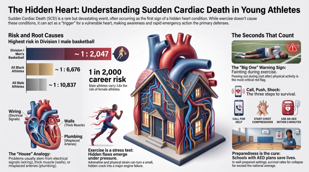
الجدول 1. معدل حدوث الموت القلبي المفاجئ حسب الطبقة الديموغرافية والرياضية، رياضيّو الرابطة الوطنية لرياضة الجامعات (NCAA)، 2002–2022 [4]
| طبقة | معدل الحدوث (لكل سنة-رياضي) | 95% CI | مخاطر مسيرة مهنية لمدة 4 سنوات | نسبة مقابل الإجمالي |
| كرة السلة للرجال للقسم الأول، أبيض | 1:5,848 | 1:2,498–1:13,691 | ~1:1,462 | 10.9 |
| رجال القسم الأول لكرة السلة، أسود | 1:7,696 | واسعة، متداخلة | ~1:1,924 | 8.3 |
| قسم الرجال الأول لكرة السلة، الإجمالي | 1:8,188 | — | ~1:2,047 | 7.8 |
| كرة السلة، جميع الأقسام | 1:19,164 | — | ~1:4,791 | 3.3 |
| كرة قدم أمريكية، بجميع الفئات | 1:31,743 | — | ~1:7,936 | 2.0 |
| جميع الرياضيين السود | 1:26,704 | 1:20,417–1:34,925 | ~1:6,676 | 2.4 |
| جميع الرياضيين الذكور | 1:43,348 | 1:36,228–1:51,867 | ~1:10,837 | 1.5 |
| جميع الرياضيين البيض | 1:74,581 | 1:60,247–1:92,326 | ~1:18,645 | 0.85 |
| جميع الرياضيات الإناث | 1:164,504 | 1:110,552–1:244,787 | ~1:41,126 | 0.39 |
| المجموعة الكلية | 1:63,682 | 1:54,065–1:75,010 | ~1:15,921 | 1.00 (مرجع) |
مخاطر المسيرة المهنية هي التقريب السنوي البسيط ذو الأبعاد الأربعة المستخدم في المنشور المصدر [4].
جنس. يتعرض الرياضيون الذكور لمعدل حدوث يقارب 3.8 أضعاف مقارنة بالرياضيات الإناث (يُقرب عادة إلى أربعة أضعاف في المصادر الثانوية) [4]. التفسير غير مكتمل التأسيس ولكنه على الأرجح متعدد العوامل: زيادة كتلة البطين الأيسر وسمك الجدار بالنسبة لحجم الجسم، والاختلافات في الاستجابة اللاوُدية وإعادة الاستقطاب، وارتفاع انتشار بعض الركائز المحدثة لاضطراب النظم القلبية, والاختلافات في شدة ونوع التعرض الرياضي6. ومن الجدير بالذكر أن فرق الجنس أصغر بالنسبة للاعتلالات القنواتية — ولا سيما متلازمة كิว تي الطويلة, ، حيث الجنس الأنثوي هو عامل خطر للأداث اللاحقة للبلوغ — مقارنة باعتلالات العضلات القلبية البنيوية.
سباق. يشهد الرياضيون السود معدل حدوث يبلغ تقريباً 2.8 ضعف معدل الرياضيين البيض [4]. تتطلب هذه النتيجة تفسيراً دقيقاً. العرق في هذه الأدبيات هو متغير اجتماعي يعمل بمثابة بديل غير دقيق لمجموعة من العوامل غير القياسية: المشاركة التفاضلية حسب الرياضة والمركز، حجم الجسم،, ارتفاع ضغط الدم الانتشار، سمة الخلايا المنجلية، الوصول الاجتماعي والاقتصادي إلى الرعاية، المحددات الهيكلية للصحة، وربما النمط الظاهري القلبي المرتبط بالأصل العرقي. الإرشادات المعاصرة تؤطر العرق صراحة باعتباره بناءً اجتماعيًا سياسيًا في هذا السياق وتدعو إلى تسجيل المحددات الاجتماعية للصحة في الأبحاث المستقبلية1النتيجة العملية هي أن “العرق الأسود” يحدد خطراً متزايداً دون تفسيره، وهو أساس ضعيف لل خوارزميات السريرية.
رياضة. تُعد كرة السلة الرياضة التي تنطوي على أعلى مستوى من المخاطر مقارنةً بجميع الرياضات التي شملتها الدراسة، ويحدث ذلك بشكل مستقل عن العوامل الأخرى: فحتى بعد إجراء تعديل متعدد المتغيرات لمراعاة الجنس والعرق، تظل ممارسة كرة السلة مرتبطة بارتفاع مخاطر الإصابة (نسبة الاحتمالات 2.75، نطاق الثقة 95٪ 1.73–4.34) [4تسهم كرة القدم الأمريكية بالعدد المطلق الأكبر من الحالات بحكم حجم قائمة اللاعبين. كما يُصنف كرة القدم ضمن الرياضات ذات معدل الإصابة الأعلى. والخصائص المشتركة لهذه الرياضات — الجهد عالي الديناميكية والقائم على التوقف والبدء مع بذل أقصى جهود متكررة والانتقالات اللاإرادية المفاجئة — يُرجح أنها ميكانيكية وليست عرضية، على الرغم من عدم اختبار هذه الفرضية بشكل مباشر.7,8].
يستحق خطأ يُكرر كثيرًا التصحيح. ففي كرة السلة للرجال بالقسم الأول، تجاوز التقدير النقطي للاعبين البيض (1:5,848) تقدير اللاعبين السود (1:7,696) [4]. تعكس المصادر الثانوية هذا عادةً. يعتمد كلا التقديرين على عدد صغير جداً من الأحداث مع تداخل واسع فترات الثقة, ولا ينبغي معاملة الترتيب داخل كرة السلة على أنه محدد في أي من الاتجاهين.
الاتجاه الزمني
انخفض معدل الإصابة بمتلازمة الموت المفاجئ (SCD) بين رياضيي الرابطة الوطنية لرياضة الجامعات (NCAA) بنحو 29% لكل فترة خمس سنوات على مدار فترة الدراسة (نسبة معدل الإصابة لكل خمس سنوات 0.71، نطاق الثقة 95% 0.61–0.82)، في حين بقي معدل الوفيات غير المرتبطة بأمراض القلب والأوعية الدموية في نفس المجموعة السكانية دون تغيير (نسبة معدل الإصابة 0.98، نطاق الثقة 95% 0.94–1.04) [4].
خصوصية الانخفاض في الوفيات القلبية الوعائية توحي بذلك، إلا أن الدراسة لم تُصمم لتحديد الآلية ويرفض مؤلفوها عزو هذا الاتجاه إلى أي تدخل منفرد. وتشمل العوامل المرجحة المحتملة الاستخدام الأوسع لتخطيط القلب الكهربائي في البرامج ذات الموارد الجيدة، وتحسين تخطيط الاستجابة للطوارئ، و جهاز مزيل الرجفان الخارجي الآلي نشر، وتحسين التعرف على أعراض التحذير المرتبطة بالمجهود، والتغيرات الزمنية في رصد الحالات. ويظل فك تشابك هذه العوامل مسألة مفتوحة، وينبغي عدم الاستشهاد بهذا الاتجاه كدليل على كفاءة أي استراتيجية وقائية معينة.
3. المسببات: طيف المرض
3.1 توزيع الأسباب
من بين 118 حالة من أصل 143 حالة تابعة للرابطة الوطنية لرياضة الجامعات (NCAA) والتي تتضمن معلومات كافية لـ حكم قضائي [4]:
- الموت الفجائي غير المفسر سلبي التشريح: 19.5%
- ضخامة البطين الأيسر مجهولة السبب أو ممكن اعتلال عضلة القلب: 16.9%
- اعتلال العضلة القلبية الضخامي: 12.7%
- تذكير: الشريان التاجي الخلقي شريان الشذوذات, اعتلال عضلة القلب البطيني المبرمج, التهاب عضلة القلب, تسلخ الأبهر, وأسباب أخرى
يختلف هذا التوزيع بشكل جوهرى عن الصورة التاريخية، التي تم فيها وصف اعتلال العضلة القلبية الضخمي باعتباره السبب المهيمن الوحيد [2,3]. يعكس هذا التحول تحسناً في الدقة الجنائية (أو التحقيقية) وليس تغيراً في بيولوجيا المرض: إذ إن الفصل المعياري للخبراء يعيد تصنيف العديد من الحالات كحالات مجهولة السبب أو غير تفسر، وهي الحالات التي تقل... مراجعة منهجية يُعزى إلى اعتلال العضلة القلبية الضخامي بناءً على سماكة جدار الحد الفاصل وحدها.
في الأدبيات العامة، تتراوح نسبة الحالات التي يفشل فيها التشريح في تحديد سبب هيكلي بين 10% و42% تقريبًا، ويُعزى هذا التباين إلى مدى دقة الفحص، وخبرة أخصائي علم الأمراض، وما إذا كانت البروتوكولات الخاصة بأمراض القلب قد تم اتباعها أم لا.
الآثار المترتبة على الوقاية مباشرة وغير مقدرة حق قدرها: في هذه البيانات، الفئة الفردية الأكبر التي تم الفصل فيها هي غياب أي نتيجة بنيوية. لذلك فإن الاستراتيجيات التي تركز حصرياً على المرض الهيكلي تترك نسبة كبيرة من الحوادث دون تفسير أو اكتشاف. وهذه النسبة ليست ثابتة، بل تختلف باختلاف المنهجية الطب شرعية، وخبرة أخصائي علم الأمراض المفحص، وما إذا كان تشريح جزيئي يتم إجراؤها — لكن أهمية الأسباب غير الهيكلية والتي تكون نتائج تشريح الجثث فيها سلبية تظل ثابتة عبر مختلف السلاسل.
3.2 لماذا يؤدي المجهود إلى حدوث النوبات
أفضل فهم للموت المفاجئ أثناء التمرين هو من خلال إطار الركيزة - المحفز - المُعدِّل. الركيزة المعرضة للخطر - وهي تشوهات تشريحيّة، أو بنيويّة، أو في القنوات الشارديّة - تُعد ضروريّة ولكنها نادراً ما تكون كافية. يوفر التمرين محفزات ومُعدِّلات تحول القابلية كامنةً للخطر إلى رجفان بطيني [7,8].
تدفق الكاتيكولامينات. يؤدي التمرين الرياضي إلى تنشيط ودي ملحوظ مع ارتفاع الكاتيكولامينات الدورية أضعافاً مضاعفة. يؤدي التحفيز الأدرينالي بيتا إلى تقصير فترة الجموح بشكل غير متجانس عبر عضلة القلب, ، يزيد من التلقائية، ويعزز تحميل الكالسيوم في الشبكة الساركوبلازمية. في حالات الخلل في التعامل مع الكالسيوم — وخاصة تسرع القلب البطيني متعدد الأشكال الناجم عن الكاتيكولامينات — يُعد هذا مسبباً مباشراً لاضطراب النظم القلبية8].
طلب نقص الدم. في تشوهات الشرايين التاجية، أو الجسر العضلي القلبية، أو الانسداد الشديد في مخرج البطين الأيسر، فإن الزيادات الناجمة عن الجهد في الطلب على الأكسجين في عضلة القلب لا يقابلها العرض. وبدوره يؤدي نقص التروية إلى إبطاء التوصيل الإقليمي وتشتت إعادة الاستقطاب، وهي الركيزة الأساسية لإعادة الدخول.
الإجهاد الميكانيكي والديناميكي الدموي. يؤدي زيادة إجهاد الجدار، وتوسع حجرات القلب، والتقلص القوي ضد انسداد إلى شد عضلة القلب بشكل حاد. توفر التغذية الراجعة الكهروميكانيكية - وهي قنوات أيونية تنشط بالشد وتغير جهد الغلاف الخلوي - آلية محتملة تربط الحمل الميكانيكي عدم الاستقرار الكهربائي، وقد تكون ذات صلة باعتلال عضلة القلب المسبب لاضطراب النظم، حيث يبدو أن التمرين يسرع المرض بالإضافة إلى تحفيز النوبات.
تحولات الكهارل وتوازن الحمض والقاعدة. التمرين يؤدي إلى خروج البوتاسيوم من العضلات العاملة، وحموضة داخل الخلايا، ونقص الحجم، وفي الجهد المطول نقص صوديوم الدم. كل منها يغير التوصيل وإعادة الاستقطاب9].
انتقال ذاتي التغذية. إن الانسحاب المفاجئ للنغمة الودية والارتفاع المفاجئ في النشاط المبهم عند توقف الجهد ينتج عنه فترة من عدم التجانس الكهربائي. تحدث نسبة معتبرة من الأحداث المرتبطة بالمجهود مباشرة بعد التمرين وليس أثناءه، وقد يؤدي الهيكل المتقطع لكرة السلة، وكرة القدم، وكرة القدم الأمريكية إلى إعادة إنتاج هذه الانتقالات بشكل متكرر8].
تقدم الركيزة. في بعض الحالات، لا يُعد التمرين مجرد محفز فحسب، بل هو مسبب للمرض. وهذا ثابت بشكل أفضل في اعتلال عضلة القلب الناجم عن عدم انتظام ضربات القلب الناتج عن متغيرا اللوكوفيлин-2، حيث يرتبط حجم تمارين التحمل بظهور مبكر للنمط الظاهري، وعبء أعلى لعدم انتظام ضربات القلب، وتقدم هيكلي1,10].
3.3 اعتلالات عضلة القلب
اعتلال العضلة القلبية الضخامي. محدد بأسباب غير مجهولة تضخم البطين الأيسر, عادة ما يكون له أساس ساركوميري موروث، على الرغم من أن حوالي نصف المرضى ليس لديهم متغير مسبب يمكن تحديده. يختلف النمط الظاهري، والمسار السريري، وخطر الإصابة باحتتشاء عضلة القلب بشكل كبير. وتشمل آليات الموت المفاجئ عدم انتظام دقات القلب البطيني الناجم عن اضطراب الخلايا العضلية والتليف الخلالي، ونقص الإمداد الدموي الناتج عن اختلال وظيفة الأوعية الدقيقة, وانهيار ديناميكا الدم الناتج عن انسداد التدفق الديناميكي. يتضمن تصنيف المخاطر أقصى سمك لجدار القلب،, التاريخ العائلي الموت المفطري، والإغماء غير المفسر، وتسرع القلب البطيني غير المستمر، وحجم الأذين الأيسر، وتدرج التدفق الخارج، وتمدد الأوعية الدموية القمي، ومدى تعزيز الجادولينيوم المتأخر على التصوير بالرنين المغناطيسي للقلب. الأفراد الذين يحملون متغير ممرض بدون نمط ظاهري ضخامي ذات خطورة اضطراب نظم منخفضة1].
اعتلال العضلات القلبية المتبمّن. تتميز بانخفاض وظائف البطينين - الأيمن، أو الأيسر، أو كلاهما - مع عبء من اضطراب نظم البطين لا يتناسب مع درجة تمدد حجرة القلب أو ضعف الانقباض. معظم المتغيرات المحددة تؤثر على الجسيمات الدريوية بروتينات, ، حيث يُعد "بلاكوفيلين-2" الأكثر شيوعاً، على الرغم من أن نحو نصف الحالات تكون سلبية النمط الجيني. يُعدل النمط الجيني الخطر بشكل كبير: فالمرض الناجم عن بلاكوفيلين-2 يظهر تسارعاً واضحاً المرتبط بالتمارين الرياضية وارتفاعاً في خطر اضطراب النظم القلبية مع المشاركة في رياضات التحمل، في حين أن الأدلة على وجود خطر مماثل في المرض غير الناجم عن بلاكوفيلين-2 والسلبي النمط الجيني غير مثبتة [10يُعدّ التمييز بين اعتلال عضلة القلب اللإيقاعي المبكر وتوسع البطين الأייمن لدى الرياضيين الذين يمارسون رياضات التحمل من أعبس المشاكل في أمراض قلب الرياضيين.1].
اعتلال العضلة القلبية التوسعي. توسع البطين الأيسر أو البطينين مصحوبًا بخلل في الوظيفة الانقباضية، مع كون النمط الجيني 60% سلبيًا تقريبًا. يرتفع خطر الإصابة باضطرابات النظم القلبي مع انخفاض كسر القذف, ، والأعراض، وعبء الندبات، وتكون مرتفعة بشكل غير متناسب في أنواع فرعية جينية محددة - مثل لامين A/C، وديسموبلاكين، وفيلامين C - والتي تستوجب مراقبة أقرب بغض النظر عن الكسر القذفي. تشير الأدلة الأولية إلى أن التعرض التراكمي العالي للتمارين الرياضية طوال العمر يرتبط بانخفاض الكسر القذفي في المرض المرتبط بـ لامين A/C، مما يثير احتمال أن تساهم التمارين في التطور فضلاً عن التحفيز وعدم انتظام ضربات القلب [1].
فرط التشقاق البطيني الأيسر. Prominent الشعيرات with deep intertrabecular recesses, formerly termed left ventricular noncompaction. It is no longer considered a distinct cardiomyopathy. In the absence of coexisting hypertrophic or dilated phenotype, ventricular arrhythmia, or symptoms, adverse events have not been demonstrated, and isolated hypertrabeculation in an asymptomatic person is best regarded as a morphological variant [1].
اعتلالات القنوات الصوديومية والبوتاسيومية والكالسيومية (أو: اعتلالات القنوات الأيونية)
تters متلازمة عدم انتظام ضربات القلب الموروثة يمكن أن تؤدي إلى الموت المفاجئ في قلب طبيعي من الناحية الهيكلية، وتُعد سبباً هاماً يمكن تحديده للموت المفاجئ السلبي في تشريح الجثة.
متلازمة كิว-تي الطويلة. تأخر إعادة الاستقطاب البطيني المؤهب لتورط دوامة الرؤوس (torsades de pointes). النمط الجيني يتنبأ بالمحفز: أحداث متلازمة كيو تي الطويلة من النمط 1 (LQT1 - KCNQ1) تتميز بأنها تحدث مع الجهد، وتعتبر السباحة محفزاً مميزاً؛ أحداث النمط 2 (LQT2 - KCNH2) ترتبط بالصدمة السمعية، والانفعال، وفترة ما بعد الولادة؛ أحداث النمط 3 (SCN5A) تحدث بشكل أساسي أثناء الراحة أو النوم. يعتمد التشخيص على فترة كيو تي المُصححة (QTc)، التاريخ المرضي للأعراض، التاريخ العائلي، والاختبار الجيني. هناك محذوران مهمان سريرياً: عتبات فترة (QTc) لدى الرياضيين تختلف عن تلك الموجودة في عامة الناس، وقد تتطبيع فترة كيو تي بشكل متقطع، بحيث أن فترة (QTc) الطبيعية الواحدة أثناء الراحة لا تنفي التشخيص. متلازمة كيو تي الطويلة المستترة -إيجابية النمط الجيني مع فترة (QTc) طبيعية باستمرار- تحدث لدى أقلية كبيرة من حاملي الطفرات [1].
تسرع القلب البطيني القلبي الكاتيكولاميني المتعدد الشكل. تنتج في الغالب عن متغیرات مستقبِل الريانودين (RYR2) التي تؤدي إلى إطلاق غير طبيعي للكالسيوم في مرحلة انبساط القلب تحت الإجهاد الأدريناليني. وهي اعتلال القنوات ترتبط بشكل أساسي بالمجهود البدني، وتتميز عادةً بحدوث تسرع القلب البطيني ثنائي الاتجاه أو متعدد الأشكال عند عتبات معدل ضربات قلب يمكن تكرارها أثناء التمرين. عادة ما يكون تخطيط القلب الكهربائي في حال الراحة طبيعياً، مما يجعل هذه الحالة خارج نطاق الكشف عن طريق تخطيط القلب في حال الراحة. يمتلك اختبار الجهد حصيلة تشخيصية عالية عندما يُشتبه إكلينيكياً بوجود تسرع القلب البطيني التفاعلي catecholaminergic polymorphic ventricular tachycardia (CPVT)، حيث يُظهر اضطراب النظم بانتظام عند عتبة معدل ضربات قلب ثابتة، وهو الاختبار المناسب لشخص يعاني من غشي مرتبط بالمجهود وتخطيط قلب طبيعي في حال الراحة1].
متلازمة بروجادا. يتميز بارتفاع مقطع ST المحدب في المسيرات الصدرية اليمنى، والمرتبط بمتغيرات جينات SCN5A في قلة من الحالات. تحدث النوبات بشكل رئيسي أثناء الراحة، أو النوم، أو مع الحمى بدلاً من الجهد، على الرغم من أن الحمى المصاحبة للتمارين الرياضية تُعد محفزاً معروفاً [1].
متلازمة وولف-باركينسون-وايت. مسار أذيني بطيني إضافي. يحدث الموت المفاجئ عندما الرجفان الأذيني ينقل بسرعة عبر مسار ذي فترة عصيان قصيرة، متدهوراً إلى رجفان بطيني. على عكس معظم الأسباب التي تمت مناقشتها هنا، تُعد هذه حالة قابلة للاكتشاف بسهولة وعلاجها حاسم، ورسم القلب الكهربائي أثناء الراحة يُعد تشخيصياً [1].
3.5 التشുوهات الخلقية للشرايين التاجية
يعتبر المنشأ الشاذ للأبهر للشريان التاجي من أبرز أسباب الموت المفاجئ المرتبط بالجهد ومن الأقل قابلية للاكتشاف بالفحوصات التقليدية.
المتغير الأعلى خطورة هو المنشأ الشاذ للشريان التاجي الأيسر من الجيب الأيمن مع مسار بين الشرايين بين الوريد الأبهر والشريان الرئوي. وتشمل السمات عالية الخطورة المقترحة وجود قطاع داخل جدار الشريان الأبهر، وفوهة قريبة تشبه الشق، وزاوية انطلاق حادة، وطول أكبر داخل الجدار. ويعتقد أن الآلية تنطوي على ضغط ديناميكي وتشوه الفوهة أثناء المجهود مع تمدد الأوعية الكبيرة، مما ينتج نقص تخدس الدم المتقطع الذي قد لا يترك أي تشوه ثابتاً بين النوبات [1].
منشا الشريان التاجي الأيمن الشاذ مع مسار بين الشرايين هو الأكثر شيوعاً ويحمل مخاطر أقل؛ ويبقى العديد من الأفراد المصابين بدون أعراض. أما المتغيرات الأخرى - المسارات داخل الحاجز، أو خلف الأبهر، أو أمام الشريان الرئوي - فهي حميدة عموماً.
التحدي السريري يكمن في أن الأفراد المصابين غالباً ما يكونون عديمي الأعراض حتى وقوع الحدث المؤشر، وعادة يكون تخطيط القلب الكهربائي أثناء الراحة طبيعياً، وقد يفشل اختبار الإجهاد البدني في إثارة نقص تروية القلب حتى في حالات التشريح شديد الخطورة. يتطلب التشخيص القطعي التصوير التشريحي، وغالباً ما يكون ذلك عن طريق تصوير الأوعية المقطعي المحوسب للشرايين التاجية أو التصوير بالرنين المغناطيسي للقلب [1].
جسور عضلي قلبي, حيث يمر جزء من الشريان التاجي عبر عضلة القلب، وهو أمر شائع وعادة ما يكون عرضياً. ويصبح مهمًا سريرياً فقط عندما تتسبب الأجزاء العميقة أو الطويلة والمطمورة في حدوث نقص مرئي في التروية الدموية1].
3.6 التهاب العضل القلبى والحالات المكتسبة
التهاب عضلة القلب يسبب الموت المفاجئ من خلال عدم انتظام نبضات القلب الناجم عن حالة حادة التهاب, ، والوذمة، والتليف اللاحق. وتكمن أهميتها جزئيًا في كونها قد تكون عابرة: فالتمارين القوية أثناء التهاب عضلة القلب النشط تكون مسببة لاضطراب النظم القلبية، ولكن الخطر يزول إلى حد كبير مع زوال الالتهاب. وهذا هو أساس التوصية القياسية بالامتناع عن التძل البدني حتى تختفي الأعراض والأدلة الموضوعية على الالتهاب. تشير بيانات التصوير بالرنين المغناطيسي للقلب الحديثة إلى أن رياضيين مختارين قد يعودون بأمان في وقت مبكر قبل فترة الثلاثة إلى ستة أشهر التي افترضتها التوجيهات السابقة، على الرغم من أن قاعدة الأدلة لا تزال محدودة، ويجب أن تحكم العودة الزوال الموضوعي للالتهاب وليس فقط الوقت المنقضي [1,11].
اعتلال الأهرط. متلازمة مارفان, متلازمة لويس-دياتز, ، وتتسبب متلازمة إهلرز-دانلوس الأوعية الدموية، ومرض الشريان الأبهر المرتبط بصمام الأبهر ثنائي الشرف، في الاستعداد للإصابة بالتسلخ الحادي للأبهر. هذا سبب نادر للموت المفاجئ لدى الشباب ولكنه مميز لكونه قابلاً للكشف عن طريق الفحص البدني والتصوير، ولأن الخطر يرتبط بحجم الشريان الأبهر بطريقة تسمح بالإدارة القائمة على العتبات. إن التضخم الملحوظ في الشريان الأبهر نادر لدى الرياضيين الشباب - حيث تعتبر الأقطار التي تزيد عن 42 مم لدى الذكور و40 مم لدى الإناث غير معتادة بغض النظر عن حجم الجسم - لذا فإن مثل هذه النتيجة تبرر تقييم اعتلال الأبهر الكامن بدلاً من عزوها إلى التدريب [1].
هبوط الصمام التاجي اللا نظميات. معظم حالات تددل الصمام التاجي حميدة، ولكن مجموعة فرعية منها - وتتميز بتدل الورقتين مع انفصال الحلقة التاجية، وتعزيز الجادولينيوم المتأخر الجانبي السفلي، والانبثاث البطيني المعقد - ترتبط بالموت المفاجئ. يوفر شد العضلات الحليمية والتليف الإقليمي الناتج عن ذلك ركيزة عدم انتظام ضربات القلب المعقولة [1].
ارتجاج القلب. رجفان بطيني ناجم عن صدمة صدرية كليلة خلال مرحلة الاستقطاب الضعيفة، في قلب سليم من الناحية الهيكلّية. وهو ظاهرة ميكانيكية وليست مرضية، ولا يتأثر بأي استراتيجية فحص، ويمكن النجاة منه بإزالة الفرجفة الفورية [1].
سمة الخلايا المنجلية. مرتبطانه بالانهيار الجهدي والموت، لا سيما أثناء التدريب المكثف في الحرارة، من خلال آليات تنطوي على الإجهاد الجهدي انحلال الربيدات and metabolic derangement rather than primary arrhythmia. It is included here because it presents as sudden collapse during exertion and is relevant to differential diagnosis and to prevention protocols [1].
3.7 Genetic architecture: penetrance, modifiers, and the limits of genotype
Most of the conditions described above are inherited, but inheritance in this field behaves far less deterministically than the term “genetic heart disease” suggests, and the gap between genotype and outcome is where much of the clinical difficulty lies.
Incomplete penetrance and variable expressivity. Carrying a pathogenic variant does not reliably produce disease, and where it does, severity varies widely within families sharing an identical variant. Penetrance estimates derived from clinically ascertained families — identified because someone was affected — substantially overstate risk when applied to variants found incidentally or through population screening. Estimates from unselected cohorts are consistently lower. This has direct consequences: an individual who is genotype-positive and phenotype-negative for hypertrophic cardiomyopathy carries low arrhythmic risk and is treated very differently from one with an expressed phenotype [1].
Age-dependent expression. Phenotype in hypertrophic, arrhythmogenic, and dilated cardiomyopathy typically emerges over adolescence and early adulthood rather than being present from birth. A normal evaluation at 14 does not exclude disease at 24. This underlies the requirement for serial evaluation of genotype-positive relatives rather than single-timepoint clearance, and it explains why screening cohorts followed long enough eventually record deaths in individuals who screened normal.
Modifier genes and polygenic background. The same variant produces different phenotypes in different genetic backgrounds. Common variants of individually small effect, aggregated as الدرجات متعددة الجينات, appear to modify penetrance and severity in the cardiomyopathies — potentially explaining part of the within-family variability that monogenic models cannot. This work is at an earlier stage than the equivalent literature in coronary disease, and polygenic scores are not yet clinically actionable in this setting, but the direction is toward a model in which a rare pathogenic variant sets susceptibility and common variation, environment, and training load determine whether and when disease appears.
Genotype-negative disease. Roughly half of hypertrophic and arrhythmogenic cardiomyopathy cases and approximately 60% of dilated cardiomyopathy cases have no identifiable causal variant. A negative genetic test in an affected individual does not exclude an inherited condition, and does not obviate clinical screening of relatives.
Variants of uncertain significance. These are the most common result of broad panel testing in the absence of a clear phenotype, and they are frequently misinterpreted as intermediate-risk findings. They are not: they are uninformative pending إعادة تصنيف, which may occur in either direction as evidence accumulates. Testing in individuals without phenotype or family history predominantly generates them, which is the principal argument against genetic testing as a primary screening tool [1].
3.8 Autopsy-negative sudden unexplained death and molecular autopsy
When comprehensive autopsy including toxicology and histology identifies no cause, the death is classified as autopsy-negative sudden unexplained death — or, in some series, sudden arrhythmic death syndrome. This classification is provisional rather than terminal.
Postmortem genetic testing identifies a clinically actionable pathogenic or likely pathogenic variant in a meaningful minority of these cases. Applying contemporary ACMG classification criteria to 302 expertly adjudicated cases, an actionable variant was identified in 13% of decedents, predominantly catecholaminergic polymorphic ventricular tachycardia and long QT syndrome, with RYR2 the most implicated gene [12]. A clinically relevant diagnosis was established in a substantially larger proportion of families when postmortem علم الوراثة was combined with clinical evaluation of surviving relatives.
Reported yields vary widely across series — from under 4% to approximately 30% — and depend heavily on the variant-classification framework applied. Series adhering strictly to ACMG criteria report lower yields; those using looser thresholds report higher ones, largely by counting variants of uncertain significance as diagnostic. Cross-series comparison is therefore unreliable unless classification criteria are matched [12].
Three points follow. First, the proportion of truly unexplained deaths is lower than autopsy-negative rates suggest. Second, molecular autopsy requires appropriate specimen retention, which depends on medical examiner practice and is frequently not performed. Third, and most important clinically, a diagnosis in the decedent enables cascade screening of living relatives — making postmortem evaluation a prevention strategy for the family, not merely a determination of cause.
4. The Athlete’s Heart: Adaptation Versus Disease
Sustained training produces cardiac remodeling that overlaps phenotypically with the diseases described above. Distinguishing adaptation from pathology is the central diagnostic problem in this field, and misclassification carries costs in both directions.
4.1 Structural adaptation
Endurance training produces predominantly إعادة تشكيل لامركزية — chamber enlargement with proportionate wall thickening — driven by sustained volume loading. Strength training produces relatively more concentric change. Most athletes exhibit a mixed pattern reflecting the actual demands of their sport.
Adaptation is modified by sport, sex, body size, ethnicity, and training duration. Left ventricular wall thickness in the 13–15 mm range in men, and right ventricular dilation in endurance athletes, fall into diagnostic gray zones overlapping hypertrophic and arrhythmogenic cardiomyopathy respectively. Left ventricular end-diastolic dimensions of 60 mm or more occur in a meaningful minority of trained athletes without valvular or myocardial disease [1,11].
Features favoring physiological adaptation include symmetric enlargement of all four chambers, normal or supranormal diastolic function, absence of late gadolinium enhancement, normal functional capacity, appropriate ضغط الدم response to exercise, and regression with detraining. Features favoring pathology include asymmetric hypertrophy, impaired diastolic function, late gadolinium enhancement, a family history of cardiomyopathy or premature sudden death, and marked ECG abnormality out of proportion to the structural findings [1].
4.2 Electrical adaptation
Training produces a reproducible set of ECG changes: sinus bradycardia and sinus arrhythmia from increased التوتر المبهمي, ectopic atrial and junctional rhythms, first-degree and Mobitz type I atrioventricular block, incomplete right bundle branch block, isolated voltage criteria for ventricular hypertrophy, and early repolarization. These are physiological, require no evaluation in an asymptomatic person without concerning family history, and their misclassification as pathological was the principal historical barrier to ECG-based screening [13].
Remodeling and repolarization vary by ethnicity, not only between Black and White athletes. Athletes of African and Afro-Caribbean descent show greater left ventricular wall thickness for a given body size and a distinctive repolarization pattern — J-point elevation with convex ST-segment elevation followed by T-wave inversion confined to leads V1–V4 — which is benign. Applying criteria derived from White European cohorts without this accommodation more than doubles the false-positive rate in Black athletes [14]. Distinct patterns have also been described in athletes of Middle Eastern, South Asian, and East Asian descent, and normative data for these groups remain comparatively sparse — a gap that propagates directly into screening performance wherever population-specific reference values do not exist.
Age matters similarly: T-wave inversion in leads V1–V3 in athletes aged 16 or younger represents a persistent juvenile pattern rather than disease [13].
5. Clinical Presentation and Warning Symptoms
The defining clinical feature of these conditions is that most affected individuals are asymptomatic until the index event. Where symptoms do occur, they are frequently attributed to deconditioning, dehydration, anxiety, or normal exertional limitation — by the individual, by coaching staff, and often by clinicians.
Symptoms warranting evaluation before further participation:
Exertional syncope or near-syncope. The single most important warning symptom. Syncope during exertion, as distinct from immediately after cessation, should be presumed cardiac until proven otherwise. Post-exertional syncope is more often neurally mediated but does not exclude cardiac causes.
Exertional chest pain. Particularly if reproducible at a consistent workload, suggesting demand ischemia from anomalous coronary anatomy or outflow obstruction.
Exertional dyspnea disproportionate to conditioning, especially where it represents a change from established baseline capacity.
Palpitations with exertion, particularly abrupt in onset and offset, or associated with lightheadedness.
Unexplained decline in performance not attributable to training load, illness, or injury.
The family history is as informative as the personal history: premature sudden death before age 50 in a قريب من الدرجة الأولى, known inherited cardiomyopathy or arrhythmia syndrome, unexplained drowning, single-vehicle accidents without explanation, or unexplained seizure disorder — the last because long QT syndrome and CPVT events are frequently misdiagnosed as epilepsy [1,15].
6. Diagnostic Evaluation
6.1 Electrocardiography and the evolution of interpretation criteria
The resting تخطيط القلب الكهربائي بـ 12 اتجاهاً detects the electrical signatures of cardiomyopathy and channelopathy, and is the single most informative low-cost test in this population. Its historical limitation was not sensitivity but specificity: applying general-population criteria to trained athletes generated false-positive rates that made systematic use impractical.
Interpretation criteria have been refined substantially over fifteen years.
Table 2. Evolution of athlete ECG interpretation criteria
| Criteria set | السنة | False positives, White athletes | False positives, Black athletes | Principal change |
| ESC recommendations [16] | 2010 | 16.2% | 40.4% | Training-related versus unrelated dichotomy; no ethnicity-specific criteria; most T-wave inversion classified abnormal |
| Seattle Criteria [17] | 2013 | 7.1% | 18.4% | Black athlete repolarization pattern recognized as physiological; QTc thresholds raised to ≥470 ms (male) and ≥480 ms (female) |
| Refined Criteria [14] | 2014 | 5.3% | 11.5% | Isolated axis deviation and isolated atrial enlargement reclassified as non-triggering |
| International Criteria [13] | 2017 | ~1.3–3.0% | ~4.2–6.8% | Formal normal/borderline/abnormal categories; two or more borderline findings required to trigger evaluation |
The 2017 International Criteria define exactly five borderline findings: left axis deviation, right axis deviation, تضخم الأذين الأيسر, right atrial enlargement, and complete right bundle branch block [13]. Any single one of these in isolation does not warrant evaluation in an asymptomatic athlete without concerning family history; two or more do. Isolated voltage criteria for left or right ventricular hypertrophy are classified as normal, not borderline — a point frequently misstated.
Two caveats matter. First, the reported false-positive rates derive from expert or specialist-supervised interpretation; rates under non-specialist reading are consistently higher, and the magnitude of that gap has not been established at scale. Second, gains in specificity have not been free: in masters and pediatric cohorts, the International Criteria have missed diagnoses that earlier criteria would have flagged, including dilated cardiomyopathy presenting with isolated left axis deviation.
6.2 Diagnostic versus screening sensitivity
An important distinction is frequently elided. Diagnostic sensitivity is the probability that a person with established, fully expressed disease has an abnormal ECG. Screening sensitivity is the probability that an asymptomatic adolescent with early, incomplete, or concealed expression is correctly identified in a mass-screening setting.
The second is systematically lower than the first. Adolescent phenotype is often immature — both hypertrophic and arrhythmogenic cardiomyopathy express progressively — dynamic conditions are intermittently normal at rest, and field acquisition and interpretation conditions are inferior to those of a referral laboratory. Sensitivity figures quoted from referral cohorts of patients with known disease, commonly cited near 98% for hypertrophic cardiomyopathy, should not be presented as screening performance [18].
The most instructive evidence comes from long-term follow-up of a screened cohort. Among 11,168 adolescent English Football Association players screened with questionnaire, examination, ECG, and echocardiography, conditions associated with sudden death were identified in 0.38%, with ECG abnormal in 86% of those individuals versus 7% for history and 5% for physical examination [19]. Over a mean 10.6 years of follow-up, however, eight athletes died of cardiac causes — six of whom had screened normal at age 16, at a mean of 6.8 years after screening, most from cardiomyopathies not detectable at the time of testing.
A normal screen is not durable clearance. This is a property of progressive disease, not a failure of the test.
6.3 Secondary evaluation
An abnormal primary screen or concerning symptom initiates further testing, selected by the suspected condition rather than applied as a fixed panel.
Transthoracic echocardiography assesses wall thickness, chamber dimensions, systolic and diastolic function, valvular structure, aortic dimensions, and in many cases coronary origins.
Cardiac magnetic resonance provides superior tissue characterization. Late gadolinium enhancement identifies fibrosis and scar; T2-weighted imaging and parametric mapping identify edema and active inflammation. It is often decisive in distinguishing physiological hypertrophy from cardiomyopathy, in evaluating the right ventricle for arrhythmogenic cardiomyopathy, and in diagnosing myocarditis. Late gadolinium enhancement confined to right ventricular insertion points is a recognized finding without established adverse prognostic significance.
Exercise testing should be sport-specific and designed to provoke symptoms at the intensity actually encountered in competition, rather than terminated at arbitrary heart-rate targets or performed pharmacologically. It is essential in suspected CPVT, in evaluating ventricular ectopy, and in assessing ischemia in anomalous coronary anatomy.
مراقبة نظم القلب في العيادة الخارجية characterizes arrhythmia burden and its relationship to activity; extended monitoring is often required given the intermittency of clinically relevant events.
تصوير الشريان التاجي by CT angiography or magnetic resonance is required to define coronary origin and course when anomalous anatomy is suspected.
Genetic testing confirms diagnosis where the phenotype is established, informs risk stratification in specific conditions — plakophilin-2 in arrhythmogenic cardiomyopathy, lamin A/C in dilated cardiomyopathy, genotype in long QT syndrome — and enables cascade screening of relatives. It performs poorly as a primary diagnostic test in the absence of phenotype or family history, where it principally generates variants of uncertain significance.
Notably, cardiac imaging, exercise testing, and ambulatory monitoring have insufficient evidence to support their use as primary screening tools in asymptomatic individuals [1]. Their role is in secondary evaluation.
6.4 Emerging technologies
Artificial intelligence applied to the ECG. Deep learning models trained on large ECG corpora can identify conditions that lack a ممرض العلامة signature to the human eye. Convolutional neural networks have been developed to detect hypertrophic cardiomyopathy from the 12-lead ECG alone, with performance maintained on external validation across diverse international cohorts and in pediatric and adolescent populations [20]. Comparable models identify electrocardiographically concealed long QT syndrome — genotype-positive individuals with a normal measured QTc — which conventional interval measurement cannot detect by definition [21].
The potential relevance to this field is obvious: the principal limitation of ECG screening is not cost but interpretive accuracy, and an algorithm that improves specificity without sacrificing sensitivity would change the calculus directly. Several important caveats apply. Models trained predominantly on clinical populations may perform differently in trained athletes, whose baseline ECGs differ systematically from those of the general population, and athlete-specific validation remains limited. Performance in the demographic groups with the highest false-positive rates under conventional criteria has not been separately established. And an algorithm that flags disease without an accessible pathway to secondary evaluation reproduces the equity problem described in Section 7.5 rather than solving it. AI-ECG is best understood at present as a promising adjunct under active validation rather than an established screening tool.
Wearable and consumer devices. Smartwatches, adhesive patch monitors, and consumer single-lead and multi-lead ECG devices are now widely used by athletes, and increasingly generate cardiac data that reaches clinicians unsolicited. Their genuine strengths are duration and opportunism: a patch monitor worn for two weeks or a smartwatch worn continuously may capture a symptomatic paroxysmal arrhythmia that a resting ECG and a 24-hour Holter both miss, and this is a real diagnostic contribution in a person with intermittent palpitations.
Their limitations are equally clear. Single-lead recordings cannot assess axis, chamber enlargement, repolarization across the precordium, or most of the criteria on which athlete ECG interpretation depends. Automated rhythm classification is optimized for atrial fibrillation detection in older populations and performs poorly for the arrhythmias relevant here. Signal quality during exercise — precisely when it would be most valuable — is frequently inadequate. And the false-positive burden generated by consumer devices in young, healthy, highly motivated users is substantial and largely unquantified.
Wearables complement rather than replace structured evaluation. A consumer device recording during a symptomatic episode is valuable evidence; a normal consumer recording is not clearance, and should not be treated as one by the athlete or the clinician.
7. Screening: What It Achieves and What It Does Not
Screening is one component of prevention. This section assesses it on its own terms rather than treating it as the organizing question of the field.
7.1 The rationale and its limits
Preparticipation cardiovascular screening aims to identify people with unrecognized disease in time for management that reduces risk. Its effectiveness depends on a chain of conditions: the disease must be detectable before the event, the test must detect it at acceptable cost and false-positive burden, effective management must exist, and affected individuals must have access to it.
That chain has weak links. Resting ECG has very limited sensitivity for anomalous coronary origin, catecholaminergic polymorphic ventricular tachycardia, early or concealed arrhythmogenic cardiomyopathy, aortopathy, and significant valve disease, and by definition contributes nothing in cases that prove autopsy-negative — the largest single category. Screening addresses a subset of the problem.
7.2 Comparative performance of screening components
Meta-analytic estimates in athletic populations give ECG a sensitivity approaching 90–94% for conditions detectable by ECG, with specificity near 93%, against approximately 20% sensitivity for history and 9% for physical examination [18]. Evidence drawn specifically from NCAA populations has since been synthesized separately [22]. The qualifier matters: this figure describes performance against cardiomyopathies and channelopathies with electrical signatures, not against the full spectrum of causes, and should not be read as 94% sensitivity for sudden death risk overall. Contemporary guidance cites a comparable range, placing history and physical sensitivity for silent cardiac conditions at 10–20% and noting that adding ECG raises the sensitivity of the preparticipation evaluation to approximately 94% [1].
The argument for ECG is therefore not that it performs well in absolute terms; it is that history and physical examination alone detect only a minority of relevant conditions. This is not an argument against the history and physical examination, which identifies symptomatic individuals, elicits family history, and detects Marfan stigmata, pathological murmurs, diminished femoral pulses, and hypertension — none of which an ECG reliably captures.
7.3 Guideline positions
European guidance has long recommended ECG-inclusive screening [11,16]. North American guidance historically endorsed a standardized 14-point history and physical examination without recommending universal ECG [15].
That divide has narrowed substantially. The 2025 American Heart Association / American College of Cardiology scientific statement holds that inclusion of a resting 12-lead ECG is reasonable, because it improves detection of underlying cardiac conditions in asymptomatic athletes relative to history and physical examination alone. The endorsement carries three conditions: clinicians must be adequately trained in contemporary athlete-specific interpretation criteria; programs must ensure access to secondary evaluation, including the financial and logistical resources for systematic downstream assessment; and because no approach provides absolute protection, an خطة العمل للطوارئ must be in place wherever people train and compete [1].
The unresolved questions are therefore not whether ECG may be used, but whether it should be universally implemented, how programs should be resourced to use it safely, and whether incremental benefit justifies downstream cost and harm.
7.4 Health economics
Published cost-effectiveness estimates for adding ECG to الscreening السابق للمشاركة (أو الفحص الطبي السابق للمشاركة الرياضية) span a wide range, driven more by assumption choice than by data.
Table 3. Published economic evaluations of ECG-inclusive screening
| Analysis | Comparison | Result |
| Wheeler et al. (2010) [23] | ECG + H&P vs. H&P alone | $42,900 per life-year saved (95% CI $21,200–$71,300) |
| Wheeler et al. (2010) [23] | ECG + H&P vs. no screening | $76,100 per life-year saved ($62,400–$130,000) |
| Schoenbaum et al. (2012) [24] | H&P then ECG vs. H&P alone | $68,800 per QALY |
| Schoenbaum et al. (2012) [24] | ECG alone vs. H&P alone | $37,700 per QALY |
| Halkin et al. (2012) [25] | National program, US extrapolation | $10.6–14.4 million per life saved |
Two of the three principal analyses return figures within commonly cited US willingness-to-pay thresholds. The third, returning figures orders of magnitude higher, does so partly by loading recurring annual history and physical costs into the ECG arm — an accounting choice contested in subsequent literature [25].
Every one of these analyses is governed by a single assumption: the الخطر النسبي reduction conferred by detection and subsequent management. That parameter is not well established, and as discussed in Section 8 the evidence underlying it has shifted.
7.5 Equity
Screening programs can widen the inequities they are intended to reduce. The mechanism is the secondary evaluation, not the primary screen.
An abnormal screening result initiates a diagnostic cascade — echocardiography, cardiac magnetic resonance, ambulatory monitoring, genetic evaluation, subspecialty consultation — that in unassisted settings the family must fund. Uninsured and underinsured individuals face two consequences: they may exit the pathway without completing evaluation, continuing to participate with uncharacterized risk; and their absence from outcome data systematically biases estimates of disease prevalence, specificity, and cost-effectiveness in a favorable direction.
This is compounded by the ethnic disparity in false-positive rates. Even under contemporary criteria, false-positive findings remain more frequent among Black athletes [14], meaning that the population most likely to be referred for costly secondary evaluation overlaps with the population least likely to be able to afford it. Contemporary guidance states directly that screening programs without appropriate downstream resources have the potential to harm athletes from underrepresented racial and ethnic groups [1].
A screening program without a funded pathway to secondary evaluation is not a neutral intervention. It is a mechanism for identifying risk in people who cannot then act on it.
7.6 Psychological consequences
Screening programs are evaluated almost exclusively on detection and cost. The psychological consequences are real, are borne disproportionately by people who turn out not to have disease, and are rarely measured.
The false-positive interval. Between an abnormal screening result and its resolution, an athlete is a person who has been told their heart may be dangerously abnormal. That interval is frequently weeks and sometimes months, determined by appointment availability, insurance authorization, and imaging capacity rather than by clinical urgency. Documented consequences include anxiety, intrusive thoughts about dying during exertion, sleep disruption, and withdrawal from training during a period in which no restriction has actually been imposed. Where the athlete’s identity is substantially organized around sport — as it is for many at collegiate and elite level — the threat is to selfhood as much as to health.
Resolution is incomplete. Reassurance after a negative workup does not reliably return people to baseline. Residual health anxiety, continued symptom vigilance, and reduced training intensity have been described persisting after formal clearance, a pattern familiar from other screening contexts. Because false positives outnumber true positives by a large factor at any plausible operating point, this is not a marginal harm affecting a handful of people; it is the modal experience of an abnormal screen.
True positives carry their own burden. A diagnosis in a young person may bring restriction or modification of participation, loss of athletic identity, disrupted scholarship or professional prospects, defibrillator carriage in adolescence, and the knowledge of inherited risk extending to siblings and future children. Depression and anxiety are recognized sequelae of disqualification, and the transition out of competitive sport is itself a period of elevated psychological risk independent of the cardiac diagnosis.
Implications. Three follow directly. Time from abnormal screen to definitive resolution is a clinically meaningful quality metric, not merely an operational one, and shortening it is a genuine intervention. Communication of an abnormal result should convey the base rate — that most abnormal screens resolve without disease — rather than deferring all interpretation to the specialist. And psychological support should be planned into screening programs rather than improvised, particularly for athletes who are restricted or who transition out of competition.
8. Management and Sports Participation
8.1 The shift from disqualification to shared decision-making
Historical practice restricted individuals with identified أمراض القلب والأوعية الدموية from competitive sport more or less categorically. That paradigm has been substantially abandoned, on both ethical and empirical grounds [1,11].
The empirical case rests on outcome data:
- In a prospective multinational دراسة رصديَّة of individuals with hypertrophic cardiomyopathy — 1,534 patients plus 126 genotype-positive, phenotype-negative individuals across 42 centers — those engaging in vigorous exercise, including competitive athletes, did not experience higher rates of death, resuscitated arrest, appropriate defibrillator shock, or arrhythmic syncope than moderate exercisers or sedentary participants [26].
- A prospective multinational registry of athletes with implantable cardioverter-defibrillators found no deaths, resuscitated arrests, or arrhythmia-related injuries during sport over long-term follow-up [27].
- Contemporary cohorts of elite athletes with genetic heart disease who returned to competition under expert supervision report low rates of breakthrough events [28].
The ethical case is that the historical model rested on the premise that athletes cannot make informed decisions about their own risk — a position that is neither supported by evidence nor consistent with the standards applied elsewhere in medicine.
اتخاذ القرار المشترك does not imply equal risk across conditions. It is a process for incorporating an individual’s values into a decision under uncertainty, not a conclusion that all diagnoses carry comparable danger or that all participation requests should be accommodated. The risk attached to genotype-positive, phenotype-negative status differs by orders of magnitude from that attached to plakophilin-2-mediated arrhythmogenic cardiomyopathy in an endurance athlete, and the framework is designed to make that difference explicit rather than to dissolve it.
The 2025 AHA/ACC statement is explicit that it does not issue disqualification recommendations, but rather clinical considerations to inform shared decision-making. Under this framework a uniform approach of restriction should not be applied to individuals with cardiomyopathy; participation is instead determined through a process incorporating accurate diagnosis, condition-specific risk stratification, guideline-directed treatment, disclosure of known and unknown risks, and the individual’s own values and risk tolerance. For those under 18, parents or guardians participate directly [1].
8.2 Where risk remains prohibitive
Shared decision-making does not mean that all participation is endorsed. Situations where risk is understood to outweigh benefit include arrhythmogenic cardiomyopathy caused by plakophilin-2 variants, particularly with endurance sport; active myocarditis or pericarditis; unrepaired anomalous origin of the left coronary artery with an interarterial course; severe symptomatic تضيق الأبهر; heritable thoracic aortic disease with aortic dilation; and prior aortic dissection. Participation is also generally deferred during diagnostic evaluation and until guideline-directed therapy is optimized [1].
8.3 Condition-specific management
Management is disease-specific and is the mechanism by which detection produces benefit: beta-blockade in long QT syndrome and CPVT, with flecainide and sympathetic denervation in selected CPVT cases; defibrillator implantation where risk stratification indicates, though never solely to enable sport participation; الاستئصال القلبي القسطاري for accessory pathways and selected ventricular arrhythmias; surgical reimplantation or unroofing for high-risk coronary anomalies; septal reduction for obstructive hypertrophic cardiomyopathy; aortic surgery at guideline thresholds; and exercise prescription modification where disease progression is exercise-associated [1].
Two points deserve emphasis. First, individuals who discontinue competitive sport should be counseled on the established health benefits of continued recreational physical activity — a transition, not a cessation. Second, longitudinal surveillance is required regardless of the participation decision, because phenotype evolves and the original decision may require revisiting.
8.4 Return to play and longitudinal surveillance
A participation decision is a point on a trajectory rather than a conclusion, and the surveillance that follows is what makes continued participation defensible.
Serial imaging at intervals determined by condition and rate of change. Genotype-positive, phenotype-negative individuals in conditions where exercise may precipitate phenotypic conversion — plakophilin-2-mediated arrhythmogenic cardiomyopathy most clearly — warrant close longitudinal surveillance, with imaging intervals individualized to phenotype, exercise exposure, and evidence of disease progression, while continuing to compete [1]. Aortic dimensions in aortopathy are followed on a schedule set by absolute diameter and rate of growth, with side-by-side comparison of images rather than reliance on prior reports, since inter-study measurement variability can exceed true annual change.
Repeat exercise testing, sport-specific and to the intensity actually encountered in competition, to confirm continued absence of provoked arrhythmia or ischemia and to verify the efficacy of pharmacological suppression where it has been prescribed.
مراقبة نظم القلب في العيادة الخارجية to track arrhythmia burden over time, and device interrogation where a defibrillator is present, including appropriate and inappropriate therapy history.
Structured re-evaluation of the decision itself. The shared decision-making conversation should be revisited periodically rather than treated as settled — because the evidence base is changing, because the individual’s own risk tolerance may change, and because phenotype progression may move a person from one risk category to another without symptoms.
Post-intervention return — after surgical coronary reimplantation, aortic repair, ablation, or device implantation — follows condition-specific intervals governed by healing, demonstrated absence of ischemia or inducible arrhythmia, and normal ventricular function, rather than by elapsed time alone.
9. Secondary Prevention: Emergency Response
Because no screening strategy detects all disease, and because some causes are undetectable in principle, survival in a substantial proportion of events is determined entirely by what happens in the first minutes.
9.1 Determinants of survival
Survival depends on rapid recognition, immediate high-quality chest compressions, and early defibrillation, and additionally on rhythm at collapse, underlying substrate, arrest location, emergency medical services interval, airway management, post-arrest care, and neurological injury.
Recognition is the most common failure point in athletic settings, and the one most amenable to training. Bystanders are primed to interpret the collapse of a young athlete as anything other than سكتة قلبية. Agonal breathing is mistaken for breathing; seizure-like activity, common in the first seconds of arrest, is mistaken for a primary neurological event. A venue with excellent defibrillator coverage can still lose several minutes to a delayed recognition decision — which is why time-to-first-compression should be measured and drilled separately from time-to-shock, since the two fail independently.
9.2 Emergency action plans
An adequate plan comprises a written, venue-specific document reviewed at least annually with local emergency medical services and physically accessible at each site; a designated coordinator responsible for oversight; defibrillator placement supporting a collapse-to-shock interval of three minutes or less from any point of athletic activity; documented CPR and defibrillator training among athletic trainers, coaching staff, and strength staff; rehearsal drills with time-to-first-compression and time-to-shock recorded; and a coordinated transport plan to a designated receiving facility [1].
Where such plans have been prospectively studied in high school settings, survival to hospital discharge has substantially exceeded rates observed for out-of-hospital cardiac arrest generally [29] — a difference attributable to witnessed collapse, trained responders, and immediate defibrillator availability rather than to any characteristic of the individuals.
A final observation on policy. Sudden cardiac arrest and death will continue to occur irrespective of screening strategy and participation decisions, and their occurrence should not be interpreted as evidence that a shared decision-making approach has failed. Sound policy is not well made in the immediate aftermath of an individual event.
10. Family Evaluation
A diagnosis of inherited heart disease is a diagnosis about a family. Where a proband is identified — living or deceased — first-degree relatives warrant clinical evaluation, and cascade genetic testing where a pathogenic variant has been identified [1].
This applies with particular force after an unexplained death. Postmortem specimen retention adequate for genetic analysis is not universal practice among medical examiners, and its absence forecloses the family’s diagnostic pathway permanently. Where molecular autopsy is combined with clinical evaluation of surviving relatives, a clinically relevant diagnosis is established in a substantially higher proportion of families than by either approach alone [12].
Cascade evaluation identifies relatives who carry risk before it manifests — the only circumstance in this field where prevention operates on a clearly identified population rather than an unselected one, and correspondingly the setting where the yield of evaluation is highest.
11. Knowledge Gaps
Several questions central to this field remain unresolved, and are unlikely to be settled by the study designs typically proposed.
Whether screening reduces mortality cannot be established by randomized trial. At an incidence near 1 per 63,682 athlete-years [4], demonstrating a 50% relative reduction with conventional power would require on the order of ten million athlete-years of observation. The Italian regional experience and the contradictory Israeli national experience are both observational [30,31], both confounded by secular trend and ascertainment change, and both are why the question remains open after four decades. This is a structural feature of studying a rare outcome, not a deficiency of effort.
The real-world false-positive rate is unknown. Published rates derive from expert interpretation. The rate achieved by clinicians who would actually staff population screening has not been established at scale, and it is the parameter that determines whether ECG-inclusive screening is feasible outside academic centers.
The magnitude of benefit from detection is uncertain and has probably narrowed. Legacy economic models assume that detection leads to restriction and that restriction prevents death. The second link has weakened considerably. Benefit now plausibly derives more from disease-specific treatment, family cascade screening, and targeted emergency preparedness than from removal from sport.
The true cost and completion rate of the diagnostic cascade are not established, because existing data derive from settings in which financially constrained individuals exit the pathway before completion.
The mechanism of the observed decline in incidence is unknown, and attributing it to screening is not supported by the data.
Sudden death in non-athletic young people is comparatively uncharacterized. The population is larger, the ascertainment poorer, and the preventive infrastructure absent.
12. Summary
Sudden cardiac death in the young is rare, concentrated, and heterogeneous in cause. Risk varies several-fold by sex, race, and sport, with Division I male basketball players the highest-risk studied group at roughly 1 in 2,000 over a four-year career. The etiologic picture is no longer dominated by hypertrophic cardiomyopathy: autopsy-negative sudden unexplained death is the most common single finding, and a meaningful fraction of these cases prove on molecular autopsy to be inherited arrhythmia syndromes.
Exercise acts as a trigger through catecholaminergic surge, demand ischemia, mechanical stress, electrolyte shift, and abrupt autonomic transition — and in at least one condition, plakophilin-2-mediated arrhythmogenic cardiomyopathy, as a driver of disease progression rather than merely a trigger.
Distinguishing physiological cardiac adaptation from disease is the central diagnostic challenge, and criteria for doing so on the ECG have improved markedly, reducing false-positive rates from roughly 40% to under 7% in Black athletes across fifteen years of refinement. Contemporary guidance on both sides of the Atlantic now regards ECG-inclusive screening as reasonable, conditioned on trained interpretation, assured access to secondary evaluation, and emergency preparedness.
Management has moved from categorical disqualification to shared decision-making, supported by outcome data showing lower risk from continued participation than was historically assumed. Detection produces benefit principally through disease-specific treatment, family cascade screening, and targeted preparedness rather than through removal from sport.
No screening strategy prevents all events. Emergency action planning, rapid recognition, immediate compressions, and early defibrillation remain the last and most reliable line of prevention, and in well-prepared settings save a majority of those who arrest.
المراجع
- Kim JH, Baggish AL, Levine BD, et al. Clinical Considerations for Competitive Sports Participation for Athletes With Cardiovascular Abnormalities: A Scientific Statement From the American Heart Association and American College of Cardiology. J Am Coll Cardiol. 2025;85(10):1059-1108. doi:10.1016/j.jacc.2024.12.025
- Maron BJ, Doerer JJ, Haas TS, Tierney DM, Mueller FO. Sudden deaths in young competitive athletes: analysis of 1866 deaths in the United States, 1980-2006. Circulation. 2009;119(8):1085-1092. doi:10.1161/CIRCULATIONAHA.108.804617
- Harmon KG, Asif IM, Maleszewski JJ, et al. Incidence, Cause, and Comparative Frequency of Sudden Cardiac Death in National Collegiate Athletic Association Athletes: A Decade in Review. Circulation. 2015;132(1):10-19. doi:10.1161/CIRCULATIONAHA.115.015431
- Petek BJ, Churchill TW, Moulson N, et al. Sudden Cardiac Death in National Collegiate Athletic Association Athletes: A 20-Year Study. Circulation. 2024;149(2):80-90. doi:10.1161/CIRCULATIONAHA.123.065908
- Moulson N, Petek BJ, Ackerman MJ, et al. Rationale and Design of the ORCCA (Outcomes Registry for Cardiac Conditions in Athletes) Study. J Am Heart Assoc. 2023;12(11):e029052. doi:10.1161/JAHA.122.029052
- Pelliccia A, Maron BJ, Culasso F, Spataro A, Caselli G. Athlete’s heart in women. Echocardiographic characterization of highly trained elite female athletes. JAMA. 1996;276(3):211-215. doi:10.1001/jama.276.3.211
- Albert CM, Mittleman MA, Chae CU, Lee IM, Hennekens CH, Manson JE. Triggering of sudden death from cardiac causes by vigorous exertion. N Engl J Med. 2000;343(19):1355-1361. doi:10.1056/NEJM200011093431902
- Franklin BA, Thompson PD, Al-Zaiti SS, et al. Exercise-Related Acute Cardiovascular Events and Potential Deleterious Adaptations Following Long-Term Exercise Training: Placing the Risks Into Perspective—An Update: A Scientific Statement From the American Heart Association. Circulation. 2020;141(13):e705-e736. doi:10.1161/CIR.0000000000000749
- Sejersted OM, Sjøgaard G. Dynamics and consequences of potassium shifts in skeletal muscle and heart during exercise. Physiol Rev. 2000;80(4):1411-1481. doi:10.1152/physrev.2000.80.4.1411
- James CA, Bhonsale A, Tichnell C, et al. Exercise increases age-related penetrance and arrhythmic risk in arrhythmogenic right ventricular dysplasia/cardiomyopathy-associated desmosomal mutation carriers. J Am Coll Cardiol. 2013;62(14):1290-1297. doi:10.1016/j.jacc.2013.06.033
- Sharma S, Pelliccia A, Gati S. The ‘Ten Commandments’ for the 2020 ESC Guidelines on Sports Cardiology and Exercise in Patients with Cardiovascular Disease. Eur Heart J. 2021;42(1):6-7. doi:10.1093/eurheartj/ehaa735
- Lahrouchi N, Raju H, Lodder EM, et al. Utility of Post-Mortem Genetic Testing in Cases of Sudden Arrhythmic Death Syndrome. J Am Coll Cardiol. 2017;69(17):2134-2145. doi:10.1016/j.jacc.2017.02.046
- Drezner JA, Sharma S, Baggish A, et al. International criteria for electrocardiographic interpretation in athletes: Consensus statement. Br J Sports Med. 2017;51(9):704-731. doi:10.1136/bjsports-2016-097331
- Sheikh N, Papadakis M, Ghani S, et al. Comparison of electrocardiographic criteria for the detection of cardiac abnormalities in elite black and white athletes. Circulation. 2014;129(16):1637-1649. doi:10.1161/CIRCULATIONAHA.113.006179
- Maron BJ, Levine BD, Washington RL, et al. Eligibility and Disqualification Recommendations for Competitive Athletes With Cardiovascular Abnormalities: Task Force 2: Preparticipation Screening for Cardiovascular Disease in Competitive Athletes: A Scientific Statement From the American Heart Association and American College of Cardiology. Circulation. 2015;132(22):e267-e272. doi:10.1161/CIR.0000000000000238
- Corrado D, Pelliccia A, Heidbuchel H, et al. Recommendations for interpretation of 12-lead electrocardiogram in the athlete. Eur Heart J. 2010;31(2):243-259. doi:10.1093/eurheartj/ehp473
- Drezner JA, Ackerman MJ, Anderson J, et al. Electrocardiographic interpretation in athletes: the ‘Seattle criteria’. Br J Sports Med. 2013;47(3):122-124. doi:10.1136/bjsports-2012-092067
- Harmon KG, Zigman M, Drezner JA. The effectiveness of screening history, physical exam, and ECG to detect potentially lethal cardiac disorders in athletes: a systematic review/meta-analysis. J Electrocardiol. 2015;48(3):329-338. doi:10.1016/j.jelectrocard.2015.02.001
- Malhotra A, Dhutia H, Finocchiaro G, et al. Outcomes of Cardiac Screening in Adolescent Soccer Players. N Engl J Med. 2018;379(6):524-534. doi:10.1056/NEJMoa1714719
- Siontis KC, Wieczorek MA, Maanja M, et al. Hypertrophic cardiomyopathy detection with artificial intelligence electrocardiography in international cohorts: an external validation study. Eur Heart J Digit Health. 2024;5(4):416-426. Published 2024 Apr 15. doi:10.1093/ehjdh/ztae029
- Bos JM, Attia ZI, Albert DE, Noseworthy PA, Friedman PA, Ackerman MJ. Use of Artificial Intelligence and Deep Neural Networks in Evaluation of Patients With Electrocardiographically Concealed Long QT Syndrome From the Surface 12-Lead Electrocardiogram. JAMA Cardiol. 2021;6(5):532-538. doi:10.1001/jamacardio.2020.7422
- Conway JJ, Bennett D, Asif IM, Teramoto M, Toresdahl BG. Pre-participation cardiovascular screening among NCAA athletes: a systematic review and meta-analysis of 27 891 athletes. Br J Sports Med. 2026;60(5):379-387. Published 2026 Mar 12. doi:10.1136/bjsports-2025-110791
- Wheeler MT, Heidenreich PA, Froelicher VF, Hlatky MA, Ashley EA. Cost-effectiveness of preparticipation screening for prevention of sudden cardiac death in young athletes. Ann Intern Med. 2010;152(5):276-286. doi:10.7326/0003-4819-152-5-201003020-00005
- Schoenbaum M, Denchev P, Vitiello B, Kaltman JR. Economic evaluation of strategies to reduce sudden cardiac death in young athletes. Pediatrics. 2012;130(2):e380-e389. doi:10.1542/peds.2011-3241
- Halkin A, Steinvil A, Rosso R, Adler A, Rozovski U, Viskin S. Preventing sudden death of athletes with electrocardiographic screening: what is the absolute benefit and how much will it cost?. J Am Coll Cardiol. 2012;60(22):2271-2276. doi:10.1016/j.jacc.2012.09.003
- Lampert R, Ackerman MJ, Marino BS, et al. Vigorous Exercise in Patients With Hypertrophic Cardiomyopathy. JAMA Cardiol. 2023;8(6):595-605. doi:10.1001/jamacardio.2023.1042
- Lampert R, Olshansky B, Heidbuchel H, et al. Safety of Sports for Athletes With Implantable Cardioverter-Defibrillators: Long-Term Results of a Prospective Multinational Registry. Circulation. 2017;135(23):2310-2312. doi:10.1161/CIRCULATIONAHA.117.027828
- Martinez KA, Bos JM, Baggish AL, et al. Return-to-Play for Elite Athletes With Genetic Heart Diseases Predisposing to Sudden Cardiac Death. J Am Coll Cardiol. 2023;82(8):661-670. doi:10.1016/j.jacc.2023.05.059
- Drezner JA, Toresdahl BG, Rao AL, Huszti E, Harmon KG. Outcomes from sudden cardiac arrest in US high schools: a 2-year prospective study from the National Registry for AED Use in Sports. Br J Sports Med. 2013;47(18):1179-1183. doi:10.1136/bjsports-2013-092786
- Corrado D, Basso C, Pavei A, Michieli P, Schiavon M, Thiene G. Trends in sudden cardiovascular death in young competitive athletes after implementation of a preparticipation screening program. JAMA. 2006;296(13):1593-1601. doi:10.1001/jama.296.13.1593
- Steinvil A, Chundadze T, Zeltser D, et al. Mandatory electrocardiographic screening of athletes to reduce their risk for sudden death proven fact or wishful thinking?. J Am Coll Cardiol. 2011;57(11):1291-1296. doi:10.1016/j.jacc.2010.10.037