
Trayectorias clínicas y factores fisiopatológicos determinantes de la mortalidad temprana en la hipercolesterolemia familiar homocigota
Un análisis de investigación exhaustivo
Investigación cardiovascular basada en la evidencia
Descripción general y definiciones
Hipercolesterolemia familiar homocigota (HoFH, por sus siglas en inglés) representa la manifestación más catastrófica y fenotípicamente extrema de los trastornos lipídicos hereditarios. Es una condición genética ultrarraria caracterizada por la incapacidad casi total del hígado para eliminar la lipoproteína de baja densidad lipoproteína (LDL) partículas de la circulación sistémica, dando lugar al plasma colesterol niveles que están profundamente elevados desde el nacimiento.[1]
Históricamente, la HoFH se definía por una tríada clínica clásica: no tratada colesterol LDL concentraciones de (c-LDL) superiores a 500 mg/dL (13 mmol/L), la presencia de patognomónico cutáneo y tendinoso xantomas apareciendo dentro de la primera década de vida, y evidencia de hipercolesterolemia familiar heterocigota (HeFH) en ambos padres biológicos.[1]
La comprensión científica de la enfermedad ha experimentado un cambio de paradigma significativo en la última década. El análisis genético ha revelado un espectro fenotípico más amplio de lo que se apreciaba anteriormente, lo que ha llevado a organismos clínicos como la Comisión Europea Ateroesclerosis Sociedad (EAS) para refinar los umbrales diagnósticos. La declaración de consenso de la EAS de 2023 recomienda que se sospeche clínicamente de AHoF en cualquier individuo con niveles de c-LDL no tratado superiores a 400 mg/dL (~10 mmol/L), ya que muchos pacientes con mutaciones bialélicas confirmadas genéticamente presentan niveles por debajo del límite tradicional de 500 mg/dL.[5] Esto es particularmente relevante en poblaciones pediátricas, donde los niveles de c-LDL pueden ser más bajos debido a la dieta o al crecimiento fisiológico, pero aun así representan una amenaza cardiovascular extrema.[6]
La arquitectura genética de la HoFH se caracteriza predominantemente por mutaciones bialélicas en el LDLR gen, que codifica el receptor de LDL responsable de la captación hepática de partículas ricas en colesterol.[8] Estos estados bialélicos pueden ser homocigotos verdaderos, lo que implica la herencia de la misma variante patógena de ambos padres, o heterocigoto compuesto, que involucra dos mutaciones distintas en el mismo gen.[8] Más allá del LDLR, las formas más raras de HoFH son causadas por variantes patogénicas en el apolipoproteína B (APOBgen), el cual altera la capacidad de la partícula de LDL para unirse a su receptor, y mutaciones de ganancia de función en el proproteína convertasa subtilisina/kexina tipo 9 (PCSK9) gen, que acelera la degradación intracelular del receptor de LDL.[10] Adicionalmente, una forma autosómica recesiva de la enfermedad (ARH) resulta de variantes bialélicas de pérdida de función en el gen LDLRAP1.[8]
Tradicionalmente, la prevalencia de la HoFH se estimaba en 1 de cada 1 000 000 de personas, pero los datos epidemiológicos contemporáneos y los hallazgos de registros sugieren una frecuencia mayor, de aproximadamente 1 de cada 160 000 a 1 de cada 300 000.[1,6] En poblaciones específicas con efectos fundador —como los franco-canadienses, los cristianos libaneses, los afrikáneres sudafricanos y ciertas comunidades judías asquenazíes—, la prevalencia puede ser significativamente mayor.[16]
El diagnóstico diferencial es fundamental. Sitosterolemia (fitosterolemia), causado por variantes biandélicas en los genes ABCG5/ABCG8, se presenta con elevaciones extremas de c-LDL y xantomas de inicio temprano, pero responde notablemente bien a la modificación dietética y ezetimiba. La deficiencia de lipasa ácida lisosomal (LAL-D) también conduce a hipercolesterolemia grave y ateroesclerosis prematura, pero se maneja con terapia de reemplazo enzimático. La xantomatosis cerebrotendinosa (XCT) puede presentarse con xantomas similares a los de la HoFH, aunque los niveles de colesterol suelen ser normales o levemente elevados.[1]
Mecanismo de riesgo extremo en la vida temprana
El riesgo cardiovascular extremo en la HoFH es impulsado por la acumulación implacable de colesterol dentro de la pared arterial, que comienza in utero. El concepto central para cuantificar este riesgo es la “carga de colesterol acumulada”, definida como la masa total de c-LDL a la que la arteria íntima ha estado expuesto con el tiempo.[19]
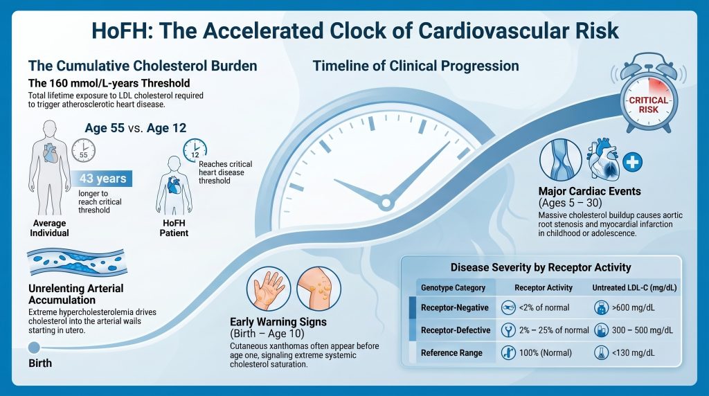
La carga acumulativa se expresa en mmol/L-años. La evidencia clínica indica que el inicio de la aterosclerosis clínica enfermedad cardiovascular (ASCVD) en la población general corresponde a un umbral acumulativo de aproximadamente 125 mmol/L-años (~4800 mg/dL-años).[62,63], mientras que el umbral para el primero infarto de miocardio es de aproximadamente 160 mmol/L-años (~6,000 mg/dL-años). Un individuo normolipidémico normalmente alcanza la carga de 160 mmol/L-año alrededor de los 55 años. En contraste, un paciente con HoFH no tratado con un C-LDL de ~500 mg/dL (~13 mmol/L) alcanza este mismo umbral aproximadamente a los 12 años, lo que explica por qué los niños con las mutaciones de receptor nulo más graves a menudo experimentan infartos de miocardio o muerte súbita cardíaca antes de los 10 años.[19,63]
La patología vascular de la HoFH pediátrica
La fisiopatología de la aterosclerosis en la HoFH pediátrica implica una progresión rápida desde la activación endotelial hasta lesiones avanzadas placa formación. Bajo la influencia de la extrema hipercolesterolemia, las partículas de LDL penetran en la íntima arterial, donde se oxidan y son engullidas por macrófagos formar células espumosas.[24] En la población general, este proceso tarda décadas en progresar desde estrías grasas a placas obstructivas. En la HoFH, el flujo absoluto de lipoproteínas hacia la pared arterial es tan alto que estas estrías grasas evolucionan hacia placas complejas, ricas en lípidos y altamente inflamatorias en los primeros años de vida.[19]
Un rasgo distintivo de la enfermedad vascular asociada a la HoFH es su predilección anatómica por el raíz aórtica y los ostios coronarios.[27] La naturaleza rica en colesterol de estos lesiones con frecuencia resulta en estenosis aórtica supravalvular. A diferencia de la valvulopatía degenerativa calcificación visto en poblaciones de edad avanzada, la EAA en la FHHo se caracteriza por una infiltración masiva de la pared aórtica y de las valvas valvulares por tejido xantomatosos.[20,21] Además, el depósito de placa en los ostios coronarios puede provocar una oclusión súbita y total oclusión incluso en niños que aún no tienen difusa enfermedad de las arterias coronarias.[27]
El papel de los modificadores de riesgo secundarios
Además de la elevación primaria del C-LDL, muchos pacientes con HoFH presentan niveles significativamente elevados de Lipoproteína(a) [Lp(a)]. La Lp(a) es una partícula similar a las LDL con una fracción de apolipoproteína(a) añadida, lo que le confiere propiedades protrombóticas y proinflamatorias. Debido a que la Lp(a) se elimina principalmente a través del receptor de LDL, sus niveles son frecuentemente dos veces más altos en pacientes con HoFH en comparación con la población general.[2] La presencia de Lp(a) elevada actúa como un multiplicador de riesgo, acelerando aún más la aterosclerosis y aumentando la probabilidad de ruptura de placa o trombosis a temprana edad.[11]
El desarrollo de xantomas cutáneos antes de los 4 años de edad se asocia con un riesgo marcadamente aumentado de enfermedad infantil enfermedad coronaria y la mortalidad prematura.[20] Estos depósitos no son meramente cosméticos; representan el desbordamiento sistémico de colesterol que el sistema vascular ya no puede contener.[2]
Magnitud de las anomalías lipídicas
La magnitud de la hipercolesterolemia en la HoFH no tiene paralelo en la medicina clínica. No tratado colesterol total los niveles suelen oscilar entre 460 y 1160 mg/dL (12–30 mmol/L), y los niveles de c-LDL superan sistemáticamente los 400 mg/dL (10 mmol/L) en la mayoría de los casos confirmados genéticamente.[4,5] En casos extremos, particularmente en individuos con mutaciones del receptor nulo, el c-LDL puede alcanzar niveles superiores a 1,000 mg/dL (26 mmol/L) y la sangre puede adquirir un aspecto visualmente distinto y opaco debido a la extrema concentración de lipoproteínas.[2]
Apolipoproteína B y métricas de partículas
La apolipoproteína B (ApoB) es la principal estructura proteína de todas las lipoproteínas aterogénicas, incluidas VLDL, IDL, Lp(a) y LDL. Debido a que hay exactamente una molécula de ApoB por partícula, su medición proporciona un recuento directo del número total de partículas aterogénicas en circulación.[35] El rango de referencia normal para la ApoB en adultos es inferior a 130 mg/dL, con una mediana en la población de EE. UU. de aproximadamente 93 mg/dL. En la HoFH, los niveles de ApoB suelen ser de cuatro a seis veces más altos que este rango de referencia.[35]
La fisiopatología de la ApoB en la HoFH se define por dos factores: la sobreproducción y un aclaramiento severamente afectado. La ausencia de receptores de LDL funcionales conduce a un tiempo de residencia prolongado de las partículas de LDL en la sangre —aproximadamente de 5 a 6 días en comparación con los 2.5 días normales.[70] Este tiempo de circulación prolongado da como resultado que las partículas se modifiquen cada vez más. Si bien el Patrón B (LDL pequeña y densa) es generalmente más aterogénico porque penetra más fácilmente en la pared arterial, en la HoFH, la masa abrumadora de partículas de LDL aún más grandes impulsa un flujo constante hacia el espacio subendotelial.[39]
Comparación del perfil lipídico por genotipo
La gravedad de la anomalía lipídica está intrínsecamente relacionada con el estado funcional del receptor de LDL. Los pacientes se clasifican como «receptor negativo» (menos del 2% de la actividad normal del LDLR) o «receptor defectuoso» (entre el 2% y el 25% de la actividad normal).[1,56]
| Parámetro | HoFH negativo a receptores | HoFH defectivo en receptores | Rango de referencia |
| C-LDL no tratado | >600 mg/dL (>15.5 mmol/L) | 300–500 mg/dL (7.8–12.9 mmol/L) | <130 mg/dL (<3.4 mmol/L) |
| ApoB no tratada | >400 mg/dL (~4–6 veces lo normal) | 200–400 mg/dL (~2–4× lo normal) | <130 mg/dL |
| Lp(a) no tratada | Frecuentemente marcadamente elevado | Elevado | <30 mg/dL |
| Actividad de LDLR | <2% de lo normal | 2%–25% de normal | 100% (normal) |
| Respuesta a Estatinas | Mínima (reducción de ~141 TP9T en el colesterol LDL) | Moderada (reducción del colesterol LDL de aproximadamente 231 TP9T) | Alto (reducción de 40 a 551 TP9T) |
Tabla 1. Perfil lipídico comparación por genotipo de LDLR. Fuente: [1,3,41,71]
Un estudio de Registro CASCADE FH en los Estados Unidos demostraron que los niveles de c-LDL no tratado eran significativamente más altos en los pacientes reclutados como niños (mediana de 776 mg/dL, RIC 704-892) en comparación con aquellos reclutados como adultos (mediana de 533 mg/dL, RIC 467-702; p=0.001).[31] Esta discrepancia probablemente refleja un sesgo de supervivencia, donde los niños con las elevaciones más extremas son diagnosticados temprano porque desarrollan síntomas visibles, como xantomas o signos tempranos eventos cardíacos—mientras que aquellos con elevaciones más leves pueden pasar desapercibidos hasta la edad adulta.[23]
La relación C-LDL:ApoB y la discordancia
La proporción de c-LDL:ApoB es un indicador indirecto del tamaño de las partículas de LDL y de su contenido de colesterol. Una proporción inferior a 1.2 (expresada como mg/dL:mg/dL) indica un predominio de partículas de LDL pequeñas y densas, las cuales son altamente susceptibles a la oxidación y se quedan atrapadas con mayor facilidad dentro de los proteoglucanos arteriales.[38] Sin embargo, en la verdadera HoFH, el C-LDL y la ApoB suelen estar elevados de forma concordante, lo que significa que el riesgo extremo está impulsado principalmente por el volumen absoluto de la masa de colesterol y el número de partículas, más que por un cambio en el tamaño de las partículas por sí solo.[18]
Gravedad clínica y evolución natural (sin tratamiento)
La historia natural de la HoFH no tratada se caracteriza por una ateroesclerosis rápida y progresiva y una esperanza de vida notablemente acortada. Sin intervención, históricamente se ha informado que la edad promedio de muerte es de aproximadamente 18 años, y algunos niños sucumben a un infarto de miocardio a la temprana edad de 5 años.[17,31]
La evolución de las manifestaciones clínicas
Los primeros signos clínicos suelen ser dermatológicos. Los xantomas cutáneos suelen aparecer en el primer año de vida, presentándose como lesiones nodulares blandas y amarillentas en sitios de fricción, como los codos, las rodillas y las nalgas.[8] Los xantomas tendinosos implican el engrosamiento del tendón de Aquiles y de los tendones extensores de las manos.[8]
A medida que aumenta la carga de colesterol, surgen manifestaciones vasculares y valvulares. El arco corneal lipoide y los xantomas se observan con frecuencia antes de los 10 años.[15] Para la segunda década, la mayoría de los pacientes no tratados ha desarrollado coronariopatía sintomática arteria enfermedad y/o enfermedad de la raíz aórtica.[2]
Evidencia de caso histórico
En 1889, G. Lehzen y K. Knauss describieron a dos hermanas que hoy en día se reconocen como los que probablemente sean los primeros casos documentados de AHoF.[28] La hermana de 11 años desarrolló múltiples manchas y bultos amarillos a partir de los 3 años. A los 11 años, presentó tumores “similares a huevos” en sus manos y xantomas masivos en sus tendones de Aquiles. El examen clínico reveló un soplo sistólico soplante prolongado y falleció poco después de una cirugía.[28]
Los hallazgos de la autopsia documentaron:
- AortaEngrosado con tejido adiposo y placas escleróticas.
- Válvula aórticaEstenótico debido a cambios masivos en la íntima.
- Arteria carótidaLa carótida izquierda estaba casi completamente ocluida.
- Arterias coronarias: Ambas mostraron múltiples placas, estando la izquierda gravemente afectada.[28]
Observaciones basadas en registros sobre la historia natural
Los datos de registros contemporáneos en países de ingresos no altos, donde el acceso a terapias avanzadas es limitado, reflejan este patrón histórico. En un estudio de 751 pacientes con HoFH en 38 países, aquellos en regiones de ingresos no altos experimentaron su primera evento cardiovascular adverso mayor una década antes que los de los países de ingresos altos (edad mediana de 24 frente a 35 años).[47]
| Característica clínica | Edad típica de inicio (sin tratamiento) | Pathological Significance |
| Cutaneous Xanthomas | <1 year | Marker of extreme systemic saturation |
| Corneal Arcus | <10 years | Early indicator of lipid spillover |
| Tendon Xanthomas | 5–15 years | Cumulative tissue deposition |
| Aortic Root Estenosis | 5–20 years | Primary cause of non-ischemic cardiac death |
| Infarto de miocardio | 5–30 years | Result of ostial or diffuse CAD |
| Sudden Cardiac Death | Variable; can occur in childhood | Fatal arrhythmia or total ostial occlusion |
Table 2. Clinical progression of untreated HoFH. Source: [2,19,47]
Why Some Patients Survive Longer Than Others
Despite the severe nature of the disease, there is significant inter-individual variability in survival. Some patients succumb to paro cardíaco in early childhood, while others, even with identical genetic mutations, may live into their 50s or 60s. This heterogeneity is driven by a complex interplay of residual receptor activity, genetic modifiers, and the timing of therapeutic intervention.[1]
Residual LDLR Activity and Mutation Type
The most powerful predictor of clinical outcome is the residual activity of the LDL receptor. Receptor-negative individuals (null/null; <2% activity) exhibit the highest LDL-C levels, the poorest response to traditional medications, and the earliest onset of ASCVD.[3,56] Receptor-defective patients (2%–25% activity) often have LDL-C levels approximately 18% lower than receptor-negative patients and respond more vigorously to pharmacological up-regulation of the receptor.[41] This genetic dosing effect can delay the onset of cardiovascular events by a decade or more.[6]
Modificadores genéticos protectores
PCSK9 Loss-of-Function (LOF)
PCSK9 is a protein that binds to the LDL receptor and targets it for lysosomal degradation. Individuals who co-inherit a loss-of-function variant in PCSK9 have naturally lower circulating PCSK9 levels, leading to a higher density of LDL receptors on the hepatocyte surface.[13] Population studies show that PCSK9 LOF variants are associated with approximately 28% lower LDL-C and up to 88% reduction in CHD risk.[69] In the context of HoFH, a PCSK9 LOF variant can significantly counteract the effect of a pathogenic LDLR variant, resulting in a much milder clinical phenotype than would otherwise be expected.[14]
ANGPTL3 Loss-of-Function
Angiopoietin-like protein 3 (ANGPTL3) inhibits lipoproteína lipasa and endothelial lipase. LOF variants in ANGPTL3 lead to lower levels of LDL, VLDL, and HDL through mechanisms largely independent of the LDL receptor.[9] Because this pathway does not rely on LDLR, it acts as a potent modifier even in receptor-negative HoFH patients.[9]
APOB Truncations
Some patients carry hypobetalipoproteinemia variants in the APOB gene that lead to reduced production of LDL particles. If a patient with an LDLR mutation also carries one of these variants, the liver produces fewer atherogenic vehicles, limiting the maximum LDL-C level achievable.[54]
Therapeutic Era and Intervention Thresholds
Before the 1980s, treatment was limited to low-fat diets and early ácido biliar sequestrants, which were largely ineffective.[17] The introduction of statins, and later LDL apheresis, began to extend the life expectancy of HoFH patients into the late 20s and 30s.[17]
The modern era, characterized by the availability of receptor-independent therapies, has fundamentally changed the prognosis. Lomitapida (an MTP inhibitor that reduces VLDL/LDL production) reduces LDL-C by approximately 50% independent of receptor genotype.[55,72] Evinacumab (an ANGPTL3 inhibitor) reduces LDL-C by 43–53% even in null-receptor patients.[32] The combination of these agents, if started in early childhood, allows many patients to keep their cumulative cholesterol burden below critical thresholds for much longer.[19]
Longest-Lived HoFH Cases
While the historic life expectancy was under 20 years, contemporary literature now documents survivors living into their 50s, 60s, and beyond.
The 57-Year-Old Tokyo Case (Komuro et al., 1987)
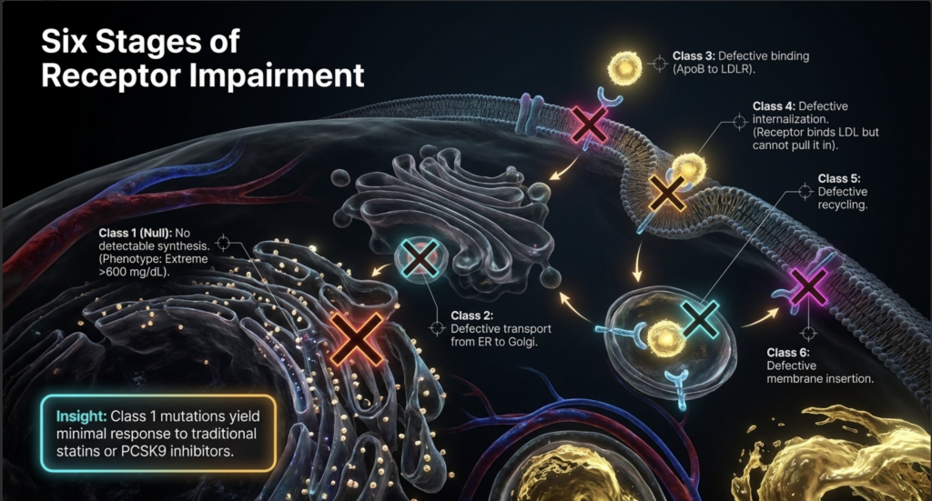
One of the first longest-lived cases reported was a 57-year-old Japanese male described in 1987.[64] This patient was homozygous for an internalization defect in the LDL receptor (a Class 4 mutation), meaning the receptors could bind LDL but could not pull it into the cell. His LDL-C was 461 mg/dL—lower than typical null-receptor patients—which allowed him to survive into his late 50s despite the absence of modern statins during much of his life.[64]
The 59-Year-Old Czech Case (Novák et al., 2025)
A 2025 study reported on several atypical Czech HoFH patients, including a 59-year-old male compound heterozygote for the p.Phe114Ile and p.Gly592Glu LDLR variants.[49] This patient was not diagnosed with HoFH until age 41. Although he eventually required a coronary artery bypass graft at age 57 and had significant carotid narrowing, his survival was exceptional given his genetic makeup. His specific compound heterozygous combination evidently conferred enough residual LDLR activity to prevent childhood mortality despite decades of extreme LDL-C elevation.[49]
The 72-Year-Old Heterozygous FH Case (Johnson et al., 2018)
An instructive case involves a 72-year-old male with a pathogenic LDLR variant (p.Val827Ile) who maintained untreated LDL-C consistently around 487 mg/dL throughout his life.[37] Despite this extreme elevation and lifelong lack of treatment, he had an Agatston puntaje de calcio of 0 on multiple scans—implying a complete absence of coronary artery calcification. His survival was attributed to an exceptionally high HDL-C (~68 mg/dL) and a Pattern A LDL (large, buoyant particles), which are less prone to oxidation and arterial retention.[37] Editorial note: This patient has been confirmed as heterozygous FH, not HoFH, based on the primary publication.[37] The case is cited here as an extreme FH phenotype illustrating the protective role of HDL-C and LDL particle size.
Contemporary Pediatric Successes
Registry data now show that children diagnosed at age 2 and started on aggressive therapy—including liver transplantation or apheresis—are reaching adulthood with minimal atherosclerotic burden.[23] Case reports document patients managed with weekly plasmapheresis and LDL apheresis from their teens into their 30s, maintaining a high quality of life and illustrating that mechanical clearance of cholesterol can effectively substitute for missing hepatic receptors.[59]
Summary Diagnostic and Clinical Tables
Table 3: Refined Clinical and Genetic Criteria for HoFH Diagnosis
| Categoría | Diagnostic Threshold / Feature | Rationale |
| C-LDL no tratado | >400 mg/dL (>10 mmol/L) | Proposed by 2023 EAS to capture broader spectrum |
| Treated LDL-C | >300 mg/dL (>8 mmol/L) on statin + ezetimibe | Historical threshold; used when baseline is unknown |
| Genetic Criteria | Bi-allelic variants (LDLR, APOB, PCSK9, LDLRAP1) | Gold standard for confirmation |
| Physical Findings | Xanthomas before age 10; Corneal Arcus | Pathognomonic for extreme cumulative burden |
| Family History | HeFH in both biological parents | Consistent with autosomal codominant pattern |
| Non-Genetic Mimics | Sitosterolaemia; LAL-D; CTX | Must be excluded to ensure appropriate therapy |
Table 4: Functional Classification of LDLR Mutations in HoFH
| Clase | Mechanism of Defect | Phenotypic Severity | Therapeutic Implication |
| Class 1 | No detectable synthesis (Null) | Extreme (>600 mg/dL) | No response to Statins/PCSK9i |
| Class 2 | Defective transport (ER to Golgi) | Grave | Minimal drug response |
| Class 3 | Defective binding (ApoB to LDLR) | Variable | May respond to PCSK9i |
| Class 4 | Defective internalization | Moderate to Severe | Some residual clearance possible |
| Class 5 | Defective recycling | Moderado | Often responds to drug up-regulation |
| Class 6 | Defective membrane insertion | Variable | Depends on density of insertion |
Source: [3]
Table 5: Pediatric vs. Adult HoFH — CASCADE FH Registry (Cuchel et al., 2023)
| Métrica | Children (n=16) Median (IQR) | Adults (n=51) Median (IQR) |
| Age at Diagnosis (years) | 2.5 (1–6) | 23 (13–34) |
| Untreated LDL-C (mg/dL) | 776 (704–892) | 533 (467–702) |
| ASCVD at Enrollment | 43.8% | 78.4% |
| Aortic Valve Stenosis at Enrollment | 18.8% | 25.5% |
| CABG (any) | 12.5% | 41.2% |
Source: [31]
Evidence Quality and Uncertainty
The scientific community’s understanding of HoFH has evolved from descriptive case studies to comprehensive international registries, but several critical areas of uncertainty remain.
Limitations of Current Evidence
The rarity of HoFH makes large-scale, ensayos controlados aleatorizados (RCTs) extremely difficult. Much of the evidence for long-term survival and treatment efficacy is derived from retrospective registry data (such as CASCADE FH or the Worldwide HoFH Study) or open-label phase 2 and phase 3 trials.[17] While registries provide invaluable real-world data, they are subject to selection bias—patients who are more severely affected or who have access to specialized care are more likely to be enrolled.[1]
A significant area of uncertainty is the missing genetic cause in approximately 20–40% of patients with a clinical diagnosis of HoFH.[7,66] This suggests that there are either unidentified FH genes or that a polygenic mechanism—the accumulation of many small-effect variants—can mimic the severity of monogenic HoFH. The clinical management of these mutation-negative patients remains a challenge.[67,68]
Gaps in Pediatric Management
While current guidelines recommend universal lipid screening in children, the optimal age for initiating advanced therapies (such as evinacumab or lomitapide) in toddlers is still under investigation.[1] The long-term safety of these agents in developing children is a concern, yet the risk of waiting for more data is the development of irreversible aortic root disease.[27]
Furthermore, the threshold hypothesis of cumulative cholesterol burden (expressed in mmol/L-years) is an elegant model but has not been prospectively validated as a definitive point of no return.[19] There is debate over whether lowering LDL-C can actually regress existing pediatric plaques or merely prevent the formation of new ones.[25]
Direcciones futuras
The future of HoFH research lies in gene therapy and base editing. Agents intended to directly alter the LDLR or PCSK9 genes in the liver—including in vivo CRISPR base editing—are currently in preclinical and early ensayos clínicos.[52,53] These one-and-done therapies could theoretically eliminate the need for lifelong infusions and daily pills, but the long-term genomic stability and safety of these approaches remain the primary uncertainties of the coming decade.[52,53]
Conclusión
HoFH is a life-threatening emergency that manifests in the first years of life. The magnitude of the lipid abnormality is so great that it overcomes standard biological repair mechanisms, necessitating a multi-hit, receptor-independent therapeutic approach. Survival variability is driven by the specific nature of the genetic defect and the presence of protective modifiers, but the most important determinant of life or death remains the timing of diagnosis and the intensity of LDL-C lowering achieved in the first decade of life.[1,19,31]
Referencias
- Cuchel M, Bruckert E, Ginsberg HN, et al. Homozygous familial hypercholesterolaemia: new insights and guidance for clinicians to improve detection and clinical management. A position paper from the Consensus Panel on Familial Hypercholesterolaemia of the European Atherosclerosis Society. Eur Heart J. 2014;35(32):2146-2157. doi:10.1093/eurheartj/ehu274
- Rahman A, Ahmed MU, Islam AK, Karim A, Sarker SA. A young male with familial hypercholesterolemia. J Saudi Heart Assoc. 2012;24(4):261-264. doi:10.1016/j.jsha.2012.06.264
- Suryawanshi YN, Warbhe RA. Familial Hypercholesterolemia: A Literature Review of the Pathophysiology and Current and Novel Treatments. Cureus. 2023;15(11):e49121. Published 2023 Nov 20. doi:10.7759/cureus.49121
- Ziajka PE. Management of patients with homozygous familial hypercholesterolemia. Am J Manag Care. 2013;19(13 Suppl):.
- Cuchel M, Raal FJ, Hegele RA, et al. 2023 Update on European Atherosclerosis Society Consensus Statement on Homozygous Familial Hypercholesterolaemia: new treatments and clinical guidance. Eur Heart J. 2023;44(25):2277-2291. doi:10.1093/eurheartj/ehad197
- Sjouke B, Kusters DM, Kindt I, et al. Homozygous autosomal dominant hypercholesterolaemia in the Netherlands: prevalence, genotype-phenotype relationship, and clinical outcome. Eur Heart J. 2015;36(9):560-565. doi:10.1093/eurheartj/ehu058
- Sturm AC, Knowles JW, Gidding SS, et al. Clinical Genetic Testing for Familial Hypercholesterolemia: JACC Scientific Expert Panel. J Am Coll Cardiol. 2018;72(6):662-680. doi:10.1016/j.jacc.2018.05.044
- Marais AD. Familial hypercholesterolaemia. Clin Biochem Rev. 2004;25(1):49-68.
- van den Bosch SE, Corpeleijn WE, Hutten BA, Wiegman A. How Genetic Variants in Children with Familial Hypercholesterolemia Not Only Guide Detection, but Also Treatment. Genes (Basel). 2023;14(3):669. Published 2023 Mar 7. doi:10.3390/genes14030669
- Zhao L, Gao Y, Liu G, et al. Zhonghua Xin Xue Guan Bing Za Zhi. 2022;50(6):585-590. doi:10.3760/cma.j.cn112148-20210715-00591
- Warden BA, Fazio S, Shapiro MD. Familial Hypercholesterolemia: Genes and Beyond. In: Feingold KR, Adler RA, Ahmed SF, et al., eds. Endotext. South Dartmouth (MA): MDText.com, Inc.; September 23, 2024.
- Razman AZ, Chua YA, Mohd Kasim NA, et al. Genetic Spectrum of Familial Hypercholesterolaemia in the Malaysian Community: Identification of Pathogenic Gene Variants Using Targeted Next-Generation Sequencing. Int J Mol Sci. 2022;23(23):14971. Published 2022 Nov 29. doi:10.3390/ijms232314971
- Saavedra YG, Dufour R, Davignon J, Baass A. PCSK9 R46L, lower LDL, and cardiovascular disease risk in familial hypercholesterolemia: a cross-sectional cohort study. Arterioscler Thromb Vasc Biol. 2014;34(12):2700-2705. doi:10.1161/ATVBAHA.114.304406
- Bayona A, Arrieta F, Rodríguez-Jiménez C, et al. Loss-of-function mutation of PCSK9 as a protective factor in the clinical expression of familial hypercholesterolemia: A case report. Medicine (Baltimore). 2020;99(34):e21754. doi:10.1097/MD.0000000000021754
- Gidding SS, Champagne MA, de Ferranti SD, et al. The Agenda for Familial Hypercholesterolemia: A Scientific Statement From the American Heart Association. Circulation. 2015;132(22):2167-2192. doi:10.1161/CIR.0000000000000297
- Defesche JC, Gidding SS, Harada-Shiba M, Hegele RA, Santos RD, Wierzbicki AS. Familial hypercholesterolaemia. Nat Rev Dis Primers. 2017;3:17093. Published 2017 Dec 7. doi:10.1038/nrdp.2017.93
- Raal FJ, Pilcher GJ, Panz VR, et al. Reduction in mortality in subjects with homozygous familial hypercholesterolemia associated with advances in lipid-lowering therapy. Circulation. 2011;124(20):2202-2207. doi:10.1161/CIRCULATIONAHA.111.042523
- Goldberg AC, Hopkins PN, Toth PP, et al. Familial hypercholesterolemia: screening, diagnosis and management of pediatric and adult patients: clinical guidance from the National Lipid Association Expert Panel on Familial Hypercholesterolemia. J Clin Lipidol. 2011;5(3 Suppl):S1-S8. doi:10.1016/j.jacl.2011.04.003
- Wiegman A, Gidding SS, Watts GF, et al. Familial hypercholesterolaemia in children and adolescents: gaining decades of life by optimizing detection and treatment. Eur Heart J. 2015;36(36):2425-2437. doi:10.1093/eurheartj/ehv157
- Bélanger AM, Akioyamen LE, Ruel I, Hales L, Genest J. Aortic stenosis in homozygous familial hypercholesterolaemia: a paradigm shift over a century. Eur Heart J. 2022;43(34):3227-3239. doi:10.1093/eurheartj/ehac339
- Rocha VZ, Santos RD. Past, Present, and Future of Familial Hypercholesterolemia Management. Methodist Debakey Cardiovasc J. 2021;17(4):28-35. Published 2021 Sep 24. doi:10.14797/mdcvj.887
- Cuchel M, Raal FJ, Hegele RA, et al. 2023 Update on European Atherosclerosis Society Consensus Statement on Homozygous Familial Hypercholesterolaemia: new treatments and clinical guidance. Eur Heart J. 2023;44(25):2277-2291. doi:10.1093/eurheartj/ehad197
- Family Heart Foundation, “Family Heart Foundation study shows some children with HoFH miss out on decades of life-saving treatment,” EurekAlert!, 2022. [Press release; cited for registry context only.]
- Hong YM. Atherosclerotic cardiovascular disease beginning in childhood. Korean Circ J. 2010;40(1):1-9. doi:10.4070/kcj.2010.40.1.1
- Tousoulis D, Kampoli AM, Papageorgiou N, et al. Pathophysiology of atherosclerosis: the role of inflammation. Curr Pharm Des. 2011;17(37):4089-4110. doi:10.2174/138161211798764843
- Wiegman A, Gidding SS, Watts GF, et al. Familial hypercholesterolaemia in children and adolescents: gaining decades of life by optimizing detection and treatment. Eur Heart J. 2015;36(36):2425-2437. doi:10.1093/eurheartj/ehv157
- Cuchel M, Raal FJ, Hegele RA, et al. 2023 Update on European Atherosclerosis Society Consensus Statement on Homozygous Familial Hypercholesterolaemia: new treatments and clinical guidance. Eur Heart J. 2023;44(25):2277-2291. doi:10.1093/eurheartj/ehad197
- Ballantyne CM, Gellis L, Tardif JC, et al. Efficacy and Safety of Oral PCSK9 Inhibitor Enlicitide in Adults With Heterozygous Familial Hypercholesterolemia: A Randomized Clinical Trial. JAMA. 2026;335(2):129-139. doi:10.1001/jama.2025.20620
- Maldar SB, Pinto CJ. Homozygous familial hypercholesterolaemia in a patient presenting with hypertensive encephalopathy. BMJ Case Rep. 2022;15(10):e250265. Published 2022 Oct 31. doi:10.1136/bcr-2022-250265
- Suresh Kumar G, Mathbout MF, Fahsah I, Ghafghazi S. Case of homozygous familial hypercholesterolaemia with premature coronary artery disease. BMJ Case Rep. 2021;14(5):e242114. Published 2021 May 19. doi:10.1136/bcr-2021-242114
- Cuchel M, Lee PC, Hudgins LC, et al. Contemporary Homozygous Familial Hypercholesterolemia in the United States: Insights From the CASCADE FH Registry. J Am Heart Assoc. 2023;12(9):e029175. doi:10.1161/JAHA.122.029175
- Raal FJ, Rosenson RS, Reeskamp LF, et al. Evinacumab for Homozygous Familial Hypercholesterolemia. N Engl J Med. 2020;383(8):711-720. doi:10.1056/NEJMoa2004215
- Palacio CH, Harring TR, Nguyen NT, Goss JA, O’Mahony CA. Homozygous familial hypercholesterolemia: case series and review of the literature. Case Rep Transplant. 2011;2011:154908. doi:10.1155/2011/154908
- Raal FJ, Santos RD. Homozygous familial hypercholesterolemia: current perspectives on diagnosis and treatment. Atherosclerosis. 2012;223(2):262-268. doi:10.1016/j.atherosclerosis.2012.02.019
- Contois JH, McConnell JP, Sethi AA, et al. Apolipoprotein B and cardiovascular disease risk: position statement from the AACC Lipoproteins and Vascular Diseases Division Working Group on Best Practices. Clin Chem. 2009;55(3):407-419. doi:10.1373/clinchem.2008.118356
- Cuchel M, Bruckert E, Ginsberg HN, et al. Homozygous familial hypercholesterolaemia: new insights and guidance for clinicians to improve detection and clinical management. A position paper from the Consensus Panel on Familial Hypercholesterolaemia of the European Atherosclerosis Society. Eur Heart J. 2014;35(32):2146-2157. doi:10.1093/eurheartj/ehu274
- Johnson KW, Dudley JT, Bobe JR. A 72-Year-Old Patient with Longstanding, Untreated Familial Hypercholesterolemia but no Coronary Artery Calcification: A Case Report. Cureus. 2018;10(4):e2452. Published 2018 Apr 9. doi:10.7759/cureus.2452
- Nordestgaard BG, Langsted A, Mora S, et al. Fasting is not routinely required for determination of a lipid profile: clinical and laboratory implications including flagging at desirable concentration cut-points-a joint consensus statement from the European Atherosclerosis Society and European Federation of Clinical Chemistry and Laboratory Medicine. Eur Heart J. 2016;37(25):1944-1958. doi:10.1093/eurheartj/ehw152
- Pencina KM, Pencina MJ, Lawler PR, et al. Interplay of Atherogenic Particle Number and Particle Size and the Risk of Coronary Heart Disease. Clin Chem. 2023;69(1):48-55. doi:10.1093/clinchem/hvac172
- Drouin-Chartier JP, Tremblay AJ, Bergeron J, Lamarche B, Couture P. The Low-Density Lipoprotein Receptor Genotype Is a Significant Determinant of the Rebound in Low-Density Lipoprotein Cholesterol Concentration After Lipoprotein Apheresis Among Patients With Homozygous Familial Hypercholesterolemia. Circulation. 2017;136(9):880-882. doi:10.1161/CIRCULATIONAHA.117.029435
- Bertolini S, Cantafora A, Averna M, et al. Clinical expression of familial hypercholesterolemia in clusters of mutations of the LDL receptor gene that cause a receptor-defective or receptor-negative phenotype. Arterioscler Thromb Vasc Biol. 2000;20(9):E41-E52. doi:10.1161/01.atv.20.9.e41
- Jayaram S, Meera S, Kadi S, Sreenivasa N. An Interesting Case of Familial Homozygous Hypercholesterolemia-A Brief Review. Indian J Clin Biochem. 2012;27(3):309-313. doi:10.1007/s12291-011-0165-8
- Bensabbahia D, El Achiwi M, Atrassi M, Abkari A, Widad G. Homozygous Familial Hypercholesterolemia in a Seven-Year-Old: A Case Study Highlighting the Importance of Early Diagnosis. Cureus. 2025;17(6):e86219. Published 2025 Jun 17. doi:10.7759/cureus.86219
- Mainieri F, Tagi VM, Chiarelli F. Recent Advances on Familial Hypercholesterolemia in Children and Adolescents. Biomedicines. 2022;10(5):1043. Published 2022 Apr 30. doi:10.3390/biomedicines10051043
- Grundy SM, Stone NJ; Guideline Writing Committee for the 2018 Cholesterol Guidelines. 2018 Cholesterol Clinical Practice Guidelines: Synopsis of the 2018 American Heart Association/American College of Cardiology/Multisociety Cholesterol Guideline. Ann Intern Med. 2019;170(11):779-783. doi:10.7326/M19-0365
- Defesche JC, Gidding SS, Harada-Shiba M, Hegele RA, Santos RD, Wierzbicki AS. Familial hypercholesterolaemia. Nat Rev Dis Primers. 2017;3:17093. Published 2017 Dec 7. doi:10.1038/nrdp.2017.93
- Tromp TR, Hartgers ML, Hovingh GK, et al. Worldwide experience of homozygous familial hypercholesterolaemia: retrospective cohort study. Lancet. 2022;399(10326):719-728. doi:10.1016/S0140-6736(21)02001-8
- Vladimirova-Kitova L, Kitov S, Ganev M, Chochkova-Bukova L. Case Report: Difficulties in the Treatment of a 12-Year-Old Patient With Homozygous Familial Hypercholesterolemia, Compound Heterozygous Form – 5 Years Follow-Up. Front Cardiovasc Med. 2021;8:743341. Published 2021 Oct 8. doi:10.3389/fcvm.2021.743341
- Zlatohlávek L, Becherová Beňová J, Foglarová T, Dudková T, Hubáček JA. Case Report: Beating the assumed prognosis: homozygous familial hypercholesterolemia with unexpected long survival. Front Cardiovasc Med. 2025;12:1643771. Published 2025 Oct 20. doi:10.3389/fcvm.2025.1643771
- Guo Q, Feng X, Zhou Y. PCSK9 Variants in Familial Hypercholesterolemia: A Comprehensive Synopsis. Front Genet. 2020;11:1020. Published 2020 Sep 23. doi:10.3389/fgene.2020.01020
- Bayona A, Arrieta F, Rodríguez-Jiménez C, et al. Loss-of-function mutation of PCSK9 as a protective factor in the clinical expression of familial hypercholesterolemia: A case report. Medicine (Baltimore). 2020;99(34):e21754. doi:10.1097/MD.0000000000021754
- Musunuru K, Chadwick AC, Mizoguchi T, et al. In vivo CRISPR base editing of PCSK9 durably lowers cholesterol in primates. Nature. 2021;593(7859):429-434. doi:10.1038/s41586-021-03534-y
- Lee RG, Mazzola AM, Braun MC, et al. Efficacy and Safety of an Investigational Single-Course CRISPR Base-Editing Therapy Targeting PCSK9 in Nonhuman Primate and Mouse Models. Circulation. 2023;147(3):242-253. doi:10.1161/CIRCULATIONAHA.122.062132
- Lacaze P, Riaz M, Sebra R, et al. Protective lipid-lowering variants in healthy older individuals without coronary heart disease. Open Heart. 2021;8(2):e001710. doi:10.1136/openhrt-2021-001710
- Cuchel M, Meagher EA, du Toit Theron H, et al. Efficacy and safety of a microsomal triglyceride transfer protein inhibitor in patients with homozygous familial hypercholesterolaemia: a single-arm, open-label, phase 3 study. Lancet. 2013;381(9860):40-46. doi:10.1016/S0140-6736(12)61731-0
- Norata GD, Tibolla G, Catapano AL. PCSK9 inhibition for the treatment of hypercholesterolemia: promises and emerging challenges. Vascul Pharmacol. 2014;62(2):103-111. doi:10.1016/j.vph.2014.05.011
- Bytyçi I, Henein MY, Bytyqi S, et al. PCSK9 and ANGPTL3 Inhibitors in Homozygous Familial Hypercholesterolemia: A Meta-analysis of Randomized Clinical Trials. Drugs. 2026;86(2):231-242. doi:10.1007/s40265-025-02272-z
- Banerjee P, Chan KC, Tarabocchia M, et al. Functional Analysis of LDLR (Low-Density Lipoprotein Receptor) Variants in Patient Lymphocytes to Assess the Effect of Evinacumab in Homozygous Familial Hypercholesterolemia Patients With a Spectrum of LDLR Activity. Arterioscler Thromb Vasc Biol. 2019;39(11):2248-2260. doi:10.1161/ATVBAHA.119.313051
- Alicezah MK, Razali R, Rahman T, et al. Homozygous familial hypercholesterolemia. Malays J Pathol. 2014;36(2):131-137.
- Harada-Shiba M, Ohtake A, Sugiyama D, et al. Guidelines for the Diagnosis and Treatment of Pediatric Familial Hypercholesterolemia 2022. J Atheroscler Thromb. 2023;30(5):531-557. doi:10.5551/jat.CR006
- Bélanger AM, Akioyamen L, Alothman L, Genest J. Evidence for improved survival with treatment of homozygous familial hypercholesterolemia. Curr Opin Lipidol. 2020;31(4):176-181. doi:10.1097/MOL.0000000000000686
- Ference BA, Graham I, Tokgozoglu L, Catapano AL. Impact of Lipids on Cardiovascular Health: JACC Health Promotion Series. J Am Coll Cardiol. 2018;72(10):1141-1156. doi:10.1016/j.jacc.2018.06.046
- Nordestgaard BG, Chapman MJ, Humphries SE, et al. Familial hypercholesterolaemia is underdiagnosed and undertreated in the general population: guidance for clinicians to prevent coronary heart disease: consensus statement of the European Atherosclerosis Society. Eur Heart J. 2013;34(45):3478-90a. doi:10.1093/eurheartj/eht273
- Komuro I, Kato H, Nakagawa T, et al. The longest-lived patient with homozygous familial hypercholesterolemia secondary to a defect in internalization of the LDL receptor. Am J Med Sci. 1987;294(5):341-345. doi:10.1097/00000441-198711000-00008
- Beheshti SO, Madsen CM, Varbo A, Nordestgaard BG. Worldwide Prevalence of Familial Hypercholesterolemia: Meta-Analyses of 11 Million Subjects. J Am Coll Cardiol. 2020;75(20):2553-2566. doi:10.1016/j.jacc.2020.03.057
- Futema M, Taylor-Beadling A, Williams M, Humphries SE. Genetic testing for familial hypercholesterolemia-past, present, and future. J Lipid Res. 2021;62:100139. doi:10.1016/j.jlr.2021.100139
- Vrablik M, Tichý L, Freiberger T, Blaha V, Satny M, Hubacek JA. Genetics of Familial Hypercholesterolemia: New Insights. Front Genet. 2020;11:574474. Published 2020 Oct 7. doi:10.3389/fgene.2020.574474
- Di Taranto MD, Giacobbe C, Fortunato G. Familial hypercholesterolemia: A complex genetic disease with variable phenotypes. Eur J Med Genet. 2020;63(4):103831. doi:10.1016/j.ejmg.2019.103831
- Cohen JC, Boerwinkle E, Mosley TH Jr, Hobbs HH. Sequence variations in PCSK9, low LDL, and protection against coronary heart disease. N Engl J Med. 2006;354(12):1264-1272. doi:10.1056/NEJMoa054013
- Bilheimer DW, Stone NJ, Grundy SM. Metabolic studies in familial hypercholesterolemia. Evidence for a gene-dosage effect in vivo. J Clin Invest. 1979;64(2):524-533. doi:10.1172/JCI109490
- Stein EA, Gipe D, Bergeron J, et al. Effect of a monoclonal antibody to PCSK9, REGN727/SAR236553, to reduce low-density lipoprotein cholesterol in patients with heterozygous familial hypercholesterolaemia on stable statin dose with or without ezetimibe therapy: a phase 2 randomised controlled trial. Lancet. 2012;380(9836):29-36. doi:10.1016/S0140-6736(12)60771-5
- Blom DJ, Averna MR, Meagher EA, et al. Long-Term Efficacy and Safety of the Microsomal Triglyceride Transfer Protein Inhibitor Lomitapide in Patients With Homozygous Familial Hypercholesterolemia. Circulation. 2017;136(3):332-335. doi:10.1161/CIRCULATIONAHA.117.028208

