
Klinische trajecten en pathofysiologische drijfveren van vroege mortaliteit bij homozygote familiaire hypercholesterolemie
Een uitgebreide onderzoeksanalyse
Evidence-based cardiovasculair onderzoek
Overzicht en definities
homozygote familiaire hypercholesterolemie (HoFH) vertegenwoordigt de meest catastrofale en fenotypisch extreme manifestatie van erfelijke lipidenstoornissen. Het is een ultrazeldzame genetische aandoening gekenmerkt door het bijna volledig onvermogen van de lever om lagedichtheids- lipoproteïne (LDL) deeltjes uit de systemische circulatie, wat resulteert in plasma cholesterol niveaus die vanaf de geboorte aanzienlijk zijn verhoogd.[1]
Historisch gezien werd HoFH gedefinieerd door een klassieke klinische triade: onbehandeld LDL-cholesterol (LDL-C) concentraties van meer dan 500 mg/dL (13 mmol/L), de aanwezigheid van pathognomonisch cutaan en pees xanthemen verschijnend in het eerste decennium van het leven, en bewijs van heterozygoot familiair hypercholesterolemie bij beide biologische ouders.[1]
Het wetenschappelijke begrip van de ziekte heeft de afgelopen decennia een aanzienlijke paradigmaverschuiving ondergaan. Genetisch onderzoek heeft een breder fenotypisch spectrum aan het licht gebracht dan voorheen werd erkend, wat klinische instanties zoals de Europese Aderverkalking Society (EAS) om de diagnostische drempelwaarden te verfijnen. De consensusverklaring van de EAS uit 2023 beveelt aan dat HoFH klinisch wordt vermoed bij elk individu met onbehandelde LDL-C-waarden boven 400 mg/dL (~10 mmol/L), aangezien veel patiënten met genetisch bevestigde biallelische mutaties zich presenteren met waarden onder de traditionele afkapwaarde van 500 mg/dL.[5] Dit is met name relevant bij pediatrische populaties, waar de LDL-C-waarden lager kunnen zijn als gevolg van voeding of fysiologische groei, maar toch een extreem cardiovasculair risico vormen.[6]
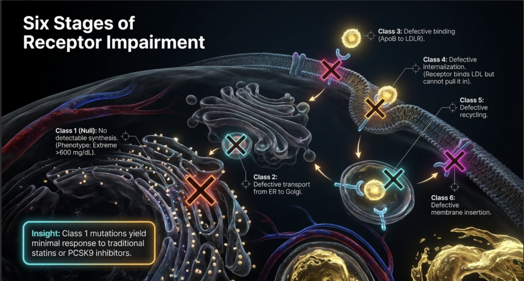
De genetische architectuur van HoFH wordt voornamelijk gekenmerkt door biallelische mutaties in het LDLR gen, dat codeert voor de LDL-receptor verantwoordelijk voor de hepatische opname van cholesterolrijke deeltjes.[8] Deze biallelicaire toestanden kunnen echt homozygoot zijn—waarbij de erferving van dezelfde pathogene variante van beide ouders—of compound heterozygoot, waarbij twee verschillende mutaties in hetzelfde gen betrokken zijn.[8] Naast LDLR worden zeldzamere vormen van HoFH veroorzaakt door pathogene varianten in de apolipoproteïne B (APOB) gen, dat het vermogen van het LDL-deeltje aantast om aan zijn receptor te binden, en "gain-of-function"-mutaties in het proproteineconvertase subtilisine/kexine type 9 (PCSK9) gen, dat de intracellulaire afbraak van de LDL-receptor versnelt.[10] Daarnaast resulteert een autosomaal recessieve vorm van de ziekte (ARH) in biallelic loss-of-function varianten in het LDLRAP1-gen.[8]
De prevalentie van HoFH werd traditioneel geschat op 1 op de 1.000.000 personen, maar hedendaagse epidemiologische gegevens en bevindingen uit registers suggereren een hogere frequentie van ongeveer 1 op de 160.000 tot 1 op de 300.000.[1,6] In specifieke populaties met stichterseffecten, zoals Frans-Canadezen, Libanese christenen, Zuid-Afrikaanse Afrikaners en bepaalde Asjkenazisch-Joodse gemeenschappen, kan de prevalentie aanzienlijk hoger zijn.[16]
Differentiële diagnose is van cruciaal belang. Sitosterolemie (fytosterolemie), veroorzaakt door biallelische varianten in de ABCG5/ABCG8-genen, presenteert zich met extreme verhogingen van LDL-C en vroege xanthomen, maar reageert opmerkelijk goed op dieetaanpassingen en ezetimib. Lysosomale zuur-lipasedeficiëntie (LAL-D) leidt ook tot ernstige hypercholesterolemie en premature atherosclerose maar wordt behandeld met enzymvervangende therapie. Cerebrotendineuze xanthomatose (CTX) kan zich manifesteren met xanthomen die vergelijkbaar zijn met HoFH, hoewel de cholesterolspiegels typisch normaal tot mild verhoogd zijn.[1]
Mechanisme van extreem risico in de vroegste levensfase
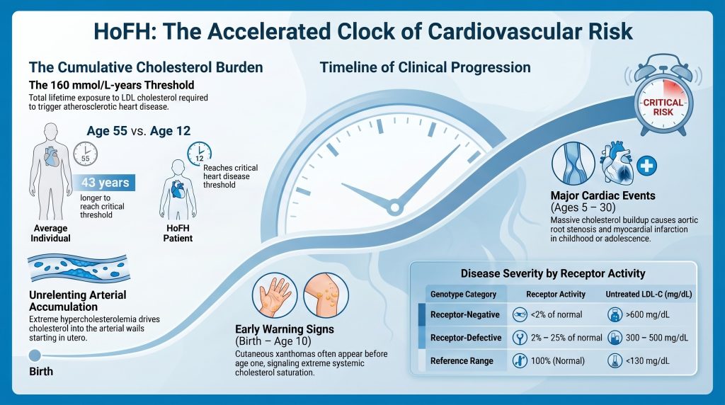
Het extreme cardiovasculaire risico bij HoFH wordt gedreven door de onophoudelijke opeenhoping van cholesterol in de vaatwand, die al in utero begint. Het centrale concept bij het kwantificeren van dit risico is de “cumulatieve cholesterolbelasting”, gedefinieerd als de totale massa LDL-C waaraan de slagader intima is in de loop der tijd blootgesteld.[19]
De cumulatieve last wordt uitgedrukt in mmol/l-jaar. Klinisch bewijs geeft aan dat het optreden van klinische atherosclerotische hart- en vaatziekten (ASCVD) in de algemene bevolking komt overeen met een cumulatieve drempelwaarde van ongeveer 125 mmol/L-jaar (~4.800 mg/dL-jaar).[62,63], terwijl de drempel voor de eerste myocardinfarct is ongeveer 160 mmol/L-jaar (~6.000 mg/dL-jaar). Een normolipidemisch individu bereikt de last van 160 mmol/L-jaar typisch rond de leeftijd van 55 jaar. Daarentegen bereikt een onbehandelde HoFH-patiënt met een LDL-C van ~500 mg/dL (~13 mmol/L) ditzelfde drempelniveau op ongeveer 12-jarige leeftijd, wat verklaart waarom kinderen met de meest ernstige nul-receptormutaties vaak een myocard infarct ondergaan of plotselinge hartdood vóór de leeftijd van 10 jaar.[19,63]
De vasculaire pathologie van pediatrische HoFH
De pathofysiologie van atherosclerose bij pediatrische HoFH omvat een snelle progressie van endotheelactivatie naar gevorderde tandplak vorming. Onder invloed van extreme hypercholesterolemie, LDL-deeltjes dringen door in de arteriële intima, waar ze worden geoxideerd en opgenomen door macrofagen vormen schuimcellen.[24] In de algemene bevolking duurt dit proces tientallen jaren om zich te ontwikkelen van vetstrepen tot obstructieve plaques. Bij HoFH is de pure instroom van lipoproteïnen in de vaatwand zo hoog dat deze vetvlekken binnen de eerste levensjaren evolueren tot complexe, lipidenrijke en zeer inflammatoire plaques.[19]
Een onderscheidend kenmerk van met HoFH geassocieerde vaatziekte is de anatomische voorliefde voor de aortawortel en de coronairostia.[27] Het cholesterolrijke karakter van deze laesies resulteert vaak in supravalvulaire aortastenose. In tegenstelling tot de degeneratieve klepaandoening verkalking gezien bij oudere populaties, wordt SVAS bij HoFH gekenmerkt door massale infiltratie van de aortawand en klepblaadjes door xanthomateus weefsel.[20,21] Bovendien kan plaqué-afzetting bij de kransslagaderoskia leiden tot een plotselinge, volledige occlusie zelfs bij kinderen die nog geen diffuse kransslagaderziekte.[27]
De rol van secundaire risicomodificatoren
Naast de primaire LDL-C-verhoging hebben veel HoFH-patiënten significant verhoogde spiegels van Lipoproteïne(a) [Lp(a)]. Lp(a) is een op LDL lijkend deeltje waaraan een apolipoproteïne(a)-eenheid is toegevoegd, wat pro-trombotische en pro-inflammatoire eigenschappen verleent. Omdat Lp(a) voornamelijk via de LDL-receptor wordt geklaard, zijn de spiegels bij HoFH-patiënten vaak twee zo hoog in vergelijking met de algemene bevolking.[2] De aanwezigheid van een hoog Lp(a) fungeert als een risicomultiplicator, die aderverkalking (atherosclerose) verder vers 武田 en de waarschijnlijkheid vergroot van plaqueruptuur of trombose op jonge leeftijd.[11]
De ontwikkeling van cutane xanthomen vóór de leeftijd van 4 jaar is geassocieerd met een aanmerkelijk verhoogd risico op jeugdige kransslagaderziekte en vroege sterfte.[20] Deze afzettingen zijn niet louter cosmetisch; ze vertegenwoordigen de systemische overloop van cholesterol die het vaatstelsel niet langer kan bevatten.[2]
Omvang van lipidenefwijkingen
De ernst van hypercholesterolemie bij HoFH is ongeëvenaard in de klinische geneeskunde. Onbehandeld totaal cholesterol de waarden variëren meestal van 460 tot 1.160 mg/dL (12–30 mmol/L), waarbij de LDL-C-waarden in de meeste genetisch bevestigde gevallen consequent hoger zijn dan 400 mg/dL (10 mmol/L).[4,5] In extreme gevallen, met name bij personen met een null-receptormutatie, kan het LDL-C niveaus bereiken van meer dan 1.000 mg/dL (26 mmol/L), en kan het bloed door de extreme concentratie aan lipoproteïnen een visueel onderscheidend, ondoorzichtig uiterlijk krijgen.[2]
Apolipoproteïne B en deeltjesmetingen
Apolipoproteïne B (ApoB) is de primaire structurele eiwit van alle atherogene lipoproteïnen, inclusief VLDL, IDL, Lp(a) en LDL. Omdat er precies één ApoB-molecuul per deeltje is, biedt de meting hiervan een directe telling van het totale aantal atherogene deeltjes in de omloop.[35] Het normale referentiebereik voor ApoB bij volwassenen is lager dan 130 mg/dL, met een mediaan voor de Amerikaanse bevolking van ongeveer 93 mg/dL. Bij HoFH zijn de ApoB-waarden typisch vier tot zes keer hoger dan dit referentiebereik.[35]
De pathofysiologie van ApoB bij HoFH wordt gedefinieerd door twee factoren: overproductie en ernstig verminderde klaring. De afwezigheid van functionele LDL-receptoren leidt tot een verlengde verblijftijd van LDL-deeltjes in het bloed – ongeveer 5–6 dagen vergeleken met de normale 2,5 dagen.[70] Deze langere circulatietijd zorgt ervoor dat de deeltjes in toenemende mate worden gemodificeerd. Hoewel Patroon B (kleine, dichte LDL) over het algemeen atherogener is omdat het gemakkelijker de vaatwand binnendringt, drijft bij HoFH de overweldigende massa van zelfs grotere LDL-deeltjes een constante flux naar de subendotheliale ruimte.[39]
Vergelijking van lipidenprofielen per genotype
De ernst van de lipidenafwijking hangt nauw samen met de functionele toestand van de LDL-receptor. Patiënten worden ingedeeld in de categorieën ‘receptornegatief’ (minder dan 2% van de normale LDLR-activiteit) of ‘receptordeficiënt’ (2% tot 25% van de normale activiteit).[1,56]
| Parameter | Receptor-negatieve HoFH | Receptor-defective HoFH | Referentiewaarde |
| Onbehandeld LDL-C | >600 mg/dL (>15,5 mmol/L) | 300–500 mg/dL (7,8–12,9 mmol/L) | <130 mg/dL (<3,4 mmol/L) |
| Onbehandeld ApoB | >400 mg/dL (~4–6× normaal) | 200–400 mg/dL (~2–4× normaal) | < 130 mg/dL |
| Onbehandelde Lp(a) | Vaak aanzienlijk verhoogd | Verhoogd | <30 mg/dL |
| LDLR-activiteit | <2% van normaal | 2%–25% van normaal | 100% (normaal) |
| Reactie op Statines | Minimaal (~141 TP9T LDL-C-verlaging) | Matig (~23% LDL-C-verlaging) | Hoog (40–551 TP9T-reductie) |
Tabel 1. Lipidenspectrum vergelijking op LDLR-genotype. Bron: [1,3,41,71]
Een studie van de CASCADE FH Register in de Verenigde Staten aantoonden dat onbehandelde LDL-C-waarden significant hoger waren bij patiënten die als kind werden ingeschreven (medediaan 776 mg/dL, IQR 704–892) vergeleken met degenen die als volwassene werden ingeschreven (mediaan 533 mg/dL, IQR 467–702; p=0,001).[31] Deze discrepantie weerspiegelt waarschijnlijk een overlevingsbias, waarbij kinderen met de meest extreme verhogingen vroeg worden gediagnosticeerd omdat ze zichtbare symptomen ontwikkelen—xanthomen of vroege cardiaque voorvallen—terwijl personen met mildere verhogingen tot de volwassen leeftijd niet gediagnosticeerd kunnen blijven.[23]
De LDL-C:ApoB-ratio en discordantie
De LDL-C:ApoB-verhouding is een surrogaat voor de LDL-deeltjesgrootte en het cholesterolgehalte. Een verhouding onder 1,2 (uitgedrukt als mg/dL:mg/dL) duidt op een overwicht van kleine, dichte LDL-deeltjes, die zeer vatbaar zijn voor oxidatie en gemakkelijker gevangen raken in arteriële proteoglycanen.[38] In klassieke HoFH zijn LDL-C en ApoB echter doorgaans beide concordant hoog, wat betekent dat het extreme risico primair wordt gedreven door de enorme hoeveelheid cholesterolmassa en het aantal deeltjes in plaats van enkel door een verschuiving in deeltjesgrootte.[18]
Klinische ernst en natuurlijk beloop (onbehandeld)
De natuurlijke geschiedenis van onbehandelde HoFH wordt gekenmerkt door snelle, progressieve atherosclerose en een sterk verkorte levensverwachting. Zonder ingrijpen is de gemiddelde leeftijd bij overlijden historisch gezien gerapporteerd als ongeveer 18 jaar, waarbij sommige kinderen al op 5-jarige leeftijd bezwijken aan een myocardinfarct.[17,31]
De evolutie van klinische manifestaties
De eerste klinische verschijnselen zijn meestal dermatologisch. Cutane xanthomen verschijnen vaak in het eerste levensjaar en presenteren zich als zachte, gele nodulaire letsels op plaatsen met wrijving, zoals de ellebogen, knieën en billen.[8] Peestoxen omvatten de verdikking van de achillespees en de extensorpezen van de handen.[8]
Naarmate de cholesterolbelasting toenemend stijgt, ontstaan er vasculaire en valvulaire manifestaties. Arcus lipoides corneae en xanthelasma worden vaak waargenomen vóór de leeftijd van 10 jaar.[15] Tegen het tweede decennium heeft de meerderheid van de onbehandelde patiënten symptomatische coronaire aandoeningen ontwikkeld slagader ziekte en/of aortawortelziekte.[2]
Historisch zaakbewijs
In 1889 beschreven G. Lehzen en K. Knauss twee zusters die nu worden erkend als vermoedelijk de eerste gedocumenteerde gevallen van HoFH.[28] Het 11-jarige zusje ontwikkelde vanaf de leeftijd van 3 jaar meerdere gele vlekken en knobbeltjes. Op 11-jarige leeftijd presenteerde zij zich met “eivormige” tumoren op haar handen en massale xanthomen op haar achillespezen. Bij klinisch onderzoek werd een lang blazend systolisch ruisje vastgesteld, en zij overleed kort na een operatie.[28]
De autopsiebevindingen toonden aan:
- AortaVerdikt met vetbevattend weefsel en sclerotische plaques.
- AortaklepStenotisch als gevolg van massieve intimale veranderingen.
- HalsaderDe linker arteria carotis was bijna volledig afgesloten.
- KransslagadersBeide vertoonden meerdere plaques, waarbij de linker ernstig was aangedaan.[28]
Registergebaseerde observaties over de natuurlijke historie
Gegevens uit hedendaagse registers in landen met een laag tot middeninkomen, waar de toegang tot geavanceerde therapieën beperkt is, weerspiegelen dit historische patroon. In een studie van 751 HoFH-patiënten in 38 landen kregen degenen in regio's met een laag tot middeninkomen hun eerste ernstige nadelige cardiovasculaire gebeurtenis een decennium eerder dan die in landen met een hoog inkomen (mediane leeftijd 24 versus 35 jaar).[47]
| Klinisch kenmerk | Typische leeftijd van begin (onbehandeld) | Pathologische betekenis |
| Cutane xanthomen | <1 jaar | Markeerder van extreme systemische verzadiging |
| Boogseniel | < 10 jaar | Vroege indicator van lipidenoverloop |
| Peesxanthomen | 5–15 jaar | Cumulatieve weefselafzetting |
| Aortawortel Stenose | 5–20 jaar | Primaire oorzaak van niet-ischemische hartdood |
| Myocardinfarct | 5–30 jaar | Resultaat van ostiaal of diffuus coronair lijden |
| Plotselinge hartdood | Variabel; kan optreden in de kindertijd | Fatale aritmie of totale ostiale occlusie |
Tabel 2. Klinische progressie van onbehandelde HoFH. Bron: [2,19,47]
Waarom sommige patiënten langer overleven dan anderen
Ondanks de ernstige aard van de ziekte is er aanzienlijke interindividuele variabiliteit in overleving. Sommige patiënten bezwijken aan hartstilstand in de vroege kindertijd, terwijl anderen, zelfs met identieke genetische mutaties, tot hun vijftigste of zestigste kunnen leven. Deze heterogeniteit wordt gedreven door een complex samenspel van resterende receptoractiviteit, genetische modificatoren en het tijdstip van de therapeutische interventie.[1]
Residuele LDLR-activiteit en mutatietype
De sterkste voorspeller van de klinische uitkomst is de resterende activiteit van de LDL-receptor. Personen zonder receptor (null/null; <2%-activiteit) vertonen de hoogste LDL-C-waarden, reageren het slechtst op traditionele medicatie en ontwikkelen het vroegst ASCVD.[3,56] Patiënten met een receptordefect (2%–25%-activiteit) hebben vaak LDL-C-waarden die ongeveer 18% lager liggen dan bij receptor-negatieve patiënten en reageren sterker op farmacologische opregulatie van de receptor.[41] Dit genetische doseringseffect kan het optreden van cardiovasculaire aandoeningen met een decennium of meer vertragen.[6]
Beschermende genetische modificatoren
PCSK9-functieverlies (LOF)
PCSK9 is een eiwit dat bindt aan de LDL-receptor en deze markeert voor lysosomale degradatie. Individuen die samen een functieverliesvariant in PCSK9 overerven, hebben van nature lagere circulerende PCSK9-spiegels, wat leidt tot een hogere dichtheid van LDL-receptoren op het hepatocytoppervlak.[13] Uit bevolkingsonderzoeken blijkt dat PCSK9-LOF-varianten gepaard gaan met een lager LDL-C-gehalte van ongeveer 28% en een afname van het risico op coronaire hartziekte (CHD) tot 88%.[69] In de context van HoFH kan een PCSK9 LOF-variant het effect van een pathogene LDLR-variant aanzienlijk tegengaan, wat resulteert in een veel milder klinisch fenotype dan anders zou worden verwacht.[14]
ANGPTL3-functieverlies
Angiopoëtine-achtige eiwit 3 (ANGPTL3) remt lipoproteïnelipase en endotheliale lipase. LOF-varianten in ANGPTL3 leiden tot lagere spiegels van LDL, VLDL en HDL via mechanismen die grotendeels onafhankelijk zijn van de LDL-receptor.[9] Omdat deze route niet afhankelijk is van LDLR, fungeert deze als een krachtige modifier, zelfs bij receptornegatieve HoFH-patiënten.[9]
APOB-trunceringen
Sommige patiënten dragen hypobetalipoproteïnemie-varianten in het APOB-gen die leiden tot een verminderde productie van LDL-partikels. Als een patiënt met een LDLR-mutatie ook een van deze varianten draagt, produceert de lever minder atherogene dragers, wat het maximale bereikbare LDL-C-gehalte beperkt.[54]
Therapeutisch Tijdperk en Interventiedrempels
Vóór de jaren 1980 was de behandeling beperkt tot vetarme diëten en vroege galzuur sequestranten, die grotendeels ineffectief waren.[17] De introductie van statines, en later LDL-aferese, begonnen de levensverwachting van HoFH-patiënten te verlengen tot achter in de jaren twintig en dertig.[17]
Het moderne tijdperk, gekenmerkt door de beschikbaarheid van receptoronafhankelijke therapieën, heeft de prognose fundamenteel veranderd. Lomitapide (een MTP-remmer die de productie van VLDL/LDL vermindert) verlaagt het LDL-C-gehalte met ongeveer 50%, ongeacht het receptorgenotype.[55,72] Evinacumab (een ANGPTL3-remmer) verlaagt het LDL-C-gehalte met 43–53%, zelfs bij patiënten met een null-receptor.[32] De combinatie van deze middelen, indien gestart in de vroege kinderjaren, stelt veel patiënten in staat om hun cumulatieve cholesterolbelasting gedurende veel langere tijd onder kritische drempels te houden.[19]
Langst levende HoFH-gevallen
Hoewel de historische levensverwachting onder de 20 jaar lag, beschrijft hedendaagse literatuur nu overlevenden die de leeftijd van 50, 60 en zelfs daarna bereiken.
De 57 jaar oude zaak in Tokio (Komuro et al., 1987)
Een van de eerst gerapporteerde langstlevende gevallen betrof een 57-jarige Japanse man die in 1987 werd beschreven.[64] Deze patiënt was homozygoot voor een internalisatiedefect in de LDL-receptor (een klasse 4-mutatie), wat betekent dat de receptoren wel LDL konden binden maar het niet de cel in konden trekken. Zijn LDL-C was 461 mg/dL—lager dan typische patiënten met nul-receptoren—waardoor hij tot zijn late vijftiger jaren kon overleven ondanks de afwezigheid van moderne statines gedurende een groot deel van zijn leven.[64]
De 59-jarige Tsjechische zaak (Novák et al., 2025)
Een studie uit 2025 rapporteerde over verschillende atypische Tsjechische HoFH-patiënten, waaronder een 59-jarige mannelijke compound heterozygoot voor de p.Phe114Ile- en p.Gly592Glu LDLR-varianten.[49] Deze patiënt werd pas op 41-jarige leeftijd gediagnosticeerd met HoFH. Hoewel hij uiteindelijk op 57-jarige leeftijd een coronaire bypassoperatie nodig had en een aanzienlijke vernauwing van de halsslagader had, was zijn overleving uitzonderlijk gezien zijn genetische opbouw. Zijn specifieke compound heterozygote combinatie verschafte blijkbaar voldoende resterende LDLR-activiteit om kindersterfte te voorkomen, ondanks tientallen jaren van extreme LDL-C-verhoging.[49]
De 72-jarige heterozygote FH-casus (Johnson et al., 2018)
Een leerzaam geval betreft een 72-jarige man met een pathogene LDLR-variant (p.Val827Ile) die gedurende zijn hele leven een onbehandeld LDL-C-gehalte had dat steevast rond de 487 mg/dL bleef.[37] Ondanks deze extreme verhoging en een levenslang gebrek aan behandeling, had hij een Agatston-score calciumscore van 0 op meerdere scans — wat duidt op een volledige afwezigheid van kransslagaderverkalking. Zijn overleving werd toegeschreven aan een uitzonderlijk hoog HDL-C (~68 mg/dL) en een patroon A LDL (grote, drijvende deeltjes), die minder gevoelig zijn voor oxidatie en arteriële retentie.[37] Noot van de redactie: Bij deze patiënt is op basis van de primaire publicatie bevestigd dat er sprake is van heterozygote FH, en niet van HoFH.[37] De zaak wordt hier aangehaald als een extreem FH-fenotype dat de beschermende rol van HDL-C en de grootte van LDL-deeltjes illustreert.
Hedendaagse pediatrische successen
Gegevens uit registers tonen nu aan dat kinderen die op 2-jarige leeftijd worden gediagnosticeerd en met een agressieve therapie worden gestart — waaronder levertransplantatie of aferese — de volwassenheid bereiken met een minimale atherosclerotische last.[23] Casusbeschrijvingen documenteren patiënten die vanaf hun tienerjaren tot in hun dertigjaren werden behandeld met wekelijkse plasmaferese en LDL-aferese, waarbij ze een hoge levenskwaliteit behouden en laten zien dat mechanische klaring van cholesterol de afwezige leverreceptoren effectief kan vervangen.[59]
Samenvatting diagnostische en klinische tabellen
Tabel 3: Verfijnde klinische en genetische criteria voor de diagnose van HoFH
| Categorie | Diagnostische drempel / Functie | Motivatie |
| Onbehandeld LDL-C | >400 mg/dL (>10 mmol/L) | Voorgesteld door de EAS in 2023 om een breder spectrum te vangen |
| Behandeld LDL-C | >300 mg/dL (>8 mmol/L) op statine + ezetimibe | Historische drempelwaarde; gebruikt wanneer de basislijn onbekend is |
| Genetische criteria | Bi-allelische varianten (LDLR, APOB, PCSK9, LDLRAP1) | Gouden standaard voor bevestiging |
| Lichamelijke bevindingen | Xanthomen voor de leeftijd van 10 jaar; Corneal arcus | Pathognomonisch voor een extreme cumulatieve belasting |
| Familiegeschiedenis | HeFH bij beide biologische ouders | Consistent met een autosomaal codominant patroon |
| Niet-genetische mimics | Sitosterolemie; LAL-D; CTX | Moet worden uitgesloten om een passende behandeling te waarborgen |
Tabel 4: Functionele classificatie van LDLR-mutaties bij HoFH
| Klas | Mechanisme van het defect | Fenotypische ernst | Therapeutische implicatie |
| Klasse 1 | Geen aantoonbare synthese (Nul) | Extreem (>600 mg/dL) | Geen respons op statines/PCSK9i |
| Klasse 2 | Defect transport (ER naar Golgi) | Ernstig | Minimale geneesmiddelrespons |
| Klasse 3 | Defecte binding (ApoB aan LDLR) | Variabele | Mag reageren op PCSK9i |
| Klasse 4 | Gebrekkige internalisatie | Matig tot ernstig | Enige residuele klaring mogelijk |
| Klas 5 | Defecte recycling | Gematigd | Reageert vaak op medicijn-upregulatie |
| Groep 6 | Defectieve membraaninsertering | Variabele | Hangt af van de plaatsingsdichtheid |
Bron:3]
Tabel 5: Pediatrische versus volwassen HoFH — CASCADE FH-register (Cuchel et al., 2023)
| Metrisch | Kinderen (n=16) Mediaan (IQR) | Volwassenen (n=51) Mediaan (IQR) |
| Leeftijd bij diagnose (jaren) | 2.5 (1–6) | 23 (13–34) |
| Onbehandeld LDL-C (mg/dL) | 776 (704–892) | 533 (467–702) |
| ASCVD bij inclusie | 43.8% | 78.4% |
| Aortaklepstenose bij inclusie | 18.8% | 25.5% |
| CABG (elk type) | 12.5% | 41.2% |
Bron:31]
Bewijskwaliteit en Onzekerheid
Het begrip van HoFH binnen de wetenschappelijke gemeenschap is geëvolueerd van beschrijvende casestudies naar uitgebreide internationale registers, maar er blijven diverse cruciale onzekerheden bestaan.
Beperkingen van het huidige bewijs
De zeldzaamheid van HoFH maakt grootschalige, gerandomiseerde gecontroleerde onderzoeken (RCT's) uiterst moeilijk. Veel van het bewijs voor langetermijnoverleving en behandelingseffectiviteit is afkomstig van retrospectieve registergegevens (zoals CASCADE FH of de Worldwide HoFH Study) of open-label fase 2- en fase 3-onderzoeken.[17] Hoewel registers waardevolle gegevens uit de praktijk opleveren, zijn ze onderhevig aan selectiebias: patiënten die ernstigst aangedaan zijn of toegang hebben tot gespecialiseerde zorg, worden vaker ingeschreven.[1]
Een belangrijk punt van onzekerheid is de ontbrekende genetische oorzaak bij ongeveer 20–40% van de patiënten met een klinische diagnose van HoFH.[7,66] Dit suggereert dat er ofwel niet-geïdentificeerde FH-genen zijn, ofwel dat een polygeen mechanisme—de accumulatie van velevarianten met een klein effect—de ernst van monogene HoFH kan nabootsen. Het klinische beheer van deze mutatieroratieve patiënten blijft een uitdaging.[67,68]
Hiaten in de pediatrische behandeling
Hoewel de huidige richtlijnen universele lipidenscreening bij kinderen aanbevelen, wordt de optimale leeftijd voor het starten van geavanceerde therapieën (zoals evinacumab of lomitapide) bij peuters nog onderzocht.[1] De veiligheid op lange termijn van deze middelen bij groeiende kinderen is een zorg, maar het risico van wachten op meer gegevens is de ontwikkeling van onomkeerbare aorta-wortelziekte.[27]
Bovendien is de drempelhypothese van de cumulatieve cholesterolbelasting (uitgedrukt in mmol/L-jaar) een elegant model, maar dit is niet prospectief gevalideerd als een definitief point of no return.[19] Er is discussie over de vraag of het verlagen van LDL-C bestaande pediatrische plaque daadwerkelijk kan doen afnemen of enkel de vorming van nieuwe kan voorkomen.[25]
Toekomstige richtingen
De toekomst van HoFH-onderzoek ligt in gentherapie en base-editing. Middelen die bedoeld zijn om de LDLR- of PCSK9-genen in de lever direct te veranderen—inclusief in vivo CRISPR-base-editing—zich momenteel in de preklinische en vroege klinische onderzoeken.[52,53] Deze eenmalige therapieën zouden theoretisch de noodzaak van levenslange infusen en dagelijkse pillen overbodig kunnen maken, maar de langetermijn-genomische stabiliteit en veiligheid van deze benaderingen blijven de belangrijkste onzekerheden van het komende decennium.[52,53]
Conclusie
HoFH is een levensbedreigende aandoening die zich in de eerste levensjaren manifesteert. De mate van de lipidenafwijking is zo groot dat deze de standaard biologische herstelmechanismen overwint, waardoor een multi-hit, receptoronafhankelijke therapeutische benadering noodzakelijk is. De variabiliteit in overleving wordt gedreven door de specifieke aard van het genetische defect en de aanwezigheid van beschermende modificatoren, maar de belangrijkste determinant van leven of dood blijft het tijdstip van diagnose en de intensiteit van de LDL-C-verlaging die in het eerste decennium van het leven wordt bereikt.[1,19,31]
Referenties
- Cuchel M, Bruckert E, Ginsberg HN, et al. Homozygous familial hypercholesterolaemia: new insights and guidance for clinicians to improve detection and clinical management. A position paper from the Consensus Panel on Familial Hypercholesterolaemia of the European Atherosclerosis Society. Eur Heart J. 2014;35(32):2146-2157. doi:10.1093/eurheartj/ehu274
- Rahman A, Ahmed MU, Islam AK, Karim A, Sarker SA. A young male with familial hypercholesterolemia. J Saudi Heart Assoc. 2012;24(4):261-264. doi:10.1016/j.jsha.2012.06.264
- Suryawanshi YN, Warbhe RA. Familial Hypercholesterolemia: A Literature Review of the Pathophysiology and Current and Novel Treatments. Cureus. 2023;15(11):e49121. Published 2023 Nov 20. doi:10.7759/cureus.49121
- Ziajka PE. Management of patients with homozygous familial hypercholesterolemia. Am J Manag Care. 2013;19(13 Suppl):.
- Cuchel M, Raal FJ, Hegele RA, et al. 2023 Update on European Atherosclerosis Society Consensus Statement on Homozygous Familial Hypercholesterolaemia: new treatments and clinical guidance. Eur Heart J. 2023;44(25):2277-2291. doi:10.1093/eurheartj/ehad197
- Sjouke B, Kusters DM, Kindt I, et al. Homozygous autosomal dominant hypercholesterolaemia in the Netherlands: prevalence, genotype-phenotype relationship, and clinical outcome. Eur Heart J. 2015;36(9):560-565. doi:10.1093/eurheartj/ehu058
- Sturm AC, Knowles JW, Gidding SS, et al. Clinical Genetic Testing for Familial Hypercholesterolemia: JACC Scientific Expert Panel. J Am Coll Cardiol. 2018;72(6):662-680. doi:10.1016/j.jacc.2018.05.044
- Marais AD. Familial hypercholesterolaemia. Clin Biochem Rev. 2004;25(1):49-68.
- van den Bosch SE, Corpeleijn WE, Hutten BA, Wiegman A. How Genetic Variants in Children with Familial Hypercholesterolemia Not Only Guide Detection, but Also Treatment. Genes (Basel). 2023;14(3):669. Published 2023 Mar 7. doi:10.3390/genes14030669
- Zhao L, Gao Y, Liu G, et al. Zhonghua Xin Xue Guan Bing Za Zhi. 2022;50(6):585-590. doi:10.3760/cma.j.cn112148-20210715-00591
- Warden BA, Fazio S, Shapiro MD. Familial Hypercholesterolemia: Genes and Beyond. In: Feingold KR, Adler RA, Ahmed SF, et al., eds. Endotext. South Dartmouth (MA): MDText.com, Inc.; September 23, 2024.
- Razman AZ, Chua YA, Mohd Kasim NA, et al. Genetic Spectrum of Familial Hypercholesterolaemia in the Malaysian Community: Identification of Pathogenic Gene Variants Using Targeted Next-Generation Sequencing. Int J Mol Sci. 2022;23(23):14971. Published 2022 Nov 29. doi:10.3390/ijms232314971
- Saavedra YG, Dufour R, Davignon J, Baass A. PCSK9 R46L, lower LDL, and cardiovascular disease risk in familial hypercholesterolemia: a cross-sectional cohort study. Arterioscler Thromb Vasc Biol. 2014;34(12):2700-2705. doi:10.1161/ATVBAHA.114.304406
- Bayona A, Arrieta F, Rodríguez-Jiménez C, et al. Loss-of-function mutation of PCSK9 as a protective factor in the clinical expression of familial hypercholesterolemia: A case report. Medicine (Baltimore). 2020;99(34):e21754. doi:10.1097/MD.0000000000021754
- Gidding SS, Champagne MA, de Ferranti SD, et al. The Agenda for Familial Hypercholesterolemia: A Scientific Statement From the American Heart Association. Circulation. 2015;132(22):2167-2192. doi:10.1161/CIR.0000000000000297
- Defesche JC, Gidding SS, Harada-Shiba M, Hegele RA, Santos RD, Wierzbicki AS. Familial hypercholesterolaemia. Nat Rev Dis Primers. 2017;3:17093. Published 2017 Dec 7. doi:10.1038/nrdp.2017.93
- Raal FJ, Pilcher GJ, Panz VR, et al. Reduction in mortality in subjects with homozygous familial hypercholesterolemia associated with advances in lipid-lowering therapy. Circulation. 2011;124(20):2202-2207. doi:10.1161/CIRCULATIONAHA.111.042523
- Goldberg AC, Hopkins PN, Toth PP, et al. Familial hypercholesterolemia: screening, diagnosis and management of pediatric and adult patients: clinical guidance from the National Lipid Association Expert Panel on Familial Hypercholesterolemia. J Clin Lipidol. 2011;5(3 Suppl):S1-S8. doi:10.1016/j.jacl.2011.04.003
- Wiegman A, Gidding SS, Watts GF, et al. Familial hypercholesterolaemia in children and adolescents: gaining decades of life by optimizing detection and treatment. Eur Heart J. 2015;36(36):2425-2437. doi:10.1093/eurheartj/ehv157
- Bélanger AM, Akioyamen LE, Ruel I, Hales L, Genest J. Aortic stenosis in homozygous familial hypercholesterolaemia: a paradigm shift over a century. Eur Heart J. 2022;43(34):3227-3239. doi:10.1093/eurheartj/ehac339
- Rocha VZ, Santos RD. Past, Present, and Future of Familial Hypercholesterolemia Management. Methodist Debakey Cardiovasc J. 2021;17(4):28-35. Published 2021 Sep 24. doi:10.14797/mdcvj.887
- Cuchel M, Raal FJ, Hegele RA, et al. 2023 Update on European Atherosclerosis Society Consensus Statement on Homozygous Familial Hypercholesterolaemia: new treatments and clinical guidance. Eur Heart J. 2023;44(25):2277-2291. doi:10.1093/eurheartj/ehad197
- Family Heart Foundation, “Family Heart Foundation study shows some children with HoFH miss out on decades of life-saving treatment,” EurekAlert!, 2022. [Press release; cited for registry context only.]
- Hong YM. Atherosclerotic cardiovascular disease beginning in childhood. Korean Circ J. 2010;40(1):1-9. doi:10.4070/kcj.2010.40.1.1
- Tousoulis D, Kampoli AM, Papageorgiou N, et al. Pathophysiology of atherosclerosis: the role of inflammation. Curr Pharm Des. 2011;17(37):4089-4110. doi:10.2174/138161211798764843
- Wiegman A, Gidding SS, Watts GF, et al. Familial hypercholesterolaemia in children and adolescents: gaining decades of life by optimizing detection and treatment. Eur Heart J. 2015;36(36):2425-2437. doi:10.1093/eurheartj/ehv157
- Cuchel M, Raal FJ, Hegele RA, et al. 2023 Update on European Atherosclerosis Society Consensus Statement on Homozygous Familial Hypercholesterolaemia: new treatments and clinical guidance. Eur Heart J. 2023;44(25):2277-2291. doi:10.1093/eurheartj/ehad197
- Ballantyne CM, Gellis L, Tardif JC, et al. Efficacy and Safety of Oral PCSK9 Inhibitor Enlicitide in Adults With Heterozygous Familial Hypercholesterolemia: A Randomized Clinical Trial. JAMA. 2026;335(2):129-139. doi:10.1001/jama.2025.20620
- Maldar SB, Pinto CJ. Homozygous familial hypercholesterolaemia in a patient presenting with hypertensive encephalopathy. BMJ Case Rep. 2022;15(10):e250265. Published 2022 Oct 31. doi:10.1136/bcr-2022-250265
- Suresh Kumar G, Mathbout MF, Fahsah I, Ghafghazi S. Case of homozygous familial hypercholesterolaemia with premature coronary artery disease. BMJ Case Rep. 2021;14(5):e242114. Published 2021 May 19. doi:10.1136/bcr-2021-242114
- Cuchel M, Lee PC, Hudgins LC, et al. Contemporary Homozygous Familial Hypercholesterolemia in the United States: Insights From the CASCADE FH Registry. J Am Heart Assoc. 2023;12(9):e029175. doi:10.1161/JAHA.122.029175
- Raal FJ, Rosenson RS, Reeskamp LF, et al. Evinacumab for Homozygous Familial Hypercholesterolemia. N Engl J Med. 2020;383(8):711-720. doi:10.1056/NEJMoa2004215
- Palacio CH, Harring TR, Nguyen NT, Goss JA, O’Mahony CA. Homozygous familial hypercholesterolemia: case series and review of the literature. Case Rep Transplant. 2011;2011:154908. doi:10.1155/2011/154908
- Raal FJ, Santos RD. Homozygous familial hypercholesterolemia: current perspectives on diagnosis and treatment. Atherosclerosis. 2012;223(2):262-268. doi:10.1016/j.atherosclerosis.2012.02.019
- Contois JH, McConnell JP, Sethi AA, et al. Apolipoprotein B and cardiovascular disease risk: position statement from the AACC Lipoproteins and Vascular Diseases Division Working Group on Best Practices. Clin Chem. 2009;55(3):407-419. doi:10.1373/clinchem.2008.118356
- Cuchel M, Bruckert E, Ginsberg HN, et al. Homozygous familial hypercholesterolaemia: new insights and guidance for clinicians to improve detection and clinical management. A position paper from the Consensus Panel on Familial Hypercholesterolaemia of the European Atherosclerosis Society. Eur Heart J. 2014;35(32):2146-2157. doi:10.1093/eurheartj/ehu274
- Johnson KW, Dudley JT, Bobe JR. A 72-Year-Old Patient with Longstanding, Untreated Familial Hypercholesterolemia but no Coronary Artery Calcification: A Case Report. Cureus. 2018;10(4):e2452. Published 2018 Apr 9. doi:10.7759/cureus.2452
- Nordestgaard BG, Langsted A, Mora S, et al. Fasting is not routinely required for determination of a lipid profile: clinical and laboratory implications including flagging at desirable concentration cut-points-a joint consensus statement from the European Atherosclerosis Society and European Federation of Clinical Chemistry and Laboratory Medicine. Eur Heart J. 2016;37(25):1944-1958. doi:10.1093/eurheartj/ehw152
- Pencina KM, Pencina MJ, Lawler PR, et al. Interplay of Atherogenic Particle Number and Particle Size and the Risk of Coronary Heart Disease. Clin Chem. 2023;69(1):48-55. doi:10.1093/clinchem/hvac172
- Drouin-Chartier JP, Tremblay AJ, Bergeron J, Lamarche B, Couture P. The Low-Density Lipoprotein Receptor Genotype Is a Significant Determinant of the Rebound in Low-Density Lipoprotein Cholesterol Concentration After Lipoprotein Apheresis Among Patients With Homozygous Familial Hypercholesterolemia. Circulation. 2017;136(9):880-882. doi:10.1161/CIRCULATIONAHA.117.029435
- Bertolini S, Cantafora A, Averna M, et al. Clinical expression of familial hypercholesterolemia in clusters of mutations of the LDL receptor gene that cause a receptor-defective or receptor-negative phenotype. Arterioscler Thromb Vasc Biol. 2000;20(9):E41-E52. doi:10.1161/01.atv.20.9.e41
- Jayaram S, Meera S, Kadi S, Sreenivasa N. An Interesting Case of Familial Homozygous Hypercholesterolemia-A Brief Review. Indian J Clin Biochem. 2012;27(3):309-313. doi:10.1007/s12291-011-0165-8
- Bensabbahia D, El Achiwi M, Atrassi M, Abkari A, Widad G. Homozygous Familial Hypercholesterolemia in a Seven-Year-Old: A Case Study Highlighting the Importance of Early Diagnosis. Cureus. 2025;17(6):e86219. Published 2025 Jun 17. doi:10.7759/cureus.86219
- Mainieri F, Tagi VM, Chiarelli F. Recent Advances on Familial Hypercholesterolemia in Children and Adolescents. Biomedicines. 2022;10(5):1043. Published 2022 Apr 30. doi:10.3390/biomedicines10051043
- Grundy SM, Stone NJ; Guideline Writing Committee for the 2018 Cholesterol Guidelines. 2018 Cholesterol Clinical Practice Guidelines: Synopsis of the 2018 American Heart Association/American College of Cardiology/Multisociety Cholesterol Guideline. Ann Intern Med. 2019;170(11):779-783. doi:10.7326/M19-0365
- Defesche JC, Gidding SS, Harada-Shiba M, Hegele RA, Santos RD, Wierzbicki AS. Familial hypercholesterolaemia. Nat Rev Dis Primers. 2017;3:17093. Published 2017 Dec 7. doi:10.1038/nrdp.2017.93
- Tromp TR, Hartgers ML, Hovingh GK, et al. Worldwide experience of homozygous familial hypercholesterolaemia: retrospective cohort study. Lancet. 2022;399(10326):719-728. doi:10.1016/S0140-6736(21)02001-8
- Vladimirova-Kitova L, Kitov S, Ganev M, Chochkova-Bukova L. Case Report: Difficulties in the Treatment of a 12-Year-Old Patient With Homozygous Familial Hypercholesterolemia, Compound Heterozygous Form – 5 Years Follow-Up. Front Cardiovasc Med. 2021;8:743341. Published 2021 Oct 8. doi:10.3389/fcvm.2021.743341
- Zlatohlávek L, Becherová Beňová J, Foglarová T, Dudková T, Hubáček JA. Case Report: Beating the assumed prognosis: homozygous familial hypercholesterolemia with unexpected long survival. Front Cardiovasc Med. 2025;12:1643771. Published 2025 Oct 20. doi:10.3389/fcvm.2025.1643771
- Guo Q, Feng X, Zhou Y. PCSK9 Variants in Familial Hypercholesterolemia: A Comprehensive Synopsis. Front Genet. 2020;11:1020. Published 2020 Sep 23. doi:10.3389/fgene.2020.01020
- Bayona A, Arrieta F, Rodríguez-Jiménez C, et al. Loss-of-function mutation of PCSK9 as a protective factor in the clinical expression of familial hypercholesterolemia: A case report. Medicine (Baltimore). 2020;99(34):e21754. doi:10.1097/MD.0000000000021754
- Musunuru K, Chadwick AC, Mizoguchi T, et al. In vivo CRISPR base editing of PCSK9 durably lowers cholesterol in primates. Nature. 2021;593(7859):429-434. doi:10.1038/s41586-021-03534-y
- Lee RG, Mazzola AM, Braun MC, et al. Efficacy and Safety of an Investigational Single-Course CRISPR Base-Editing Therapy Targeting PCSK9 in Nonhuman Primate and Mouse Models. Circulation. 2023;147(3):242-253. doi:10.1161/CIRCULATIONAHA.122.062132
- Lacaze P, Riaz M, Sebra R, et al. Protective lipid-lowering variants in healthy older individuals without coronary heart disease. Open Heart. 2021;8(2):e001710. doi:10.1136/openhrt-2021-001710
- Cuchel M, Meagher EA, du Toit Theron H, et al. Efficacy and safety of a microsomal triglyceride transfer protein inhibitor in patients with homozygous familial hypercholesterolaemia: a single-arm, open-label, phase 3 study. Lancet. 2013;381(9860):40-46. doi:10.1016/S0140-6736(12)61731-0
- Norata GD, Tibolla G, Catapano AL. PCSK9 inhibition for the treatment of hypercholesterolemia: promises and emerging challenges. Vascul Pharmacol. 2014;62(2):103-111. doi:10.1016/j.vph.2014.05.011
- Bytyçi I, Henein MY, Bytyqi S, et al. PCSK9 and ANGPTL3 Inhibitors in Homozygous Familial Hypercholesterolemia: A Meta-analysis of Randomized Clinical Trials. Drugs. 2026;86(2):231-242. doi:10.1007/s40265-025-02272-z
- Banerjee P, Chan KC, Tarabocchia M, et al. Functional Analysis of LDLR (Low-Density Lipoprotein Receptor) Variants in Patient Lymphocytes to Assess the Effect of Evinacumab in Homozygous Familial Hypercholesterolemia Patients With a Spectrum of LDLR Activity. Arterioscler Thromb Vasc Biol. 2019;39(11):2248-2260. doi:10.1161/ATVBAHA.119.313051
- Alicezah MK, Razali R, Rahman T, et al. Homozygous familial hypercholesterolemia. Malays J Pathol. 2014;36(2):131-137.
- Harada-Shiba M, Ohtake A, Sugiyama D, et al. Guidelines for the Diagnosis and Treatment of Pediatric Familial Hypercholesterolemia 2022. J Atheroscler Thromb. 2023;30(5):531-557. doi:10.5551/jat.CR006
- Bélanger AM, Akioyamen L, Alothman L, Genest J. Evidence for improved survival with treatment of homozygous familial hypercholesterolemia. Curr Opin Lipidol. 2020;31(4):176-181. doi:10.1097/MOL.0000000000000686
- Ference BA, Graham I, Tokgozoglu L, Catapano AL. Impact of Lipids on Cardiovascular Health: JACC Health Promotion Series. J Am Coll Cardiol. 2018;72(10):1141-1156. doi:10.1016/j.jacc.2018.06.046
- Nordestgaard BG, Chapman MJ, Humphries SE, et al. Familial hypercholesterolaemia is underdiagnosed and undertreated in the general population: guidance for clinicians to prevent coronary heart disease: consensus statement of the European Atherosclerosis Society. Eur Heart J. 2013;34(45):3478-90a. doi:10.1093/eurheartj/eht273
- Komuro I, Kato H, Nakagawa T, et al. The longest-lived patient with homozygous familial hypercholesterolemia secondary to a defect in internalization of the LDL receptor. Am J Med Sci. 1987;294(5):341-345. doi:10.1097/00000441-198711000-00008
- Beheshti SO, Madsen CM, Varbo A, Nordestgaard BG. Worldwide Prevalence of Familial Hypercholesterolemia: Meta-Analyses of 11 Million Subjects. J Am Coll Cardiol. 2020;75(20):2553-2566. doi:10.1016/j.jacc.2020.03.057
- Futema M, Taylor-Beadling A, Williams M, Humphries SE. Genetic testing for familial hypercholesterolemia-past, present, and future. J Lipid Res. 2021;62:100139. doi:10.1016/j.jlr.2021.100139
- Vrablik M, Tichý L, Freiberger T, Blaha V, Satny M, Hubacek JA. Genetics of Familial Hypercholesterolemia: New Insights. Front Genet. 2020;11:574474. Published 2020 Oct 7. doi:10.3389/fgene.2020.574474
- Di Taranto MD, Giacobbe C, Fortunato G. Familial hypercholesterolemia: A complex genetic disease with variable phenotypes. Eur J Med Genet. 2020;63(4):103831. doi:10.1016/j.ejmg.2019.103831
- Cohen JC, Boerwinkle E, Mosley TH Jr, Hobbs HH. Sequence variations in PCSK9, low LDL, and protection against coronary heart disease. N Engl J Med. 2006;354(12):1264-1272. doi:10.1056/NEJMoa054013
- Bilheimer DW, Stone NJ, Grundy SM. Metabolic studies in familial hypercholesterolemia. Evidence for a gene-dosage effect in vivo. J Clin Invest. 1979;64(2):524-533. doi:10.1172/JCI109490
- Stein EA, Gipe D, Bergeron J, et al. Effect of a monoclonal antibody to PCSK9, REGN727/SAR236553, to reduce low-density lipoprotein cholesterol in patients with heterozygous familial hypercholesterolaemia on stable statin dose with or without ezetimibe therapy: a phase 2 randomised controlled trial. Lancet. 2012;380(9836):29-36. doi:10.1016/S0140-6736(12)60771-5
- Blom DJ, Averna MR, Meagher EA, et al. Long-Term Efficacy and Safety of the Microsomal Triglyceride Transfer Protein Inhibitor Lomitapide in Patients With Homozygous Familial Hypercholesterolemia. Circulation. 2017;136(3):332-335. doi:10.1161/CIRCULATIONAHA.117.028208

