
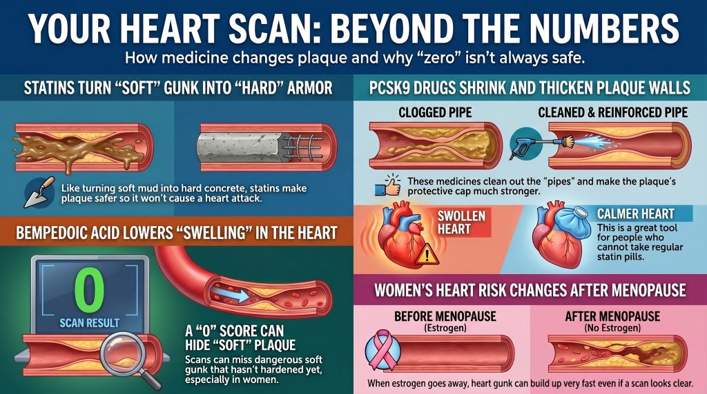
Lipidtherapie, CAC en Seksverschillen
Mechanistische en klinische verschillen tussen PCSK9-remmers, statines en bempedoiczuur bij coronaire arteriële calciumprogressie, plaquebiologie en geslachtsgebonden cardiovasculair risico
De evolutie van het atherosclerotische paradigma
Historisch gezien, aderverkalking werd geconceptualiseerd als een simplistisch, lineair proces van cholesterol ophoping in de vaatwand die leidt tot mechanische obstructie van de bloedstroom. Eigentijds onderzoek heeft dit begrip fundamenteel verschoven naar een immunointflammatoir paradigma. Atherosclerose wordt nu gekarakteriseerd als een chronische ontstekingsziekte, geïnitieerd door de subendotheliale afzetting van apolipoproteïne B (apoB)-houdend lipoproteïnen zoals low-density lipoproteïne (LDL), die oxidatieve modificatie ondergaan (ox-LDL) om te activeren endotheeldisfunctie en immuunactivatie.[1,2]
In dit kader is ziekteprogressie niet slechts een kwestie van plaquevolume maar van tandplak samenstelling en stabiliteit. De reis van initiële endotheelbeschadiging tot een levensbedreigende myocardinfarct omvat een complex samenspel van lipidedepositie, macrofaag infiltratie, fenotypische omschakeling van vaten gladde spiercellen (VSMC's), en de uiteindelijke vorming van een necrotische kern.[1,3] De uiteindelijke klinische manifestatie — hetzij stabiel angina of een acuut coronair syndroom (ACS)—wordt gedefinieerd door de mechanische integriteit van de plaque fibreuze kap en de biologische staat van verkalking.[4,9]
Het standaard niet-invasieve instrument voor het beoordelen van de atherosclerotische belasting is al lang de kransslagaderkalk (CAC) score. De Agatston-score is een van de krachtigste voorspellers van toekomstige cardiovasculaire events, maar de relatie met farmacologische interventie is complexer dan op het eerste gezicht lijkt.[5] De opkomst van intensieve cholesterolverlagende therapieën, waaronder krachtige statines, PCSK9-remmers, en bempedoïnezuur—heeft betekenisvolle verschillen benadrukt in de wijze waarop deze middelen de plaquebiologie en radiografische verkalking beïnvloeden, met belangrijke implicaties voor hoe CAC-scores worden geïnterpreteerd bij behandelde patiënten.[5,9,13]
De statine-CAC-paradox: Plaquestabilisatie en calciumdensificatie
Observationele cohorten hebben vaak aangetoond dat statine gebruikers hebben in de loop der tijd hogere Agatston-scores in vergelijking met gematchte onbehandelde personen, zelfs wanneer hun percentage klinische gebeurtenissen aanzienlijk daalt – een patroon dat soms het “statine-CAC-paradox” wordt genoemd.”[5,7] Gerandomiseerd gecontroleerd onderzoek Gegevens over deze vraag zijn minder consistent: heterogene methoden voor CAC-kwantificering en beperkte follow-upduur betekenen dat RCT's geen statistisch significant algeheel effect van statines op de CAC-progressie hebben aangetoond.[7] Het sterkste en best onderbouwde beeldvormingssignaal is geen algemene versnelling van calcificatie, maar een verschuiving in de aard van het calcium zelf: statines worden geassocieerd met afnames in plaquecomponenten met een lage demping en fibro-vetachtige componenten, naast een sterkere theorievorming oftewel progressie van hogedichte calciumfenotypes.[5,6,45]
Mechanistische basis van plaque-stabilisatie
De “genezingshypothese” stelt dat intensieve lipidenverlagend transformeert instabiele, lipidenrijke, ontstoken plaques in meer fibrotische en mechanisch stabiele laesies.[5,8] Naarmate lipide en necrotische componenten worden opgeruimd, ondergaat de vaatwand een hermodellering die wordt gekenmerkt door de afzetting van dichte calcium, dat dient als structurele versteviging rond de necrotische kern, waardoor de mechanische stress op de fibreuze kap wordt verminderd en pro-trombotisch materiaal wordt afgeschermd van de bloedbaan.[8]
Seriële CCTA-gegevens van de PARADIGM-studie ondersteun dit model: statinetherapie was geassocieerd met afname van plaque met lage demping en fibro-vetachtige plaque en met grotere progressie van hoge dichtheid verkalkte plaque, terwijl de toename in het totale volume van verkalkte plaque werd verzacht in laesies zonder lipidenrijke componenten.[45] Een serieel ongecontrasteerd CT-onderzoek uit 2025 toonde eveneens een statine-geassocieerde verschuiving naar dichtere calciumstrata aan bij asymptomatische individuen.[6] Deze bevindingen zorgen voor een nauwkeurigere typering dan simpelweg te stellen dat statines de progressie van CAC versnellen.
Bij sommige patiënten kan een stijgende Agatston-score onder effectieve lipidenverlaging daarom gedeeltelijk een afspiegeling zijn van plaque-rijping en -verdichting in plaats van nieuwe atherogenese. Echter kan seriële CAC op zichzelf genezing niet betrouwbaar onderscheiden van doorlopende ziekteactiviteit op individueel patiëntniveau, en deze interpretatie mag niet worden gegeneraliseerd zonder ondersteunende klinische context.[5]
Voorgestelde mechanismen: Vitamine K2- en macrofaagroutes
Verschillende mechanismen zijn voorgesteld om statine-geassocieerde calciumdensificatie te verklaren. Eén hypothese betreft de remming van Vitamine K2 synthese: Vitamine K2 is een cofactor voor Matrix Gla-eiwit, een fysiologische rem op vasculaire verkalking, en een verminderde biologische beschikbaarheid van vitamine K2 zouden theoretisch een rem van het verkalkingsproces kunnen wegnemen.[7,8] Door macrofagen gemedieerde trajecten die normaliter calciumafzetting remmen, kunnen eveneens worden onderdrukt. Dit blijven mechanistische hypothesen die grotendeels zijn afgeleid uit preklinisch werk; ze zijn niet vastgesteld als dominante trajecten in gerandomiseerde humane studies, en de literatuur over statines en verkalking beschrijft deze biologische processen expliciet als nog steeds omstreden.[7,8]
Tabel 1: Plaque-kenmerken: Onbehandelde Progressie vs. Statin-geassocieerde Remodellering
| Functie | Ziekteverloop zonder behandeling | Statin-geassocieerde remodellering |
| Plaquesamenstelling | Hoog lipidegehalte, grote necrotische kern | Fibrotisch, verminderde lipiderijke componenten |
| Calciumfenotype | Vlekkerig, lage dichtheid microverkalking | Bijbehorende verschuiving naar dichtere, meer samengevloeide calciumlagen |
| Vezelige kap | Dun, ontstoken, ruptuurgevoelig | Verdikt, stabiel, verminderde mechanische stress |
| Agatston-scoretrend | Stijgend als gevolg van de vorming van nieuwe laesies | Kan stijgen als gevolg van calciumdichtheid; vereist een zorgvuldige klinische interpretatie |
| Klinisch risico | Zeer verhoogd | Aanzienlijk verminderd ondanks mogelijke calciumscore stijgen |
PCSK9-remmers: Plaque-regressie en herstel van de fibreuze kap bij gelijktijdige statinetherapie
PCSK9 remmers zoals evolocumab en alirocumab een diepgaande verlaging van LDL-C bereiken door de hepatische afbraak van LDL-receptoren te voorkomenLDLR).[10,11] De belangrijkste onderzoeken naar plaque-beeldvorming toonden een zinvolle regressie van atheroom volume en verbetering in plaque-morfologie complementair aan achtergrondtherapie met statines. Gedurende deze sectie is het belangrijk op te merken dat Glagov, Huygens, en PACMAN-AMI alle bestudeerde PCSK9-remming toegevoegd aan statines; hun bevindingen kunnen niet rechtstreeks worden toegeschreven aan PCSK9-remming als een op zichzelf staand geneesmiddelklasse-effect. Direct bewijs over de progressie van CAC met PCSK9-remmers is beperkt en minder rijp dan bewijs voor plaque-regressie uit IVUS- en OCT-onderzoeken; de beschikbare gegevens omvatten een kleine gerandomiseerde secundaire analyse van alirocumab-plus-statinetherapie die een vertraagde CAC-progressie aantoont ten opzichte van standaard statine-gebaseerde zorg, maar er is geen grote, gerichte CAC-progresiestudie gerapporteerd die vergelijkbaar is met GLAGOV of HUYGENS.[14,15]
Plaque-regressie en verdikking van het fibreuze kapsel
Uit het GLAGOV-onderzoek bleek dat de toevoeging van evolocumab aan een statinebehandeling leidde tot een significante afname van het volume van coronaire atheromen: een nominale verandering van −0,95% in atheroompercentage op basis van volume (PAV) tegenover een stijging van +0,051 TP9T in de placebo groep (P < 0,001), waarbij 64,31 TP9T van de met evolocumab behandelde patiënten een PAV-regressie vertoonden, tegenover 47,31 TP9T in de placebogroep (P < 0,001). Het gemiddelde bereikte LDL-C-gehalte was 36,6 mg/dL, tegenover 93,0 mg/dL in de placebogroep.[14]
De HUYGENS-studie, waarbij gebruik wordt gemaakt van seriële optische coherentietomografie (OCT) na een acuut myocardinfarct, toonde aan dat evolocumab een significant grotere toename van de minimale dikte van het fibreuze kapsel (+42,7 μm vs. +21,5 μm; P=0,015) en een grotere afname van de maximale lipidnevel (−57,5° vs. −31,4°; P=0,04) opleverde in vergelijking met placebo.[15]
Uit de PACMAN-AMI-studie bleek dat alirocumab, gestart binnen 24 uur na een acuut myocardinfarct, leidde tot een significant grotere afname van het procentuele atheromavolume (PAV −2,13% vs. −0,92%, P < 0,001) en een grotere afname van het genormaliseerde totale atheromavolume (−26,12 mm³ versus −14,97 mm³), evenals een grotere verdikking van de fibreuze kap, in vergelijking met monotherapie met statines.[11]
Vasculaire biologie: gevestigde en opkomende signalen
Preklinische studies hebben potentiële LDLR-onafhankelijke effecten van PCSK9 op atherogenese geïdentificeerd, waaronder remming van monocytenadhesie aan gestimuleerde humane kransslagader slagader endotheelcellen (HCAEC's) via de VCAM-1 en ICAM-1 trajecten.[12,16] In grote cardiovasculaire uitkomstonderzoeken heeft PCSK9-remming echter over het algemeen minimale verandering in hsCRP laten zien, wat aangeeft dat het klinisch vastgestelde primaire werkingsmechanisme een diepgaande verlaging van apoB/LDL is in plaats van een systemisch ontstekingsremmend effect. Pleiotrope of lokale vasculaire effecten die door preklinisch werk worden gesuggereerd, kunnen bijdragen, maar dit moet niet worden overdreven gezien het grotendeels neutrale hsCRP-signaal in menselijke onderzoeksgegevens.[17]
Lipoproteïne(a): Een Belangrijke Onafhankelijke Risicofactor
Een zinvol voordeel van PCSK9-remmers is hun vermogen om te verlagen Lipoproteïne(a) [Lp(a)], een onafhankelijke en genetisch bepaald cardiovasculair risicofactor grotendeels onbeïnvloed door statinetherapie.[10] PCSK9-remmers verlagen het Lp(a)-gehalte met ongeveer 25–27% (mediaan 26,9% in de Fourier evolocumab-cohort). Het verband tussen Lp(a)-waarden en de CAC-last is inconsistent in verschillende onderzoeken; Lp(a) en CAC lijken onafhankelijk informatieve risicomarkers te zijn in plaats van sterk gecorreleerde maten. Clinici moeten daarom onderscheid maken tussen het robuuste bewijs dat een verhoogd Lp(a) een onafhankelijke ASCVD-risicofactor is en het zwakkere en minder consistente bewijs dat Lp(a) de CAC-last direct voorspelt.[10,17]
Bempedoicinezuur: Bewezen resultaten, beperkt bewijs uit plaque-beeldvorming
Bempedoïnezuur is een oraal ATP-citraatlyase (ACL)-remmer die stroomopwaarts werkt van HMG-CoA-reductase in de cholesterolbiosyntheseroute.[19,23] Als prodrug, wordt het omgezet in zijn actieve metaboliet, bempedoyl-CoA, door het enzym very long-chain acyl-CoA synthetase 1 (ACSVL1). Dit activerende enzym komt tot expressie in de lever maar is afwezig in skeletspierweefsel, wat de mechanistische basis vormt voor de lage incidentie van spiergerelateerde bijwerkingen van het geneesmiddel en het een waardevolle optie maakt voor statine-intolerante patiënten.[20,23]
Cardiovasculaire Resultaten: De CLEAR-onderzoeken
De CLEAR Outcomes klinische werkzaamheid aangetoond bij patiënten met een hoog risico en statine-intolerantie.[21] Gedurende een mediane follow-up van 3,4 jaar zorgde bempedoïnezuur voor een placebo-gecorrigeerde daling van het LDL-C-gehalte van 21,1% en een statistisch significante daling van 13% relatief risico verlaging van de primaire MACE-4 eindpunt [HR 0,87, 95%-betrouwbaarheidsinterval 0,79–0,96; P = 0,004].[21] Uit een vooraf gespecificeerde analyse van het totale aantal voorvallen bleek een afname van 20% in het totale aantal MACE-4-voorvallen [HR 0,80, 95% BI 0,72–0,89; P < 0,001].[22]
Tabel 2: CLEAR Outcomes-studie: Samenvatting van Cardiovasculaire Eindpunten (Nissen et al., NEJM 2023; JAMA 2024)
| Resultaat (CLEAR Outcomes) | HR | 95% CI |
| MACE-4 — Primair Eindpunt | 0.87 | 0,79–0,96 (P = 0,004) |
| MACE-3 (CV-sterfte / MI / Beroerte) | 0.85 | 0.76–0.96 |
| Fatale of niet-fatale hartinfarcten | 0.77 | 0.66–0.91 |
| kransslagader- Revascularisatie | 0.81 | 0.72–0.92 |
| Totaal aantal MACE-4-gebeurtenissen (vooraf gespecificeerd) | 0.80 | 0,72–0,89 (P < 0,001) |
Ontsteking, mechanisme en beeldvormend bewijs
Een opvallend kenmerk van bempedoïnezuur is de consistente verlaging van het hsCRP-gehalte: in de CLEAR Outcomes-studie werd na 6 maanden een placebo-gecorrigeerde mediane daling van 22% waargenomen.[19,21] Activatie van het AMP-geactiveerde eiwit kinase (AMPK)-route is voorgesteld als een bijdragend cellulair mechanisme, voornamelijk op basis van preklinische en mechanistische overzichtsgegevens.[20,23] Recent werk suggereert dat de biologie van het geneesmiddel complexer kan zijn dan aanvankelijk werd gekarakteriseerd, met voorlopig bewijs van potentiële directe PPARα-activatie; deze mechanistische vragen blijven onderwerp van actief onderzoek and mogen niet worden gepresenteerd als vaststaande biologie.
Bempedoecidinezuur heeft bewezen voordelen voor de uitkomsten en verlaging van hsCRP bij statine-intolerante patiënten, maar de directe effecten op de morfologie van coronaire plaque blijven grotendeels onbewezen in afwachting van specifieke beeldvormingsonderzoeken.[24] Het enige gepubliceerde plaque-beeldvormende bewijs is een enkel casusrapport dat de krimp beschrijft van laag-attenuatie plaque met langetermijn-bempedoiczuurmonotherapie—een hypothese-genererende waarneming, geen bewijs van een plaque-regressieklasse-effect. Een fase 3-beeldvormingsstudie geregistreerd in 2026 evalueert de combinatie van bempedoiczuur, ezetimib, en intensieve statinetherapie op coronaire plaque; het ontwerp ervan test drievoudige combinatiebehandeling in plaats van bempedoïnezuur op zichzelf, en de resultaten zijn nog niet gerapporteerd.
Beperkingen en blinde vlekken van CAC = 0: Voorbij een binaire interpretatie
De constatering dat een CAC-score van 0 een zeer lage voorspelt 10-jaars cardiovasculair risico—de “kracht van nul”—heeft CAC gevestigd als een nuttige poortwachter bij risicostratificatie.[25,27,29] CAC = 0 is in de meeste klinische situaties een werkelijk krachtige kortetermijnnegatieve risicomarker. Het heeft echter belangrijke en goed gekarakteriseerde blinde vlekken die een zorgvuldige interpretatie vereisen, met name bij symptomatische patiënten, jongere personen en vrouwen in de middelbare leeftijd.[30]
Niet-verkalkte plaque: Het onderkende onderdeel
Een CAC-score van 0 sluit niet uit kransslagaderziekte. Niet-verkalkte plaque (NCP), dat rijk is aan lipiden en zeer kwetsbaar is voor scheuring, is volledig onzichtbaar op een CT-scan zonder contrast.[25,28] Uit onderzoeken met behulp van CCTA is gebleken dat ongeveer 15–17% van de personen met een laag tot gemiddeld risico en een CAC-waarde van 0 aantoonbare atherosclerose vertonen, waarbij ongeveer 3,5% een obstructieve aandoening hebben (>50% stenose) ondanks een nul calciumniveau.[28,30]
In een divers, symptomatisch Montefiore-cohort dat door Rozanski et al. werd geanalyseerd, behield een CAC-score van nul een zeer hoge negatieve voorspellende waarde voor obstructieve coronaire aandoeningen en ongunstige gebeurtenissen op korte termijn, en slechts een klein percentage van de patiënten met CAC = 0 — ongeveer 7% — vertoonde aantoonbare niet-verkalkte plaque op CCTA.[30] Losse prospectieve gegevens van populaties met stabiele pijn op de borst tonen eveneens zeer lage MACE-gebeurtenispercentages wanneer CAC = 0, hoewel obstructief coronair lijden niet volledig wordt uitgesloten. Deze gegevens bevestigen tezamen dat CAC = 0 een oprechte en krachtige geruststelling op de korte termijn biedt, terwijl er toch een klinisch betekenisvolle minderheid overblijft met niet-verkalkte plaque die de calciumscore niet kan detecteren.
De mechanismen van klinische gebeurtenissen bij individuen met CAC = 0 omvatten typisch ruptuur of erosie van niet-verkalkte laesies, systemische ontsteking, of coronair microvasculaire disfunctie.[27,31] Plaque-erosie—verstoring van het endotheeloppervlak over een lipiderijke, niet-verkalkte plaque—komt met name veel voor bij vrouwen en jongere personen.[27]
Prevalentieramingen hangen cruciaal af van cohort en methode
In de SCAPIS (Swedish Cardiopulmonary Bioimage Study): onder volwassenen van middelbare leeftijd uit de algemene bevolking met CAC = 0 had ongeveer 5,5% een coronaire plaque die op de CCTA waarneembaar was en had 0,4% een significante stenose.[29] Het vaak aangehaalde cijfer van 9,21 TP9T voor de prevalentie van plaque uit SCAPIS heeft specifiek betrekking op de subgroep met CAC = 0 en een gemiddeld 10-jaars ASCVD-risico — een belangrijk onderscheid, omdat het toepassen van deze voorwaardelijke schatting op de gehele algemene populatie met een CAC van nul de prevalentie van de ziekte aanzienlijk zou overschatten.[29]
AI-QCT-registers van symptomatische patiënten die zijn doorverwezen voor een klinische CCTA, melden aanzienlijk hogere percentages voor het opsporen van plaque bij personen met CAC = 0 — variërend van ongeveer 54% tot 95% in sommige geselecteerde cohorten.[26] Deze cijfers zijn niet generaliseerbaar naar bevolkingsonderzoeken; ze weerspiegelen de hogere pre-test-waarschijnlijkheid van klinische verwijzingscohorten evenals variatie in AI-QCT-protocollen, drempelwaarden en definities tussen studies. Prevalentjeschattingen op basis van symptomatische, asymptomatische, algemene-populatie- en AI-QCT-registratiestudies mogen niet worden gecombineerd zonder zorgvuldige populatiespecifieke stratificatie.[30]
Tabel 3: Prevalentie van niet-verkalkte plaque bij patiënten met CAC = 0: Contextafhankelijke schattingen in studiepopulaties
| Studie / Cohort | Bevolking | Belangrijkste bevinding voor patiënten met een CAC = 0 |
| Cheng et al. | Laag tot matig risico, asymptomatisch | ~16% met waarneembare, niet-verkalkte plaque op CCTA |
| Rubinshtein et al. | Symptomatisch | ~14,51 TP9T met tandplak; ~31 TP9T met obstructieve aandoening |
| Rozanski et al. (JCCT 2023) | Symptomatische stabiele pijn op de borst, diverse populatie | ~7% NCP op CCTA; zeer hoge NPV voor obstructieve aandoeningen en gebeurtenissen op korte termijn; divers symptomatisch Montefiore-cohort |
| SCAPIS (Bergström et al.) | Algemene bevolking van middelbare leeftijd | ~5,51 TP9T voor plaque in het algemeen; ~9,21 TP9T specifiek in de subgroep met CAC=0 en gemiddeld risico [29] |
| AI-QCT-registers (geselecteerde klinische CCTA-cohorten) | Symptomatisch / hoge pre-test kans | 54–95%: sterk afhankelijk van de selectie van de onderzoeksgroep, de indicatie voor doorverwijzing en het AI-QCT-protocol. Niet generaliseerbaar naar screeningspopulaties [26] |
Geslachtsspecifiek Cardiovasculair Risico en Plaquebiologie
Vrouwen vertonen belangrijke verschillen in de presentatie en pathofysiologie van atherosclerotische hart- en vaatziekten (ASCVD) vergeleken met mannen. Vrouwen ontwikkelen klinisch manifeste coronaire hartziekte over het algemeen ongeveer een decennium later dan mannen, een fenomeen dat grotendeels wordt toegeschreven aan de vaatbeschermende effecten van endogene oestrogeen.[37,39]
De beschermende rol van oestrogeen en de overgang naar de menopauze
Oestrogeen handhaaft de vaatgezondheid via meerdere routes: het bevordert endotheelafhankelijke vasodilatatie door het stimuleren van stikstofoxide (NO) en de synthese van prostacycline, remt de contractie en proliferatie van vasculaire gladde spiercellen en oefent ontstekingsremmende effecten uit op de vaatwand.[35,38] Oestrogeen zorgt er ook voor dat de verhoudingen gunstiger blijven lipidenprofielen gedurende de premenopauzale jaren.[35]
De menopauze de overgang wordt geassocieerd met een sterke afname van circulerend oestradiol, wat een versnelde trigger is vasculaire veroudering. Deze verschuiving leidt tot meetbare verminderingen in endotheelfunctie zoals beoordeeld door flow-mediated dilatatie (FMD), verhoogd systemisch oxidatieve stress, en progressieve verergering van arteriële stijfheid, bloeddruk, en atherogene dyslipidemie.[34,36] De FMD bij vrouwen in de late postmenopauze is ongeveer 50% lager dan bij premenopauzale vrouwen van vergelijkbare leeftijd, waarbij de meest significante afname plaatsvindt tijdens de perimenopauzale overgang.[34]
Risicoversnelling na de menopauze
Naarmate vrouwen hun zestiger jaren bereiken, versnelt het cardiovasculaire risico aanzienlijk, waarbij de incidentie van hart- en vaatziekten die van leeftijdsgenoten van het mannelijk geslacht benadert.[37,38] Uit longitudinale gegevens van de Healthy Women Study blijkt dat bij postmenopauzale vrouwen met een CAC-waarde van 0 bij aanvang van het onderzoek, ongeveer 33% in de daaropvolgende periode van zes jaar nieuwe verkalkingen ontwikkelen, waarbij de LDL-C- en HDL-C-waarden vóór de menopauze sterke voorspellende factoren zijn.[39]
Vroege menopauze en langetermijnrisico
Vrouwen die te maken krijgen met vroege menopauze (VM, gedefinieerd als menopauze vóór de leeftijd van 45 jaar) lopen aanzienlijk grotere cardiovasculaire risico's op de lange termijn. In de Multi-Ethnic Study of Atherosclerosis (MESA), meer dan de helft van de postmenopauzale vrouwen met een vroegtijdige menopauze had een baseline CAC-score van 0.[40] Bij de vrouwen met een CAC-waarde van 0 was de 10-jaarsincidentie van ASCVD laag tot grenswaardig, maar bij vrouwen met een vroege menopauze was het 15-jaarsrisico significant hoger dan bij vrouwen zonder vroege menopauze (gecorrigeerde HR 1,96, 95%-betrouwbaarheidsinterval 1,26–3,04).[40] Dit illustreert het centrale diagnostische probleem: dezelfde CAC = 0-uitslag die na 10 jaar geruststellend lijkt, kan een aanzienlijk verhoogd langetermijnrisicoproces maskeren bij vrouwen met vervroegde menopauze, gedreven door een langdurig oestrogeentekort dat de subklinische plaque-accumulatie na de initiële scan versnelt.
Systematische meta-analyses bevestigen dat een vroege menopauze onafhankelijk geassocieerd is met een toename van 50–60% gedurende het hele leven kransslagaderziekte risico, een effect dat slechts gedeeltelijk wordt verminderd door traditionele risicofactorcorrectie.[41]
Sex Differences in Plaque Morphology and Detection
Women generally have lower absolute CAC burden than men and, in several cohorts, lower absolute plaquebelasting as well; however, recent AI-QCT data suggest that the relative cardiovascular risk associated with a given increase in plaque burden is greater in women than in men.[27,33] Women are significantly more likely to develop non-calcified, lipiderijke plaques prone to endothelial erosion rather than fibrous cap rupture—yet both mechanisms trigger MACE. Because CAC scoring captures only the calcified component of disease, it may be a less complete risk indicator in women, who often carry a greater proportion of their total plaque burden in non-calcified form. Women with any detectable CAC face disproportionately higher relative cardiovascular risk: CAC Consortium data show approximately 1.3-fold higher hazard of cardiovascular death in women than in men at equivalent CAC burden.[27]
In their 40s and 50s, women may accumulate a substantial burden of lipid-rich NCP that will only become radiographically detectable as calcium after the post-menopausal acceleration of vascular calcification.[27,33] This creates a diagnostic window during midlife when traditional risk factors are rising and inflammatory plaque is accumulating, yet CAC remains at zero. The absence of calcium may be an especially incomplete risk signal precisely in this population—younger, perimenopausal women—where near-term absoluut risico appears lowest but medium-term risk may be substantially higher than the calcium score suggests.
Table 4: Sex-Specific Cardiovascular Risk per Unit Plaque Volume: Selected Findings from the CONFIRM2-register (Choi et al., Circ Cardiovasc Imaging 2025 [33])
| Finding — CONFIRM2 Registry (Choi et al. 2025 [33]) | Vrouwen | Mannen |
| Relative risk increase per 50 mm³ total plaque volume | +17.7% (RR ≈1.18) | +5.3% (RR ≈1.05) |
| Pattern across NCP, calcified plaque, and high-risk plaque subtypes | Larger relative risk increments across all subtypes | Smaller relative risk increments across all subtypes |
| Principal published conclusion | Substantially higher relative cardiovascular risk per unit plaque | Lower relative risk per equivalent plaque unit |
The total plaque volume risk increment (+17.7% per 50 mm³ in women vs +5.3% in men) is reported from the published CONFIRM2 results. The directional pattern across non-calcified, calcified, and high-risk plaque subtypes (larger increments in women) is the principal published conclusion of that paper; exact subtype risicoverhoudingen should be transcribed from the published results table before any submission requiring those specific values.
Advanced Imaging: The Role of AI-Enhanced CCTA
Artificial intelligence–based quantitative CT (AI-QCT) represents a meaningful advance in non-invasive cardiovascular risk assessment. By automating measurement of total plaque burden—including calcified, non-calcified, and low-attenuation components—AI-QCT provides a more comprehensive characterization of disease than traditional CAC scoring alone.[26,33] These tools are rapidly evolving, but broad standardization across institutions and vendors, external validation in diverse populations, sex-specific risk thresholds, and integration into clinical guidelines are still ongoing processes. AI-QCT is best understood as a promising emerging modality rather than a fully standardized replacement for current CAC and CCTA frameworks.
AI-QCT, CAC, and Reclassification in Symptomatic Cohorts
In symptomatic patients referred for clinical CCTA, research demonstrates only moderate categorical agreement between traditional CAC scores and AI-derived total plaque burden, with discordantie particularly pronounced in women.[42] AI-QCT frequently identifies significant NCP in patients with zero Agatston scores in these referral populations. Prevalence varies widely by cohort, scanner protocol, and AI-QCT algorithm, and figures from clinical CCTA registries should not be extrapolated to general screening populations. Nonetheless, the herclassificatie is clinically meaningful for symptomatic patients in whom a zero calcium score might otherwise prematurely terminate the diagnostic workup, especially women with additional risk features.[26,30,42]
AI-QCT further enables quantification of high-risk plaque (HRP) features including low-attenuation plaque (LAP) and positive coronary remodeling. Data from the CONFIRM2 registry demonstrate that these features carry significantly higher relative risk in women than in men per unit of plaque volume, consistent with the clinical observation that women’s smaller-caliber coronary vessels may render each unit of high-risk plaque more hemodynamically consequential.[33]
Integrating Epicardial Adipose Tissue Analysis
Automated analysis of epicardial adipose tissue (EAT) volume and attenuation from routine calcium-scoring CT scans has been shown to significantly enhance MACE prediction, with particularly strong incremental value in women.[43] EAT is metabolically active and exerts paracrine pro-inflammatory and pro-atherogenic effects on adjacent kransslagaders through the secretion of cytokines and adipokines,[32] and integrating quantitative EAT features with CAC scoring and conventional risk factors provides a more biologically comprehensive sex-specific risk assessment than calcified plaque metrics alone, though this approach requires further standardization and prospective validation before routine clinical implementation.[43]
An Integrated Framework for Therapeutic Mapping
A useful framework maps atherosclerosis progression through specific biological stages and identifies where different therapeutic classes exert their primary mechanistic influence. This mapping should be understood as directionally plausible based on available mechanistic and imaging data, not as a clinically proven stage-by-stage model validated by head-to-head drug-class trials.[1,4,9]
Stages of Plaque Development and Therapeutic Positioning
Stage 1 — Endothelial Activation: Endothelial dysfunction, reduced NO bioavailability, and subendotheliale retentie of apoB-containing lipoproteins. This stage is substantially modulated by endogenous estrogen and is the proposed target of early PCSK9 inhibition based on preclinical evidence.[2,12]
Stage 2 — Early Non-Calcified Plaque: Macrophage schuimcel formation establishes the earliest atheromatous lesion. Bempedoic acid and statins reduce the circulating lipid pool available for subendothelial accumulation and dampen systemic inflammation.[1,20]
Stage 3 — Intermediate / Mixed Plaque: Extracellular lipid accumulation and microcalcifications emerge within necrotic core zones. High-intensity statins and PCSK9i (when added to statins) drive plaqueregressie and fibrous cap maturation, with the strongest direct human trial evidence at this stage.[1,9,14]
Stage 4 — Fibrocalcific / Stable Plaque: Dense, coalescent calcification with a thick collagen-rich fibrous cap, representing the histopathological end-state of healed plaque, associated primarily with intensive statin therapy and the calcium densification process.[5,8,9]
Comparative Therapeutic Profile
Statins are best characterized as plaque stabilizers associated with calcium densification in observational and serial CCTA data. They have the strongest and broadest evidence base for reducing lipid-rich plaque and promoting transformation of vulnerable lesions toward denser, more stable phenotypes. A rising Agatston score under statin therapy should be interpreted in the context of documented densification and clinical improvement rather than as evidence of therapeutic failure.[5,7,8,45]
PCSK9 Inhibitors are best characterized as plaque regressionors and fibrous cap restorers when added to statin therapy in high-risk imaging cohorts. Their principal established mechanism of clinical benefit is profound apoB/LDL lowering, with meaningful incremental plaque regression and cap thickening beyond statins alone. Pleiotropic effects suggested by preclinical data have not translated into a clear systemic anti-inflammatory signal in human outcome trials.[11,12,14,15]
Bempedoic Acid functions as an upstream lipid-lowering and hsCRP-lowering agent with proven outcomes benefit in statin-intolerant patients. Its plaque-modification effects are mechanistically plausible but supported by very limited imaging data; robust serial plaque imaging evidence has not yet been reported.[19,20,21]
Clinical Implications and Guideline Directions
The integration of pharmacologic mechanisms, advanced imaging, and sex-specific plaque biology highlights important gaps in current clinical practice and supports a shift toward biologically informed, individualized risk assessment.
Interpreting CAC = 0 in Symptomatic and Mid-Life Women
The cumulative evidence supports a calibrated interpretation of CAC = 0: it is a powerful short-term negative risk marker that should not be dismissed, yet it should not automatically end risk assessment in younger or symptomatic women—especially in the presence of early menopause, strong familiegeschiedenis, diabetes, elevated Lp(a), or persistent inflammatory risk.[27,40,41] A CAC of 0 in a woman with these characteristics may correctly predict low 10-year event risk while substantially underestimating 15-year risk driven by ongoing non-calcified plaque accumulation and the biological effects of prolonged estrogen deficiency. In these settings, a CAC of 0 should invite further characterization of risk rather than conclude risk stratification.
A More Comprehensive Approach to Sex-Conscious Risk Assessment
A multi-modal, sex-conscious risk assessment framework incorporates several complementary elements.
Sex-specific risk factors—including age at menopause, duration of estrogen deficiency, and reproductive history—should be formally integrated into clinical risk discussions, as they carry independent prognostic weight beyond traditional Framingham-based variables.[37,41]
Advanced imaging via CCTA should be considered when CAC scoring may underestimate true atherosclerotic burden, particularly in symptomatic women with CAC = 0 and additional risk features. The 2024 ESC Guidelines for the Management of Chronic Coronary Syndromes recommend first-line noninvasive anatomic or functional imaging for suspected CCS, selected according to pretest likelihood, patient characteristics, and local expertise. Within that framework, CCTA is specifically preferred for ruling out obstructive CAD and identifying nonobstructive disease, and the guidelines explicitly incorporate age, sex, and symptom character into pretest likelihood models.[26,31,33]
Integrated biomarker-imaging models combining Lp(a), hsCRP, and emerging EAT fat-omics analysis represent a biologically more complete framework for sex-specific risk stratification. These approaches require further standardization and prospective validation before routine implementation.[43]
Limitations and Open Questions
The evidence reviewed here has several important limitations that must be recognized when applying these concepts clinically or in academic scholarship.
Most plaque-imaging evidence for PCSK9 inhibitors derives from studies of add-on therapy to background statins, not from clean mechanistic comparisons between drug classes. GLAGOV, HUYGENS, and PACMAN-AMI do not establish what PCSK9 inhibitors achieve independently of statins, and their imaging endpoints cannot be directly compared with statin-only or bempedoic acid-only imaging data.[11,14,15]
The bempedoic acid plaque-imaging literature currently consists of a single case report and one recently registered phase 3 imaging trial that has not yet reported results. Outcomes-based conclusions about bempedoic acid are well supported; morphologic plaque-imaging conclusions are not.[24]
AI-QCT plaque detection rates vary substantially by cohort selection, referral indication, and AI-QCT protocol. Plaque prevalence figures from symptomatic CCTA referral populations should not be extrapolated to general screening populations. Standardized thresholds, validated sex-specific risk cutoffs, and prospective outcome data for AI-QCT remain incomplete.[26,33,42]
The field still lacks clean head-to-head randomized trials designed to compare drug class effects on CAC progression or plaque morphology using sex-stratified pre-specified endpoints and the same imaging platform. The mechanistic pathways proposed for statin-associated calcium densification (Vitamin K2 inhibition, macrophage suppression) and for bempedoic acid’s anti-inflammatory effects (AMPK, possible PPARα activation) are supported primarily by preclinical evidence and remain incompletely validated in human studies.[7,8,20,23]
Conclusie
Intensive apoB-lowering therapy reduces cardiovascular risk across drug classes, but its imaging footprints differ in important ways. Statins have the strongest and longest-established evidence for reducing lipid-rich plaque and promoting a denser, more stable calcium phenotype; their association with rising Agatston scores is best understood as calcium densification rather than uncomplicated disease progression. PCSK9 inhibitors provide additional plaque regression, lipid arc reduction, and fibrous cap thickening when added to statins, with a primarily LDL-lowering mechanism driving outcome benefit rather than a systemic anti-inflammatory one. Bempedoic acid offers proven event reduction and meaningful hsCRP lowering in statin-intolerant patients, but its effects on coronary plaque morphology remain largely unproven pending dedicated imaging trials.
CAC remains a powerful marker of calcified plaque burden and near-term prognosis, but it incompletely captures non-calcified atherosclerosis—particularly in symptomatic patients and women in mid-life. CAC = 0 should be interpreted as one component of a biologically informed risk assessment rather than as a stand-alone arbiter of disease absence. In most clinical contexts it provides powerful short-term reassurance; in specific contexts—early menopause, symptomatic presentations, elevated Lp(a), family history, or persistent inflammatory risk—it should prompt further rather than conclude risk stratification.
For women specifically, the distinct biology of plaque formation—characterized by early lipid-rich non-calcified lesions, a sensitive period of post-menopausal risk acceleration, and significantly higher relative cardiovascular risk per unit of plaque volume—demands an individualized approach to imaging and lipid-lowering therapy. A sex-conscious, multi-modal risk assessment framework incorporating CCTA, sex-specific biomarkers, and comprehensive clinical context offers the most evidence-consistent path toward equitable and effective cardiovascular disease prevention.
Referenties
- Libby P. Inflammation in atherosclerosis. Nature. 2002;420(6917):868-874. doi:10.1038/nature01323
- Han Y, Xu H, Yao X, et al. Molecular mechanisms and therapeutic progress in atherosclerosis: bridging immune inflammation and precision medicine. Front Immunol. 2026;16:1737662. Published 2026 Jan 5. doi:10.3389/fimmu.2025.1737662
- Moore KJ, Tabas I. Macrophages in the pathogenesis of atherosclerosis. Cell. 2011;145(3):341-355. doi:10.1016/j.cell.2011.04.005
- Papafaklis MI, Koros R, Tsigkas G, Karanasos A, Moulias A, Davlouros P. Reversal of Atherosclerotic Plaque Growth and Vulnerability: Effects of Lipid-Modifying and Anti-Inflammatory Therapeutic Agents. Biomedicines. 2024;12(11):2435. Published 2024 Oct 23. doi:10.3390/biomedicines12112435
- Khanna S, Nerlekar N, Bhat A. Reconciling Coronary Artery Calcification in the Lipid-Lowering Era. JACC Adv. 2026;5(1):102506. doi:10.1016/j.jacadv.2025.102506
- Giovannucci J, Shanbhag A, Hong W, et al. Impact of statins on progression of coronary artery calcium composition and density as assessed by noncontrast CT. Int J Cardiovasc Imaging. 2025;41(12):2481-2492. doi:10.1007/s10554-025-03561-0
- Shahraki MN, Jouabadi SM, Bos D, Stricker BH, Ahmadizar F. Statin Use and Coronary Artery Calcification: a Systematic Review and Meta-analysis of Observational Studies and Randomized Controlled Trials. Curr Atheroscler Rep. 2023;25(11):769-784. doi:10.1007/s11883-023-01151-w
- Kadoglou NP, Stasinopoulou M, Velidakis N, et al. The Complex Mechanisms and the Potential Effects of Statins on Vascular Calcification: A Narrative Review. Rev Cardiovasc Med. 2024;25(2):51. Published 2024 Jan 30. doi:10.31083/j.rcm2502051
- Papafaklis MI, Koros R, Tsigkas G, Karanasos A, Moulias A, Davlouros P. Reversal of Atherosclerotic Plaque Growth and Vulnerability: Effects of Lipid-Modifying and Anti-Inflammatory Therapeutic Agents. Biomedicines. 2024;12(11):2435. Published 2024 Oct 23. doi:10.3390/biomedicines12112435
- Wu Z, Gao L, Lin Z. Can proprotein convertase subtilisin/kexin type 9 (PCSK9) inhibitors regress coronary atherosclerotic plaque? A systematic review and meta-analysis. Am J Transl Res. 2023;15(1):452-465. Published 2023 Jan 15.
- Räber L, Ueki Y, Otsuka T, et al. Effect of Alirocumab Added to High-Intensity Statin Therapy on Coronary Atherosclerosis in Patients With Acute Myocardial Infarction: The PACMAN-AMI Randomized Clinical Trial. JAMA. 2022;327(18):1771-1781. doi:10.1001/jama.2022.5218
- Zulkapli R, Muid SA, Wang SM, Nawawi H. PCSK9 Inhibitors Reduce PCSK9 and Early Atherogenic Biomarkers in Stimulated Human Coronary Artery Endothelial Cells. Int J Mol Sci. 2023;24(6):5098. Published 2023 Mar 7. doi:10.3390/ijms24065098
- Ahmadi A, Argulian E, Leipsic J, Newby DE, Narula J. From Subclinical Atherosclerosis to Plaque Progression and Acute Coronary Events: JACC State-of-the-Art Review. J Am Coll Cardiol. 2019;74(12):1608-1617. doi:10.1016/j.jacc.2019.08.012
- Nicholls SJ, Puri R, Anderson T, et al. Effect of Evolocumab on Progression of Coronary Disease in Statin-Treated Patients: The GLAGOV Randomized Clinical Trial. JAMA. 2016;316(22):2373-2384. doi:10.1001/jama.2016.16951
- Nicholls SJ, Kataoka Y, Nissen SE, et al. Effect of Evolocumab on Coronary Plaque Phenotype and Burden in Statin-Treated Patients Following Myocardial Infarction. JACC Cardiovasc Imaging. 2022;15(7):1308-1321. doi:10.1016/j.jcmg.2022.03.002
- Barbieri L, Tumminello G, Fichtner I, et al. PCSK9 and Coronary Artery Plaque-New Opportunity or Red Herring?. Curr Atheroscler Rep. 2024;26(10):589-602. doi:10.1007/s11883-024-01230-6
- Pradhan AD, Aday AW, Rose LM, Ridker PM. Residual Inflammatory Risk on Treatment With PCSK9 Inhibition and Statin Therapy. Circulation. 2018;138(2):141-149. doi:10.1161/CIRCULATIONAHA.118.034645
- Chen H, Chen X. PCSK9 inhibitors for acute coronary syndrome: the era of early implementation. Front Cardiovasc Med. 2023;10:1138787. Published 2023 May 2. doi:10.3389/fcvm.2023.1138787
- Jia X, Virani SS. CLEAR Serenity Trial: More Clarity for the Future of Bempedoic Acid in Patients Unable to Take Statins?. J Am Heart Assoc. 2019;8(7):e012352. doi:10.1161/JAHA.119.012352
- Pinkosky SL, Newton RS, Day EA, et al. Liver-specific ATP-citrate lyase inhibition by bempedoic acid decreases LDL-C and attenuates atherosclerosis. Nat Commun. 2016;7:13457. Published 2016 Nov 28. doi:10.1038/ncomms13457
- Nissen SE, Lincoff AM, Brennan D, et al. Bempedoic Acid and Cardiovascular Outcomes in Statin-Intolerant Patients. N Engl J Med. 2023;388(15):1353-1364. doi:10.1056/NEJMoa2215024
- Nicholls SJ, Nelson AJ, Lincoff AM, et al. Impact of Bempedoic Acid on Total Cardiovascular Events: A Prespecified Analysis of the CLEAR Outcomes Randomized Clinical Trial. JAMA Cardiol. 2024;9(3):245-253. doi:10.1001/jamacardio.2023.5155
- Biolo G, Vinci P, Mangogna A, et al. Mechanism of action and therapeutic use of bempedoic acid in atherosclerosis and metabolic syndrome. Front Cardiovasc Med. 2022;9:1028355. Published 2022 Oct 28. doi:10.3389/fcvm.2022.1028355
- Korosoglou G, Giesen A, Geiss E, Stach K. Case report: Strong low-density-cholesterol reduction accompanied by shrinkage of low-attenuation coronary plaque during lipid-lowering treatment with bempedoic acid-serial evaluation by coronary computed tomography angiography. Front Cardiovasc Med. 2023;10:1203832. Published 2023 Aug 4. doi:10.3389/fcvm.2023.1203832
- Koulaouzidis G, Charisopoulou D, Jenkins PJ, Koulaouzidis A, McArthur T. Prevalence of noncalcified coronary plaque in patients with calcium score of 0: the silent enemy. Angiology. 2013;64(3):205-210. doi:10.1177/0003319712440618
- Khan NA, Wesbey III G, Cobb G, et al. Using AI-Quantitative CT to evaluate the relationship between coronary artery calcium and segment involvement scores in quantifying coronary plaque burden. Int J Cardiovasc Imaging. 2026;42(1):49-59. doi:10.1007/s10554-025-03569-6
- Shaw LJ, Min JK, Nasir K, et al. Sex differences in calcified plaque and long-term cardiovascular mortality: observations from the CAC Consortium. Eur Heart J. 2018;39(41):3727-3735. doi:10.1093/eurheartj/ehy534
- Cho I, Suh JW, Chang HJ, et al. Prevalence and prognostic implication of non-calcified plaque in asymptomatic population with coronary artery calcium score of zero. Korean Circ J. 2013;43(3):154-160. doi:10.4070/kcj.2013.43.3.154
- Bergström G, Persson M, Adiels M, et al. Prevalence of Subclinical Coronary Artery Atherosclerosis in the General Population. Circulation. 2021;144(12):916-929. doi:10.1161/CIRCULATIONAHA.121.055340
- Fattouh M, Kuno T, Pina P, et al. Interplay Between Zero CAC, Quantitative Plaque Analysis, and Adverse Events in a Diverse Patient Cohort. Circ Cardiovasc Imaging. 2023;16(8):e015236. doi:10.1161/CIRCIMAGING.123.015236
- Vrints C, Andreotti F, Koskinas KC, et al. 2024 ESC Guidelines for the management of chronic coronary syndromes. Eur Heart J. 2024;45(36):3415-3537. doi:10.1093/eurheartj/ehae177
- Antonopoulos AS, Sanna F, Sabharwal N, et al. Detecting human coronary inflammation by imaging perivascular fat. Sci Transl Med. 2017;9(398):eaal2658. doi:10.1126/scitranslmed.aal2658
- Feuchtner GM, Lacaita PG, Bax JJ, et al. AI-Quantitative CT Coronary Plaque Features Associate With a Higher Relative Risk in Women: CONFIRM2 Registry. Circ Cardiovasc Imaging. 2025;18(6):e018235. doi:10.1161/CIRCIMAGING.125.018235
- Moreau KL, Hildreth KL, Meditz AL, Deane KD, Kohrt WM. Endothelial function is impaired across the stages of the menopause transition in healthy women. J Clin Endocrinol Metab. 2012;97(12):4692-4700. doi:10.1210/jc.2012-2244
- Reslan OM, Khalil RA. Vascular effects of estrogenic menopausal hormone therapy. Rev Recent Clin Trials. 2012;7(1):47-70. doi:10.2174/157488712799363253
- Moreau KL, Hildreth KL, Klawitter J, Blatchford P, Kohrt WM. Decline in endothelial function across the menopause transition in healthy women is related to decreased estradiol and increased oxidative stress. Geroscience. 2020;42(6):1699-1714. doi:10.1007/s11357-020-00236-7
- Mishra T, Pasnoor DS, Gandhi M, et al. Interplay of menopause, coronary artery calcium score and cardiovascular disease risk. World J Cardiol. 2025;17(11):109627. doi:10.4330/wjc.v17.i11.109627
- SenthilKumar G, Katunaric B, Bordas-Murphy H, Sarvaideo J, Freed JK. Estrogen and the Vascular Endothelium: The Unanswered Questions. Endocrinology. 2023;164(6):bqad079. doi:10.1210/endocr/bqad079
- Kuller LH, Matthews KA, Edmundowicz D, Chang Y. Incident coronary artery calcium among postmenopausal women. Atherosclerosis. 2008;200(2):278-285. doi:10.1016/j.atherosclerosis.2007.12.057
- Chu JH, Michos ED, Ouyang P, et al. Coronary artery calcium and atherosclerotic cardiovascular disease risk in women with early menopause: The Multi-Ethnic Study of Atherosclerosis (MESA). Am J Prev Cardiol. 2022;11:100362. Published 2022 Jun 13. doi:10.1016/j.ajpc.2022.100362
- Muka T, Oliver-Williams C, Kunutsor S, et al. Association of Age at Onset of Menopause and Time Since Onset of Menopause With Cardiovascular Outcomes, Intermediate Vascular Traits, and All-Cause Mortality: A Systematic Review and Meta-analysis. JAMA Cardiol. 2016;1(7):767-776. doi:10.1001/jamacardio.2016.2415
- Khan NA, Wesbey III G, Cobb G, et al. Using AI-Quantitative CT to evaluate the relationship between coronary artery calcium and segment involvement scores in quantifying coronary plaque burden. Int J Cardiovasc Imaging. 2026;42(1):49-59. doi:10.1007/s10554-025-03569-6
- Singh P, Hu T, Hoori A, Al-Kindi S, Wilson DL, Rajagopalan S. Sex-specific cardiovascular risk prediction using AI-derived epicardial adipose tissue measurements on CT calcium scoring exams. Am J Prev Cardiol. 2025;25:101367. Published 2025 Dec 8. doi:10.1016/j.ajpc.2025.101367
- Narula J, Chandrashekhar Y, Ahmadi A, et al. SCCT 2021 Expert Consensus Document on Coronary Computed Tomographic Angiography: A Report of the Society of Cardiovascular Computed Tomography. J Cardiovasc Comput Tomogr. 2021;15(3):192-217. doi:10.1016/j.jcct.2020.11.001
- Lee SE, Chang HJ, Sung JM, et al. Effects of Statins on Coronary Atherosclerotic Plaques: The PARADIGM Study. JACC Cardiovasc Imaging. 2018;11(10):1475-1484. doi:10.1016/j.jcmg.2018.04.015

