
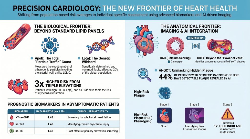
Paradigmas Avançados na Avaliação de Risco Cardiovascular:
Integração de Biomarcadores e Imagem em Cardiologia de Precisão
Resumo
Doença cardiovascular (DCV) continua sendo a principal causa de morbidade e mortalidade em todo o mundo, apesar dos grandes avanços na prevenção, farmacoterapia e imagem. As ferramentas tradicionais de avaliação de risco, incluindo as Escore de Risco de Framingham e o Equações de Coortes Reunidas da ACC/AHA, forneceram a base da cardiologia preventiva por décadas, embora suas limitações em nível individual tenham se tornado cada vez mais evidentes. Esses modelos estimam a probabilidade de eventos futuros com base em dados demográficos e clínicos fatores de risco, mas eles não quantificam diretamente a carga aterosclerótica, placa biologia ou lesão miocárdica subclínica. Como resultado, uma proporção substancial de eventos cardiovasculares continua a ocorrer em pessoas categorizadas como de baixo ou moderado risco, enquanto alguns indivíduos designados como de alto risco pelos modelos convencionais podem apresentar pouca doença demonstrável.
Essa discrepância impulsionou o surgimento da cardiologia de precisão — uma abordagem que integra marcadores lipídicos avançados, biomarcadores inflamatórios e metabólicos, predisposição genética, marcadores específicos cardíacos e exames de imagem anatômica para produzir uma avaliação de risco mais individualizada e biologicamente fundamentada. Este manuscrito revisa biomarcadores sanguíneos avançados, incluindo apolipoproteína B (ApoB), lipoproteína(a) [Lp(a)], baixa densidade pequena e densa lipoproteína (sdLDL), lipoproteína de baixa densidade oxidada (oxLDL), proteína C-reativa de alta sensibilidade (PCR-us), fosfolipase A2 associada à lipoproteína (Lp-PLA2), jejum insulina e resistência à insulina índices, troponina de alta sensibilidade (hs-Tn), e NT-proBNP, bem como modalidades de imagem, incluindo escore de cálcio da artéria coronária (CAC) e tomografia computadorizada angiografia coronariana (CCTA). Particular ênfase é colocada na integração dessas ferramentas dentro de uma estrutura em camadas. Evidências atuais sugerem que a combinação de informações biológicas e anatômicas melhora a discriminação, reclassificação, e alocação de tratamento preventivo. A transição da predição baseada na população para a caracterização individualizada da doença representa uma grande mudança de paradigma na prevenção cardiovascular e oferece uma oportunidade de reduzir o ônus global da doença aterosclerótica por meio de uma intervenção mais precoce e precisa [1]–[27].

Introdução
A história moderna da prevenção cardiovascular é inseparável do desenvolvimento da epidemiologia previsão de risco. Estudos longitudinais de referência identificaram idade, sexo, pressão arterial, tabagismo, colesterol total, e diabetes como os principais determinantes de futuros eventos cardiovasculares e permitiram a construção de ferramentas pragmáticas para estimar o risco na prática clínica1], [2]. Esses modelos transformaram o cuidado preventivo ao permitir que os clínicos passassem de uma abordagem puramente reativa para uma baseada em probabilidade antecipada, e também estabeleceram a estrutura para limites de tratamento direcionados por diretrizes para estatinas e anti-hipertensivo terapia. No entanto, à medida que essas ferramentas foram amplamente adotadas, suas fraquezas tornaram-se mais visíveis: elas funcionam bem como estimativas em nível populacional, mas o desempenho em nível populacional não garante uma caracterização precisa do risco em qualquer pessoa específica.
Na prática clínica rotineira, os clínicos encontram indivíduos com valores lipídicos aparentemente aceitáveis e risco calculado moderado que posteriormente apresentam infarto do miocárdio, assim como pacientes com múltiplos fatores de risco convencionais cuja imagem revela pouca ou nenhuma carga aterosclerótica. Os calculadores tradicionais não medem diretamente número de partículas aterogênicas, estados pró-aterogênicos herdados, como Lp(a) elevada, atividade inflamatória, fenótipo da placa, ou estresse miocárdico. Portanto, eles comprimem um processo biologicamente complexo em um número limitado de variáveis de entrada. A consequência é previsível: a sub-reconhecimento de risco residual, apreciação incompleta da heterogeneidade da doença e correspondência menos precisa da terapia preventiva com a necessidade biológica real [3], [4].
O Equações PREVENT representam um passo significativo adiante porque melhoram a calibração, incorporam a função renal e removem variáveis baseadas em raça3]. Mesmo assim, o PREVENT continua sendo um modelo de estimativa probabilística, em vez de detecção direta de doença. Ele não responde à pergunta cada vez mais central da cardiologia preventiva moderna: o que está realmente acontecendo nas artérias deste paciente, miocárdio, e as vias biológicas subjacentes agora?
Essa pergunta tornou-se mais passível de resposta porque os avanços na lipidologia, em biomarcadores moleculares e na imagem não invasiva agora permitem que os clínicos examine o risco cardiovascular através de múltiplas lentes complementares. A ApoB oferece uma contagem direta de partículas aterogênicas em vez de uma estimativa em massa de colesterol conteúdo6], [7]. A Lp(a) identifica um fator determinante geneticamente mediado e frequentemente silencioso de doença prematura [8]–[11]. A proteína C-reativa de alta sensibilidade (hs-CRP) reflete o eixo inflamatório do risco residual [13]. Alta sensibilidade troponina e o NT-proBNP detectam lesão miocárdica subclínica e estresse hemodinâmico antes que a doença clínica evidente apareça16]–[18]. A pontuação de CAC quantifica a carga de placa calcificada, enquanto a CCTA avalia não apenas estenose mas a composição da placa e a morfologia de alto risco [19]–[23]. A inteligência artificial expande o valor da imagem médica ao aumentar a reprodutibilidade e permitir a análise quantitativa de placas em escala [24], [25].
O propósito deste manuscrito é sintetizar esses desenvolvimentos dentro de um arcabouço coerente para a avaliação de risco cardiovascular. Em vez de tratar biomarcadores e exames de imagem como ferramentas isoladas, a discussão os apresenta como componentes de um modelo integrado biológica e anatomicamente. O argumento central é que o futuro da avaliação de risco não reside no abandono dos fatores convencionais, mas em situá-los dentro de uma arquitetura mais ampla e precisa que reflita a verdadeira patobiologia da doença aterosclerótica.
As bases biológicas do risco de aterosclerose
Aterosclerose começa muito antes do surgimento dos sintomas e muito antes de serem ultrapassados os limites convencionais da doença clínica. É desencadeado pela retenção de lipoproteínas contendo ApoB no espaço subendotelial de segmentos arteriais suscetíveis — o evento desencadeante principal [5]. Uma vez preso no íntima, essas partículas sofrem modificação oxidativa e enzimática, o que as torna potentes gatilhos de ativação endotelial e sinalização imune inata. Os monócitos migram para a parede do vaso, diferenciam-se em macrófagos, e ingerem lipídios modificados através de receptores scavenger, produzindo células espumosas e estrias gordurosas. Com o tempo, o lesão evolui para uma placa complexa contendo "pools" de lipídios, detritos necróticos, células musculares lisas, calcificação, infiltrados inflamatórios, matriz extracelular reforma e um cápula fibrosa de espessura variável5].
Várias implicações decorrem dessa biologia. Primeiro, a concentração de colesterol isoladamente não é um descritor suficiente da carga aterogênica porque o número de partículas e o comportamento importam de forma independente. Segundo, inflamação não é um fenômeno secundário de espectador, mas uma característica central da iniciação, progressão e desestabilização da placa. Terceiro, ruptura de placa depende não apenas do tamanho da placa, mas também de sua composição e atividade biológica — placas pequenas com grande núcleos necróticos ricos em lipídios e capas fibrosas finas se rompem mais facilmente do que lesões grandes, calcificadas e estáveis. Quarto, o próprio miocárdio pode sinalizar estresse ou lesão bem antes de um paciente desenvolver angina, insuficiência cardíaca, ou eventos isquêmicos evidentes. Em suma, o risco cardiovascular é o produto cumulativo da exposição a partículas, suscetibilidade herdada, tônus inflamatório, disfunção metabólica, fenótipo da placa e resposta miocárdica.
A inadequação dos modelos simplificados de estimativa de risco está zak raiz na complexidade da própria doença. Se a aterosclerose é impulsionada por vários eixos biológicos parcialmente independentes, então a avaliação precisa do risco deve investigar esses eixos diretamente. Esta é a justificativa fundamental para expandir a avaliação cardiovascular além do C-LDL e dos fatores de risco convencionais.
Escores de Risco Tradicionais: Utilidade e Limites
Os escores de risco tradicionais continuam indispensáveis porque são acessíveis, validados e fáceis de implementar. Eles fornecem um ponto de partida para estimar risco absoluto e ajudar a identificar amplos grupos que se beneficiam da terapia preventiva1], [2]. Além disso, elas contribuem para a estratégia de saúde pública ao transformar décadas de evidências epidemiológicas em uma ferramenta prática para uso à beira do leito. Seria um erro desconsiderá-las.
No entanto, seus pontos fortes são inseparáveis de suas simplificações. A idade exerce um peso enorme nesses modelos, frequentemente eclipsando os fatores de risco biológicos em adultos mais jovens que já podem albergar uma doença subclínica significativa. Por outro lado, a idade avançada pode elevar as estimativas de risco mesmo quando os perfis de imagem e biomarcadores sugerem uma atividade biológica relativamente baixa. Os modelos geralmente assumem que os fatores de risco mensurados representam adequadamente o processo da doença subjacente, o que frequentemente não é o caso. O LDL-C é tratado como a principal variável lipídica, apesar da substancial variação interindividual no número e na composição das partículas. A inflamação, as anomalias lipoproteicas hereditárias e o fenótipo da placa não são capturados. Histórico familiar é incorporado de forma inconsistente ou subponderado3], [4].
Estas limitações são especialmente consequentes em certos fenótipos. Pacientes com síndrome metabólica ou o diabetes tipo 2 frequentemente apresentam LDL-C enganosamente normal, apesar de níveis elevados de ApoB e carga de remanescentes aterogênicos. Indivíduos com Lp(a) muito alta podem parecer de baixo risco em uma calculadora tradicional até sofrerem um evento prematuro. Mulheres e adultos mais jovens podem ter placa não calcificada que é invisível no escore de cálcio, mas clinicamente significativo na angiotomografia coronariana. Pacientes com ativação inflamatória persistente podem permanecer em risco elevado, apesar de uma conduta agressiva hipolipemiante. As pontuações tradicionais não falham por serem inúteis — elas falham porque são incompletas. As estratégias de biomarcadores e exames de imagem discutidas abaixo devem, portanto, ser vistas como ferramentas para resolver a incerteza, refinar a estimativa e detectar o que os modelos básicos não conseguem ver.
Apolipoproteína B e o Paradigma do Número de Partículas
A ApoB tornou-se um dos marcadores mais atraentes na cardiologia preventiva moderna porque se alinha mais intimamente com a biologia aterogênica do que as medidas convencionais de colesterol [6], [7]. Toda partícula aterogênica capaz de entrar na parede arterial — incluindo LDL, VLDL restos, IDL, e a lipoproteína(a) — carrega exatamente uma molécula de ApoB. A medição da ApoB, portanto, fornece uma contagem direta da carga total circulante de partículas aterogênicas. Do ponto de vista mecanístico, isso é altamente relevante: são as partículas, e não a massa total de colesterol que elas contêm, que interagem com a parede arterial e iniciam a formação da placa [5].
Esta distinção é clinicamente importante porque o LDL-C e a ApoB podem divergir substancialmente. Na resistência à insulina, obesidade, síndrome metabólica e diabetes tipo 2, as partículas de LDL frequentemente se tornam menores e com menor teor de colesterol — o clássico LDL pequeno e denso fenótipo. Uma pessoa pode, portanto, ter um valor de LDL-C que parece estar próximo da meta, mas ainda assim carregar um grande número de partículas aterogênicas. Este é o discordância estado. Em tais pacientes, a ApoB permanece elevada e reflete melhor a verdadeira oportunidade de retenção de partículas na parede do vaso. Grandes análises de coortes abrangendo centenas de milhares de participantes confirmaram que a ApoB frequentemente supera o C-LDL na previsão de eventos cardiovasculares quando há discordância [6], [7].
A ApoB também apresenta vantagens práticas no direcionamento terapêutico. Ela integra várias classes de partículas aterogênicas em uma única métrica e, portanto, pode refletir melhor o risco residual em pessoas com hipertrigliceridemia ou dislipidemia mista dislipidemia. Para os clínicos, a ApoB pode resolver a ambiguidade quando o LDL-C e o não-HDL-C parecem aceitáveis, mas o fenótipo clínico mais amplo sugere risco elevado. Em um paciente com obesidade abdominal, o aumento triglicerídeos, baixo HDL-C e histórico familiar de doença coronariana prematura, a ApoB frequentemente esclarece se valores de colesterol aparentemente controlados estão mascarando uma aterogenicidade substancial carga de partículas. À medida que a obesidade, a resistência à insulina e a disfunção metabólica se tornam mais prevalentes, o valor clínico da avaliação do número de partículas provavelmente aumentará ainda mais.
Lipoproteína(a): A Curinga Genética
Entre os marcadores lipídicos avançados, a Lp(a) ocupa uma posição singularmente importante porque capta uma forma herdada de risco que frequentemente passa totalmente despercebida pela avaliação clínica de rotina8]–[11]. A Lp(a) é estruturalmente composta por uma partícula semelhante à LDL covalentemente ligada à apolipoproteína(a), uma altamente polimórfica proteína com homologia estrutural com plasminogênio. Essa arquitetura explica por que a Lp(a) não é meramente mais uma fração de colesterol: ela é simultaneamente aterogênica e pró-trombótica, e carrega fosfolipídios oxidados pró-inflamatórios que amplificam a lesão vascular [8].
A importância clínica da Lp(a) é ampliada por três características determinantes. Em primeiro lugar, seus níveis são determinados geneticamente em cerca de 80–90% e permanecem relativamente estáveis ao longo da vida [9]. Em segundo lugar, é comum: aproximadamente uma em cada cinco pessoas — o que representa cerca de 20–25% da população mundial — apresenta um nível elevado, de acordo com os parâmetros atuais, superior a 50 mg/dL ou superior a 125 nmol/L [8Terceiro, ela é pouco modificada por mudanças no estilo de vida ou pela terapia padrão com estatinas. Tomadas em conjunto, essas propriedades fazem da Lp(a) um fator de risco oculto arquetípico: uma pessoa pode manter um estilo de vida saudável, apresentar lipídios de rotina aceitáveis e ainda carregar um risco cardiovascular vitalício acentuadamente elevado devido ao excesso hereditário de Lp(a).
A importância da Lp(a) torna-se especialmente clara na doença cardiovascular precoce, histórico familiar forte, eventos recorrentes apesar do bom controle do C-LDL e calcificação inexplicada valva aórtica doença. Níveis muito altos de Lp(a) — particularmente acima de 180 mg/dL (acima de aproximadamente 430 nmol/L) — estão associados a uma carga de risco comparável à heterozigótica hipercolesterolemia familiar [10]. Na prática, um paciente com Lp(a) acentuadamente elevada merece uma postura preventiva agressiva, mesmo quando os parâmetros convencionais calculadoras de risco parecer reconfortante.
A crescente ênfase na realização do teste de Lp(a) uma única vez na vida reflete essa realidade clínica. Uma única medição pode revelar uma grande vulnerabilidade hereditária e alterar materialmente o tratamento a longo prazo — influenciando a agressividade da redução do LDL, o limite para exames de imagem, a intensidade do aconselhamento sobre o estilo de vida e a urgência do rastreamento familiar. Além disso, o rápido desenvolvimento de terapias direcionadas para reduzir a Lp(a), incluindo oligonucleotídeos antisense e agentes de RNA de interferência pequeno atualmente em estágio avançado ensaios clínicos, torna a identificação dos pacientes afetados ainda mais relevante11À medida que esses tratamentos avançam, a Lp(a) provavelmente passará de um marcador que primariamente intensifica a conscientização sobre o risco para um que orienta diretamente a farmacoterapia específica para a doença.
LDL Pequena e Densa e LDL Oxidada
Embora a ApoB e a Lp(a) tenham se aproximado da prática convencional, a LDL pequena e densa (sdLDL) e LDL oxidada (oxLDL) continuam sendo medidas mais especializadas que, no entanto, são biologicamente informativas e conceitualmente importantes [12]. As partículas de sdLDL apresentam menor afinidade pelo receptor, tempo de residência plasmática prolongado, maior penetrabilidade endotelial e maior suscetibilidade à modificação oxidativa — propriedades que as tornam participantes particularmente eficientes na aterogênese. O seu pequeno diâmetro permite uma passagem mais fácil através da barreira endotelial, e a sua redução Receptor de LDL a afinidade os mantém em circulação por mais tempo, aumentando a probabilidade cumulativa de entrada e retenção na parede arterial.
A LDL oxidadas são ainda mais diretamente patogênicas5], [12]. Uma vez que a LDL se torna modificada oxidativamente no espaço subendotelial, ela é prontamente internalizada por receptores Scavenger de macrófagos — particularmente LOX-1 e CD36 — impulsionando a formação de células espumosas e a ativação inflamatória. Os lipídios oxidados também contribuem para disfunção endotelial, liberação de citocinas pró-inflamatórias, metaloproteinase da matriz atividade e afinamento da capa fibrosa. O seu papel faz a ponte entre a carga lipídica e a biologia inflamatória, ajudando a explicar por que a qualidade das partículas importa tanto quanto a quantidade de partículas.
O desafio prático é que a medição rotineira de sdLDL e oxLDL é menos padronizada, menos amplamente disponível e menos claramente vinculada a algoritmos de tratamento do que a ApoB ou a Lp(a). Para a maioria dos clínicos, é improvável que esses marcadores se tornem testes de primeira linha para rastreamento de população em massa. Seu papel mais plausível é em pacientes selecionados com risco residual inexplicado, disfunção metabólica avançada ou avaliação voltada para a pesquisa. Mesmo que não se tornem rotina, eles reforçam a mensagem conceitual central: as características qualitativas das lipoproteínas importam, e não apenas o colesterol total ou o número de partículas isoladamente.
Inflamação e Risco Inflamatório Residual
A tendência histórica de ver a aterosclerose principalmente como uma doença de depósito de lipídios tem dado lugar gradualmente a uma compreensão mais completa de sua base inflamatória. A hs-CRP tornou-se o marcador prototípico dessa dimensão porque é facilmente mensurável, reprodutível e fortemente associada a eventos cardiovasculares [13]. A PCR-us elevada não identifica uma lesão vascular específica, mas sinaliza um ambiente inflamatório sistêmico no qual a progressão e a desestabilização da placa são mais prováveis.
O conceito de risco inflamatório residual é especialmente útil em pacientes que já alcançaram uma redução substancial de lipídios. Tais pacientes podem parecer idealmente tratados de acordo com os parâmetros lipídicos convencionais, mas ainda continuam a carregar um risco excessivo de eventos porque a inflamação vascular permanece ativa. A hs-CRP também é útil em pacientes com risco limítrofe ou intermediário cuja decisão sobre a terapia com estatinas é incerta: um nível igual ou superior a 2,0 mg/L apoia a visão de que o risco biológico é significativamente maior do que a calculadora básica sozinha sugere13].
Existem limitações. A proteína C-reativa de alta sensibilidade (hs-CRP) não é específica para os vasos sanguíneos e pode estar elevada devido à obesidade, infecção, doenças autoimunes e muitos outros estados inflamatórios. No entanto, sob uma perspectiva cardiovascular, isso pode não ser inteiramente uma fraqueza. A inflamação associada à obesidade, por exemplo, faz parte da via metabólica que impulsiona a aterosclerose. Nesse sentido, a hs-CRP captura um aspecto importante do ambiente biológico mais amplo no qual a placa se desenvolve e se torna instável. Sua utilidade clínica não reside em substituir a avaliação lipídica, mas em revelar outro eixo da atividade da doença — um que pode, quando elevado juntamente com anomalias lipídicas modestas, alterar substancialmente o cálculo da intervenção preventiva.
Lp-PLA2, Homocisteína e Marcadores Metabólicos
A Lp-PLA2 tem sido atraente há muito tempo porque reflete a biologia inflamatória ao nível da placa de forma mais específica do que a hs-CRP [14]. Produzida principalmente por macrófagos e células espumosas dentro de lesões ateroscleróticas, a Lp-PLA2 participa na hidrólise de fosfolipídios oxidados na superfície de partículas aterogênicas, gerando mediadores pró-inflamatórios, incluindo a lisofosfatidilcolina. Estudos observacionais associaram a elevação da Lp-PLA2 a acidente vascular cerebral e eventos coronarianos, sugerindo que ele pode funcionar como um marcador de inflamação vascular específica de lesão. No entanto, o entusiasmo foi substancialmente moderado quando a inibição terapêutica da Lp-PLA2 não conseguiu reduzir os desfechos cardiovasculares em ensaios clínicos randomizados [14]. Isso não elimina sua relevância biológica, mas reduz seu interesse para triagem rotineira ampla. Seu papel mais plausível é como marcador seletivo em pacientes complexos com inflamação vascular residual inexplicada.
Homocisteína ocupa uma posição um tanto semelhante. A homocisteína elevada tem sido associada à disfunção endotelial, estresse oxidativo, e aumento do risco aterosclerótico, mas o histórico da terapia de redução da homocisteína com vitaminas B tem sido decepcionante em termos de redução de eventos clínicos. Assim como com a Lp-PLA2, as associações biológicas permanecem intactas, embora a utilidade terapêutica direta não tenha sido demonstrada.
Em contrapartida, marcadores metabólicos como a insulina em jejum e o Modelo de Avaliação Homeostática da Resistência à Insulina (HOMA-IR) pode ganhar importância à medida que as doenças cardiometabólicas continuam a dominar a cardiologia preventiva [15]. A resistência à insulina está na origem de várias anormalidades aterogênicas, incluindo hipertrigliceridemia, baixos níveis de HDL-C e superprodução hepática de partículas contendo ApoB, e maior prevalência de sdLDL. Também está fortemente associada à inflamação sistêmica, à disfunção endotelial e à adiposidade visceral. Em muitos pacientes, a disfunção metabólica é o substrato comum no qual convergem a carga de partículas aterogênicas e a ativação inflamatória — e a avaliação da resistência à insulina pode revelar esse substrato mais cedo do que o jejum glicose ou apenas diabetes manifesto.
Troponina de alta sensibilidade e a detecção de lesão miocárdica subclínica
Um dos avanços conceituais mais importantes dos últimos anos tem sido o reconhecimento de que lesões miocárdicas crônicas de baixo grau podem ser detectadas antes que a doença cardiovascular sintomática se torne clinicamente evidente. Os exames de troponina de alta sensibilidade tornaram isso possível [16], [17]. A troponina é tradicionalmente associada ao infarto agudo do miocárdio, mas exames de alta sensibilidade revelam que mesmo indivíduos assintomáticos podem apresentar concentrações mensuráveis e prognosticamente significativas de troponina no contexto crônico.
Essa descoberta redefine a troponina, transformando-a de um marcador puramente diagnóstico de quadro agudo em um marcador de risco crônico. Um aumento de baixo grau pode refletir um quadro subclínico isquemia, disfunção microvascular, fibrose miocárdica, remodelação hipertensiva, cardiopatia estrutural ou o impacto cumulativo da carga aterosclerótica. Independentemente do mecanismo, a presença de lesão miocárdica crônica mensurável identifica pacientes cujo sistema cardiovascular já se encontra sob estresse — o próprio “boletim” do miocárdio, de certa forma.
Meta-análises que integram a troponina de alta sensibilidade aos modelos tradicionais de risco sugerem que a inclusão da hs-Tn pode aumentar a precisão preditiva em aproximadamente 10–15%, especialmente quando combinada com avaliações baseadas em exames de imagem [16]. O valor da troponina de alta sensibilidade na avaliação do risco preventivo não reside, portanto, em indicar com precisão onde se localiza a patologia, mas sim em sinalizar as consequências biológicas da doença subjacente. Em um paciente com risco clínico intermediário, ApoB elevada e histórico familiar de doença coronariana precoce, uma troponina de alta sensibilidade detectável ou elevada pode reforçar a indicação para exames de imagem ou intensificação da terapia preventiva. A troponina não substitui os exames de imagem anatômica, mas acrescenta um sinal biológicamente significativo à avaliação do risco.
NT-proBNP e estresse hemodinâmico pré-clínico
O NT-proBNP é tradicionalmente analisado sob a ótica da insuficiência cardíaca manifesta, mas seu papel preventivo é substancialmente mais amplo [18]. Liberado em resposta ao estresse da parede miocárdica, o NT-proBNP reflete a tensão hemodinâmica e a carga ventricular. Níveis elevados em indivíduos assintomáticos estão associados a um risco aumentado de insuficiência cardíaca incidente, morte cardiovascular e resultados cardiovasculares adversos mais amplos.
Na cardiologia preventiva, o NT-proBNP pode ser entendido como um marcador de estresse cardiovascular pré-clínico — um marcador que reflete as consequências fisiológicas de hipertensão, disfunção diastólica, isquemia subclínica, rigidez miocárdica, relacionada à obesidade cardiomiopatia, e outros fatores estruturais. Em idosos, o método melhora significativamente a previsão de risco em relação aos modelos tradicionais, resultando em uma melhoria na reclassificação líquida na faixa de 6–10% em populações assintomáticas [18]. Em pacientes com diabetes ou doença cardiometabólica, a combinação de NT-proBNP e troponina de alta sensibilidade permite identificar aqueles que apresentam risco particularmente elevado de progressão para insuficiência cardíaca manifesta e morte cardiovascular.
O NT-proBNP, portanto, amplia o escopo da avaliação do risco cardiovascular para além da aterosclerose isoladamente. A prevenção abrangente não se resume exclusivamente a evitar a ruptura da placa; trata-se também de prevenir as consequências miocárdicas cumulativas das doenças vasculares e metabólicas. Quando o NT-proBNP está elevado em um paciente assintomático, os médicos são alertados não apenas para o risco estatístico, mas para um estresse fisiológico já em andamento — um sinal que justifica maior atenção e, potencialmente, uma intervenção farmacológica mais precoce.
Tabela 1: Biomarcadores de estresse cardíaco — Resumo das evidências prognósticas
| Marcador | Mecanismo-chave | Sinal prognóstico | Aplicação clínica |
| NT-proBNP | Sinal de distensão da parede ventricular | HR ~1,9/DP; NRI ~6–10% em adultos assintomáticos [18] | Triagem de insuficiência cardíaca subclínica; risco de IC em pacientes com DM |
| hs-TnT | Marcador de lesão miocárdica crônica | HR ~1,7/DP; especialmente preditivo em pacientes com DM | Identifica o estresse miocárdico em andamento; orienta os exames de imagem |
| hs-TnI | Marcador de lesão miocárdica crônica | ~3–5% Ganho da estatística C; boa relação custo-benefício em adultos assintomáticos [16] | Prevenção primária triagem; combinada com ApoB/imagem |
Pontuação de cálcio nas artérias coronárias e o poder do zero
A pontuação CAC tornou-se uma das ferramentas de imagem mais influentes na cardiologia preventiva, pois oferece uma medida simples, rápida e altamente validada da calcificação coronariana carga de placa [19], [20]. Um escore CAC igual a zero está associado a um risco muito baixo de eventos a curto e médio prazo em muitas populações, o que levou ao conceito amplamente citado do “poder do zero”: a ausência de cálcio coronariano pode ser tranquilizadora em determinados pacientes e pode justificar o adiamento da terapia com estatinas quando o risco clínico é limítrofe [20].
No entanto, o sucesso clínico do CAC também tem incentivado uma interpretação exagerada. O cálcio é um marcador de aterosclerose, mas não é sinônimo de toda a aterosclerose. Ele reflete a placa calcificada, que muitas vezes representa uma fase mais avançada ou mais estabilizada do processo da doença. Ele não detecta placas não calcificadas, lesões precoces ricas em lipídios nem muitas características de vulnerabilidade da placa. Assim, um escore de CAC igual a zero deve ser interpretado como um baixo risco observado no curto prazo em contextos apropriados — e não como prova da ausência de doença coronariana [24], [25].
Essa nuance é importante especialmente em adultos mais jovens, mulheres e pacientes com alto risco hereditário ou metabólico. Esses indivíduos podem apresentar placa não calcificada mesmo com pontuação zero de cálcio, e seu risco a longo prazo pode ser significativamente maior do que o resultado do CAC por si só sugere. Uma pontuação zero deve diminuir a preocupação, mas não descartá-la quando outros sinais biológicos continuarem preocupantes. Por outro lado, pontuações de CAC acima de 100 ou acima do 75º percentil para idade e sexo têm fortes implicações para a intensificação do tratamento e têm demonstrado melhorar a resposta à medicação adesão ao traduzir o risco estatístico em uma carga visível da doença — uma função motivacional que constitui um dos pontos fortes subestimados do CAC [19], [20].
Angiografia coronária por tomografia computadorizada: da carga ao fenótipo
Se o CAC indica aos médicos a presença de placa, a CCTA informa a eles que tipo de placa está presente, onde ela está localizada, qual o grau de estenose que ela causa e se apresenta características associadas à instabilidade biológica [21]–[23]. A CCTA representa, portanto, um importante avanço conceitual: ela transforma a avaliação do risco coronariano de uma inferência indireta em uma caracterização anatômica direta tanto da estenose quanto da composição da placa.
O Estudo SCOT-HEART foi fundamental porque demonstrou que a adição da CCTA ao tratamento padrão reduziu o infarto do miocárdio em 41% em relação ao tratamento padrão — não apenas porque o exame identificou estenoses que limitavam o fluxo, mas porque melhorou o reconhecimento da doença em todo o seu espectro e levou à aplicação mais adequada da terapia preventiva [21]. Esse é um ponto crucial. O benefício dos exames de imagem na prevenção muitas vezes reside menos nos resultados imediatos revascularização do que em um diagnóstico mais preciso, uma comunicação de riscos mais eficaz e o início mais precoce do tratamento médico.
A CCTA é especialmente valiosa porque permite visualizar placas não calcificadas. Em um paciente com sintomas torácicos, forte histórico familiar, níveis elevados de Lp(a) ou biomarcadores discordantes, a CCTA pode revelar uma doença que a CAC deixaria de detectar por completo. Ela também identifica características das placas associadas a maior risco: placas de baixa atenuação, remodelação positiva, morfologia em anel de guardanapo e calcificação pontual [22], [23]. Essas características são importantes porque os eventos coronários agudos geralmente se originam de placas que não apresentam estenose grave, mas que são biologicamente instáveis — as chamadas placa vulnerável. A CCTA permite essa distinção e, ao fazê-lo, alinha o exame de imagem anatômica de forma muito mais próxima às realidades biológicas da aterosclerose do que a avaliação focada apenas na estenose.
Fenotipagem de placas de alto risco
Durante décadas, a gravidade da doença coronariana foi concebida principalmente em termos de estreitamento luminal. Esse modelo luminal era clinicamente intuitivo, mas biologicamente incompleto: muitos síndromes coronarianas agudas surgem de placas que anteriormente não limitavam o fluxo [22], [23]. O que importa não é apenas a quantidade de placa que ocupa o vaso, mas se a placa é frágil, rica em lipídios, inflamada e estruturalmente predisposta à ruptura.
As características das placas de alto risco identificadas pela CCTA oferecem uma visão não invasiva dessa vulnerabilidade. A placa de baixa atenuação — definida como placa com densidade tomográfica inferior a 30 unidades de Hounsfield — indica uma placa rica em lipídios núcleo necrótico. A remodelação positiva, definida como um índice de remodelação superior a 1,10, reflete uma expansão do vaso para fora que preserva o diâmetro luminal, ao mesmo tempo em que oculta uma grande carga aterosclerótica. A calcificação pontual — focos de cálcio menores que 3 mm dentro de uma placa predominantemente não calcificada — pode representar um estado de instabilidade microestrutural. O sinal do anel de guardanapo está relacionado a fibroateroma de teto fino arquitetura e está associada a um risco substancialmente maior de eventos coronarianos agudos [22], [23].
A consequência clínica é que o significado da doença coronariana moderada muda radicalmente com a fenotipagem da placa. Um artéria uma artéria que apresente apenas um estreitamento moderado, mas que contenha múltiplas características de placa de alto risco, pode representar um alvo muito mais significativo para a prevenção médica agressiva do que uma artéria com estenose calcificada mais acentuada, mas com morfologia de placa estável. Meta-análises confirmaram que a presença de duas ou mais características de placa de alto risco na CCTA está associada a um aumento de 3 a 12 vezes no risco de síndrome coronariana aguda futura, em comparação com pacientes com morfologia de placa estável. Na cardiologia preventiva, a quantificação da carga total de placa e de sua vulnerabilidade provavelmente se tornará cada vez mais central — substituindo a dependência exclusiva de limites de estenose derivados de uma abordagem orientada para a isquemia.
Tabela 2: Características das placas de alto risco na CCTA e seu significado clínico
| Recurso | Definição | Limite de TC | Significado Clínico |
| Placa de baixa atenuação (LAP) | Núcleo necrótico rico em lipídios | <30 unidades de Hounsfield | O indicador mais forte de placa vulnerável; aumento do risco de SCA em mais de três vezes [22] |
| Sinal do Anel de Guardanapo (NRS) | Núcleo de baixa atenuação com borda de maior atenuação | Característica qualitativa da tomografia computadorizada | Está relacionado com a capitalização de mercado reduzida fibroateroma Arquitetura (TCFA) [23] |
| Remodelação Positiva (PR) | Expansão do vaso para fora no local da placa | Índice de reforma > 1,10 | A carga de placa nas máscaras está associada a um fenótipo instável [22] |
| Calcificação pontual (SC) | Pequenos focos de cálcio dentro da placa não calcificada | Focos <3 mm | Instabilidade microestrutural; associada ao risco de ruptura da placa [23] |
Tabela 3: Pontuação CAC versus CCTA — Comparação clínica
| Recurso | Pontuação CAC | CCTA |
| Medição primária | Calcificado volume da placa (Escore de Agatston) | Estenose, placa calcificada e não calcificada |
| Contexto clínico | Triagem de indivíduos assintomáticos de risco intermediário | Pacientes sintomáticos; perfis genéticos/de biomarcadores de alto risco |
| Principal vantagem | Baixo custo, sem contraste, baixa radiação | Fenotipagem completa da placa; descarta estenose significativa |
| Limitação principal | Não detecta placa ‘mole’ não calcificada | Custo mais elevado, exposição ao meio de contraste, maior radiação |
| Capacidade preditiva | Eficaz para a estratificação de risco a longo prazo | Eficaz na previsão de eventos agudos no curto prazo [21] |
Inteligência Artificial e Avaliação Quantitativa da Placa Dentária
A inteligência artificial começou a transformar o campo da imagem cardiovascular, tornando a análise detalhada de placas mais escalável, mais reproduzível e mais quantitativa [24], [25]. A interpretação humana continua sendo essencial, mas os sistemas baseados em IA podem segmentar rapidamente a placa, classificar a composição do tecido, estimar o volume total da placa e identificar fenótipos de alto risco — o que, de outra forma, exigiria tempo considerável e conhecimento especializado.
Uma das contribuições clinicamente mais instigantes da análise da CCTA com recurso à IA tem sido a demonstração de que muitos indivíduos com pontuação de CAC igual a zero ainda apresentam placa não calcificada. Estudos que utilizaram plataformas quantitativas de IA constataram que aproximadamente 25% a 50% dos pacientes com CAC zero apresentam placa coronariana não calcificada detectável — uma proporção que é particularmente alta entre mulheres e adultos mais jovens [24], [25]. Essa constatação não invalida a pontuação do CAC, mas exige uma interpretação mais cuidadosa de seus limites, especialmente quando outros sinais de risco — como níveis elevados de Lp(a), ApoB alta ou hs-Tn elevado — sugerem atividade aterogênica em andamento.
A IA também oferece uma oportunidade de direcionar a prevenção para trajetórias quantificadas da doença. Em vez de simplesmente informar se há ou não placa, os médicos poderão, cada vez mais, acompanhar a carga da placa, sua composição e a resposta ao tratamento ao longo do tempo. Esse monitoramento quantitativo poderá, eventualmente, tornar-se tão essencial para a cardiologia preventiva quanto o monitoramento serial da HbA1c é para o controle do diabetes. A trajetória rumo a esse futuro é clara, mesmo que a implementação generalizada ainda não tenha sido plenamente concretizada [24], [25].
Uma estrutura em múltiplas camadas para integração
O maior valor da avaliação cardiovascular avançada não decorre de um único exame, mas da integração de várias medidas complementares [26]. O futuro cardiovascular de um paciente é determinado por vias que interagem entre si: risco hereditário, carga de partículas, ativação inflamatória, disfunção metabólica, placa existente, fenótipo da placa e resposta miocárdica. Nenhum biomarcador ou modalidade de imagem, por si só, abrange todas essas dimensões. Uma abordagem em camadas é, portanto, mais fiel à biologia da doença.
Um modelo prático para implementação clínica começa com uma pontuação de risco tradicional como estrutura inicial [1]–[3]. A próxima camada leva em conta a suscetibilidade genética — especialmente a Lp(a) e, quando for o caso, escores de risco poligênico — para identificar indivíduos cuja biologia hereditária os coloca fora do intervalo de previsão dos modelos convencionais [8]–[11]. A terceira camada avalia a atividade biológica por meio da ApoB, da hs-CRP e de marcadores metabólicos ou cardíacos selecionados [6], [7], [13], [16]–[18]. A quarta camada permite visualizar diretamente a doença por meio da CAC ou da CCTA, dependendo do contexto clínico e do grau de incerteza remanescente após a avaliação biológica [19]–[25]. Essa abordagem não descarta as ferramentas mais antigas; ela as organiza dentro de um sistema mais abrangente e com base biológica.
Essa integração é especialmente valiosa no grande grupo de pacientes cujo risco é incerto, e não óbvio. Um paciente com risco tradicional limítrofe, mas com ApoB elevada, Lp(a) elevada e hs-CRP elevada, não está realmente na faixa limítrofe — os sinais biológicos traçam um quadro claramente mais preocupante. Por outro lado, um paciente com risco moderado relacionado à idade, mas com CAC zero, níveis baixos de ApoB, marcadores inflamatórios baixos e sem risco hereditário pode estar biologicamente em menor risco do que a calculadora sugere. Essa é a essência prática da prevenção de precisão: utilizar informações mais precisas para tomar melhores decisões para pacientes individuais, em vez de para populações [26].
Tabela 4: Estrutura integrada de avaliação de riscos em quatro camadas
| Camada | Domínio | Principais ferramentas | Impacto da decisão |
| 1 | Dados clínicos iniciais | Equações do estudo PREVENT; pressão arterial, colesterol, eGFR, tabagismo, DM | Estabelece estimativas de risco de referência para 10 e 30 anos [1]–[3] |
| 2 | Predisposição genética | Lp(a) — uma vez na vida; Pontuação de Risco Poligênico (PRS) | Identifica fenótipos hereditários de alto risco não detectados pela Camada 1 [8]–[11] |
| 3 | Atividade biológica | ApoB, hs-CRP, HOMA-IR, Lp-PLA2, hs-Tn, NT-proBNP | Quantifica a carga de partículas, o nível de inflamação e o estresse miocárdico [6],[7],[13],[16]–[18] |
| 4 | Avaliação Direta da Doença | CAC (assintomático); CCTA + AI-QCT (risco mais elevado) | Visualiza diretamente e determina o fenótipo da carga aterosclerótica [19]–[25] |
Custo-benefício e escalabilidade
Uma preocupação comum em relação à avaliação avançada de riscos é que ela possa aumentar os custos ou a complexidade sem trazer benefícios proporcionais. As evidências disponíveis sugerem que o quadro é mais matizado e, em muitos contextos, mais favorável — especialmente quando essas ferramentas são utilizadas de forma estratégica, em vez de indiscriminadamente [27]. A prevenção orientada pelo CAC parece ser custo-efetiva em diversas populações, pois melhora a alocação do tratamento, reduz a terapia desnecessária ao longo da vida em alguns pacientes e identifica outros que se beneficiam de uma intervenção mais precoce. Estudos de microsimulação que modelaram a alocação de estatinas com base no CAC em pacientes com diabetes geraram índices de custo-efetividade incremental bem dentro dos limites aceitos no sistema de saúde dos EUA de $100.000 por ano de vida ajustado pela qualidade [27].
Estratégias orientadas por biomarcadores parecem igualmente razoáveis quando alteram comprovadamente o manejo. A triagem orientada pela troponina de alta sensibilidade em adultos assintomáticos tem sido associada, em análises de custo-efetividade, a reduções nos eventos cardiovasculares modelados de aproximadamente 16% e nas mortes cardiovasculares de aproximadamente 12%, com índices de custo-efetividade que se comparam favoravelmente a outras intervenções preventivas aceitas [16].
O argumento econômico mais sutil é que uma melhor fenotipagem pode reduzir os custos a jusante, evitando o diagnóstico tardio da doença. Riscos hereditários não identificados, carga de partículas subtratada, placas não calcificadas ignoradas e estresse miocárdico não detectado — todos esses fatores contribuem para eventos que acarretam enormes custos clínicos e financeiros. Nesse sentido, a comparação econômica relevante não é entre exames avançados e a ausência de custo adicional, mas entre exames avançados e o custo cumulativo posterior decorrente da imprecisão diagnóstica [27]. A escalabilidade continua sendo um desafio real: a ApoB e a hs-CRP são relativamente fáceis de implementar em larga escala. A Lp(a) deve ser simples de realizar, como um exame único na vida. O CAC está amplamente disponível e é relativamente barato. A CCTA e a análise avançada de placas baseada em IA exigem mais recursos e, provavelmente, continuarão sendo ferramentas direcionadas, em vez de instrumentos de triagem universal, no curto prazo.
Lacunas na implementação e barreiras à prática clínica
Apesar das evidências crescentes, várias barreiras continuam a limitar a adoção de métodos avançados de avaliação de risco cardiovascular na prática rotineira. A mais persistente é a inércia no pensamento centrado nos lipídios: o LDL-C continua sendo o parâmetro terapêutico dominante em muitos contextos, mesmo quando a ApoB ou a Lp(a) caracterizariam melhor o risco. Isso se deve, em parte, a uma questão de treinamento, em parte a um atraso nas diretrizes e, em parte, às estruturas de reembolso que, historicamente, têm recompensado a medição do LDL-C e as metas de LDL-C.
A familiaridade desigual entre os profissionais da área clínica — especialmente fora das subespecialidades de lipidologia e cardiologia preventiva — representa mais um obstáculo. Médicos de atenção primária que raramente se deparam com resultados de Lp(a) ou relatórios de fenotipagem de placas podem ter dificuldade em integrar esses achados às decisões clínicas. São necessários treinamento, ferramentas de apoio à decisão clínica e orientações mais específicas nas diretrizes para preencher essa lacuna.
As restrições de reembolso e de acesso também limitam o uso de determinados exames, apesar da sólida justificativa biológica. Os exames avançados de lipoproteínas são classificados como experimentais por algumas operadoras de saúde, criando barreiras financeiras para os pacientes que mais se beneficiariam. A pontuação de CAC (calcificação da coronária) nem sempre é coberta para pacientes de risco intermediário, apesar do apoio das diretrizes. A CCTA (angiografia coronária por tomografia computadorizada) continua apresentando disponibilidade desigual geograficamente e de acordo com o tipo de unidade de atendimento.
Há também uma barreira conceitual que vale a pena reconhecer. A medicina preventiva tem se sentido historicamente à vontade com estimativas probabilísticas de risco, mas menos à vontade com a doença visível. Uma vez que a placa é observada ou quantificada, médicos e pacientes se deparam com uma conversa qualitativamente diferente — que passa de ‘você pode estar em risco algum dia’ para ‘já há uma doença mensurável ou estresse biológico em andamento’. Para muitos pacientes, essa concretude é motivadora. Para alguns médicos, ela desafia heurísticas estabelecidas e exige novos paradigmas para comunicar o risco e a justificativa do tratamento.
Recomendações clínicas
Várias conclusões práticas decorrem das evidências analisadas. Em primeiro lugar, a Lp(a) deve ser medida pelo menos uma vez em todos os adultos, pois identifica uma fonte comum, hereditária e, muitas vezes, clinicamente assintomática de risco cardiovascular substancial ao longo da vida [8]–[11]. Uma única medição é suficiente para a maioria das pessoas, dada a estabilidade dos níveis de Lp(a) ao longo da vida, e isso tem o potencial de alterar significativamente a estratégia preventiva de longo prazo.
Em segundo lugar, a ApoB deveria ser incorporada de forma mais rotineira na avaliação de pacientes com diabetes, obesidade, síndrome metabólica, hipertrigliceridemia ou discordância clínica entre o fenótipo e o LDL-C [6], [7]. Nesses indivíduos, a ApoB reflete a carga de partículas aterogênicas com muito mais precisão do que o LDL-C e deve orientar a intensidade da terapia hipolipemiante.
Em terceiro lugar, a hs-CRP é útil quando há suspeita de risco residual inflamatório ou quando as decisões sobre o tratamento com estatinas permanecem incertas após a avaliação convencional [13]. Isso acrescenta um contexto biológico à decisão sobre o risco — especialmente quando este está elevado no contexto de valores lipídicos limítrofes ou após o aparente alcance da meta lipídica.
Em quarto lugar, a troponina de alta sensibilidade e o NT-proBNP podem ser especialmente úteis em populações de alto risco, porém assintomáticas, particularmente idosos e pacientes com diabetes ou disfunção metabólica [16]–[18]. Esses marcadores podem identificar estresse miocárdico pré-clínico, o que fornece informações prognósticas independentes e pode contribuir para o início mais precoce da terapia cardioprotetora.
Em quinto lugar, o CAC continua sendo uma ferramenta eficaz para o refinamento do risco e é particularmente eficaz para esclarecer ambiguidades em pacientes com risco limítrofe ou intermediário. No entanto, um escore de CAC igual a zero não deve ser interpretado de forma dogmática como garantia de segurança na presença de fortes sinais de risco biológicos, genéticos ou inflamatórios [19], [20], [24], [25]. Deve ser interpretado como um elemento de evidência dentro de um quadro clínico mais amplo.
Em sexto lugar, a CCTA deve ser considerada com maior prontidão quando houver probabilidade de que a caracterização direta da placa altere o manejo — particularmente em pacientes sintomáticos ou indivíduos assintomáticos com marcadores de risco discordantes, Lp(a) elevada, forte histórico familiar ou incerteza persistente após a avaliação de biomarcadores e do CAC [21]–[25]. A incorporação da quantificação de placa com base em IA aumenta ainda mais o valor clínico da CCTA, melhorando a reprodutibilidade e revelando lesões não calcificadas que a avaliação visual humana pode subestimar.
A recomendação mais ampla é conceitual: a prevenção cardiovascular deve evoluir de um modelo unidimensional, dominado pela idade e pelo LDL-C, para um modelo multidimensional que incorpore a carga de partículas, o risco hereditário, o estado inflamatório, a lesão anatômica e a resposta miocárdica. Não se trata de complexidade por si só — é uma tentativa de alinhar a avaliação à realidade biológica da doença aterosclerótica [26].
Conclusão
A avaliação do risco cardiovascular está entrando em uma nova era. O modelo mais antigo de estimativa da probabilidade a partir de um conjunto limitado de variáveis tradicionais continua sendo fundamental, mas já não é suficiente para o nível de precisão atualmente disponível. A aterosclerose é uma doença impulsionada por partículas, modulada pela inflamação, influenciada geneticamente e anatomicamente heterogênea, cujos efeitos a jusante podem ser detectados antes do surgimento dos sintomas. Assim, a abordagem mais informativa para a avaliação de risco é aquela que mede diretamente esses domínios, em vez de estimá-los por meio de indicadores demográficos.
A ApoB aprimora nossa compreensão da carga de partículas aterogênicas. A Lp(a) revela uma vulnerabilidade hereditária que os exames lipídicos padrão costumam deixar de detectar. A hs-CRP captura o eixo inflamatório do risco residual. A troponina de alta sensibilidade e o NT-proBNP detectam as consequências miocárdicas subclínicas das doenças vasculares e metabólicas. A pontuação CAC quantifica a carga de placas calcificadas, enquanto a CCTA caracteriza o volume total da placa e seu fenótipo. A inteligência artificial está acelerando a transição da interpretação qualitativa para uma avaliação quantitativa e reproduzível da doença. Juntas, essas ferramentas possibilitam um modelo de prevenção menos dependente de médias populacionais e mais sensível à biologia de cada paciente.
A consequência prática é substancial. Ao identificar o risco correto no paciente certo, no momento certo, os médicos podem intervir mais cedo, intensificar a prevenção de maneira mais adequada, evitar o subtratamento de fenótipos ocultos de alto risco e reduzir o tratamento excessivo nos casos em que a carga da doença é realmente baixa. Essa é a promessa central da cardiologia de precisão. O desafio agora é a implementação — traduzir essa compreensão mais profunda da biologia cardiovascular na prática rotineira em grande escala. As evidências apoiam cada vez mais não apenas uma melhor previsão como resultado desse esforço, mas resultados significativamente melhores para os pacientes [26], [27].
Referências
| NOTA | Todas as 27 referências abaixo são fontes verificadas, submetidas à revisão por pares ou diretrizes importantes utilizadas ao longo deste manuscrito. Os números das citações no texto correspondem sequencialmente a esta lista. |
- D’Agostino RB Sr, Vasan RS, Pencina MJ, et al. General cardiovascular risk profile for use in primary care: the Framingham Heart Study. Circulation. 2008;117(6):743-753. doi:10.1161/CIRCULATIONAHA.107.699579
- Arnett DK, Blumenthal RS, Albert MA, et al. 2019 ACC/AHA Guideline on the Primary Prevention of Cardiovascular Disease: A Report of the American College of Cardiology/American Heart Association Task Force on Clinical Practice Guidelines. Circulation. 2019;140(11):e596-e646. doi:10.1161/CIR.0000000000000678
- Faridi KF, Malik D, Essa M, et al. 10-Year and 30-Year Risks of Cardiovascular Disease in the U.S. Population. J Am Coll Cardiol. 2025;85(23):2239-2249. doi:10.1016/j.jacc.2025.03.546
- Lüscher TF, Wenzl FA, D’Ascenzo F, Friedman PA, Antoniades C. Artificial intelligence in cardiovascular medicine: clinical applications. Eur Heart J. 2024;45(40):4291-4304. doi:10.1093/eurheartj/ehae465
- Tabas I, García-Cardeña G, Owens GK. Recent insights into the cellular biology of atherosclerosis. J Cell Biol. 2015;209(1):13-22. doi:10.1083/jcb.201412052
- Sniderman AD, Thanassoulis G, Glavinovic T, et al. Apolipoprotein B Particles and Cardiovascular Disease: A Narrative Review. JAMA Cardiol. 2019;4(12):1287-1295. doi:10.1001/jamacardio.2019.3780
- Ference BA, Ginsberg HN, Graham I, et al. Low-density lipoproteins cause atherosclerotic cardiovascular disease. 1. Evidence from genetic, epidemiologic, and clinical studies. A consensus statement from the European Atherosclerosis Society Consensus Panel. Eur Heart J. 2017;38(32):2459-2472. doi:10.1093/eurheartj/ehx144
- Zheng WC, Chan W, Dart A, Shaw JA. Novel therapeutic targets and emerging treatments for atherosclerotic cardiovascular disease. Eur Heart J Cardiovasc Pharmacother. 2024;10(1):53-67. doi:10.1093/ehjcvp/pvad074
- Nordestgaard BG, Chapman MJ, Ray K, et al. Lipoprotein(a) as a cardiovascular risk factor: current status. Eur Heart J. 2010;31(23):2844-2853. doi:10.1093/eurheartj/ehq386
- Kamstrup PR, Benn M, Tybjaerg-Hansen A, Nordestgaard BG. Extreme lipoprotein(a) levels and risk of myocardial infarction in the general population: the Copenhagen City Heart Study. Circulation. 2008;117(2):176-184. doi:10.1161/CIRCULATIONAHA.107.715698
- Viney NJ, van Capelleveen JC, Geary RS, et al. Antisense oligonucleotides targeting apolipoprotein(a) in people with raised lipoprotein(a): two randomised, double-blind, placebo-controlled, dose-ranging trials. Lancet. 2016;388(10057):2239-2253. doi:10.1016/S0140-6736(16)31009-1
- Hirano T. Pathophysiology of Diabetic Dyslipidemia. J Atheroscler Thromb. 2018;25(9):771-782. doi:10.5551/jat.RV17023
- Ridker PM. Cardiology Patient Page. C-reactive protein: a simple test to help predict risk of heart attack and stroke. Circulation. 2003;108(12):e81-e85. doi:10.1161/01.CIR.0000093381.57779.67
- O’Donoghue ML, Braunwald E, White HD, et al. Effect of darapladib on major coronary events after an acute coronary syndrome: the SOLID-TIMI 52 randomized clinical trial. JAMA. 2014;312(10):1006-1015. doi:10.1001/jama.2014.11061
- Hill MA, Yang Y, Zhang L, et al. Insulin resistance, cardiovascular stiffening and cardiovascular disease. Metabolism. 2021;119:154766. doi:10.1016/j.metabol.2021.154766
- De Lemos JA, Drazner MH, Omland T, et al. Association of troponin T detected with a highly sensitive assay and cardiac structure and mortality risk in the general population. JAMA. 2010;304(22):2503-2512. doi:10.1001/jama.2010.1768
- Neumann JT, Twerenbold R, Weimann J, et al. Prognostic Value of Cardiovascular Biomarkers in the Population. JAMA. 2024;331(22):1898-1909. doi:10.1001/jama.2024.5596
- Willeit P, Ridker PM, Nestel PJ, et al. Baseline and on-statin treatment lipoprotein(a) levels for prediction of cardiovascular events: individual patient-data meta-analysis of statin outcome trials. Lancet. 2018;392(10155):1311-1320. doi:10.1016/S0140-6736(18)31652-0
- Gupta A, Bera K, Kikano E, et al. Coronary Artery Calcium Scoring: Current Status and Future Directions. Radiographics. 2022;42(4):947-967. doi:10.1148/rg.210122
- Shah NR, Coulter SA. An evidence-based guide for coronary calcium scoring in asymptomatic patients without coronary heart disease. Tex Heart Inst J. 2012;39(2):240-242.
- SCOT-HEART investigators. CT coronary angiography in patients with suspected angina due to coronary heart disease (SCOT-HEART): an open-label, parallel-group, multicentre trial. Lancet. 2015;385(9985):2383-2391. doi:10.1016/S0140-6736(15)60291-4
- Motoyama S, Kondo T, Sarai M, et al. Multislice computed tomographic characteristics of coronary lesions in acute coronary syndromes. J Am Coll Cardiol. 2007;50(4):319-326. doi:10.1016/j.jacc.2007.03.044
- Otsuka K, Fukuda S, Tanaka A, et al. Napkin-ring sign on coronary CT angiography for the prediction of acute coronary syndrome. JACC Cardiovasc Imaging. 2013;6(4):448-457. doi:10.1016/j.jcmg.2012.09.016
- Nurmohamed NS, Bom MJ, Jukema RA, et al. AI-Guided Quantitative Plaque Staging Predicts Long-Term Cardiovascular Outcomes in Patients at Risk for Atherosclerotic CVD. JACC Cardiovasc Imaging. 2024;17(3):269-280. doi:10.1016/j.jcmg.2023.05.020
- Feuchtner GM, Lacaita PG, Bax JJ, et al. AI-Quantitative CT Coronary Plaque Features Associate With a Higher Relative Risk in Women: CONFIRM2 Registry. Circ Cardiovasc Imaging. 2025;18(6):e018235. doi:10.1161/CIRCIMAGING.125.018235
- Lüscher TF, Wenzl FA, D’Ascenzo F, Friedman PA, Antoniades C. Artificial intelligence in cardiovascular medicine: clinical applications. Eur Heart J. 2024;45(40):4291-4304. doi:10.1093/eurheartj/ehae465
- Grundy SM, Stone NJ, Bailey AL, et al. 2018 AHA/ACC/AACVPR/AAPA/ABC/ACPM/ADA/AGS/APhA/ASPC/NLA/PCNA Guideline on the Management of Blood Cholesterol: A Report of the American College of Cardiology/American Heart Association Task Force on Clinical Practice Guidelines. Circulation. 2019;139(25):e1082-e1143. doi:10.1161/CIR.0000000000000625

