

Die frühkindlichen Ursprünge der Atherosklerose: Lipidverläufe, pathobiologische Determinanten und die lebenslange Last des kardiovaskulären Risikos ab dem Säuglingsalter
Einleitung
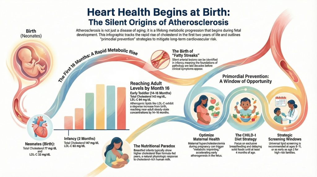
Das Verständnis der Atherosklerose Herz-Kreislauf-Erkrankung (ASCVD) hat im Laufe des letzten Jahrhunderts einen tiefgreifenden Wandel durchlaufen und sich von einer wahrgenommenen unvermeidlichen Folge des Alterns zu einem anerkannten lebenslangen metabolischen Verlauf entwickelt, der während der Fetalentwicklung und des Säuglingsalters beginnt.1Während die klinischen Manifestationen der Krankheit, wie Myokardinfarkt, Schlaganfall, und periphere vaskuläre Syndrome treten typischerweise im mittleren oder späten Erwachsenenalter auf, wobei die stille Inkubation dieser Erkrankungen sich über Jahrzehnte erstreckt.2–4Dieses Entwicklungsparadigma wird durch longitudinale epidemiologische Kohorten und postmortale Untersuchungen gestützt, die insgesamt darauf hindeuten, dass die Grundlagen der kardiovaskulären Pathologie in den frühesten Stadien der menschlichen Existenz gelegt werden.2–5] Brauchen Babys also StatineMal sehen!
Physiologische Verläufe von Plasmalipiden in den ersten zwei Lebensjahren
Die Neonatalperiode und das frühe Säuglingsalter stellen eine Phase dramatischer metabolischer Umstellung dar, die durch eine rasche physiologische Anpassung an das extrauterine Leben gekennzeichnet ist. Bei der Geburt, Cholesterin Die Konzentrationen im Nabelschnurblut sind wesentlich niedriger als die in erwachsenen Populationen beobachteten.6Diese Werte sind jedoch hochgradig dynamisch und folgen in den ersten 24 Lebensmonaten bestimmten Verläufen.
Schrittweise Anstiege und Erreichung früher Ausgangswerte
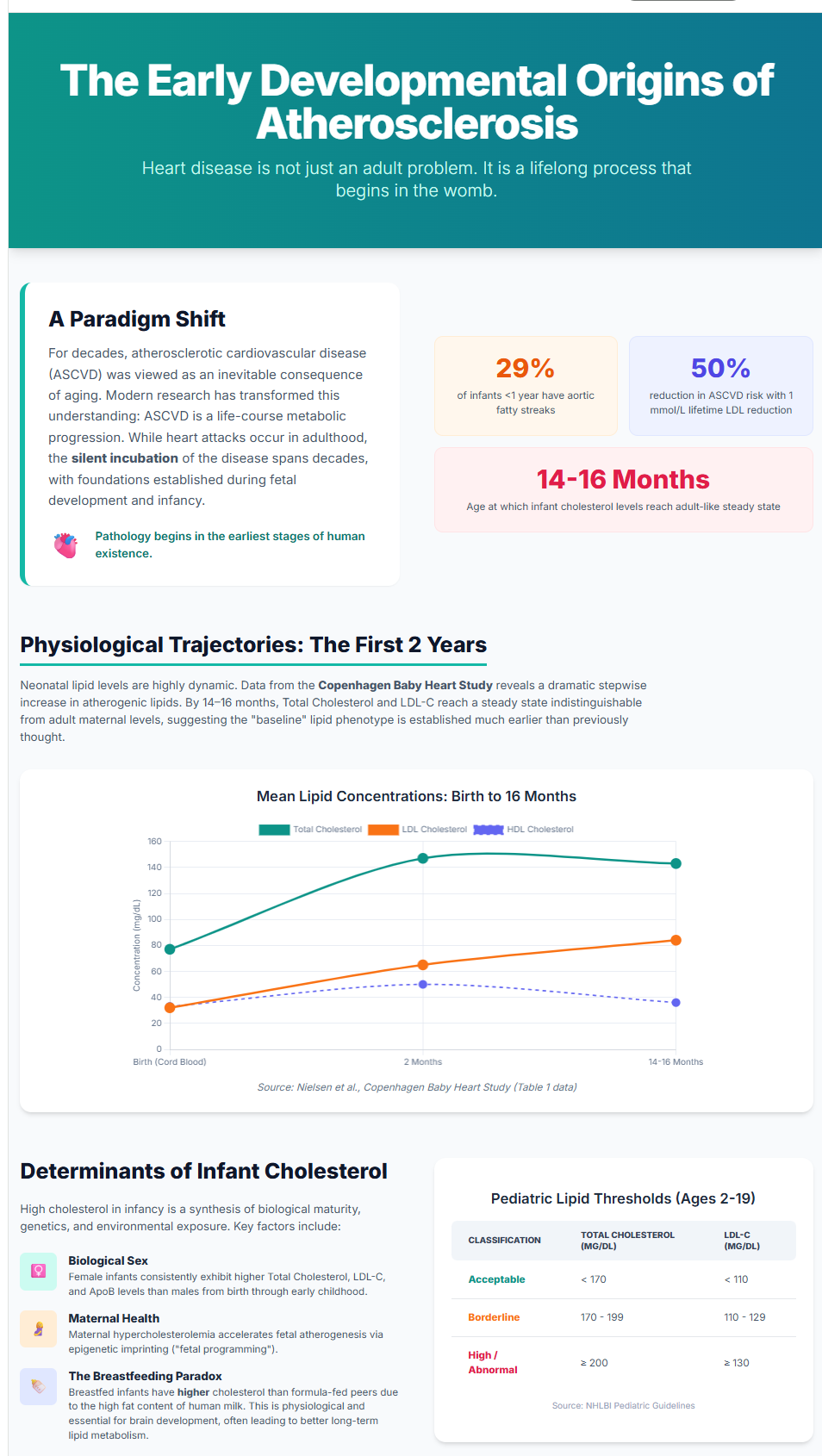
Konzentrationen atherogener Lipidmerkmale – einschließlich Low-Density Lipoprotein Cholesterin (LDL-C), Non-HDL-C, Apolipoprotein B und Lipoprotein(a)—weisen von der Geburt bis zu den ersten 16 Lebensmonaten einen durchgehend schrittweisen Anstieg auf.6] Daten aus den Kopenhagener Herzstudie bei Säuglingen, unter Einbeziehung von 13.354 Nabelschnurblutproben mit longitudinalem venösen Follow-up, zeigen, dass bis zum Alter von 14–16 Monaten, Gesamtcholesterin und die LDL-C-Konzentrationen erreichen Fließgleichgewichtswerte, die denen ähneln, die im Erwachsenenalter beobachtet werden.1,6]
Diese Beobachtung legt nahe, dass der Lipid-Basisphaenotyp viel früher als bisher angenommen festgelegt sein könnte, was eine vernachlässigte Ressource für die frühe Identifizierung kardiovaskulärer Risiken darstellt.1]
Variabilität nicht-atherogener Merkmale
Nicht alle Lipidparameter zeigen einen linearen Anstieg. Gesamtcholesterin und Triglyceride steigen im frühen Säuglingsalter stark an und stabilisieren sich anschließend, während HDL-C während der Ernährungsumstellung von lipidreicher Milch zu Mischnahrung anfangs ansteigen und mäßig abfallen kann.6Diese Variabilität spiegelt die metabolische Plastizität des Säuglingsalters wider, wenn sich die Nährsubstrate verändern.
Normale Referenzwerte für Säuglinge und Kinder
Da sich die Lipidwerte im frühen Leben rasch verändern, ist für die Interpretation die Verwendung altersspezifischer Referenzbereiche erforderlich.6,7]
Lipidwerte bei Neugeborenen und Säuglingen (0–24 Monate)
Die Lipidspiegel bei der Geburt liegen deutlich unter den Schwellenwerten für Erwachsene, steigen jedoch im Säuglingsalter rasch an und erreichen im Alter von 14 bis 16 Monaten nahezu stabile Konzentrationen wie bei Erwachsenen.1,6]
Tabelle 1. Neonatale und Säuglings-Lipid-Referenzwerte (Copenhagen Baby Heart Study)6]
| Altersgruppe | Gesamtcholesterin (mg/dL) Median (IQR) | LDL-C (mg/dL) Median (IQR) | HDL-C (mg/dL) Median (IQR) | Triglyceride (mg/dL) Median (IQR) |
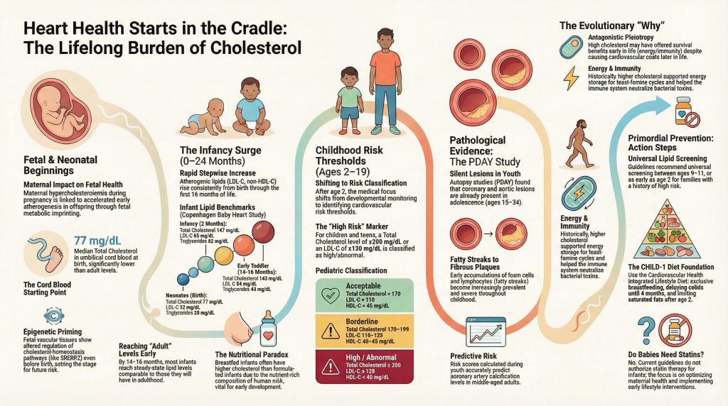
| Neugeborene (Geburt) | 77 (70–93) | 32 (24–39) | 33 (27–41) | 28 (24–34) |
| Säuglingsalter (2 Monate) | 147 (128–166) | 65 (52–79) | 50 (41–62) | 62 (48–84) |
| Frühes Kleinkindalter (14–16 Monate) | 143 (124–162) | 84 (70–103) | 36 (30–45) | 43 (30–60) |
Hinweis: Die in Nielsen et al. in mmol/L angegebenen Werte wurden in mg/dL umgerechnet.6]
Klassifikation von Pädiatrischen Lipiden (Alter 2–19 Jahre)
Nach dem 2. Lebensjahr verschiebt sich die Lipidinterpretation von der Entwicklungsphysiologie zu kardiovaskulären Risikoschwellenwerten. Die pädiatrischen Leitlinien des NHLBI definieren die folgenden Kategorien.7]
Tabelle 2. Schwellenwerte für die pädiatrische Lipidklassifikation (2–19 Jahre)7]
| Klassifikation | Gesamtcholesterin (mg/dL) | LDL-C (mg/dL) | HDL-C (mg/dL) | Triglycerides (mg/dL) |
| Acceptable | <170 | <110 | >45 | <75 (0–9y) / <90 (10–19y) |
| Borderline | 170–199 | 110–129 | 40–45 | 75–99 (0–9y) / 90–129 (10–19y) |
| High/Abnormal | ≥200 | ≥130 | <40 | ≥100 (0–9y) / ≥130 (10–19y) |
Biological and Environmental Determinants of Infantile Lipid Levels
High cholesterol in infancy reflects a synthesis of biological maturity, genetic inheritance, and early environmental exposure.
Biological Sex and Maternal Health
Female infants exhibit higher total cholesterol, LDL-C, and ApoB concentrations than males, differences detectable from birth and persisting through early childhood.[1] Maternal Hypercholesterinämie during pregnancy is associated with accelerated early Atherogenese in offspring.[8] Epigenetic analyses of fetal vascular tissue show altered epigenetic regulation of cholesterol-homeostasis pathways (including regulatory regions of SREBP2) in association with early fetal atherosclerotic Läsionen, supporting fetal metabolic imprinting as a mechanistic framework.[9]
The Nutritional Paradox of Breastfeeding
Breastfed infants often show higher cholesterol concentrations than formula-fed peers, a physiologic response to the cholesterol-rich composition of human milk.[6] Early-life lipid exposure is consistent with lipid tracking across the life course.[10]
Atherosclerosis in Infancy and Youth: Pathological Evidence
Arteriosklerose begins as Fettstreifen—subintimal accumulations of Schaumzellen and lymphocytes—that can be identified early in life.[2–4] Autopsy studies demonstrate that early lesions are detectable in infancy and become increasingly prevalent through childhood.[4,11,12]
Pathobiological Determinants of Atherosclerosis in Youth (PDAY)
Der PDAY-Studie remains foundational in pediatric cardiovascular pathology. PDAY collected arterial specimens from 2,876 individuals aged 15–34 years who died of external causes.[2] Coronary and aortic lesions were present in adolescence, and lesion severity correlated strongly with modifiable Risikofaktoren.[2–4]
Risk scoring derived from PDAY predicts later coronary Arterie Verkalkung in adulthood, validating the clinical relevance of early-life risk burden.[13]
Historical Trends in Pediatric Lipids
Mean total cholesterol levels among US youths declined between 1988 and 2010, with reductions in both mean levels and prevalence of elevated total cholesterol documented in NHANES analyses.[14] However, these improvements are counterbalanced by the Adipositas epidemic, which amplifies adverse lipid phenotypes in vulnerable subgroups.[7]
Mechanisms of Age-Related Cholesterol Elevation
Plasma cholesterol rises with aging largely due to reduced fractional clearance of LDL particles.[15] Aging is associated with declining hepatic LDL-Rezeptor activity and altered endocrine regulation. Growth hormone influences Galle Säure synthesis through cholesterol 7α-hydroxylase activity, providing mechanistic links between senescence and Dyslipidämie.[16]
The Evolutionary Heritage of High Plasma Cholesterol
High circulating cholesterol, while harmful in modern environments of caloric abundance, may have conferred survival advantages in ancestral contexts characterized by nutritional scarcity and infectious burden.[17]
Evolutionary Advantages
Energy Security. Genetic variants favoring higher circulating cholesterol may have supported energy storage and reproductive fitness during feast-famine cycles.[18]
Immune Modulation. Membrane cholesterol organizes Lipid Rafts that cluster antigen receptors on memory T cells, enhancing immune responsiveness.[19] HDL also binds and neutralizes bacterial toxins such as lipopolysaccharides during infection.[20]
Endocrine Readiness. Cholesterol is the obligate precursor for steroid hormone synthesis, including cortisol, and is essential for adrenal stress adaptation.[21]
Antagonistic Pleiotropy. Traits conferring early-life survival benefit may persist despite late-life cardiovascular costs.[17]
The Lifelong Burden of Cumulative LDL Exposure
Cardiovascular risk reflects cumulative arterial exposure to atherogenic lipoproteins rather than single time-point measurements.[1,22] Genetic, epidemiologic, and clinical evidence establishes LDL-C as a causal driver of ASCVD, emphasizing the importance of early prevention.[22]
Clinical Management and the Window of Opportunity
Universal lifestyle promotion and targeted screening are central to prevention. NHLBI guidelines recommend universal lipid screening at ages 9–11 years and earlier screening beginning at age 2 in high-risk families.[7]
Universal Lifestyle Interventions
The Cardiovascular Health Integrated Lifestyle Diet (CHILD-1) provides the foundation of Primärprävention beginning in early childhood.[7]
- Exclusive breastfeeding when possible[6]
- Delaying solid foods until at least 4 months[7]
- Transition to reduced-fat dairy after 12 months in appropriate contexts[7]
- Total fat intake of ~25–30% of calories after age 2 with gesättigtes Fett limitation[7]
Pharmacologic therapy is reserved for older children with severe dyslipidemia, particularly familiäre Hypercholesterinämie.[7]
Fazit
Atherosclerosis is a lifelong process beginning in fetal development and infancy. Early lipid trajectories establish kumulative Exposition burdens that drive lesion progression from fatty streaks in childhood to fibrous Placken in adulthood.[2–4] Prevention must therefore begin early—optimizing maternal health, identifying high-risk lipid phenotypes in infancy, and implementing primordial prevention during the developmental window when vascular injury remains minimal and modifiable.[1,7,8] Infants are not eligible for statin therapy at this time but there certainly are things the mother can do when pregnant and after birth to protect her child for years to come.
Referenzen
- Holven KB. Plasma cholesterol levels in infancy: a neglected resource. Eur Heart J. 2023;44(42):4419-4421. doi:10.1093/eurheartj/ehad558
- Natural history of aortic and coronary atherosclerotic lesions in youth. Findings from the PDAY Study. Pathobiological Determinants of Atherosclerosis in Youth (PDAY) Research Group. Arterioscler Thromb. 1993;13(9):1291-1298. doi:10.1161/01.atv.13.9.1291
- McGill HC Jr, McMahan CA, Zieske AW, et al. Association of Coronary Heart Disease Risk Factors with microscopic qualities of coronary atherosclerosis in youth. Circulation. 2000;102(4):374-379. doi:10.1161/01.cir.102.4.374
- Strong JP, Malcom GT, McMahan CA, et al. Prevalence and extent of atherosclerosis in adolescents and young adults: implications for prevention from the Pathobiological Determinants of Atherosclerosis in Youth Study. JAMA. 1999;281(8):727-735. doi:10.1001/jama.281.8.727
- Luca AC, David SG, David AG, et al. Atherosclerosis from Newborn to Adult-Epidemiology, Pathological Aspects, and Risk Factors. Life (Basel). 2023;13(10):2056. Published 2023 Oct 14. doi:10.3390/life13102056
- Taageby Nielsen S, Mohr Lytsen R, Strandkjær N, et al. Significance of lipids, lipoproteins, and apolipoproteins during the first 14-16 months of life. Eur Heart J. 2023;44(42):4408-4418. doi:10.1093/eurheartj/ehad547
- Expert Panel on Integrated Guidelines for Cardiovascular Health and Risk Reduction in Children and Adolescents; National Heart, Lung, and Blood Institute. Expert panel on integrated guidelines for cardiovascular health and risk reduction in children and adolescents: summary report. Pediatrics. 2011;128 Suppl 5(Suppl 5):S213-S256. doi:10.1542/peds.2009-2107C
- Napoli C, Glass CK, Witztum JL, Deutsch R, D’Armiento FP, Palinski W. Influence of maternal hypercholesterolaemia during pregnancy on progression of early atherosclerotic lesions in childhood: Fate of Early Lesions in Children (FELIC) study. Lancet. 1999;354(9186):1234-1241. doi:10.1016/S0140-6736(99)02131-5
- de Nigris F, Cacciatore F, Mancini FP, et al. Epigenetic Hallmarks of Fetal Early Atherosclerotic Lesions in Humans. JAMA Cardiol. 2018;3(12):1184-1191. doi:10.1001/jamacardio.2018.3546
- Øyri LKL, Bogsrud MP, Kristiansen AL, et al. Cholesterol at ages 6, 12 and 24 months: Tracking and associations with diet and maternal cholesterol in the Infant Cholesterol Study. Atherosclerosis. 2021;326:11-16. doi:10.1016/j.atherosclerosis.2021.04.017
- Tanaka K, Masuda J, Imamura T, et al. A nation-wide study of atherosclerosis in infants, children and young adults in Japan. Atherosclerosis. 1988;72(2-3):143-156. doi:10.1016/0021-9150(88)90075-5
- Françoso LA, Coates V. Anatomicopathological evidence of the beginning of atherosclerosis in infancy and adolescence. Arq Bras Cardiol. 2002;78(1):131-142.
- Gidding SS, Rana JS, Prendergast C, et al. Pathobiological Determinants of Atherosclerosis in Youth (PDAY) Risk Score in Young Adults Predicts Coronary Artery and Abdominal Aorta Calcium in Middle Age: The CARDIA Study. Circulation. 2016;133(2):139-146. doi:10.1161/CIRCULATIONAHA.115.018042
- Kit BK, Carroll MD, Lacher DA, Sorlie PD, DeJesus JM, Ogden C. Trends in serum lipids among US youths aged 6 to 19 years, 1988-2010. JAMA. 2012;308(6):591-600. doi:10.1001/jama.2012.9136
- Ericsson S, Eriksson M, Vitols S, Einarsson K, Berglund L, Angelin B. Influence of age on the metabolism of plasma low density lipoproteins in healthy males. J Clin Invest. 1991;87(2):591-596. doi:10.1172/JCI115034
- Rudling M, Parini P, Angelin B. Growth hormone and bile acid synthesis. Key role for the activity of hepatic microsomal cholesterol 7alpha-hydroxylase in the rat. J Clin Invest. 1997;99(9):2239-2245. doi:10.1172/JCI119398
- Lea AJ, Clark AG, Dahl AW, et al. Applying an evolutionary mismatch framework to understand disease susceptibility. PLoS Biol. 2023;21(9):e3002311. Published 2023 Sep 11. doi:10.1371/journal.pbio.3002311
- Brassington L, Arner AM, Watowich MM, et al. Integrating the Thrifty Genotype and Evolutionary Mismatch Hypotheses to understand variation in cardiometabolic disease risk. Evol Med Public Health. 2024;12(1):214-226. Published 2024 Jul 31. doi:10.1093/emph/eoae014
- Bietz A, Zhu H, Xue M, Xu C. Cholesterol Metabolism in T Cells. Front Immunol. 2017;8:1664. Published 2017 Nov 27. doi:10.3389/fimmu.2017.01664
- Meilhac O, Tanaka S, Couret D. High-Density Lipoproteins Are Bug Scavengers. Biomolecules. 2020;10(4):598. Published 2020 Apr 12. doi:10.3390/biom10040598
- Gomez-Sanchez CE, Gomez-Sanchez EP. Cholesterol Availability and Adrenal Steroidogenesis. Endocrinology. 2024;165(4):bqae032. doi:10.1210/endocr/bqae032
- Ference BA, Ginsberg HN, Graham I, et al. Low-density lipoproteins cause atherosclerotic cardiovascular disease. 1. Evidence from genetic, epidemiologic, and clinical studies. A consensus statement from the European Atherosclerosis Society Consensus Panel. Eur Heart J. 2017;38(32):2459-2472. doi:10.1093/eurheartj/ehx144

