
This page has been automatically translated. If there is any discrepancy, the English version is authoritative.

Revised: July 16, 2026

Do Babies Need Statins?

By: Peter Megdal PhD

How to Use This Article

Medical disclaimer: This article is for education only and is not medical advice. Always consult your clinician for personal guidance.

Easy Read

Introduction: The Silent Incubation

For decades, the public has viewed heart disease as a “late-life accident”—a sudden collision of age and lifestyle that strikes in the golden years. However, as a preventive health scientist, I can tell you that the modern medical paradigm has shifted: atherosclerotic cardiovascular diseaseCardiovascular disease is the umbrella term for problems with the heart and blood vessels, including heart attacks, strokes, and blocked leg arteries. (ASCVD) is a life-course metabolic progression that begins decades before the first symptom appears.

While clinical events like heart attacksA heart attack happens when blood flow to part of the heart muscle is cut off and that muscle starts to die. emerge in middle age, the silent incubation of these conditions spans nearly a century. The biological clock doesn’t start at fifty; it starts in the womb. To understand heart health, we must investigate the cradle, where the foundations of cardiovascular pathology are established during the most formative stages of human development.

The 16-Month Milestone: Adult Levels in Diapers

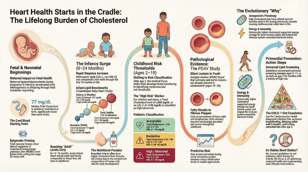
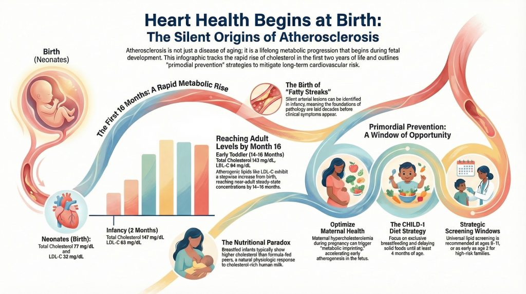
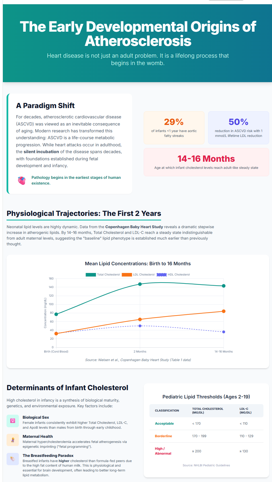
The neonatal period is a phase of radical metabolic transition. At birth, cholesterolCholesterol is a waxy substance your body needs. It goes into cell walls, hormones, vitamin D, and the bile that digests your food. You would die without it. concentrations in umbilical cord blood are significantly lower than adult levels, but this “clean slate” is short-lived. Data from the Copenhagen Baby Heart StudyA large Danish cohort study that analyzed lipid and lipoprotein concentrations in over 13,000 cord blood samples with longitudinal follow-up, providing normative reference data for cholesterol trajectories from birth through the first 14–16 months of life., which analyzed over 13,000 samples, reveals a rapid, stepwise increase in atherogenic lipids like LDL-C and apolipoprotein BApoB is a protein that sits on the outside of every cholesterol particle that can get stuck in your artery wall and cause plaque. Each of those particles carries exactly one ApoB. (apo B) immediately after birth.

Remarkably, by 14–16 months of age—and potentially as early as 6 months—these lipid concentrations reach a steady state that is statistically indistinguishable from adult maternal levels. This “adult baseline” is established while the child is still in diapers. Intriguingly, female infants generally exhibit significantly higher concentrations of total cholesterolTotal cholesterol adds together the cholesterol in all your particles, harmful and helpful alike., LDL-C, and apo B than their male counterparts from birth, a disparity that persists through the first two years.

Perhaps most critical for parents is the early footprint of lipoprotein(a) [Lp(a)]. If an infant’s Lp(a) levels are above the 90th percentile at birth, it serves as a robust predictor that they will reach adult-range high-risk concentrations as early as 14 months of age. This highlights a counter-intuitive reality: the metabolic trajectory of a toddler can mirror the long-term risk profile of an adult before they have even mastered walking.

Fetal Programming: The Aorta’s First Lessons

The cardiovascular journey begins before the first breath, dictated by a biological aging set-point established in utero. The maternal environment acts as the primary instructor for fetal metabolism. When a mother experiences hypercholesterolemiaHypercholesterolemia is an abnormally elevated level of cholesterol-carrying particles in the blood, typically caused in primate experiments by feeding a diet high in dietary cholesterol and saturated fat, and associated with accelerated plaque formation in artery walls. during pregnancy, it acts as a potent stimulus for early atherogenesisAtherogenesis is the step-by-step process of a plaque forming. in the fetus. Findings from the FELIC studyThe Fate of Early Lesions in Children (FELIC) study examined aortic tissue from children aged 1 to 13 who died of non-cardiovascular causes and compared lesion sizes with those seen in fetal specimens, providing indirect evidence that some fetal fatty streaks regress after birth while also showing that children of hypercholesterolemic mothers accumulate lesions faster through childhood. in Naples demonstrated that maternal LDLLDL, or low-density lipoprotein, is the main particle that carries cholesterol through your blood — and the main one that gets stuck in artery walls. levels correlate directly with the size of atherosclerotic lesionsIn cardiology, a lesion refers to a discrete area of atherosclerotic plaque narrowing a coronary artery, typically described by the percentage of luminal obstruction it causes. The article describes four residual lesions too small in vessel diameter to accept a stent after the most critical one was treated. in the fetal aortaThe aorta is the biggest artery in your body. It carries blood out of the heart and down through the chest and belly, sending branches everywhere..

This is more than a temporary exposure; it is a permanent metabolic reprogramming. As the research indicates:
“This association is likely mediated through epigenetic mechanisms… maternal cholesterol levels are positively associated with the methylation of the promoter in fetal aortas… This ‘fetal programmingThe process by which the intrauterine environment—particularly maternal nutrition and metabolic state—permanently alters gene expression and metabolic set-points in the developing fetus, with lasting consequences for cardiovascular and metabolic health in later life.’ may lead to a permanent alteration in the offspring’s metabolic set-point.”

Further evidence of this biological clock is found in telomere length (TL)The length of protective repetitive DNA sequences capping chromosomes; shorter telomeres are a marker of cellular aging, and individuals born with shorter telomeres have been shown to carry significantly higher risks of midlife hypercholesterolemia and arterial damage.. Shorter telomeres at birth are not just markers of cellular age; they are predictive of midlife health. A 40-year longitudinal studyA research design that follows the same individuals over an extended period to observe how exposures or traits at one time point relate to outcomes—such as mortality—years or decades later. found that individuals born with shorter telomeres had a 3.24× increased risk of midlife hypercholesterolemia and significantly higher risk scores for arterial damage by age 42.

The Pathological Reality: Fatty Streaks in Infancy

The most jarring evidence for early intervention comes from autopsy studies. AtherosclerosisAtherosclerosis is the disease behind most heart attacks and many strokes. Cholesterol particles get stuck in the wall of an artery, the body sends immune cells to clean up, and over years that mess hardens into plaque. begins as fatty streaks—microscopic accumulations of lipid-engorged macrophage foam cells in the arterial wall. A Japanese nationwide study revealed that 29% of infants younger than 1 year already possessed these fatty streaks in their aortas. In some regions, such as Taiwan, pathological examinations have even shown a 17% narrowing of the left coronary arteryAn artery is a blood vessel that carries blood away from the heart to the rest of the body. in infants.

Early Warning Signs

Age Group Pathological Prevalence Pathological Feature
Infants (< 1 year) 29% (Aorta) Sudan IV–positive fatty streaks; macrophage foam cells
Infants (< 1 year) 17% (Left coronary) Intimal proliferation; narrowing of the vessel
Children (Age 3) Ubiquitous Global presence of aortic fatty streaksA fatty streak is the earliest visible stage of atherosclerosis — a flat yellow smear of cholesterol-filled immune cells just under the artery lining.
Adolescents (15–19) 50% (Coronary) Initial lipid accumulation; progressing lesions

The Breastfeeding Paradox: When High Cholesterol is Healthy

There is a nutritional paradox in early life: breastfed infants consistently show higher total cholesterol and LDL-C than those who are formula-fed. In an adult, these numbers would trigger alarm, but in an infant, this is often a salutogenicHealth-promoting, as opposed to pathogenic; used in this article to describe the physiologically beneficial role of high cholesterol in breast-fed infants, where lipids support brain myelination and cell membrane synthesis rather than driving disease. (health-promoting) effect.

Human milk is naturally high in fat and cholesterol to support rapid myelin formation in the brain and cell membrane synthesis. This reflects the unique metabolic plasticity of infancy. As the infant transitions from a primarily lipid-based fuel source (milk) to a carbohydrate-inclusive diet, their body handles these lipids as essential building blocks rather than pathological waste. This physiological response is distinct from the chronic, dysfunctional high cholesterol seen in adults.

Vascular Memory: The Legacy of Childhood Lipids

The lipids circulating in a child’s blood leave a permanent imprint on their arteries through vascular memoryThe concept that cumulative exposure of arterial walls to atherogenic lipoproteins during childhood leaves a structural imprint—detectable years later as increased carotid intima-media thickness—that persists even if risk factors subsequently improve.. Data from the Bogalusa Heart StudyThe Bogalusa Heart Study examined the arteries of children and young adults who died in accidents in a Louisiana town. and the Young Finns Study have shown that childhood LDL-C and BMI are highly predictive of carotid arteryThe carotid arteries run up either side of your neck and supply blood to your brain. intima-media thicknessIntima-media thickness, or IMT, is a measurement of how thick the inner layers of an artery have become, usually taken in the neck with ultrasound. (cIMT) 20 to 30 years later.

This memory means the metabolic insults of youth are stored in the arterial wall, manifesting as clinical disease in adulthood. As the Bogalusa researchers famously concluded:
“Heart disease is not an adult problem but a childhood problem with adult manifestations.”

The Modern Paradox: Better Numbers, Bigger Risks

Historical data presents a deceptive picture. Mean total cholesterol in U.S. youth has dropped from 165 mg/dL in the late 1980s to roughly 158 mg/dL today, largely due to the removal of trans fatsTrans fat is an artificially altered fat once used to make processed foods last longer on the shelf. and reduced tobacco exposure.

However, this statistical success is being cannibalized by the obesityObesity means carrying enough excess body fat to affect health. epidemic. While the average child may look better on paper, the high-risk obesity tail of the population is larger and more vulnerable than ever. Furthermore, modern environmental stressors—specifically chronic sleep deprivation—act as physiological disruptors. Sleep loss is not merely a lifestyle issue; it triggers a cascade of hunger hormones and inflammationInflammation is your immune system's response to injury or something it treats as an invader. It brings swelling, heat, and cleanup cells. that acts as a catalyst for metabolic syndromeMetabolic syndrome is a cluster of five problems that tend to travel together: a large waist, high triglycerides, low HDL, high blood pressure, and high blood sugar. Having three or more counts., potentially neutralizing decades of dietary progress.

The PDAY Score: Calculating the Biological Clock

The Pathobiological Determinants of Atherosclerosis in Youth (PDAYPDAY, short for Pathobiological Determinants of Atherosclerosis in Youth, examined the arteries of young people aged 15 to 34 who died of other causes.) study allows us to quantify this early damage. By correlating antemortem risk factorsA risk factor is something that raises your chance of developing a disease — high cholesterol particles, high blood pressure, smoking, diabetes, family history. with arterial findings in young people who died of external causes, researchers developed a risk score that predicts future calcificationCalcification is when calcium gets deposited into a plaque, turning part of it hard and bony. with high accuracy.

The PDAY risk score is calculated using several modifiable components:

  • Non-HDL and HDLHDL, or high-density lipoprotein, is the particle often called "good cholesterol." It picks up cholesterol from tissues and carries it back to the liver. cholesterol
  • BMI (obesity)
  • SmokingSmoking damages the lining of your blood vessels, raises blood pressure, makes blood clot more easily, and speeds up plaque growth. status
  • HypertensionHypertension is the medical term for high blood pressure.
  • HyperglycemiaAbnormally elevated blood glucose concentration; included as one of the modifiable risk factors in the PDAY scoring system because it accelerates arterial lesion progression in adolescents and young adults.

The biological clock measured by this score ticks with alarming speed. In men, the prevalence of advanced GradeGRADE (Grading of Recommendations, Assessment, Development and Evaluations) is a widely used framework for rating the certainty of evidence behind a clinical finding, classifying it as high, moderate, low, or very low based on factors such as study design, risk of bias, consistency, directness, and precision of results. 4 and 5 lesions—plaquesPlaque is the buildup of cholesterol, immune cells, scar tissue, and calcium inside an artery wall. with a distinct lipid coreThe lipid core is the soft, greasy center of a plaque, made of cholesterol and the debris of dead immune cells.—jumps from 2.4% at ages 15–19 to a staggering 20.3% by ages 30–34. This rapid acceleration is fueled by the cumulative exposure to risk factors that began in infancy.

Conclusion: The Window of Opportunity

We currently face a narrow window of opportunity—a brief period in early life when vascular damage is still minor and potentially reversible. Research suggests that maintaining an LDL-C level below 2 mmol/L throughout the life course makes ASCVD extremely rare. However, waiting until middle age to address these numbers is often too late, as calcified plaquesCalcified plaque is the hardened, calcium-filled part of a plaque. It shows up brightly on a CT scan, which is what a calcium scan measures. may have already solidified the disease.

To change the global trajectory of heart disease, we must prioritize primordial preventionPrimordial prevention is a strategy aimed at stopping the development of cardiovascular risk factors in the first place—rather than treating risk factors or existing disease—by keeping atherogenic exposures near zero from birth or early life. It is distinguished from primary prevention, which targets people who already have risk factors but no clinical disease.. This means optimizing maternal health and perhaps considering a provocative shift in policy: universal screening for conditions like familial hypercholesterolemiaFamilial hypercholesterolemia, or FH, is an inherited condition where the liver cannot clear cholesterol from the blood properly. Levels are very high from birth. in infancy. Identifying high-risk individuals in the cradle is our best chance at ensuring the ideal cardiovascular health most babies are born with is protected for a century.

References

  1. Holven KB. Plasma cholesterol levels in infancy: a neglected resource. Eur Heart J. 2023;44(42):4419-4421. doi:10.1093/eurheartj/ehad558
  2. Taageby Nielsen S, Mohr Lytsen R, Strandkjær N, et al. Significance of lipids, lipoproteins, and apolipoproteins during the first 14-16 months of life. Eur Heart J. 2023;44(42):4408-4418. doi:10.1093/eurheartj/ehad547
  3. Luca AC, David SG, David AG, et al. Atherosclerosis from Newborn to Adult-Epidemiology, Pathological Aspects, and Risk Factors. Life (Basel). 2023;13(10):2056. Published 2023 Oct 14. doi:10.3390/life13102056
  4. Napoli C, Glass CK, Witztum JL, Deutsch R, D’Armiento FP, Palinski W. Influence of maternal hypercholesterolaemia during pregnancy on progression of early atherosclerotic lesions in childhood: Fate of Early Lesions in Children (FELIC) study. Lancet. 1999;354(9186):1234-1241. doi:10.1016/S0140-6736(99)02131-5
  5. de Nigris F, Cacciatore F, Mancini FP, et al. Epigenetic Hallmarks of Fetal Early Atherosclerotic Lesions in Humans. JAMA Cardiol. 2018;3(12):1184-1191. doi:10.1001/jamacardio.2018.3546
  6. Natural history of aortic and coronary atherosclerotic lesions in youth. Findings from the PDAY Study. Pathobiological Determinants of Atherosclerosis in Youth (PDAY) Research Group. Arterioscler Thromb. 1993;13(9):1291-1298. doi:10.1161/01.atv.13.9.1291
  7. McGill HC Jr, McMahan CA, Zieske AW, et al. Association of Coronary Heart Disease Risk Factors with microscopic qualities of coronary atherosclerosis in youth. Circulation. 2000;102(4):374-379. doi:10.1161/01.cir.102.4.374
  8. Strong JP, Malcom GT, McMahan CA, et al. Prevalence and extent of atherosclerosis in adolescents and young adults: implications for prevention from the Pathobiological Determinants of Atherosclerosis in Youth Study. JAMA. 1999;281(8):727-735. doi:10.1001/jama.281.8.727
  9. Takei H, Strong JP, Yutani C, Malcom GT. Comparison of coronary and aortic atherosclerosis in youth from Japan and the USA. Atherosclerosis. 2005;180(1):171-179. doi:10.1016/j.atherosclerosis.2004.11.014
  10. Françoso LA, Coates V. Anatomicopathological evidence of the beginning of atherosclerosis in infancy and adolescence. Arq Bras Cardiol. 2002;78(1):131-142.
  11. Øyri LKL, Bogsrud MP, Kristiansen AL, et al. Cholesterol at ages 6, 12 and 24 months: Tracking and associations with diet and maternal cholesterol in the Infant Cholesterol Study. Atherosclerosis. 2021;326:11-16. doi:10.1016/j.atherosclerosis.2021.04.017
  12. Gidding SS, Rana JS, Prendergast C, et al. Pathobiological Determinants of Atherosclerosis in Youth (PDAY) Risk Score in Young Adults Predicts Coronary Artery and Abdominal Aorta Calcium in Middle Age: The CARDIA Study. Circulation. 2016;133(2):139-146. doi:10.1161/CIRCULATIONAHA.115.018042
  13. Raitakari O, Kartiosuo N, Pahkala K, et al. Lipoprotein(a) in Youth and Prediction of Major Cardiovascular Outcomes in Adulthood. Circulation. 2023;147(1):23-31. doi:10.1161/CIRCULATIONAHA.122.060667
  14. Kit BK, Carroll MD, Lacher DA, Sorlie PD, DeJesus JM, Ogden C. Trends in serum lipids among US youths aged 6 to 19 years, 1988-2010. JAMA. 2012;308(6):591-600. doi:10.1001/jama.2012.9136
  15. Expert Panel on Integrated Guidelines for Cardiovascular Health and Risk Reduction in Children and Adolescents; National Heart, Lung, and Blood Institute. Expert panel on integrated guidelines for cardiovascular health and risk reduction in children and adolescents: summary report. Pediatrics. 2011;128 Suppl 5(Suppl 5):S213-S256. doi:10.1542/peds.2009-2107C
  16. Ning H, Labarthe DR, Shay CM, et al. Status of cardiovascular health in US children up to 11 years of age: the National Health and Nutrition Examination Surveys 2003-2010. Circ Cardiovasc Qual Outcomes. 2015;8(2):164-171. doi:10.1161/CIRCOUTCOMES.114.001274
  17. Ference BA, Ginsberg HN, Graham I, et al. Low-density lipoproteins cause atherosclerotic cardiovascular disease. 1. Evidence from genetic, epidemiologic, and clinical studies. A consensus statement from the European Atherosclerosis Society Consensus Panel. Eur Heart J. 2017;38(32):2459-2472. doi:10.1093/eurheartj/ehx144

  18. Ericsson S, Eriksson M, Vitols S, Einarsson K, Berglund L, Angelin B. Influence of age on the metabolism of plasma low density lipoproteins in healthy males. J Clin Invest. 1991;87(2):591-596. doi:10.1172/JCI115034
  19. Rudling M, Parini P, Angelin B. Growth hormone and bile acid synthesis. Key role for the activity of hepatic microsomal cholesterol 7alpha-hydroxylase in the rat. J Clin Invest. 1997;99(9):2239-2245. doi:10.1172/JCI119398
  20. Lea AJ, Clark AG, Dahl AW, et al. Applying an evolutionary mismatch framework to understand disease susceptibility. PLoS Biol. 2023;21(9):e3002311. Published 2023 Sep 11. doi:10.1371/journal.pbio.3002311
  21. Brassington L, Arner AM, Watowich MM, et al. Integrating the Thrifty Genotype and Evolutionary Mismatch Hypotheses to understand variation in cardiometabolic disease risk. Evol Med Public Health. 2024;12(1):214-226. Published 2024 Jul 31. doi:10.1093/emph/eoae014
  22. Bietz A, Zhu H, Xue M, Xu C. Cholesterol Metabolism in T Cells. Front Immunol. 2017;8:1664. Published 2017 Nov 27. doi:10.3389/fimmu.2017.01664
  23. Meilhac O, Tanaka S, Couret D. High-Density Lipoproteins Are Bug Scavengers. Biomolecules. 2020;10(4):598. Published 2020 Apr 12. doi:10.3390/biom10040598
  24. Gomez-Sanchez CE, Gomez-Sanchez EP. Cholesterol Availability and Adrenal Steroidogenesis. Endocrinology. 2024;165(4):bqae032. doi:10.1210/endocr/bqae032

Deep Dive

The Early Developmental Origins of Atherosclerosis: Lipid Trajectories, Pathobiological Determinants, and the Lifelong Burden of Cardiovascular Risk from Infancy

Introduction

The understanding of atherosclerotic cardiovascular diseaseCardiovascular disease is the umbrella term for problems with the heart and blood vessels, including heart attacks, strokes, and blocked leg arteries. (ASCVD) has undergone a profound transformation over the last century, shifting from a perceived inevitable consequence of aging to a recognized life-course metabolic progression that commences during fetal development and infancy.[1] While the clinical manifestations of disease, such as myocardial infarctionSee Heart Attack for the full entry., strokeA stroke happens when blood flow to part of the brain stops, either from a blockage or from bleeding., and peripheral vascular syndromes, typically emerge in middle or late adulthood, the silent incubation of these conditions spans decades.[24] This developmental paradigm is supported by longitudinal epidemiological cohorts and post-mortem investigations, which collectively indicate that the foundations of cardiovascular pathology are established in the earliest stages of human existence.[25] So do babies need statinsA statin slows the enzyme your liver uses to make cholesterol. Your liver responds by pulling more cholesterol out of your blood, which is where the real benefit comes from.? Let’s see!

Physiological Trajectories of Plasma Lipids in the First Two Years of Life

The neonatal period and early infancy represent a phase of dramatic metabolic transition characterized by rapid physiological adaptation to extrauterine life. At birth, cholesterolCholesterol is a waxy substance your body needs. It goes into cell walls, hormones, vitamin D, and the bile that digests your food. You would die without it. concentrations in umbilical cord blood are substantially lower than those observed in adult populations.[6] However, these levels are highly dynamic and follow distinct trajectories during the first 24 months of life.

Stepwise Increases and Achievement of Early Baseline Levels

Concentrations of atherogenic lipid traits—including low-density lipoproteinA lipoprotein is a tiny package that carries fat and cholesterol through your bloodstream. Since fat won't dissolve in water, it needs a protein wrapper to travel. cholesterol (LDL-C), non-HDL-C, apolipoproteinAn apolipoprotein is a protein attached to a fat-carrying particle in your blood. Fat and water don't mix, so these proteins act like a wrapper that lets fat travel safely through the bloodstream. B, and lipoprotein(a)Lipoprotein(a), written Lp(a) and said "L-P-little-a," is an LDL-like particle with an extra sticky protein attached.—exhibit a consistent stepwise increase from birth through the first 16 months of life.[6] Data from the Copenhagen Baby Heart StudyA large Danish cohort study that analyzed lipid and lipoprotein concentrations in over 13,000 cord blood samples with longitudinal follow-up, providing normative reference data for cholesterol trajectories from birth through the first 14–16 months of life., incorporating 13,354 cord blood samples with longitudinal venous follow-up, demonstrate that by 14–16 months, total cholesterolTotal cholesterol adds together the cholesterol in all your particles, harmful and helpful alike. and LDL-C concentrations approach steady-state levels similar to those observed in adulthood.[1,6]

This observation suggests that the baseline lipid phenotype may be established far earlier than previously hypothesized, constituting a neglected resource for early cardiovascular risk identification.[1]

Variability in Non-Atherogenic Traits

Not all lipid parameters follow a linear rise. Total cholesterol and triglyceridesTriglycerides are the main form of fat in your blood and in your body's storage. increase sharply in early infancy and subsequently stabilize, whereas HDL-C may rise initially and decline modestly during dietary transition from lipid-rich milk to mixed diets.[6] This variability reflects the metabolic plasticity of infancy as nutritional substrates shift.

Normal Reference Values for Infants and Children

Because lipid levels change rapidly in early life, interpretation requires age-specific reference ranges.[6,7]

Neonatal and Infantile Lipid Ranges (0–24 Months)

Lipid levels at birth are markedly lower than adult thresholds but rise rapidly during infancy, reaching near–adult steady-state concentrations by 14–16 months.[1,6]

Table 1. Neonatal and Infant Lipid Reference Values (Copenhagen Baby Heart Study)[6]

Age Group Total Cholesterol (mg/dL) Median (IQR) LDL-C (mg/dL) Median (IQR) HDL-C (mg/dL) Median (IQR) Triglycerides (mg/dL) Median (IQR)
Neonates (birth) 77 (70–93) 32 (24–39) 33 (27–41) 28 (24–34)
Infancy (2 months) 147 (128–166) 65 (52–79) 50 (41–62) 62 (48–84)
Early toddler (14–16 months) 143 (124–162) 84 (70–103) 36 (30–45) 43 (30–60)

Note: Values reported in mmol/L in Nielsen et al. and converted to mg/dL.[6]

Pediatric Lipid Classification (Ages 2–19 Years)

After age 2 years, lipid interpretation shifts from developmental physiology to cardiovascular risk thresholds. NHLBI pediatric guidelines define the following categories.[7]

Table 2. Pediatric Lipid Classification Thresholds (2–19 Years)[7]

Classification Total Cholesterol (mg/dL) LDL-C (mg/dL) HDL-C (mg/dL) Triglycerides (mg/dL)
Acceptable <170 <110 >45 <75 (0–9y) / <90 (10–19y)
Borderline 170–199 110–129 40–45 75–99 (0–9y) / 90–129 (10–19y)
High/Abnormal ≥200 ≥130 <40 ≥100 (0–9y) / ≥130 (10–19y)

Biological and Environmental Determinants of Infantile Lipid Levels

High cholesterol in infancy reflects a synthesis of biological maturity, genetic inheritance, and early environmental exposure.

Biological Sex and Maternal Health

Female infants exhibit higher total cholesterol, LDL-C, and apoBApoB is a protein that sits on the outside of every cholesterol particle that can get stuck in your artery wall and cause plaque. Each of those particles carries exactly one ApoB. concentrations than males, differences detectable from birth and persisting through early childhood.[1] Maternal hypercholesterolemiaHypercholesterolemia is an abnormally elevated level of cholesterol-carrying particles in the blood, typically caused in primate experiments by feeding a diet high in dietary cholesterol and saturated fat, and associated with accelerated plaque formation in artery walls. during pregnancy is associated with accelerated early atherogenesisAtherogenesis is the step-by-step process of a plaque forming. in offspring.[8] Epigenetic analyses of fetal vascular tissue show altered epigenetic regulation of cholesterol-homeostasis pathways (including regulatory regions of SREBP2) in association with early fetal atherosclerotic lesionsIn cardiology, a lesion refers to a discrete area of atherosclerotic plaque narrowing a coronary artery, typically described by the percentage of luminal obstruction it causes. The article describes four residual lesions too small in vessel diameter to accept a stent after the most critical one was treated., supporting fetal metabolic imprinting as a mechanistic framework.[9]

The Nutritional Paradox of Breastfeeding

Breastfed infants often show higher cholesterol concentrations than formula-fed peers, a physiologic response to the cholesterol-rich composition of human milk.[6] Early-life lipid exposure is consistent with lipid trackingThe tendency for an individual's lipid concentrations to maintain their relative rank within a population over time, so that a child with high LDL-C is likely to remain relatively high as an adult; documented from infancy through adulthood in longitudinal studies such as the Bogalusa Heart Study and Young Finns Study. across the life course.[10]

Atherosclerosis in Infancy and Youth: Pathological Evidence

AtherosclerosisAtherosclerosis is the disease behind most heart attacks and many strokes. Cholesterol particles get stuck in the wall of an artery, the body sends immune cells to clean up, and over years that mess hardens into plaque. begins as fatty streaksA fatty streak is the earliest visible stage of atherosclerosis — a flat yellow smear of cholesterol-filled immune cells just under the artery lining.—subintimal accumulations of foam cellsA foam cell is an immune cell that has eaten so much trapped cholesterol that it swells up and looks foamy under a microscope. and lymphocytes—that can be identified early in life.[24] Autopsy studies demonstrate that early lesions are detectable in infancy and become increasingly prevalent through childhood.[4,11,12]

Pathobiological Determinants of Atherosclerosis in Youth (PDAY)

The PDAY studyThe Pathobiological Determinants of Atherosclerosis in Youth (PDAY) study was a pathological study that examined the coronary arteries and aortas of young people aged 15–34 who died from unrelated causes such as accidents; it demonstrated that early atherosclerotic lesions — fatty streaks and more advanced plaques — were already present in most adolescents and young adults, decades before any cli… remains foundational in pediatric cardiovascular pathology. PDAYPDAY, short for Pathobiological Determinants of Atherosclerosis in Youth, examined the arteries of young people aged 15 to 34 who died of other causes. collected arterial specimens from 2,876 individuals aged 15–34 years who died of external causes.[2] Coronary and aortic lesions were present in adolescence, and lesion severity correlated strongly with modifiable risk factorsA risk factor is something that raises your chance of developing a disease — high cholesterol particles, high blood pressure, smoking, diabetes, family history..[24]

Risk scoring derived from PDAY predicts later coronary arteryAn artery is a blood vessel that carries blood away from the heart to the rest of the body. calcificationCalcification is when calcium gets deposited into a plaque, turning part of it hard and bony. in adulthood, validating the clinical relevance of early-life risk burden.[13]

Historical Trends in Pediatric Lipids

Mean total cholesterol levels among US youths declined between 1988 and 2010, with reductions in both mean levels and prevalence of elevated total cholesterol documented in NHANES analyses.[14] However, these improvements are counterbalanced by the obesityObesity means carrying enough excess body fat to affect health. epidemic, which amplifies adverse lipid phenotypes in vulnerable subgroups.[7]

Mechanisms of Age-Related Cholesterol Elevation

Plasma cholesterol rises with aging largely due to reduced fractional clearance of LDLLDL, or low-density lipoprotein, is the main particle that carries cholesterol through your blood — and the main one that gets stuck in artery walls. particles.[15] Aging is associated with declining hepatic LDL receptorThe LDL receptor is a docking port on liver cells that grabs LDL particles out of the blood and pulls them in to be broken down. activity and altered endocrine regulation. Growth hormone influences bile acidBile acids are made by your liver from cholesterol and released into the gut to help digest fat. synthesis through cholesterol 7α-hydroxylase activity, providing mechanistic links between senescence and dyslipidemiaDyslipidemia is the medical word for an unhealthy pattern of fats in the blood. It can mean high LDL, high triglycerides, low HDL, or some combination..[16]

The Evolutionary Heritage of High Plasma Cholesterol

High circulating cholesterol, while harmful in modern environments of caloric abundance, may have conferred survival advantages in ancestral contexts characterized by nutritional scarcity and infectious burden.[17]

Evolutionary Advantages

Energy Security. Genetic variants favoring higher circulating cholesterol may have supported energy storage and reproductive fitness during feast-famine cycles.[18]
Immune Modulation. Membrane cholesterol organizes lipid raftsLipid rafts are cholesterol-enriched microdomains within cell membranes that act as organized platforms for receptor tyrosine kinases, G-protein-coupled receptors, and other signalling molecules. Their integrity depends on adequate membrane cholesterol, underscoring why cholesterol is maintained as a structural asset rather than a fuel source. that cluster antigen receptors on memory T cells, enhancing immune responsiveness.[19] HDLHDL, or high-density lipoprotein, is the particle often called "good cholesterol." It picks up cholesterol from tissues and carries it back to the liver. also binds and neutralizes bacterial toxins such as lipopolysaccharides during infection.[20]
Endocrine Readiness. Cholesterol is the obligate precursor for steroid hormone synthesis, including cortisol, and is essential for adrenal stress adaptation.[21]
Antagonistic PleiotropyAntagonistic pleiotropy is an evolutionary hypothesis in which a gene or biological trait confers a survival or reproductive advantage early in life while becoming harmful later; applied to cardiovascular disease, it suggests that lipid-transport systems that may have offered benefits in ancestral environments become disadvantageous across long modern lifespans of caloric abundance.. Traits conferring early-life survival benefit may persist despite late-life cardiovascular costs.[17]

The Lifelong Burden of Cumulative LDL Exposure

Cardiovascular risk reflects cumulative arterial exposure to atherogenic lipoproteins rather than single time-point measurements.[1,22] Genetic, epidemiologic, and clinical evidence establishes LDL-C as a causal driver of ASCVD, emphasizing the importance of early prevention.[22]

Clinical Management and the Window of Opportunity

Universal lifestyle promotion and targeted screening are central to prevention. NHLBI guidelines recommend universal lipid screening at ages 9–11 years and earlier screening beginning at age 2 in high-risk families.[7]

Universal Lifestyle Interventions

The Cardiovascular Health Integrated Lifestyle Diet (CHILD-1) provides the foundation of primordial preventionPrimordial prevention is a strategy aimed at stopping the development of cardiovascular risk factors in the first place—rather than treating risk factors or existing disease—by keeping atherogenic exposures near zero from birth or early life. It is distinguished from primary prevention, which targets people who already have risk factors but no clinical disease. beginning in early childhood.[7]

  • Exclusive breastfeeding when possible[6]
  • Delaying solid foods until at least 4 months[7]
  • Transition to reduced-fat dairy after 12 months in appropriate contexts[7]
  • Total fat intake of ~25–30% of calories after age 2 with saturated fatSaturated fat is the kind that stays solid at room temperature — butter, the fat in red meat, coconut oil, and palm oil. limitation[7]

Pharmacologic therapy is reserved for older children with severe dyslipidemia, particularly familial hypercholesterolemiaFamilial hypercholesterolemia, or FH, is an inherited condition where the liver cannot clear cholesterol from the blood properly. Levels are very high from birth..[7]

Conclusion

Atherosclerosis is a lifelong process beginning in fetal development and infancy. Early lipid trajectories establish cumulative exposureCumulative exposure is the total amount of harmful cholesterol particles your arteries have been soaked in across your entire life — how high, multiplied by how long. burdens that drive lesion progression from fatty streaks in childhood to fibrous plaquesPlaque is the buildup of cholesterol, immune cells, scar tissue, and calcium inside an artery wall. in adulthood.[24] Prevention must therefore begin early—optimizing maternal health, identifying high-risk lipid phenotypes in infancy, and implementing primordial prevention during the developmental window when vascular injury remains minimal and modifiable.[1,7,8] Infants are not eligible for statin therapy at this time but there certainly are things the mother can do when pregnant and after birth to protect her child for years to come.

References

  1. Holven KB. Plasma cholesterol levels in infancy: a neglected resource. Eur Heart J. 2023;44(42):4419-4421. doi:10.1093/eurheartj/ehad558
  2. Natural history of aortic and coronary atherosclerotic lesions in youth. Findings from the PDAY Study. Pathobiological Determinants of Atherosclerosis in Youth (PDAY) Research Group. Arterioscler Thromb. 1993;13(9):1291-1298. doi:10.1161/01.atv.13.9.1291
  3. McGill HC Jr, McMahan CA, Zieske AW, et al. Association of Coronary Heart Disease Risk Factors with microscopic qualities of coronary atherosclerosis in youth. Circulation. 2000;102(4):374-379. doi:10.1161/01.cir.102.4.374
  4. Strong JP, Malcom GT, McMahan CA, et al. Prevalence and extent of atherosclerosis in adolescents and young adults: implications for prevention from the Pathobiological Determinants of Atherosclerosis in Youth Study. JAMA. 1999;281(8):727-735. doi:10.1001/jama.281.8.727
  5. Luca AC, David SG, David AG, et al. Atherosclerosis from Newborn to Adult-Epidemiology, Pathological Aspects, and Risk Factors. Life (Basel). 2023;13(10):2056. Published 2023 Oct 14. doi:10.3390/life13102056
  6. Taageby Nielsen S, Mohr Lytsen R, Strandkjær N, et al. Significance of lipids, lipoproteins, and apolipoproteins during the first 14-16 months of life. Eur Heart J. 2023;44(42):4408-4418. doi:10.1093/eurheartj/ehad547
  7. Expert Panel on Integrated Guidelines for Cardiovascular Health and Risk Reduction in Children and Adolescents; National Heart, Lung, and Blood Institute. Expert panel on integrated guidelines for cardiovascular health and risk reduction in children and adolescents: summary report. Pediatrics. 2011;128 Suppl 5(Suppl 5):S213-S256. doi:10.1542/peds.2009-2107C
  8. Napoli C, Glass CK, Witztum JL, Deutsch R, D’Armiento FP, Palinski W. Influence of maternal hypercholesterolaemia during pregnancy on progression of early atherosclerotic lesions in childhood: Fate of Early Lesions in Children (FELIC) study. Lancet. 1999;354(9186):1234-1241. doi:10.1016/S0140-6736(99)02131-5
  9. de Nigris F, Cacciatore F, Mancini FP, et al. Epigenetic Hallmarks of Fetal Early Atherosclerotic Lesions in Humans. JAMA Cardiol. 2018;3(12):1184-1191. doi:10.1001/jamacardio.2018.3546
  10. Øyri LKL, Bogsrud MP, Kristiansen AL, et al. Cholesterol at ages 6, 12 and 24 months: Tracking and associations with diet and maternal cholesterol in the Infant Cholesterol Study. Atherosclerosis. 2021;326:11-16. doi:10.1016/j.atherosclerosis.2021.04.017
  11. Tanaka K, Masuda J, Imamura T, et al. A nation-wide study of atherosclerosis in infants, children and young adults in Japan. Atherosclerosis. 1988;72(2-3):143-156. doi:10.1016/0021-9150(88)90075-5
  12. Françoso LA, Coates V. Anatomicopathological evidence of the beginning of atherosclerosis in infancy and adolescence. Arq Bras Cardiol. 2002;78(1):131-142.
  13. Gidding SS, Rana JS, Prendergast C, et al. Pathobiological Determinants of Atherosclerosis in Youth (PDAY) Risk Score in Young Adults Predicts Coronary Artery and Abdominal Aorta Calcium in Middle Age: The CARDIA Study. Circulation. 2016;133(2):139-146. doi:10.1161/CIRCULATIONAHA.115.018042
  14. Kit BK, Carroll MD, Lacher DA, Sorlie PD, DeJesus JM, Ogden C. Trends in serum lipids among US youths aged 6 to 19 years, 1988-2010. JAMA. 2012;308(6):591-600. doi:10.1001/jama.2012.9136
  15. Ericsson S, Eriksson M, Vitols S, Einarsson K, Berglund L, Angelin B. Influence of age on the metabolism of plasma low density lipoproteins in healthy males. J Clin Invest. 1991;87(2):591-596. doi:10.1172/JCI115034
  16. Rudling M, Parini P, Angelin B. Growth hormone and bile acid synthesis. Key role for the activity of hepatic microsomal cholesterol 7alpha-hydroxylase in the rat. J Clin Invest. 1997;99(9):2239-2245. doi:10.1172/JCI119398
  17. Lea AJ, Clark AG, Dahl AW, et al. Applying an evolutionary mismatch framework to understand disease susceptibility. PLoS Biol. 2023;21(9):e3002311. Published 2023 Sep 11. doi:10.1371/journal.pbio.3002311
  18. Brassington L, Arner AM, Watowich MM, et al. Integrating the Thrifty Genotype and Evolutionary Mismatch Hypotheses to understand variation in cardiometabolic disease risk. Evol Med Public Health. 2024;12(1):214-226. Published 2024 Jul 31. doi:10.1093/emph/eoae014
  19. Bietz A, Zhu H, Xue M, Xu C. Cholesterol Metabolism in T Cells. Front Immunol. 2017;8:1664. Published 2017 Nov 27. doi:10.3389/fimmu.2017.01664
  20. Meilhac O, Tanaka S, Couret D. High-Density Lipoproteins Are Bug Scavengers. Biomolecules. 2020;10(4):598. Published 2020 Apr 12. doi:10.3390/biom10040598
  21. Gomez-Sanchez CE, Gomez-Sanchez EP. Cholesterol Availability and Adrenal Steroidogenesis. Endocrinology. 2024;165(4):bqae032. doi:10.1210/endocr/bqae032
  22.  Ference BA, Ginsberg HN, Graham I, et al. Low-density lipoproteins cause atherosclerotic cardiovascular disease. 1. Evidence from genetic, epidemiologic, and clinical studies. A consensus statement from the European Atherosclerosis Society Consensus Panel. Eur Heart J. 2017;38(32):2459-2472. doi:10.1093/eurheartj/ehx144

Transparency Note: This blog post was created with assistance from AI tools. The final content has been carefully reviewed and edited by the author, who is responsible for its accuracy. The information provided is for educational purposes only and does not constitute medical advice.

AI App

Heart Risk Calulator

Educational family-history heart risk calculator with H-score insights, visual family tree input and shareable PDF reports.

Read why this app is so important here.