
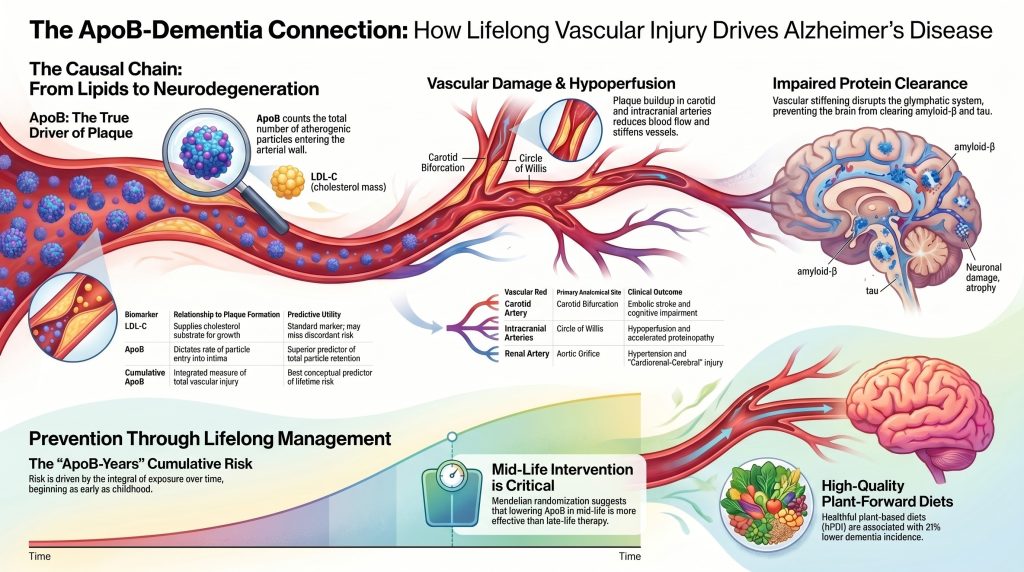
Apolipoprotein B-Driven Atherosclerosis as a Causal Basis for Alzheimer’s Disease
A Multidisciplinary Perspective on the Cardiorenal-Cerebral Axis
Abstract
Background. The dominant neurocentric framework defines Alzheimer’s disease (AD) biologically by amyloid and tau biomarkersA biomarker is something measurable in the body that tells you about health or disease — a lab value, a scan result, a blood pressure reading. and treats vascular pathology as a frequent but secondary modifier. Genetic, epidemiological, and imaging evidence increasingly support an alternative reading in which lifelong exposure to apolipoproteinAn apolipoprotein is a protein attached to a fat-carrying particle in your blood. Fat and water don't mix, so these proteins act like a wrapper that lets fat travel safely through the bloodstream. B (ApoBApoB is a protein that sits on the outside of every cholesterol particle that can get stuck in your artery wall and cause plaque. Each of those particles carries exactly one ApoB.)-containing lipoproteinsA lipoprotein is a tiny package that carries fat and cholesterol through your bloodstream. Since fat won't dissolve in water, it needs a protein wrapper to travel. is a causal upstream driver of the cerebrovascular and small-vessel injury that, in a substantial subset of patients, precedes and accelerates AD-defining pathology.
Objective. To synthesize current evidence linking lifelong ApoB exposure to atherosclerotic injury across the carotid, intracranial, and renal vascular beds and, in turn, to the cognitive syndrome clinically diagnosed as Alzheimer’s disease.
Methods. Structured narrative reviewA narrative review is a type of scientific article that synthesizes existing research on a topic through expert selection and interpretation rather than through a pre-registered, exhaustive search with formal bias scoring; unlike a systematic review or meta-analysis, its conclusions can reflect the authors' editorial judgment in choosing which studies to emphasize. of PubMed-indexed primary studies, Mendelian randomizationMendelian randomization is a clever research method that uses the genes people were born with as a natural experiment. analyses, longitudinal imaging cohorts, randomized cardiovascular and dementia-prevention trials, and current consensus statements from the American Heart Association, the National Lipid Association, and the Alzheimer’s Association. Causal claims were evaluated against STROBE-MR reporting standards.[29]
Conclusions. Lifelong ApoB-containing lipoprotein exposure is causally established as a driver of atherosclerotic vascular disease. Converging evidence from drug-target Mendelian randomizationRandomization is the process of assigning trial participants to treatment or control groups by chance, ensuring that known and unknown confounding factors are evenly distributed; when randomization fails—as auditors found occurred in PREDIMED—the groups may differ in ways that distort the apparent treatment effect. in over one million individuals,[7] longitudinal PET imaging of intracranial stenosisStenosis is narrowing — usually described as a percentage, like a 70 percent blockage.,[13] and randomized vascular-risk-reduction trials[14] supports a causal contribution of this vascular injury to cognitive decline, to vascular cognitive impairment and dementia (VCID)Vascular cognitive impairment and dementia is an umbrella term for cognitive decline caused primarily by cerebrovascular disease, encompassing a spectrum from mild impairment to severe dementia resulting from strokes, small-vessel disease, or chronic hypoperfusion., and to the mixed-pathology dementia phenotypes that dominate older-adult autopsy series.[2,32] The extension from cerebrovascular injury to biomarker-confirmed Alzheimer’s disease as defined under the 2024 NIA-AA revised criteriaThe 2024 NIA-AA revised criteria are diagnostic guidelines from the National Institute on Aging and Alzheimer's Association that define Alzheimer's disease biologically by the presence of core amyloid and tau biomarkers, independent of clinical symptoms.[40] is supported but with appropriately weaker confidence, and the language of this manuscript is hedged accordingly at that endpoint. The 2024 NIA-AA criteria are a diagnostic framework, not an etiologic claim; they are fully compatible with both an established independent pathogenic role for amyloid-β (most directly demonstrated by autosomal-dominant familial AD)[41] and a causal upstream role for ApoB-driven cerebrovascular injury in sporadic late-onset disease. Lifelong ApoB reduction — through high-quality plant-forward dietary patterns and, where indicated, pharmacotherapy initiated in midlife — is a defensible prevention strategy for cognitive decline and a plausible component of dementia and AD prevention.
Introduction
The prevailing neurocentric model of Alzheimer’s disease, which prioritizes the amyloid-β cascade as the primary initiator of neurodegeneration, is increasingly challenged by genetic, epidemiological, and imaging evidence indicating that vascular health is not merely a comorbid factor but a foundational upstream driver of the disease process.[1,5,17] This analysis investigates the hypothesis that lifetime exposure to elevated apolipoprotein B (ApoB)-containing lipoproteins drives progressive atherosclerotic plaquePlaque is the buildup of cholesterol, immune cells, scar tissue, and calcium inside an artery wall. accumulation in the carotid, intracranial, and renal arteries, initiating a cascade of systemic and local injuries that culminate in dementia in a substantial subset of patients.[4,7,20,22] By examining overlapping mechanisms of plaque formation across vascular beds and the causal inferences afforded by Mendelian randomization, a rigorous framework emerges for understanding Alzheimer’s disease as the late-stage neurological manifestation of decades-long vascular-lipid-inflammatory injury.[2,3,9]
Causal language is used deliberately but is calibrated to the strength of evidence at each link in the chain. ApoB is established as causal for atherosclerotic cardiovascular diseaseCardiovascular disease is the umbrella term for problems with the heart and blood vessels, including heart attacks, strokes, and blocked leg arteries. by triangulation across multivariable Mendelian randomization,[21,25] cumulative-exposure analyses,[22,33] and randomized lipid-lowering trials.[26] The extension of this causal pathway to vascular cognitive impairment and to mixed-pathology dementia is well-supported by drug-target Mendelian randomization for non-HDL-C and dementia in over one million individuals[7] and by longitudinal PET imaging linking baseline intracranial stenosis to subsequent amyloid and tau accumulation.[13] The further extension to biomarker-confirmed Alzheimer’s disease specifically — AD as defined by the 2024 NIA-AA revised criteria[40] — is supported by directionally consistent ApoB-specific Mendelian randomization[4] and by the mechanistic compatibility of vascular injury with amyloid and tau biology,[16,17,42] but at appropriately weaker strength than the cardiovascular link. The manuscript adopts hedged language at the AD endpoint accordingly and explicitly distinguishes all-cause dementia, vascular cognitive impairment, mixed-pathology dementia, and biomarker-confirmed AD throughout.
The Vascular-Atherosclerotic Hypothesis of Alzheimer’s Disease and Dementia
The vascular-atherosclerotic hypothesis posits that the clinical syndrome of Alzheimer’s disease is, in a substantial subset of patients, the downstream consequence of chronic cerebral hypoperfusion and neurovascular unit dysfunction initiated by large-artery atherosclerosisAtherosclerosis is the disease behind most heart attacks and many strokes. Cholesterol particles get stuck in the wall of an artery, the body sends immune cells to clean up, and over years that mess hardens into plaque. and propagated through the cerebral small-vessel network.[1,5,17] In this framework, focal narrowing of carotid and intracranial arteries, combined with loss of arterial elasticity, creates a hemodynamic environment hostile to neuronal survival and proteinProtein is the nutrient your body uses to build and repair muscle and tissue. clearance.[12,18]
Contemporary neuropathological evidence demonstrates that the classical categorical distinction between pure vascular dementia and pure Alzheimer’s disease is empirically untenable in older adults.[3,32] Community-based autopsy studies report that mixed neuropathologies — any combination of Alzheimer-type, Lewy body, TDP-43, and vascular lesionsIn cardiology, a lesion refers to a discrete area of atherosclerotic plaque narrowing a coronary artery, typically described by the percentage of luminal obstruction it causes. The article describes four residual lesions too small in vessel diameter to accept a stent after the most critical one was treated. — are present in the majority of aged brains, and pure proteinopathy without cerebrovascular pathology is the exception rather than the rule.[3,32] Large-vessel atherosclerosis, including carotid stenosis and intracranial atherosclerotic disease (ICAD)Intracranial atherosclerotic disease is the buildup of atherosclerotic plaque within the arteries inside the skull, narrowing vessels that supply brain tissue and contributing to stroke, chronic hypoperfusion, and cognitive impairment., commonly coexists with cerebral small-vessel disease characterized by white-matter hyperintensities, lacunar infarcts, and microbleeds.[3,8,9]
Arterial stiffnessArterial stiffness is a measure of how much an artery's wall resists expansion with each pulse of blood; it increases with age as elastin is lost and collagen accumulates, and manifests clinically as a rising systolic blood pressure alongside a falling or stable diastolic blood pressure after about age 60. — particularly in the proximal aortaThe aorta is the biggest artery in your body. It carries blood out of the heart and down through the chest and belly, sending branches everywhere. and carotid arteries — transmits high-pressure pulsatility into the delicate microvasculature of the brain.[12] This hyperpulsatile flow damages the blood-brain barrier and disrupts the glymphatic systemThe glymphatic system is a perivascular waste-clearance network in the brain that uses cerebrospinal fluid flowing along blood vessel walls to flush out metabolic byproducts, including amyloid-beta and tau, primarily during sleep., a perivascular network responsible for clearing metabolic waste, including amyloid-β and tau.[15,18] In this framework, accumulation of these proteins is, in part, a consequence of impaired clearance rather than solely a consequence of overproduction.[16,17,18]
This hypothesis is not exclusive of canonical AD genetic risk. The apolipoprotein E ε4 (APOEAPOE is a gene that comes in three common versions, labeled E2, E3, and E4. It controls how efficiently your liver clears leftover fat particles. ε4) allele — the largest common genetic risk factorA risk factor is something that raises your chance of developing a disease — high cholesterol particles, high blood pressure, smoking, diabetes, family history. for late-onset AD — operates substantially through cerebrovascular pathways. Direct human imaging evidence demonstrates that APOE ε4 carriers exhibit blood-brain barrier breakdown that precedes and predicts cognitive decline independently of amyloid and tau,[42] providing a mechanistic bridge between systemic lipoprotein biology, cerebrovascular injury, and the clinical AD phenotype.[5,16,42]
We engage explicitly with the 2024 Alzheimer’s Association revised criteria,[40] which define AD biologically by core amyloid and tau biomarkers, independent of clinical syndrome. These criteria are a diagnostic framework, not an etiologic claim, and the working group recognizes vascular and other co-pathologies as common modifiers of disease course. Our position is therefore not that the 2024 criteria are wrong or that they deny vascular contributions — they do neither. Our position is that the biomarker fingerprint they operationalize has, for a substantial subset of patients, an upstream cause: decades of ApoB-driven cerebrovascular injury interacting with intrinsic amyloid and tau biology. Defining AD by its downstream biomarker fingerprint is fully compatible with attributing that fingerprint, in part, to upstream vascular causes.
Table 1. Overlapping Pathological and Hemodynamic Features in Dementia Subtypes
| Feature | Alzheimer’s Disease (AD) | Vascular Dementia (VaD) | Mixed Dementia |
| Primary proteinopathy | Amyloid-β plaques; tau tangles | Minimal or absent | Extensive Aβ and tau |
| Vascular lesions | Often present (CAA, microbleeds) | Large infarcts, lacunae, WMH | Combined vascular and proteinopathic |
| Hemodynamics | Reduced CBF; glymphatic failure | Focal or global ischemiaIschemia is when a tissue is not getting enough blood and oxygen for what it is being asked to do. | Chronic hypoperfusion + stiffness |
| Lipid driver | ApoB link via cerebrovascular injury | Strong LDL-C / ApoB association | Lifelong high ApoB burden |
| Imaging hallmarks | Hippocampal atrophy; Aβ-PET (+) | Multiple infarcts; carotid stenosis | Atrophy, WMH, plaque burdenPlaque burden is the total amount of plaque in your arteries, everywhere — not just at the single worst spot. |
Adapted from references [3,5,9,32,40]. CAA = cerebral amyloid angiopathy; WMH = white-matter hyperintensities; CBF = cerebral blood flow.
Lipoprotein Dynamics and Lifelong Causal Exposure
Central to the development of atherosclerosis is the concentration of ApoB-containing lipoproteins in the circulation.[10,19,20] ApoB is a structural protein found on the surface of every atherogenic lipoprotein particle. Hepatically derived very-low-density lipoproteins (VLDLVLDL, or very-low-density lipoprotein, is the particle your liver makes to ship triglycerides out to the rest of the body.), intermediate-density lipoproteins (IDLIDL, or intermediate-density lipoprotein, is a particle that forms partway through the process of a big triglyceride-carrying particle shrinking down into an LDL particle.), low-density lipoproteins (LDLLDL, or low-density lipoprotein, is the main particle that carries cholesterol through your blood — and the main one that gets stuck in artery walls.), and lipoprotein(a)Lipoprotein(a), written Lp(a) and said "L-P-little-a," is an LDL-like particle with an extra sticky protein attached. each carry one molecule of apoB-100ApoB-100 is the full-length form of apolipoprotein B found on LDL, VLDL, IDL, and remnant lipoproteins; its positively charged amino-acid domains bind ionically to negatively charged proteoglycan side chains in the arterial wall, physically trapping the particle in the intima and initiating plaque formation. per particle, while chylomicronsA chylomicron is a very large particle that carries fat from a meal out of your intestines and into your bloodstream. and chylomicron remnants carry apoB-48ApoB-48 is a truncated isoform of apolipoprotein B produced in the intestine and found exclusively on chylomicrons and their remnants; unlike ApoB-100, it is not measured by standard clinical ApoB assays in the fasting state, meaning routine ApoB tests reflect atherogenic particle burden from liver-derived lipoproteins rather than dietary fat absorption. originating from the intestine.[20] In adults in the post-absorptive state, the dominant atherogenic burden reflected by plasma ApoB measurement is overwhelmingly composed of apoB-100–bearing particles.[20] Beyond the count distinction, LDL particles (apoB-100) have substantially longer intimal residence times than larger, more transiently circulating chylomicron remnants (apoB-48), so the apoB-100 fraction contributes disproportionately to cumulative arterial-wall injury and dominates the “ApoB-yearsApoB-years is a proposed research metric that represents cumulative apolipoprotein B exposure over time, expressed as the area under the apoB-versus-age curve in mg/dL·years; it is intended to capture integrated atherogenic particle burden more directly than any single measurement, but has not yet been validated as a clinical tool or treatment threshold.” integral in the cerebrovascular bed.
Whereas LDL cholesterolLDL cholesterol, or LDL-C, is the amount of cholesterol sitting inside your LDL particles. It is the number on almost every standard lab report. (LDL-C) measures the cholesterolCholesterol is a waxy substance your body needs. It goes into cell walls, hormones, vitamin D, and the bile that digests your food. You would die without it. mass within LDL particles, ApoB provides a direct count of total atherogenic particlesAtherogenic particles are the ApoB-containing lipoproteins—including LDL, IDL, VLDL, and lipoprotein(a)—that can enter and be retained in the artery wall to initiate and sustain plaque growth; the article uses the term to describe what must be lowered substantially and sustainably to achieve plaque regression..[20] This distinction is mechanistically critical: the probability of a lipoprotein particle entering the arterial intimaThe intima is the innermost layer of an artery wall, sitting just beneath the smooth lining. and being retained by extracellular proteoglycans is a function of particle concentration rather than cholesterol content per particle.[19] Multivariable Mendelian randomization analyses have established that ApoB is the dominant lipid trait in the relationship between lipoproteins and coronary heart diseaseCoronary heart disease is the narrowing or blockage of the arteries that supply blood to the heart muscle, caused by the buildup of atherosclerotic plaque; it is the leading cause of heart attack and cardiac death worldwide.; when ApoB is conditioned upon, neither LDL-C nor triglyceridesTriglycerides are the main form of fat in your blood and in your body's storage. retain independent causal information for atherosclerotic cardiovascular disease.[21,25]
The risk conferred by ApoB is cumulative — a concept now formalized as the LDL/ApoB cumulative exposureCumulative exposure is the total amount of harmful cholesterol particles your arteries have been soaked in across your entire life — how high, multiplied by how long. hypothesis.[22] AtherogenesisAtherogenesis is the step-by-step process of a plaque forming. is a slow process that typically begins in childhood with the formation of nascent fatty streaksA fatty streak is the earliest visible stage of atherosclerosis — a flat yellow smear of cholesterol-filled immune cells just under the artery lining..[19] As ApoB-containing particlesLipoproteins—including LDL, IDL, VLDL, and their remnants—that each carry one molecule of apolipoprotein B on their surface; particle number (rather than cholesterol mass alone) is a key driver of atherosclerosis because each particle can be retained in the arterial wall. remain elevated over decades, atherosclerotic plaque burden gradually accrues until critical thresholds are crossed, leading to luminal narrowing or plaque rupturePlaque rupture is when the protective cap over a plaque tears open, spilling its contents into the bloodstream..[22,33] Lifelong cumulative exposure (often described as “cholesterol-yearsCholesterol-years is a cumulative-exposure metric that multiplies a person's average LDL-C level (in mg/dL) by the number of years they have carried that level, analogous to pack-years for tobacco. The concept holds that it is the total lifetime burden of apoB-containing lipoproteins, not any single reading, that determines when and how severely atherosclerosis develops.” or “ApoB-years”) is a more biologically appropriate predictor of cardiovascular and, plausibly, dementia risk than any single mid-life or late-life measurement, which may be confounded by reverse causationReverse causation is when the arrow points the other way — the illness caused the exposure rather than the exposure causing the illness. in older adults developing frailty and weight lossWeight loss means reducing body fat, whether through food changes, exercise, medication, or surgery. in the preclinical phase of dementia.[22,24,28]
Table 2. Comparative Predictive Value of Lipid Biomarkers for Atherosclerotic Risk
| Biomarker | Definition | Relationship to plaque formation | Utility |
| LDL-C | Cholesterol mass in LDL particles | Supplies cholesterol substrate for plaque growth | Standard marker; misses risk in discordanceSee ApoB Discordance for the full entry. |
| ApoB | Total count of atherogenic particles (one per particle) | Dictates rate of particle entry into the intima | Superior predictor of particle retention |
| Non-HDL-C | Total cholesterolTotal cholesterol adds together the cholesterol in all your particles, harmful and helpful alike. minus HDL-C | Captures LDL, VLDL, IDL, and Lp(a) cholesterol | Practical surrogate for atherogenic burden |
| Lp(a) | LDL-like particle with apolipoprotein(a) | Pro-thrombotic and pro-inflammatory | Independent genetic risk factor |
| Cumulative ApoB exposure | Integral of ApoB over time (“ApoB-years”) | Integrated measure of total vascular injury | Best conceptual predictor of lifetime risk |
Adapted from references [19,20,21,22,25,33].
Review of Mendelian Randomization Evidence in Dementia
Mendelian randomization (MR) leverages the random assortment of genetic alleles during gametogenesis to estimate the causal effect of modifiable exposures on outcomes, mimicking the design of a randomized controlled trialA randomized controlled trial assigns people to a treatment or a comparison group purely by chance, then follows both groups. while avoiding many sources of observational confoundingConfounding is when a hidden third factor makes two unrelated things look connected..[29] All MR claims summarized below have been evaluated against the STROBE-MR statement on transparent reporting of Mendelian randomization investigations.[29]
MR analyses of genetic instruments for common lipid-lowering drug targets — HMGCRHMGCR is the gene for HMG-CoA reductase, the enzyme that performs the rate-limiting step in making cholesterol. It is the exact target of every statin. (statinA statin slows the enzyme your liver uses to make cholesterol. Your liver responds by pulling more cholesterol out of your blood, which is where the real benefit comes from. target), PCSK9PCSK9 is a protein made by your liver that destroys the docking ports your liver uses to pull cholesterol out of your blood. (PCSK9 inhibitorA PCSK9 inhibitor is a medicine that blocks that cholesterol-destroying protein, leaving more docking ports available to clear particles from the blood. target), NPC1L1NPC1L1 is the transporter in your intestine that absorbs cholesterol from food and bile. Ezetimibe blocks it. (ezetimibeEzetimibe is a pill that blocks your intestines from absorbing cholesterol. target), and CETPCETP is a protein that swaps cholesterol and triglycerides between HDL and the harmful ApoB particles. — provide some of the strongest current causal evidence linking lifelong lower non-HDL-C to lower dementia risk. In a one-sample MR meta-analysisA meta-analysis statistically combines the results of many separate studies into one overall estimate. of 1,091,775 individuals, the odds ratios per 1 mmol/L (≈39 mg/dL) lower non-HDL-C for all-cause dementia were 0.24 (95% CI 0.18–0.31) for HMGCR, 0.18 (0.12–0.25) for NPC1L1, 0.30 (0.26–0.34) for CETP, and 0.97 (0.70–1.35) for PCSK9.[7] An earlier Danish MR analysis reported risk ratios per 1 mmol/L lower LDL-C of 0.57 (0.27–1.17) for Alzheimer’s disease and 0.81 (0.34–1.89) for vascular dementia — directionally consistent with protection but underpowered for individual outcomes.[27]
Two points warrant explicit acknowledgement. First, the primary outcome in Nordestgaard et al. is all-cause dementia rather than biomarker-confirmed Alzheimer’s disease under the 2024 NIA-AA criteria.[40] Second, the PCSK9 estimate is compatible with the null, in contrast to robust signals for HMGCR, NPC1L1, and CETP.[7] This target-specific heterogeneity argues against a simple model in which any pharmacologic mechanism that lowers ApoB-containing lipoproteins will equivalently lower dementia risk, and points toward target-specific cerebrovascular biology that future trials must address.
MR evidence specifically for ApoB and Alzheimer’s disease is more recent. Univariable two-sample MR using UK BiobankUK Biobank holds detailed genetic, lifestyle, and health data on half a million British volunteers, linked to their medical records. ApoB instruments and IGAP AD summary statistics shows higher genetically proxied ApoB is associated with shortened healthspanHealthspan is the portion of a person's life spent in good health, free from serious disease, disability, or significant functional decline, as distinguished from total lifespan, which counts all years alive regardless of health status. and — in the authors’ own hedged language — may possibly increase risk for Alzheimer’s disease.[4] Multivariable MR conditioning on LDL-C indicates that ApoB retains its association with shortened healthspan even when LDL-C is null in the joint model.[4] We adopt the authors’ own characterization: this is supportive but preliminary causal evidence at the AD endpoint, which should be replicated against biomarker-confirmed AD outcomes as such data become available.
Limitations of Mendelian Randomization
Several limitations apply to all MR evidence presented above and must inform causal interpretation:
- Survival bias: individuals with genetically high ApoB may die of premature cardiovascular events before reaching the age at which dementia would clinically manifest, potentially attenuating the observed lipid–AD association.[4,7]
- Competitive risk of death: more specifically than general survival bias, ApoB is so strongly causal for myocardial infarctionSee Heart Attack for the full entry. and strokeA stroke happens when blood flow to part of the brain stops, either from a blockage or from bleeding. that individuals with genetically elevated ApoB who would otherwise have developed AD are disproportionately removed from the AD-eligible population by earlier cardiovascular mortality. This competing-risk structure systematically attenuates the observable lipid–AD association in standard Mendelian randomization designs, and is one specific reason the AD-endpoint signal in current MR is weaker than the cardiovascular signal — not necessarily because the causal pathway is weak.[4,27]
- Pleiotropy: genetic variants may affect the outcome through pathways other than the intended exposure. MR-Egger, weighted median, and multivariable MR estimators were used by the studies cited to assess robustness; ApoB-conditional analyses retain a directionally consistent signal.[4,21,25]
- Outcome heterogeneity: clinically diagnosed AD in administrative datasets frequently includes substantial vascular copathology;[3,32] biomarker-confirmed AD endpoints under the 2024 NIA-AA framework[40] would strengthen future MR work. The drug-target MR evidence for all-cause dementia[7] is methodologically stronger but applies to dementia broadly, not to biomarker-confirmed AD specifically.
- Ancestry and generalizability: most MR studies of lipids and dementia have been conducted in populations of European ancestry; causal estimates may differ across ancestry groups.[7]
- Lifelong-exposure interpretation: MR estimates the cumulative effect of lifelong genetic exposure, which typically exceeds the effect achievable by pharmacotherapy initiated in late life. This difference is informative for prevention timing but does not directly translate to expected effect sizes from late-life dementia trials.[22,26]
Regional Pathophysiology of Atherosclerotic Plaque Formation
Atherosclerosis is a chronic inflammatory disease initiated by lipid retention in focal areas of arteries, particularly regions of disturbed, non-laminar flow.[10,19] The molecular and cellular mechanisms of plaque formation are broadly conserved across major vascular beds, yet local anatomy and hemodynamics impart distinctive characteristics to disease in each location.[10,11,31]
Carotid Artery Atherosclerosis
The carotid bifurcation is especially vulnerable to plaque formation owing to its complex geometry and flow reversalREVERSAL compared moderate and intensive statin therapy, using intravascular ultrasound to measure what happened to coronary plaque. during the cardiac cycle.[10,11] Low and oscillatory wall shear stressWall shear stress is the frictional force exerted by flowing blood on the inner surface of an artery; low, oscillatory, or multidirectional shear stress at arterial bends and bifurcations promotes endothelial dysfunction and plaque initiation, whereas high, uniform shear stress in straight segments is generally protective. at the origin of the internal carotid arteryThe carotid arteries run up either side of your neck and supply blood to your brain. promotes endothelial dysfunctionEndothelial dysfunction is when that thin lining stops doing its job well. Vessels don't widen properly, and the barrier gets leakier. and increases vessel-wall permeability to ApoB-containing lipoproteins.[10,11,19] A distinctive feature of carotid plaque is its tendency to embolize: high-velocity flow dislodges thrombi forming on ulcerated plaque surfaces, generating recurrent microemboli rather than complete proximal occlusionOcclusion is the partial or complete blockage of a blood vessel, preventing normal blood flow; a coronary occlusion reduces or cuts off oxygen delivery to the heart muscle supplied by that artery..[11,34] Cumulative microembolic injury, lacunar infarction, and microbleeds are core substrates of vascular cognitive impairment.[2,9,34]
Intracranial Atherosclerotic Disease (ICAD)
ICAD refers to plaque formation in the large arteries at the base of the brain — including the middle cerebral arteryAn artery is a blood vessel that carries blood away from the heart to the rest of the body., basilar artery, and the Circle of Willis.[11,13] Unlike extracranial carotid disease, ICAD has been directly linked, in prospective longitudinal imaging, to subsequent deposition of amyloid-β and tau in the brain parenchyma. In a prospective cohortA prospective cohort enrolls healthy people, records their characteristics, and then waits to see what happens. with serial PiB-PET (amyloid) and AV-1451-PET (tau) imaging, the presence of any intracranial stenosis at baseline was significantly associated with greater amyloid-β accumulation over four years, and stenosis in two or more arteries predicted greater tau deposition over two years.[13] This longitudinal directionality — vascular injury preceding accelerated proteinopathy — is among the strongest current human imaging evidence that intracranial vascular health is causally upstream of AD-defining pathology rather than merely co-occurring with it.
Renal Artery Atherosclerosis and the Cardiorenal-Cerebral Axis
Renal artery stenosis (RAS) is overwhelmingly atherosclerotic in origin and concentrates at the aortic orifice or within the proximal one-third of the main renal artery.[31,37] The kidney, like the brain, is a low-resistance vascular bed sensitive to hyperpulsatile flow and ischemia.[6,12] Plaque progression in the renal artery is augmented by dysfunctional perivascular adipose tissue and expansion of the vasa vasorumThe vasa vasorum are tiny blood vessels that supply the wall of a larger artery. The name means "vessels of the vessels.", which release pro-inflammatory cytokines and extracellular vesicles.[31]
Atherosclerotic RAS activates the renin-angiotensin-aldosterone system, producing secondary hypertensionHypertension is the medical term for high blood pressure. that further accelerates damage to the cerebral microvasculature and establishes a feed-forward loop linking kidney, heart, and brain.[6,31,36,39] Chronic kidney diseaseChronic kidney disease is a lasting reduction in the kidneys' ability to filter waste from the blood. is itself characterized by systemic inflammationInflammation is your immune system's response to injury or something it treats as an invader. It brings swelling, heat, and cleanup cells., oxidative stressOxidative stress is an imbalance between damaging reactive molecules and the body's ability to neutralize them., and uremic endothelial toxicity, all of which are independently associated with cognitive decline through the cardiorenal-cerebral axis.[6,38,39]
Table 3. Comparison of Plaque Characteristics Across Vascular Beds
| Vascular bed | Primary anatomical site | Unique hemodynamic driver | Primary clinical outcome |
| Carotid artery | Carotid bifurcation; ICA origin | Flow reversal and turbulence; low/oscillatory shear | Embolic stroke and cognitive impairment |
| Intracranial arteries | MCA, basilar, Circle of Willis | Complex branching; loss of autoregulation | Hypoperfusion and accelerated proteinopathy |
| Renal artery | Aortic orifice / proximal one-third | Orifice-related shear stress | RAS, hypertension, CKD |
| Coronary artery | Proximal LAD and bifurcations | Cyclic compressive stress; bending | Myocardial infarction and anginaAngina is chest discomfort that happens when the heart muscle isn't getting enough oxygen. People describe it as pressure, tightness, squeezing, or burning, and it can spread to the arm, neck, or jaw. |
Adapted from references [10,11,13,31,37].
The Mechanistic Pathway: From Lipids to Neurodegeneration
The transition from elevated circulating ApoB to clinical dementia involves a multi-stage cascade in which subendothelial retentionSubendothelial retention is the process by which ApoB-containing lipoprotein particles that have crossed the endothelial barrier become electrostatically bound to proteoglycans in the arterial intima and are unable to diffuse back into the bloodstream; it is considered the non-redundant first step in atherosclerosis under the response-to-retention framework. of ApoB-containing lipoproteins serves as the initiating event.[19] Retained particles undergo oxidative and enzymatic modification, generating potent pro-inflammatory signals that activate endothelial cellsThe thin layer of cells lining the inner surface of all blood vessels; they regulate vascular tone, prevent clotting, and control the passage of substances into the artery wall — and their dysfunction is an early, critical step in atherosclerosis., recruit monocytes, and drive macrophageA macrophage is a large immune cell that swallows debris and invaders. The name literally means "big eater." foam-cell formation.[10,19,53]
Lipid Retention, Endothelial Activation, and Hypoperfusion
Endothelial cells respond to oxidized lipoproteins by expressing adhesion molecules (VCAM-1VCAM-1 is a sticky molecule that appears on an inflamed vessel lining and grabs passing white blood cells so they can burrow into the wall., ICAM-1ICAM-1 is a molecule that appears on the surface of the blood vessel lining and acts like Velcro, catching passing immune cells.) and releasing chemokines (MCP-1, CXCL1) and cytokines (IL-1β, IL-6Interleukin-6, or IL-6, is a signaling molecule the immune system uses to spread an inflammatory message through the body.).[10,19,53] Progressive plaque growth produces arterial narrowing and stiffening,[10,12] which in the cerebrovascular tree manifests as reduced cerebral blood flow and chronic ischemia.[12,17] Chronic hypoperfusion induces metabolic stress in neurons and glia, activates the NLRP3 inflammasomeThe NLRP3 inflammasome is an intracellular protein complex in immune cells that, when activated by cholesterol crystals, oxidized lipids, or other danger signals within an atherosclerotic plaque, triggers the release of the inflammatory cytokines interleukin-1β and interleukin-6, accelerating plaque growth and instability., and primes the brain for accelerated neurodegeneration.[17,53]
Amyloid-β in the Vascular Pathway
A central element of the vascular framing is that amyloid-β (Aβ) accumulation is, in part, both a cause and a consequence of cerebrovascular injury.[16,17] Ischemia and oxidative stress upregulate amyloid precursor protein expression and shift its processing toward the amyloidogenic pathway,[16,17] while vascular Aβ deposition (cerebral amyloid angiopathy, CAA) impairs vessel-wall contractility and reduces protein efflux from the brain.[16] This generates a feed-forward loop: vascular injury promotes Aβ accumulation, which causes additional vascular damage and neuroinflammation through innate immune activation.[16,17,53]
We are explicit, however, that amyloid is not merely downstream debris from vascular injury. Autosomal-dominant familial AD caused by mutations in APP, PSEN1, or PSEN2 establishes amyloid as a sufficient pathogenic factor in its own right — these mutations produce AD through altered amyloid biology without requiring upstream vascular insult.[41] Anti-amyloid monoclonal antibody trials in sporadic AD, discussed below in the Counterarguments section, further demonstrate that amyloid removal produces statistically robust clinical benefit, which is incompatible with a model in which amyloid is epiphenomenal. Our claim is therefore narrower and more defensible: in sporadic late-onset AD — the overwhelming majority of cases — decades of ApoB-driven cerebrovascular injury are a major upstream determinant of the cerebral environment in which intrinsic amyloid and tau biology become pathogenic. The vascular and amyloid pathways are interacting and bidirectionally amplifying rather than mutually exclusive.
Glymphatic Dysfunction and Protein Clearance
Clearance of metabolic waste from the brain is mediated in part by the glymphatic system, a perivascular CSF–interstitial fluid exchange network whose driving force depends on arterial pulsatility and stable venous outflow.[15,18] Arterial stiffening — a direct mechanical consequence of atherosclerosis and vascular agingThe progressive structural and functional deterioration of arteries over time, characterized by loss of elasticity, increased stiffness, and accumulation of microscopic damage that makes arterial walls more susceptible to lipid deposition and chronic inflammation. — impairs the pulsatile driving force,[12] and rodent and human work have shown that hypertension and aging reduce CSF influx along the perivascular space.[18] When glymphatic clearance is impaired, neurotoxic proteins including Aβ and tau accumulate, contributing to synaptic dysfunction and cognitive decline.[12,15,18] We treat the arterial-stiffness → impaired-glymphatic-clearance → AD chain as a mechanistically plausible and partially supported hypothesis rather than an established clinical pathway. Human glymphatic physiology remains incompletely validated, and the diffusion-tensor-image analysis along the perivascular space (DTI-ALPS) index is an imaging proxy for perivascular diffusivity rather than a direct measure of glymphatic flux; its reproducibility and biological specificity are the subject of active methodological work.[35]
Testing the Lifelong Protection Hypothesis
Early, sustained lipid loweringLipid lowering means reducing the harmful, ApoB-carrying particles in your blood — through food, medication, or both. is critical to preventing irreversible neurodegeneration.[22,26] MR studies effectively provide a natural model of lifelong lower LDL-C and ApoB, showing that individuals with genetically lower exposure carry substantially lower lifetime cardiovascular risk than those who initiate lipid-lowering therapy only in late life.[22,25]
Quantitatively, clinical trialsA clinical trial is a study where researchers give one group a treatment and another group a placebo or standard care, then compare what happens. in the Cholesterol Treatment Trialists’ Collaboration demonstrate approximately a 22% relative reduction in major vascular events per 1 mmol/L (≈39 mg/dL) reduction in LDL-C achieved with statin therapy, irrespective of baseline risk.[26] By contrast, MR estimates a markedly larger reduction in coronary heart disease per 1 mmol/L genetically lower lifelong LDL-C — roughly threefold the per-mmol/L benefit observed with late-life statin therapy.[22,33] We label this difference clearly as an inference about the value of duration: it argues that for dementia prevention, interventions should ideally occur in midlife, before mature plaques and the cerebrovascular substrate for cognitive decline have developed.[22,40] Direct extrapolation of the cardiovascular per-mmol/L estimate to dementia outcomes is not warranted in the absence of trial data, and we present the timing inference rather than a numerical prediction of dementia-event reduction.
Randomized vascular-risk-reduction trials provide complementary, if smaller-effect, evidence. In SPRINTSPRINT tested whether pushing systolic blood pressure below 120 was better than the usual target of under 140. MIND, intensive blood-pressure control (target SBP <120 mmHg) versus standard control (<140 mmHg) reduced the composite of mild cognitive impairment or probable dementia (hazard ratioA hazard ratio compares how quickly events happen in two groups. A ratio of 0.75 means events occurred at three-quarters the rate in the treated group. 0.85, 95% CI 0.74–0.97) and reduced MCI specifically (HR 0.81, 95% CI 0.69–0.94), although the probable-dementia primary endpoint did not reach statistical significance.[14] In the FINGER multidomain prevention trial, a two-year combined intervention of diet, exercise, cognitive training, and vascular risk monitoring improved cognitive composite scores versus control in at-risk older adults.[50] These trials demonstrate that randomized modification of vascular risk factors can produce measurable cognitive benefit, supporting the broader logic of lifelong vascular-risk reduction for dementia prevention.
The Role of Plant-Based Dietary Patterns
High-quality plant-based dietary patterns are a powerful non-pharmacological strategy for reducing lifelong ApoB exposure and systemic vascular inflammation.[23,46,49] The quality of the diet matters: a healthful plant-based dietA plant-based, or plant-predominant, diet is built mostly around vegetables, fruit, beans, whole grains, nuts, and seeds, with animal foods limited or absent. index (hPDI) emphasizing whole grains, fruits, vegetables, legumes, and nuts is associated with lower dementia risk, whereas an unhealthful plant-based pattern (uPDI) high in refined grains and sugar-sweetened beverages is associated with higher risk.[43,46]
Meta-analytic evidence from prospective cohorts (n≈208,000) shows the highest tertile of hPDI is associated with approximately 21% lower dementia incidence relative to the lowest tertile, while the highest tertile of uPDI is associated with approximately 26% higher dementia incidence.[43] In adults with established cardiometabolic disease — a population for whom the cardiorenal-cerebral axis is already activated — a healthful plant-based diet pattern combined with broader healthy-lifestyle factors substantially attenuates dementia risk in UK Biobank follow-up.[45] Among plant-forward dietary patterns, Mediterranean and MIND dietsThe MIND diet (Mediterranean-DASH Intervention for Neurodegenerative Delay) is a dietary pattern combining elements of the Mediterranean and DASH diets, emphasizing vegetables, berries, nuts, beans, fish, and whole grains, specifically designed and studied for its potential to slow cognitive decline. currently have the strongest cumulative evidence base, including randomized cardiovascular outcome data (PREDIMEDPREDIMED randomly assigned thousands of high-risk Spanish adults to a Mediterranean diet with extra olive oil, the same diet with extra nuts, or a low-fat control diet.)[49] and consistent observational associations with lower dementia risk;[44] strict whole-food plant-based or vegan patterns have weaker direct evidence for dementia or AD endpoints specifically. We therefore frame plant-forward dietary patterns broadly as a biologically well-grounded prevention strategy supported by strong observational data and (for Mediterranean dietThe Mediterranean diet emphasizes vegetables, fruit, beans, whole grains, nuts, and olive oil, with fish and little red meat.) randomized cardiovascular evidence, and we do not assert the superiority of strict whole-food plant-based or vegan diets over Mediterranean or MIND patterns on the basis of currently available dementia data.
Beyond ApoB lowering itself, neuroprotective effects of high-quality plant-based dietary patterns are mediated by complementary mechanisms: improved endothelial functionThe ability of the inner lining of blood vessels to regulate vascular tone, inflammation, and clotting; healthy endothelial cells release nitric oxide to keep arteries relaxed and resistant to plaque formation. and reduced oxidative stress;[23,46] favorable modulation of the gut microbiomeThe gut microbiome is the enormous community of bacteria living in your intestines. There are trillions of them, and they are not passive passengers. with reduced production of trimethylamine N-oxide (TMAOTMAO is a compound your gut bacteria produce from nutrients found in red meat, eggs, and some fish. Higher blood levels have been linked to heart disease.), a metabolite linked to atherosclerosis and cognitive impairment;[47,48] and improved blood pressureBlood pressure is the force of blood pushing against your artery walls. It is written as two numbers, like 120/80. The top number is the pressure when your heart squeezes, the bottom is when it relaxes., insulin sensitivityInsulin sensitivity is how well your cells respond to insulin. It is the opposite of insulin resistance., and inflammatory tone.[23,49,50] Mediterranean and MIND dietary patterns share many of these mechanisms and are supported by both observational and randomized evidence for cardiovascular outcomes and cognitive endpoints.[44,49,50]
Table 4. Impact of Dietary Patterns on Vascular and Cognitive Risk Factors
| Dietary pattern | Key components | Effect on ApoB | Effect on inflammation | Dementia risk |
| Healthful plant-based (hPDI) | Whole plants, legumes, nuts | Decreased | Decreased | Lower [43,45] |
| Mediterranean | Olive oilOlive oil is the main fat of the Mediterranean diet, rich in monounsaturated fat and, in the extra virgin form, in plant compounds called polyphenols., plants, fish, nuts | Moderately decreased | Moderately decreased | Lower [44,49] |
| MIND | Plants, berries, fish, olive oil | Moderately decreased | Decreased | Lower [44] |
| Unhealthful plant-based (uPDI) | Refined grains, sweets, fried potatoes | Variable / neutral | Increased | Higher [43] |
| Western | Red and processed meat, refined sugars | Increased | Increased | Higher [23,46] |
Adapted from references [23,43,44,45,46,49].
Counterarguments and Reconciliation with the Biological Definition of AD
The hypothesis that ApoB-driven atherosclerosis is a causal upstream contributor to Alzheimer’s disease must engage three legitimate counterarguments: (1) the late-life low-cholesterol paradox, (2) cases of neurodegeneration without overt vascular disease, and (3) the partial therapeutic success of anti-amyloid monoclonal antibodies, which appear to support an amyloid-causal model.
The Late-Life Low-Cholesterol Paradox
Observational studies frequently show that low cholesterol in adults aged ≥ 75 years is associated with higher dementia risk.[24,28] The most parsimonious explanation is reverse causationCausation means one thing actually makes another thing happen. It is different from correlation, which only means two things tend to show up together.: preclinical dementia is preceded by years of unintentional weight loss and frailty, and in late-stage neurodegeneration sometimes by hepatic synthetic dysfunction, all of which lower circulating cholesterol independent of any protective effect of low lifelong ApoB exposure.[24,28] This paradox underscores why midlife cholesterol measurements and lifelong genetic instruments — not geriatric cholesterol levels — are the appropriate substrate for causal inference about the ApoB–dementia relationship.[22,29]
Non-Vascular AD and Genetic Architecture
Predominantly neurodegenerative Alzheimer’s disease occurs through specific autosomal-dominant mutations in APP, PSEN1, and PSEN2,[41] and additional contributors include chronic infection, mitochondrial dysfunction, sleep-disordered breathing, and chronic sleep deprivation.[41] These are not refutations of a vascular-causal model; they are simply reminders that AD is a heterogeneous syndrome in which atherosclerosis is a major causal contributor in a substantial subset of patients, not the exclusive cause in all.[3,32,41] The APOE ε4 alleleThe APOE ε4 allele is a common variant of the apolipoprotein E gene and the largest known common genetic risk factor for late-onset Alzheimer's disease; it promotes cerebrovascular damage and blood-brain barrier breakdown independently of its effects on amyloid and tau. increases AD risk in significant part through cerebrovascular mechanisms — including blood-brain barrier breakdown that precedes cognitive decline,[42] impaired Aβ clearance, and increased CAA[16] — illustrating that genetic and vascular risk are intertwined rather than competing.
Anti-Amyloid Monoclonal Antibody Trials
LecanemabLecanemab is an anti-amyloid monoclonal antibody approved for early Alzheimer's disease that reduces amyloid-beta plaques in the brain and modestly slows cognitive decline, but carries risks of brain swelling and bleeding. and donanemabDonanemab is an anti-amyloid monoclonal antibody that targets a modified form of amyloid-beta and, like lecanemab, has been shown to remove plaques and modestly slow Alzheimer's progression in clinical trials, with similar risks of brain swelling and bleeding. provide the most direct test of the amyloid pathway in symptomatic disease. In CLARITY AD, lecanemab slowed CDR-SB decline by 0.45 points relative to placeboA placebo is a dummy treatment — a sugar pill or a saline injection — given so researchers can tell what a real drug actually does. over 18 months on an 18-point scale (mean 1.21 vs 1.66; 27% relative slowing), with amyloid-related imaging abnormalities (ARIA-E) in 12.6% and symptomatic ARIA-E in 2.8%.[51] In TRAILBLAZER-ALZ 2, donanemab slowed iADRS decline by 35% and CDR-SB decline by 36% over 76 weeks; ARIA-E occurred in 24.0% (symptomatic 6.1%), with three donanemab-related deaths attributable to ARIA.[52]
We accept these trials as substantive evidence that amyloid is mechanistically important in symptomatic AD: removing amyloid produces measurable, statistically robust, and consistent slowing of cognitive decline across primary and secondary endpoints. That result is not compatible with a model in which amyloid is epiphenomenal vascular debris, and we do not adopt such a model. Whether the absolute magnitude of slowing reaches a minimal clinically important difference (MCID) is genuinely contested rather than settled, with proposed CDR-SB MCIDs for early AD ranging across roughly 0.5–2.0 points and different working groups landing in different places along that range. Reasonable people disagree on whether the lecanemab and donanemab effects clear that threshold.
What the trials do not show is that amyloid removal in late-stage symptomatic disease is sufficient to halt or reverse AD progression: the effects are incremental, the ARIA safety profile is non-trivial and concentrated in APOE ε4 homozygotes, and the trials test late-stage amyloid removal rather than upstream prevention. These results are fully compatible with our framing — amyloid is mechanistically important and a legitimate therapeutic target; decades of upstream ApoB-driven cerebrovascular injury are an additional and modifiable major determinant of the substrate on which amyloid pathology operates; and a complete dementia-prevention strategy reasonably invests in both pathways.
Engagement with the 2024 NIA-AA Biological Definition of AD
The 2024 Alzheimer’s Association revised criteria operationalize AD biologically by core amyloid and tau biomarkers, independent of clinical syndrome.[40] These criteria are a diagnostic framework, not an etiologic theory. The working group does not claim that amyloid and tau are the sole upstream causes of AD and does not deny vascular contributions — indeed, vascular and other co-pathologies are explicitly recognized as common modifiers of disease course. We are therefore not arguing against the 2024 criteria; we are making a complementary etiologic argument compatible with them. For a substantial subset of patients meeting the biological criteria for AD, the upstream cause of the amyloid/tau biomarker fingerprint is, in our reading, lifelong ApoB-driven cerebrovascular atherogenesis interacting with intrinsic amyloid biology. Longitudinal evidence that intracranial atherosclerosis predicts subsequent amyloid and tau accumulation[13] is directly compatible with this reading.
Proposed Research Designs to Test the Hypothesis
Rigorous testing of the vascular-atherosclerotic causal hypothesis requires longitudinal studies that integrate cardiovascular and neurodegenerative markers across the life course.[9,40] Studies should follow individuals from their 30s and 40s with serial measurement of ApoB, Lp(a), and inflammatory markers.[20,22] High-resolution imaging — including contrast-enhanced ultrasound and vessel-wall MRI — should monitor plaque growth and regression in the carotid and intracranial arteries.[11,13] Advanced neuroimaging, including amyloid- and tau-PET and emerging perivascular-diffusivity proxies of glymphatic function, would map how vascular injury precedes protein accumulation.[13,15,18,35]
New MR studies should isolate ApoB from other lipid traits using multivariable instruments and examine its independent effect against biomarker-confirmed AD endpoints under the 2024 NIA-AA criteria.[4,40] Randomized prevention trials are needed to test whether early, aggressive ApoB lowering — initiated in midlife rather than in late life — reduces subsequent cognitive decline and neurodegenerative-protein burden.[7,22,26] Trials should stratify by APOE ε4 status and baseline cerebrovascular burden,[16,42] and should comply with STROBE-MR or CONSORT reporting as appropriate.[29]
Final Thesis and Conclusion
Cognitive decline, vascular cognitive impairment and dementia, mixed-pathology dementia, and — in a substantial subset of patients — biologically defined Alzheimer’s disease can be productively understood as late neurological manifestations of decades-long vascular-lipid-inflammatory injury operating alongside intrinsic amyloid and tau biology.[1,2,3,17,40] The central modifiable driver of this vascular pathway is lifelong exposure to ApoB-containing lipoproteins, which initiates and propagates atherosclerotic plaque formation in critical cerebral-supplying and renal arteries.[7,20,22,31] These plaques cause chronic cerebral hypoperfusion, arterial stiffening, blood-brain barrier dysfunction, and impaired glymphatic clearance, contributing to the accumulation of amyloid and tau and to clinical cognitive decline in interaction with intrinsic neurodegenerative biology.[12,13,16,17,18,42]
Our argument is therefore not that Alzheimer’s disease should be reclassified as vascular disease or that amyloid is mechanistically unimportant. Familial AD,[41] anti-amyloid trial results in sporadic AD,[51,52] and the 2024 NIA-AA biological criteria[40] each establish that amyloid biology is a legitimate and necessary component of any complete model of AD. Our argument is more focused: for a substantial subset of patients meeting biological criteria for AD, the upstream cause of the amyloid/tau fingerprint is, in important part, decades of ApoB-driven cerebrovascular injury. This framing is fully compatible with the 2024 NIA-AA biological criteria, which define AD diagnostically by amyloid and tau biomarkers without making etiologic claims about the upstream cause of those biomarkers.
The practical consequence is that prevention strategy for Alzheimer’s disease and related dementias should expand to give substantially greater weight to lifelong preservation of vascular integrity — through high-quality plant-forward dietary patterns,[43,44,45] randomized vascular-risk-reduction strategies such as those validated in SPRINT MIND and FINGER,[14,50] and, where indicated, lipid-lowering pharmacotherapy initiated in midlife rather than in late life[7,22,26] — alongside continued investment in late-stage protein-targeted therapy. Both arms of intervention are warranted by current evidence; the upstream vascular arm offers the larger plausible public-health gain on the longest time horizons, and is the focus of this manuscript.
References
- Iadecola C. The pathobiology of vascular dementia. Neuron. 2013;80(4):844-866. doi:10.1016/j.neuron.2013.10.008
- Smith EE, Aparicio HJ, Gottesman RF, et al. Vascular Contributions to Cognitive Impairment and Dementia in the United States: Prevalence and Incidence: A Scientific Statement From the American Heart Association. Stroke. 2025;56(10):e317-e330. doi:10.1161/STR.0000000000000494
- Kapasi A, DeCarli C, Schneider JA. Impact of multiple pathologies on the threshold for clinically overt dementia. Acta Neuropathol. 2017;134(2):171-186. doi:10.1007/s00401-017-1717-7
- Martin L, Boutwell BB, Messerlian C, Adams CD. Mendelian randomization reveals apolipoprotein B shortens healthspan and possibly increases risk for Alzheimer’s disease. Commun Biol. 2024;7(1):230. Published 2024 Feb 24. doi:10.1038/s42003-024-05887-2
- Sweeney MD, Sagare AP, Zlokovic BV. Blood-brain barrier breakdown in Alzheimer disease and other neurodegenerative disorders. Nat Rev Neurol. 2018;14(3):133-150. doi:10.1038/nrneurol.2017.188
- Bugnicourt JM, Godefroy O, Chillon JM, Choukroun G, Massy ZA. Cognitive disorders and dementia in CKD: the neglected kidney-brain axis. J Am Soc Nephrol. 2013;24(3):353-363. doi:10.1681/ASN.2012050536
- Nordestgaard LT, Hanson A, Sanderson E, et al. Cholesterol-lowering drug targets reduce risk of dementia: Mendelian randomization and meta-analyses of 1 million individuals. Alzheimers Dement. 2025;21(10):e70638. doi:10.1002/alz.70638
- Bos D, Vernooij MW, de Bruijn RF, et al. Atherosclerotic calcification is related to a higher risk of dementia and cognitive decline. Alzheimers Dement. 2015;11(6):639-47.e1. doi:10.1016/j.jalz.2014.05.1758
- Wardlaw JM, Smith C, Dichgans M. Small vessel disease: mechanisms and clinical implications. Lancet Neurol. 2019;18(7):684-696. doi:10.1016/S1474-4422(19)30079-1
- Libby P, Buring JE, Badimon L, et al. Atherosclerosis. Nat Rev Dis Primers. 2019;5(1):56. Published 2019 Aug 16. doi:10.1038/s41572-019-0106-z
- Yahagi K, Kolodgie FD, Lutter C, et al. Pathology of Human Coronary and Carotid Artery Atherosclerosis and Vascular Calcification in Diabetes Mellitus. Arterioscler Thromb Vasc Biol. 2017;37(2):191-204. doi:10.1161/ATVBAHA.116.306256
- Mitchell GF, van Buchem MA, Sigurdsson S, et al. Arterial stiffness, pressure and flow pulsatility and brain structure and function: the Age, Gene/Environment Susceptibility–Reykjavik study. Brain. 2011;134(Pt 11):3398-3407. doi:10.1093/brain/awr253
- Kang KM, Park C, Nam H, et al. Intracranial stenosis and longitudinal progression of Alzheimer’s disease pathologies. Alzheimers Dement. 2025;21(12):e71010. doi:10.1002/alz.71010
- SPRINT MIND Investigators for the SPRINT Research Group, Williamson JD, Pajewski NM, et al. Effect of Intensive vs Standard Blood Pressure Control on Probable Dementia: A Randomized Clinical Trial. JAMA. 2019;321(6):553-561. doi:10.1001/jama.2018.21442
- Iliff JJ, Wang M, Liao Y, et al. A paravascular pathway facilitates CSF flow through the brain parenchyma and the clearance of interstitial solutes, including amyloid β. Sci Transl Med. 2012;4(147):147ra111. doi:10.1126/scitranslmed.3003748
- Greenberg SM, Bacskai BJ, Hernandez-Guillamon M, Pruzin J, Sperling R, van Veluw SJ. Cerebral amyloid angiopathy and Alzheimer disease – one peptide, two pathways. Nat Rev Neurol. 2020;16(1):30-42. doi:10.1038/s41582-019-0281-2
- Iadecola C. The Neurovascular Unit Coming of Age: A Journey through Neurovascular Coupling in Health and Disease. Neuron. 2017;96(1):17-42. doi:10.1016/j.neuron.2017.07.030
- Mestre H, Tithof J, Du T, et al. Flow of cerebrospinal fluid is driven by arterial pulsations and is reduced in hypertension. Nat Commun. 2018;9(1):4878. Published 2018 Nov 19. doi:10.1038/s41467-018-07318-3
- Tabas I, Williams KJ, Borén J. Subendothelial lipoprotein retention as the initiating process in atherosclerosis: update and therapeutic implications. Circulation. 2007;116(16):1832-1844. doi:10.1161/CIRCULATIONAHA.106.676890
- Soffer DE, Marston NA, Maki KC, et al. Role of apolipoprotein B in the clinical management of cardiovascular risk in adults: An Expert Clinical Consensus from the National Lipid Association. J Clin Lipidol. 2024;18(5):e647-e663. doi:10.1016/j.jacl.2024.08.013
- Richardson TG, Sanderson E, Palmer TM, et al. Evaluating the relationship between circulating lipoprotein lipids and apolipoproteins with risk of coronary heart disease: A multivariable Mendelian randomisation analysis. PLoS Med. 2020;17(3):e1003062. Published 2020 Mar 23. doi:10.1371/journal.pmed.1003062
- Ference BA, Braunwald E, Catapano AL. The LDL cumulative exposure hypothesis: evidence and practical applications. Nat Rev Cardiol. 2024;21(10):701-716. doi:10.1038/s41569-024-01039-5
- Sacks FM, Lichtenstein AH, Wu JHY, et al. Dietary Fats and Cardiovascular Disease: A Presidential Advisory From the American Heart Association. Circulation. 2017;136(3):e1-e23. doi:10.1161/CIR.0000000000000510
- Mielke MM, Zandi PP, Sjögren M, et al. High total cholesterol levels in late life associated with a reduced risk of dementia. Neurology. 2005;64(10):1689-1695. doi:10.1212/01.WNL.0000161870.78572.A5
- Zuber V, Gill D, Ala-Korpela M, et al. High-throughput multivariable Mendelian randomization analysis prioritizes apolipoprotein B as key lipid risk factor for coronary artery disease. Int J Epidemiol. 2021;50(3):893-901. doi:10.1093/ije/dyaa216
- Cholesterol Treatment Trialists’ (CTT) Collaboration, Baigent C, Blackwell L, et al. Efficacy and safety of more intensive lowering of LDL cholesterol: a meta-analysis of data from 170,000 participants in 26 randomised trials. Lancet. 2010;376(9753):1670-1681. doi:10.1016/S0140-6736(10)61350-5
- Benn M, Nordestgaard BG, Frikke-Schmidt R, Tybjærg-Hansen A. Low LDL cholesterol, PCSK9 and HMGCR genetic variation, and risk of Alzheimer’s disease and Parkinson’s disease: Mendelian randomisation study. BMJ. 2017;357:j1648. Published 2017 Apr 24. doi:10.1136/bmj.j1648
- Iwagami M, Qizilbash N, Gregson J, et al. Blood cholesterol and risk of dementia in more than 1·8 million people over two decades: a retrospective cohort study. Lancet Healthy Longev. 2021;2(8):e498-e506. doi:10.1016/S2666-7568(21)00150-1
- Skrivankova VW, Richmond RC, Woolf BAR, et al. Strengthening the Reporting of Observational Studies in Epidemiology Using Mendelian Randomization: The STROBE-MR Statement. JAMA. 2021;326(16):1614-1621. doi:10.1001/jama.2021.18236
- Schwartz GG, Steg PG, Szarek M, et al. Alirocumab and Cardiovascular Outcomes after Acute Coronary Syndrome. N Engl J Med. 2018;379(22):2097-2107. doi:10.1056/NEJMoa1801174
- Safian RD. Renal artery stenosis. Prog Cardiovasc Dis. 2021;65:60-70. doi:10.1016/j.pcad.2021.03.003
- Schneider JA, Arvanitakis Z, Bang W, Bennett DA. Mixed brain pathologies account for most dementia cases in community-dwelling older persons. Neurology. 2007;69(24):2197-2204. doi:10.1212/01.wnl.0000271090.28148.24
- Domanski MJ, Tian X, Wu CO, et al. Time Course of LDL Cholesterol Exposure and Cardiovascular Disease Event Risk. J Am Coll Cardiol. 2020;76(13):1507-1516. doi:10.1016/j.jacc.2020.07.059
- Bos D, Arshi B, van den Bouwhuijsen QJA, et al. Atherosclerotic Carotid Plaque Composition and Incident Stroke and Coronary Events. J Am Coll Cardiol. 2021;77(11):1426-1435. doi:10.1016/j.jacc.2021.01.038
- Taoka T, Ito R, Nakamichi R, et al. Reproducibility of diffusion tensor image analysis along the perivascular space (DTI-ALPS) for evaluating interstitial fluid diffusivity and glymphatic function: CHanges in Alps index on Multiple conditiON acquIsition eXperiment (CHAMONIX) study. Jpn J Radiol. 2022;40(2):147-158. doi:10.1007/s11604-021-01187-5
- Toyoda K. Cerebral small vessel disease and chronic kidney disease. J Stroke. 2015;17(1):31-37. doi:10.5853/jos.2015.17.1.31
- Hansen KJ, Edwards MS, Craven TE, et al. Prevalence of renovascular disease in the elderly: a population-based study. J Vasc Surg. 2002;36(3):443-451. doi:10.1067/mva.2002.127351
- Sedaghat S, Sorond F, Yaffe K, et al. Decline in kidney function over the course of adulthood and cognitive function in midlife. Neurology. 2020;95(17):e2389-e2397. doi:10.1212/WNL.0000000000010631
- Drew DA, Weiner DE, Sarnak MJ. Cognitive Impairment in CKD: Pathophysiology, Management, and Prevention. Am J Kidney Dis. 2019;74(6):782-790. doi:10.1053/j.ajkd.2019.05.017
- Jack CR Jr, Andrews JS, Beach TG, et al. Revised criteria for diagnosis and staging of Alzheimer’s disease: Alzheimer’s Association Workgroup. Alzheimers Dement. 2024;20(8):5143-5169. doi:10.1002/alz.13859
- Knopman DS, Amieva H, Petersen RC, et al. Alzheimer disease. Nat Rev Dis Primers. 2021;7(1):33. Published 2021 May 13. doi:10.1038/s41572-021-00269-y
- Montagne A, Nation DA, Sagare AP, et al. APOE4 leads to blood-brain barrier dysfunction predicting cognitive decline. Nature. 2020;581(7806):71-76. doi:10.1038/s41586-020-2247-3
- Shen J, Chen H, Gong Y, et al. Association between plant-based diets and incident dementia: results from prospective cohort studies and a meta-analysis. J Prev Alzheimers Dis. 2026;13(2):100457. doi:10.1016/j.tjpad.2025.100457
- de Crom TOE, Mooldijk SS, Ikram MK, Ikram MA, Voortman T. MIND diet and the risk of dementia: a population-based study. Alzheimers Res Ther. 2022;14(1):8. Published 2022 Jan 12. doi:10.1186/s13195-022-00957-1
- Dunk MM, Dove A, Wang J, Sakakibara S, Carballo-Casla A, Xu W. Plant-Based Diet Quality, Healthy Lifestyle, and Dementia Risk in Older Adults With Cardiometabolic Diseases. JACC Adv. 2025;4(11 Pt 1):102229. doi:10.1016/j.jacadv.2025.102229
- Satija A, Bhupathiraju SN, Spiegelman D, et al. Healthful and Unhealthful Plant-Based Diets and the Risk of Coronary Heart Disease in U.S. Adults. J Am Coll Cardiol. 2017;70(4):411-422. doi:10.1016/j.jacc.2017.05.047
- Tang WH, Wang Z, Levison BS, et al. Intestinal microbial metabolism of phosphatidylcholine and cardiovascular risk. N Engl J Med. 2013;368(17):1575-1584. doi:10.1056/NEJMoa1109400
- Vogt NM, Romano KA, Darst BF, et al. The gut microbiota-derived metabolite trimethylamine N-oxide is elevated in Alzheimer’s disease. Alzheimers Res Ther. 2018;10(1):124. Published 2018 Dec 22. doi:10.1186/s13195-018-0451-2
- Estruch R, Ros E, Salas-Salvadó J, et al. Primary Prevention of Cardiovascular Disease with a Mediterranean Diet Supplemented with Extra-Virgin Olive Oil or Nuts. N Engl J Med. 2018;378(25):e34. doi:10.1056/NEJMoa1800389
- Ngandu T, Lehtisalo J, Solomon A, et al. A 2 year multidomain intervention of diet, exercise, cognitive training, and vascular risk monitoring versus control to prevent cognitive decline in at-risk elderly people (FINGER): a randomised controlled trial. Lancet. 2015;385(9984):2255-2263. doi:10.1016/S0140-6736(15)60461-5
- van Dyck CH, Swanson CJ, Aisen P, et al. Lecanemab in Early Alzheimer’s Disease. N Engl J Med. 2023;388(1):9-21. doi:10.1056/NEJMoa2212948
- Sims JR, Zimmer JA, Evans CD, et al. Donanemab in Early Symptomatic Alzheimer Disease: The TRAILBLAZER-ALZ 2 Randomized Clinical Trial. JAMA. 2023;330(6):512-527. doi:10.1001/jama.2023.13239
- Heneka MT, Carson MJ, El Khoury J, et al. Neuroinflammation in Alzheimer’s disease. Lancet Neurol. 2015;14(4):388-405. doi:10.1016/S1474-4422(15)70016-5

