
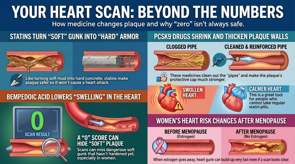
Lipid Therapy, CAC, and Sex Differences
Mechanistic and Clinical Differences Between PCSK9 Inhibitors, Statins, and Bempedoic Acid in Coronary Artery Calcium Progression, Plaque Biology, and Sex-Specific Cardiovascular Risk
The Evolution of the Atherosclerotic Paradigm
Storicamente, aterosclerosiL'aterosclerosi è la malattia responsabile della maggior parte degli attacchi cardiaci e di molti ictus. Le particelle di colesterolo rimangono intrappolate nella parete di un'arteria, il corpo invia cellule immunitarie per ripulire e, nel corso degli anni, questo accumulo si indurisce trasformandosi in placca. was conceptualized as a simplistic, linear process of colesteroloIl colesterolo è una sostanza cerosa di cui il corpo ha bisogno. Entra nelle pareti cellulari, negli ormoni, nella vitamina D e nella bile che digerisce il cibo. Moriresti senza di esso. accumulation within the arterial wall leading to mechanical obstruction of blood flow. Contemporary research has fundamentally shifted this understanding toward an immunoinflammatory paradigm. Atherosclerosis is now characterized as a chronic inflammatory disease, initiated by the subendothelial deposition of apolipoproteinaUn'apolipoproteina è una proteina legata a una particella di trasporto dei grassi nel sangue. I grassi e l'acqua non si mescolano, quindi queste proteiche agiscono come un involucro che consente ai grassi di viaggiare in sicurezza attraverso il flusso sanguigno. B (apoBL'ApoB è una proteina che si trova sulla superficie di ogni particella di colesterolo che può depositarsi nella parete arteriosa e causare la formazione di placca. Ognuna di queste particelle trasporta esattamente una molecola di ApoB.contenente lipoproteineUna lipoproteina è una piccola struttura che trasporta grassi e colesterolo attraverso il flusso sanguigno. Poiché i grassi non si dissolvono in acqua, hanno bisogno di un rivestimento proteico per viaggiare. such as low-density lipoprotein (LDLL'LDL, o lipoproteina a bassa densità, è la principale particella che trasporta il colesterolo nel sangue ed è la principale a bloccarsi nelle pareti delle arterie.), which undergo oxidative modification (ox-LDL) to trigger disfunzione endotelialeLa disfunzione endoteliale si verifica quando quel sottile rivestimento smette di fare bene il suo lavoro. I vasi non si dilatano adeguatamente e la barriera diventa più permeabile. and immune activation.[1,2]
In this framework, disease progression is not merely a matter of volume della placcaIl volume della placca è la quantità fisica totale di placca in un tratto di arteria, misurata in millimetri cubi. but of placcaLa placca è l'accumulo di colesterolo, cellule immunitarie, tessuto cicatriziale e calcio all'interno della parete di un'arteria. composition and stability. The journey from initial endothelial injury to a life-threatening infarto miocardicoVedi Infarto miocardico per la voce completa. involves a complex interplay of lipid deposition, macrofagoUn macrofago è una grande cellula immunitaria che inghiotte detriti e invasori. Il nome significa letteralmente "grande mangiatore"." infiltration, phenotypic switching of vascular cellule muscolari lisceLe cellule della muscolatura liscia costituiscono lo strato medio di un'arteria e controllano quanto il vaso si contrae o si rilassa. (VSMCs), and the eventual formation of a centro necroticoIl nucleo necrotico è il centro morto e poltaceo di una placca avanzata, costituito da cellule immunitarie che hanno ingerito colesterolo intrappolato e sono poi morte sul posto..[1,3] The final clinical manifestation—whether stable anginaL'angina è un fastidio al petto che si verifica quando il muscolo cardiaco non riceve abbastanza ossigeno. Le persone la descrivono come pressione, costrizione, oppressione o bruciore, e può estendersi al braccio, al collo o alla mascella. or an sindrome coronarica acutaLa sindrome coronarica acuta (SCA) è il termine generale per qualsiasi improvviso calo del flusso sanguigno al cuore — dall'angina instabile a un vero e proprio infarto miocardico — causato dalla rottura o dall'erosione improvvisa di una placca. (ACS)—is dictated by the mechanical integrity of the plaque’s cappuccio fibrosoIl cappuccio fibroso è il robusto strato di tessuto che riveste una placca, separandone il nucleo lipidico dal flusso sanguigno. and the biological state of its calcificazioneLa calcificazione è il processo in cui il calcio si deposita in una placca, rendendone una parte dura e simile a un osso..[4,9]
The standard non-invasive tool for assessing atherosclerotic burden has long been the punteggio di calcio coronaricoCoronary artery calcium is a measure of calcified plaque deposits in the walls of the coronary arteries, quantified by CT scan and expressed as an Agatston score; higher scores indicate greater cumulative plaque burden and predict future cardiovascular events. score. The Punteggio di AgatstonThe Agatston score is the specific formula used to turn a calcium CT scan into a single number, weighting each calcium deposit by how dense and how large it is. is one of the most powerful predictors of future cardiovascular events, yet its relationship with pharmacological intervention is more complex than it first appears.[5] The emergence of intensive lipid-lowering therapies—including statine ad alta intensitàA high-intensity statin is a dose expected to cut LDL by 50 percent or more — in practice, higher doses of atorvastatin or rosuvastatin., Inibitori di PCSK9A PCSK9 inhibitor is a medicine that blocks that cholesterol-destroying protein, leaving more docking ports available to clear particles from the blood., e acido bempedoicoL'acido bempedoico è una pillola colesterolemica che agisce nel fegato, in un punto appena precedente a quello in cui agiscono le statine.—has highlighted meaningful differences in how these agents influence plaque biology and radiographic calcification, with important implications for how CAC scores are interpreted in treated patients.[5,9,13]
The Statin–CAC Paradox: Plaque Stabilization and Calcium Densification
Observational cohorts have frequently shown that statinaUna statina rallenta l'enzima che il fegato usa per produrre il colesterolo. Il fegato risponde prelevando più colesterolo dal sangue, ed è qui che risiede il beneficio reale. users have higher Agatston scores over time compared with matched untreated individuals, even as their rate of clinical events falls markedly—a pattern sometimes called the “statin–CAC paradox.”[5,7] Randomized controlled trialUno studio clinico randomizzato e controllato assegna le persone a un gruppo di trattamento o a un gruppo di confronto puramente per caso, e successivamente segue entrambi i gruppi. data on this question are less consistent: heterogeneous CAC quantification methods and limited follow-up durations mean that RCTs have not demonstrated a statistically significant overall effect of statins on CAC progression.[7] The strongest and best-supported imaging signal is not blanket acceleration of calcification but a shift in the nature of the calcium itself: statins are associated with decreases in low-attenuation and fibro-fatty plaque components alongside greater progression of high-density calcium phenotypes.[5,6,45]
Mechanistic Basis of Plaque Stabilization
The “healing hypothesis” proposes that intensive ipolipemizzanteLa riduzione dei lipidi significa diminuire le particelle dannose trasportate dall'ApoB nel sangue, attraverso l'alimentazione, i farmaci o entrambi. transforms unstable, lipid-rich, inflamed plaques into more fibrotic and mechanically stable lesioniIn cardiologia, una lesione si riferisce a un'area distinta di placca aterosclerotica che restringe un'arteria coronaria, tipicamente descritta dalla percentuale di ostruzione luminale che provoca. L'articolo descrive quattro lesioni residue troppo piccole nel diametro del vaso per accogliere uno stent dopo che quella più critica è stata trattata..[5,8] As lipid and necrotic components are resolved, the vessel wall undergoes remodeling characterized by deposition of dense calcium that serves as structural reinforcement around the necrotic core, reducing mechanical stress on the fibrous cap and sequestering pro-thrombotic material from the bloodstream.[8]
Serial CCTA data from the PARADIGM studyPARADIGM (Progression of AtheRosclerotic PlAque DetermIned by Computed TomoGraphic Angiography IMaging) is a serial CCTA registry study that tracked changes in coronary plaque composition over time, providing key evidence that statin therapy shifts plaques toward denser, more stable calcium phenotypes. support this model: statin therapy was associated with reductions in low-attenuation and fibro-fatty plaque and with greater progression of high-density placca calcificaLa placca calcificata è la parte indurita e ricca di calcio di una placca. Appare luminosa su una tomografia computerizzata (TC), che è ciò che misura una scansione del calcio., while the increase in overall calcified plaque volume was attenuated in lesions without lipid-rich components.[45] A 2025 serial noncontrast CT study similarly found a statin-associated shift toward denser calcium strata in asymptomatic individuals.[6] These findings provide a more precise characterization than simply stating that statins accelerate CAC progression.
In some patients, therefore, a rising Agatston score under effective lipid-lowering may partly reflect plaque maturation and densification rather than new aterogenesiL'aterogenesi è il processo graduale di formazione di una placca.. However, serial CAC alone cannot reliably distinguish healing from ongoing disease activity on an individual-patient basis, and this interpretation should not be generalized without supporting clinical context.[5]
Proposed Mechanisms: Vitamin K2 and Macrophage Pathways
Several mechanisms have been proposed to explain statin-associated calcium densification. One hypothesis involves inhibition of Vitamin K2Vitamin K2 (menaquinone) is a fat-soluble cofactor required to activate Matrix Gla-protein, which inhibits vascular calcification; statin therapy may reduce vitamin K2 bioavailability as a byproduct of mevalonate pathway suppression, potentially influencing how calcium is deposited in plaques. synthesis: Vitamin K2 is a cofactor for Matrix Gla-protein (MGP)Matrix Gla-protein is a vitamin K–dependent protein secreted by vascular smooth muscle cells that acts as a physiological inhibitor of arterial calcification; it requires carboxylation by vitamin K2 to be active., a physiological inhibitor of vascular calcification, and reduced Vitamin K2 bioavailability could theoretically remove a brake on the calcification process.[7,8] Macrophage-mediated pathways that normally inhibit calcium deposition may also be suppressed. These remain mechanistic hypotheses drawn largely from preclinical work; they have not been established as dominant pathways in randomized human studies, and the statin-calcification literature explicitly frames this biology as still-disputed.[7,8]
Tabella 1: Plaque Characteristics: Untreated Progression vs. Statin-Associated Remodeling
| Funzionalità | Untreated Disease Progression | Statin-Associated Remodeling |
| Plaque Composition | High lipid content, large necrotic core | Fibrotic, reduced lipid-rich components |
| Calcium Phenotype | Spotty, low-density microcalcificazioneMicroscopic calcium deposits within atherosclerotic plaque that fall below the resolution threshold of conventional CT; unlike dense macrocalcification, microcalcifications can generate mechanical stress within the fibrous cap and increase plaque rupture susceptibility. | Associated shift toward denser, more coalescent calcium strata |
| Fibrous Cap | Thin, inflamed, rupture-prone | Thickened, stable, reduced mechanical stress |
| Agatston Score Trend | Rising due to new lesion formation | May rise due to calcium densification; requires careful clinical interpretation |
| Clinical Risk | Highly elevated | Significantly reduced despite possible punteggio di calcioUn punteggio del calcio (punteggio del calcio delle arterie coronarie) è un valore ricavato da una TAC che quantifica la quantità totale di placca calcificata nelle arterie coronarie; un punteggio pari a zero indica l'assenza di placca calcificata rilevabile, mentre punteggi più alti riflettono un maggiore carico di placca e un rischio cardiovascolare elevato. rise |
PCSK9 Inhibitors: Plaque Regression and Fibrous Cap Restoration on Background Statin Therapy
PCSK9La PCSK9 è una proteina prodotta dal fegato che distrugge i punti di attacco utilizzati dal fegato per rimuovere il colesterolo dal sangue. inhibitors such as evolocumabL'evolocumab è un farmaco iniettabile per il colesterolo appartenente alla famiglia degli inibitori della PCSK9, solitamente somministrato ogni due o quattro settimane. e alirocumabAlirocumab, sold as Praluent, is an injectable antibody that blocks PCSK9, given every two to four weeks. achieve profound reductions in LDL-C by preventing hepatic degradation of low-density lipoprotein receptors (LDLRLDLR è il gene che costruisce il recettore delle LDL, il punto di attracco che il fegato utilizza per rimuovere le particelle di colesterolo dalla circolazione.).[10,11] The principal plaque-imaging trials demonstrated meaningful regression of ateromaL'ateroma è un altro termine per indicare il deposito di grasso all'interno della parete di un'arteria, essenzialmente un sinonimo di placca, usato più spesso nella scrittura di ricerca. volume and improvement in plaque morphology incremental to background statin therapy. Throughout this section it is important to note that GLAGOVGLAGOV added a PCSK9 inhibitor to statin therapy and measured coronary plaque with intravascular ultrasound before and after., HUYGENSHUYGENS used optical coherence tomography — a very high-resolution imaging catheter — to see whether a PCSK9 inhibitor changed plaque structure after a heart attack., e PACMAN-AMIPACMAN-AMI gave a PCSK9 inhibitor to patients immediately after a heart attack and imaged their non-culprit arteries with three different catheter techniques. all studied PCSK9 inhibition added to statins; their findings cannot be straightforwardly attributed to PCSK9 inhibition as a standalone drug-class effect. Direct evidence on CAC progression with PCSK9 inhibitors is limited and less mature than plaque-regression evidence from IVUS and OCT trials; available data include a small randomized secondary analysis of alirocumab-plus-statin therapy showing attenuated CAC progression versus standard statin-based care, but no large dedicated CAC progression trial comparable to GLAGOV or HUYGENS has been reported.[14,15]
Plaque Regression and Fibrous Cap Thickening
The GLAGOV trial demonstrated that evolocumab added to statin therapy produced significant regression of coronary atheroma volume: a −0.95% nominal change in volume percentuale di ateromaPercent atheroma volume, or PAV, is the share of an artery segment taken up by plaque rather than open channel. (PAV) versus a +0.05% increase in the placeboUn placebo è un trattamento fittizio — una pillola di zucchero o un'iniezione di soluzione fisiologica — somministrato affinché i ricercatori possano capire cosa fa realmente un farmaco vero. group (P<0.001), with 64.3% of evolocumab-treated patients demonstrating PAV regression versus 47.3% in the placebo group (P<0.001). Mean achieved LDL-C was 36.6 mg/dL versus 93.0 mg/dL in the placebo group.[14]
The HUYGENS trial, using serial tomografia a coerenza otticaLa tomografia a coerenza ottica, o OCT, inserisce una sonda basata sulla luce in un'arteria coronaria. Offre un livello di dettaglio circa dieci volte superiore rispetto all'ecografia. (OCT) following acute myocardial infarction, showed that evolocumab produced significantly greater minimum fibrous cap thickness increase (+42.7 μm vs. +21.5 μm; P=0.015) and maximum lipid arc reduction (−57.5° vs. −31.4°; P=0.04) compared with placebo.[15]
The PACMAN-AMI trial demonstrated that alirocumab initiated within 24 hours of acute myocardial infarction produced significantly greater regression in percent atheroma volume (PAV −2.13% vs. −0.92%, P<0.001) and greater reduction in normalized total atheroma volume (−26.12 mm³ vs. −14.97 mm³), as well as greater fibrous cap thickening, compared with statin monotherapy.[11]
Vascular Biology: Established and Emerging Signals
Preclinical studies have identified potential LDLR-independent effects of PCSK9 on atherogenesis, including inhibition of monocyte adhesion to stimulated human coronary arteriaUn'arteria è un vaso sanguigno che trasporta il sangue dal cuore al resto del corpo. cellule endotelialiIl sottile strato di cellule che riveste la superficie interna di tutti i vasi sanguigni; esse regolano il tono vascolare, prevengono la coagulazione e controllano il passaggio delle sostanze nella parete arteriosa — e la loro disfunzione è un passo iniziale e critico nell'aterosclerosi. (HCAECs) via the VCAM-1La VCAM-1 è una molecola adesiva che compare sul rivestimento di un vaso infiammato e cattura i globuli bianchi di passaggio in modo che possano penetrare nella parete. e ICAM-1L'ICAM-1 è una molecola che compare sulla superficie del rivestimento dei vasi sanguigni e agisce come il Velcro, catturando le cellule immunitarie di passaggio. pathways.[12,16] In large cardiovascular outcome trials, however, PCSK9 inhibition has generally shown minimal change in hsCRP, indicating that the clinically established primary mechanism of benefit is profound apoB/LDL lowering rather than a systemic anti-inflammatory effect. Pleiotropic or local vascular effects suggested by preclinical work may contribute, but this should not be overstated given the largely neutral hsCRP signal in human trial data.[17]
Lipoprotein(a): An Important Independent Risk Factor
A meaningful advantage of PCSK9 inhibitors is their ability to lower Lipoproteina(a)La lipoproteina(a), scritta Lp(a) e pronunciata "L-P-piccola-a", è una particella simile alle LDL con una proteina extra appiccicosa attaccata. [Lp(a)], an independent and genetically determined cardiovascular fattore di rischioUn fattore di rischio è qualcosa che aumenta la probabilità di sviluppare una malattia: particelle di colesterolo alto, pressione alta, fumo, diabete, storia familiare. largely unaffected by statin therapy.[10] PCSK9 inhibitors reduce Lp(a) by approximately 25–27% (median 26.9% in the FourierFOURIER ha testato l'evolocumab, un inibitore di PCSK9, in pazienti che avevano già una malattia cardiovascolare ed erano in trattamento con statine. evolocumab cohort). The relationship between Lp(a) levels and CAC burden is inconsistent across studies; Lp(a) and CAC appear to be independently informative risk markers rather than tightly correlated measures. Clinicians should therefore distinguish the robust evidence that elevated Lp(a) is an independent ASCVD risk factor from the weaker and less consistent evidence that Lp(a) directly predicts CAC burden.[10,17]
Bempedoic Acid: Proven Outcomes, Limited Plaque Imaging Evidence
Bempedoic acid is an oral ATP citrate lyase (ACL) inhibitor that acts upstream of Idrossi-metilglutaril-CoA reduttasiHMG-CoA reductase is the rate-limiting enzyme in the liver's cholesterol biosynthetic (mevalonate) pathway; statins work by competitively blocking it, reducing the liver's own cholesterol production and prompting it to pull more LDL out of the bloodstream. in the cholesterol biosynthesis pathway.[19,23] As a profamacoA prodrug is a pharmacologically inactive compound that is converted into its active form by metabolic processes after administration; bempedoic acid is a prodrug activated specifically in the liver, which is why it avoids causing muscle side effects seen with statins., it is converted into its active metabolite, bempedoyl-CoA, by the enzyme very long-chain acyl-CoA synthetase 1 (ACSVL1). This activating enzyme is expressed in the liver but is absent in skeletal muscle, providing the mechanistic basis for the drug’s low incidence of muscle-related adverse effects and making it a valuable option for statin-intolerant patients.[20,23]
Cardiovascular Outcomes: The CLEAR Trials
Il Risultati CLEARCLEAR Outcomes was a large trial that tested bempedoic acid in people who couldn't tolerate statins, to see whether it lowered heart attack and stroke risk the way statins do. trial established clinical efficacy in high-risk, statin-intolerant patients.[21] Over a median follow-up of 3.4 years, bempedoic acid achieved a 21.1% placebo-corrected reduction in LDL-C and a statistically significant 13% rischio relativoIl rischio relativo confronta due gruppi: questo gruppo ha avuto il 30 percento in meno di attacchi cardiaci rispetto a quell'altro gruppo. reduction in the primary MACE-4MACE-4 (Major Adverse Cardiovascular Events, 4-component) is a composite clinical endpoint comprising cardiovascular death, non-fatal myocardial infarction, non-fatal stroke, and coronary revascularization, used as the primary endpoint in the CLEAR Outcomes trial. endpoint [HR 0.87, 95% CI 0.79–0.96; P=0.004].[21] A prespecified total-event analysis demonstrated a 20% reduction in total MACE-4 events [HR 0.80, 95% CI 0.72–0.89; P<0.001].[22]
Table 2: CLEAR Outcomes Trial: Cardiovascular Endpoint Summary (Nissen et al., NEJM 2023; JAMA 2024)
| Outcome (CLEAR Outcomes) | HR | 95% CI |
| MACE-4 — Primary Endpoint | 0.87 | 0.79–0.96 (P = 0.004) |
| MACE-3 (CV Death / MI / IctusUn ictus si verifica quando il flusso sanguigno verso una parte del cervello si interrompe, a causa di un'ostruzione o di un'emorragia.) | 0.85 | 0.76–0.96 |
| Fatal or Nonfatal MI | 0.77 | 0.66–0.91 |
| Coronarica RevascularizationRevascularization is a medical or surgical procedure—such as coronary artery bypass grafting or percutaneous coronary intervention—performed to restore blood flow through a blocked or narrowed coronary artery, addressing the physical obstruction rather than the underlying atherogenic process. | 0.81 | 0.72–0.92 |
| Total MACE-4 Events (prespecified) | 0.80 | 0.72–0.89 (P < 0.001) |
Inflammation, Mechanism, and Imaging Evidence
A distinguishing feature of bempedoic acid is its consistent reduction of hsCRP: a 22% placebo-corrected median reduction was observed at 6 months in CLEAR Outcomes.[19,21] Activation of the AMP-activated proteinaLe proteine sono il nutriente che il corpo utilizza per costruire e riparare muscoli e tessuti. kinase (AMPK) pathway has been proposed as a contributing cellular mechanism, based primarily on preclinical and mechanistic review data.[20,23] More recent work suggests the drug’s biology may be more complex than initially characterized, with preliminary evidence of potential direct PPARα activation; these mechanistic questions remain under active investigation and should not be presented as settled biology.
Bempedoic acid has proven outcomes benefit and hsCRP lowering in statin-intolerant patients, but its direct effects on coronary plaque morphology remain largely unproven pending dedicated imaging trials.[24] The only published plaque-imaging evidence is a single case report describing shrinkage of placca a bassa attenuazioneLa placca a bassa attenuazione è la placca più scura e grassa in assoluto su una scansione TC: abbastanza morbida da permettere ai raggi X di attraversarla facilmente. with long-term bempedoic acid monotherapy—a hypothesis-generating observation, not proof of a plaque-regression class effect. A phase 3 imaging trial registered in 2026 evaluates the combination of bempedoic acid, ezetimibeL'ezetimibe è una pillola che impedisce all'intestino di assorbire il colesterolo., and high-intensity statin therapy on coronary plaque; its design tests triple combination therapy rather than bempedoic acid in isolation, and results have not yet been reported.
Limitations and Blind Spots of CAC = 0: Moving Beyond a Binary Interpretation
The observation that a CAC score of 0 predicts very low rischio cardiovascolare a 10 anniTen-year cardiovascular risk is a calculated probability—expressed as a percentage—that an individual will experience a major cardiovascular event such as a heart attack or stroke within the next decade, estimated using validated equations that factor in age, sex, blood pressure, cholesterol, diabetes status, and smoking; current guidelines reserve primary-prevention aspirin consideration for adu…—the “power of zero”—has established CAC as a useful gatekeeper in risk stratification.[25,27,29] CAC = 0 is a genuinely powerful short-term negative risk marker in most clinical settings. However, it has important and well-characterized blind spots that require careful interpretation, particularly in symptomatic patients, younger individuals, and women in mid-life.[30]
Non-Calcified Plaque: The Underdetected Component
A CAC score of 0 does not exclude malattia coronaricaLa malattia coronarica è l'accumulo di placca nelle arterie che alimentano il muscolo cardiaco.. Placca non calcificaNon-calcified plaque is the soft, fatty portion of a plaque that has not hardened with calcium. It shows up dark on a CT scan. (NCP), which is lipid-rich and highly vulnerable to rupture, is entirely invisible on non-contrast CT.[25,28] Studies using CCTA have shown that approximately 15–17% of low-to-intermediate-risk individuals with CAC = 0 have detectable atherosclerosis, with roughly 3.5% harboring obstructive disease (>50% stenosiLa stenosi è un restringimento, solitamente descritto come percentuale, come un blocco del 70%.) despite a punteggio di calcio pari a zeroA zero calcium score means a CT scan found no hardened plaque in your heart's arteries at all..[28,30]
In a diverse symptomatic Montefiore cohort analyzed by Rozanski et al., a zero CAC score retained very high negative predictive value for obstructive coronary disease and near-term adverse events, and only a small percentage of CAC = 0 patients—approximately 7%—had detectable placca non calcificaDepositi aterosclerotici all'interno delle pareti arteriose che non hanno ancora subito calcificazione; talvolta chiamate placche 'molle', queste lesioni sono ricche di lipidi e strutturalmente instabili, il che le rende più inclini alla rottura e alla trombosi acuta rispetto alla placca calcificata. on CCTA.[30] Separate prospective data from stable-chest-pain populations likewise show very low MACE event rates when CAC = 0, though obstructive CAD is not completely excluded. These data together confirm that CAC = 0 is a genuinely powerful short-term reassurance signal, while still leaving a clinically meaningful minority with non-calcified plaque that calcium scoring cannot detect.
The mechanisms of clinical events in individuals with CAC = 0 typically involve rupture or erosion of non-calcified lesions, systemic infiammazioneL'infiammazione è la risposta del sistema immunitario a una lesione o a qualcosa che esso tratta come un aggressore. Porta gonfiore, calore e cellule di pulizia., or coronary disfunzione microvascolareLa disfunzione microvascolare è una malattia dei vasi sanguigni più piccoli del cuore, troppo piccoli per essere visti su qualsiasi angiografia..[27,31] Plaque erosionPlaque erosion is when the lining over a plaque simply wears away and a clot forms, without the cap tearing open.—disruption of the endothelial surface over a lipid-rich non-calcified plaque—is particularly common in women and younger individuals.[27]
Prevalence Estimates Depend Critically on Cohort and Method
Nel SCAPISSCAPIS (Swedish CArdioPulmonary bioImage Study) è un ampio studio di imaging di popolazione su oltre 25.000 adulti svedesi di età compresa tra 50 e 64 anni senza nota cardiopatia ischemica, che ha utilizzato l'angio-TC coronarica per rilevare l'aterosclerosi nel 42% dei partecipanti e una stenosi significativa in circa il 5%. (Swedish Cardiopulmonary Bioimage Study), among middle-aged adults from the general population with CAC = 0, approximately 5.5% had any coronary plaque detectable on CCTA and 0.4% had significant stenosis.[29] The frequently cited 9.2% plaque-prevalence figure from SCAPIS applies specifically to the subgroup with CAC = 0 and intermediate 10-year ASCVD risk—an important distinction, because applying this conditional estimate to the entire CAC-zero general population would materially overstate disease prevalence.[29]
AI-QCT registries of symptomatic patients referred for clinical CCTA report substantially higher plaque detection rates in CAC = 0 individuals—ranging from approximately 54% to 95% in some selected cohorts.[26] These figures are not generalizable to screening populations; they reflect the higher probabilità pre-testThe estimated likelihood that a patient has a particular condition before a diagnostic test is performed, based on symptoms, risk factors, and clinical context; in CAC interpretation, a symptomatic patient has a higher pre-test probability of coronary disease, so a zero score still leaves meaningful residual risk. of clinical referral cohorts as well as variation in AI-QCT protocols, thresholds, and definitions across studies. Prevalence estimates from symptomatic, asymptomatic, general-population, and AI-QCT registry studies should not be combined without careful population-specific stratification.[30]
Table 3: Prevalence of Non-Calcified Plaque in CAC = 0 Patients: Context-Dependent Estimates Across Study Populations
| Studio / Coorte | Popolazione | Key Finding for CAC = 0 Patients |
| Cheng et al. | Low to intermediate risk, asymptomatic | ~16% with detectable non-calcified plaque on CCTA |
| Rubinshtein et al. | Symptomatic | ~14.5% with plaque; ~3% with obstructive disease |
| Rozanski et al. (JCCT 2023) | Symptomatic stable chest pain, diverse population | ~7% NCP on CCTA; very high NPV for obstructive disease and near-term events; diverse symptomatic Montefiore cohort |
| SCAPIS (Bergström et al.) | Middle-aged general population | ~5.5% any plaque overall; ~9.2% specifically in the CAC=0 + intermediate-risk subgroup [29] |
| AI-QCT registries (selected clinical CCTA cohorts) | Symptomatic / high pre-test probability | 54–95%: highly dependent on cohort selection, referral indication, and AI-QCT protocol. Not generalizable to screening populations [26] |
Sex-Specific Cardiovascular Risk and Plaque Biology
Women exhibit important differences in the presentation and pathophysiology of atherosclerotic malattia cardiovascolareLe malattie cardiovascolari sono il termine generico per indicare i problemi al cuore e ai vasi sanguigni, tra cui infarti, ictus e ostruzione delle arterie delle gambe. (ASCVD) compared with men. Women generally develop clinically manifest coronary artery disease approximately a decade later than men, a phenomenon largely attributed to the vasoprotective effects of endogenous estrogenoEstrogen is a hormone, present at much higher levels in women before menopause, that affects blood vessels, cholesterol, and bone..[37,39]
The Protective Role of Estrogen and the Menopause Transition
Estrogen maintains vascular health through multiple pathways: it promotes endothelium-dependent vasodilation by stimulating ossido nitricoL'ossido nitrico è un gas prodotto dal rivestimento dei vasi sanguigni per dire al vaso di rilassarsi e dilatarsi. (NO) and prostacyclin synthesis, inhibits vascular smooth muscle cell contraction and proliferation, and exerts anti-inflammatory effects on the vessel wall.[35,38] Estrogen also maintains more favorable profili lipidiciUn pannello di esami del sangue che misura il colesterolo totale, il colesterolo LDL, il colesterolo HDL e i trigliceridi, utilizzato per valutare il rischio cardiovascolare e monitorare l'effetto di interventi dietetici o farmacologici. throughout the premenopausal years.[35]
Il menopausaMenopause is when a woman's periods stop permanently, usually around age 51, as estrogen levels fall. transition is associated with a precipitous decline in circulating estradiol, triggering accelerated invecchiamento vascolareIl progressivo deterioramento strutturale e funzionale delle arterie nel tempo, caratterizzato da perdita di elasticità, aumentata rigidità e accumulo di danni microscopici che rendono le pareti arteriose più suscettibili al deposito di lipidi e all'infiammazione cronica.. This shift produces measurable reductions in funzione endotelialeLa capacità del rivestimento interno dei vasi sanguigni di regolare il tono vascolare, l'infiammazione e la coagulazione; le cellule endoteliali sane rilasciano ossido nitrico per mantenere le arterie rilassate e resistenti alla formazione di placche. as assessed by dilatazione mediata dal flussoLa dilatazione mediata dal flusso è una misurazione ecografica non invasiva di quanto un'arteria di conduttura — tipicamente l'arteria brachiale — si dilata in risposta a un aumento del flusso sanguigno, fungendo da marcatore della segnalazione dell'ossido nitrico endoteliale e della funzione endoteliale., increased systemic stress ossidativoLo stress ossidativo è uno squilibrio tra molecole reattive dannose e la capacità dell'organismo di neutralizzarle., and progressive worsening of rigidezza arteriosaLa rigidità arteriosa è una misura di quanto la parete di un'arteria resiste all'espansione a ogni impulso di sangue; aumenta con l'età a causa della perdita di elastina e dell'accumulo di collagene, e si manifesta clinicamente con un aumento della pressione arteriosa sistolica accompagnata da una pressione arteriosa diastolica in calo o stabile dopo i 60 anni circa., pressione sanguignaLa pressione sanguigna è la forza del sangue che preme contro le pareti delle arterie. È scritta come due numeri, come 120/80. Il numero superiore è la pressione quando il cuore si contrae, quello inferiore quando si rilassa., e dislipidemia aterogenicaAtherogenic dyslipidemia is a lipid pattern characterized by elevated triglycerides, low HDL cholesterol, and an increased proportion of small, dense LDL particles; it is commonly seen with insulin resistance, visceral obesity, and sedentary behavior, and is associated with accelerated atherosclerosis..[34,36] FMD in late postmenopausal women is approximately 50% lower than in premenopausal women of similar age, with the most significant decline occurring during the perimenopausal transition.[34]
Risk Acceleration After Menopause
As women enter their 60s, cardiovascular risk accelerates markedly, with CVD incidence approaching that of age-matched men.[37,38] Longitudinal data from the Healthy Women Study show that among postmenopausal women with a baseline CAC of 0, approximately 33% develop new, incident calcification over a subsequent 6-year period, with premenopausal LDL-C and HDL-C as strong predictors.[39]
Early Menopause and Long-Term Risk
Women who experience early menopause (EM, defined as menopause before age 45) face substantially greater long-term cardiovascular risks. In the Studio Multietnico dell'Aterosclerosi (MESA)A large prospective cohort study of adults initially free of cardiovascular disease that has provided foundational data on coronary artery calcium scoring, demonstrating a strong graded association between CAC burden and future coronary events and validating the risk implications of a CAC score of zero., more than half of postmenopausal women with early menopause had a baseline CAC score of 0.[40] Among those women with CAC = 0, 10-year ASCVD incidence was low-to-borderline, yet those with early menopause had significantly higher 15-year risk than those without (adjusted HR 1.96, 95% CI 1.26–3.04).[40] This illustrates the core diagnostic problem: the same CAC = 0 result that appears reassuring at 10 years can mask a substantially elevated long-term risk trajectory in women with early menopause, driven by prolonged estrogen deficiency that accelerates subclinical plaque accumulation after the initial scan.
Systematic meta-analyses confirm that early menopause is independently associated with a 50–60% increase in lifetime malattia coronaricaLa cardiopatia coronarica è il restringimento o l'occlusione delle arterie che forniscono sangue al muscolo cardiaco, causato dall'accumulo di placca aterosclerotica; è la principale causa di infarto e morte cardiaca in tutto il mondo. risk, an effect only partially attenuated by traditional risk factor adjustment.[41]
Sex Differences in Plaque Morphology and Detection
Women generally have lower absolute CAC burden than men and, in several cohorts, lower absolute carico di placcaIl carico di placca è la quantità totale di placca nelle arterie, ovunque, e non solo nel punto peggiore. as well; however, recent AI-QCT data suggest that the relative cardiovascular risk associated with a given increase in plaque burden is greater in women than in men.[27,33] Women are significantly more likely to develop non-calcified, placche ricche di lipidiAn atherosclerotic lesion whose core is dominated by cholesterol esters and inflammatory lipids rather than calcium or fibrous tissue; the article notes that such plaques are highly responsive to intensive treatment and that the dramatic 65-percentage-point regression at the diagonal branch origin is consistent with reversal of a lipid-rich lesion. prone to endothelial erosion rather than fibrous cap rupture—yet both mechanisms trigger MACE. Because CAC scoring captures only the calcified component of disease, it may be a less complete risk indicator in women, who often carry a greater proportion of their total plaque burden in non-calcified form. Women with any detectable CAC face disproportionately higher relative cardiovascular risk: CAC Consortium data show approximately 1.3-fold higher hazard of cardiovascular death in women than in men at equivalent CAC burden.[27]
In their 40s and 50s, women may accumulate a substantial burden of lipid-rich NCP that will only become radiographically detectable as calcium after the post-menopausal acceleration of vascular calcification.[27,33] This creates a diagnostic window during midlife when traditional risk factors are rising and inflammatory plaque is accumulating, yet CAC remains at zero. The absence of calcium may be an especially incomplete risk signal precisely in this population—younger, perimenopausal women—where near-term rischio assolutoIl rischio assoluto è la probabilità reale che ti accada qualcosa, espressa in percentuale. Se il tuo rischio assoluto di infarto nei prossimi dieci anni è del 12%, significa che circa 12 persone su 100 con caratteristiche simili alle tue ne subirebbero uno. appears lowest but medium-term risk may be substantially higher than the calcium score suggests.
Table 4: Sex-Specific Cardiovascular Risk per Unit Plaque Volume: Selected Findings from the CONFIRM2 RegistryUn ampio registro multinazionale comprendente oltre 6.000 pazienti, nel quale sono state valutate le caratteristiche delle placche coronariche quantificate tramite intelligenza artificiale (IA) in relazione alla loro capacità di prevedere eventi cardiovascolari avversi maggiori; il registro ha dimostrato che l’analisi delle placche derivata dall’IA ha migliorato l’area sotto la curva ROC per la previsione degli eventi da 0,62 a 0,75 rispetto ai metodi standard. (Choi et al., Circ Cardiovasc Imaging 2025 [33])
| Finding — CONFIRM2 Registry (Choi et al. 2025 [33]) | Women | Men |
| Relative risk increase per 50 mm³ total plaque volume | +17.7% (RR ≈1.18) | +5.3% (RR ≈1.05) |
| Pattern across NCP, calcified plaque, and high-risk plaque subtypes | Larger relative risk increments across all subtypes | Smaller relative risk increments across all subtypes |
| Principal published conclusion | Substantially higher relative cardiovascular risk per unit plaque | Lower relative risk per equivalent plaque unit |
The total plaque volume risk increment (+17.7% per 50 mm³ in women vs +5.3% in men) is reported from the published CONFIRM2 results. The directional pattern across non-calcified, calcified, and high-risk plaque subtypes (larger increments in women) is the principal published conclusion of that paper; exact subtype hazard ratiosIl rapporto di rischio mette a confronto la frequenza con cui si verificano gli eventi in due gruppi. Un rapporto pari a 0,75 indica che nel gruppo trattato gli eventi si sono verificati a una frequenza pari a tre quarti rispetto al gruppo di controllo. should be transcribed from the published results table before any submission requiring those specific values.
Advanced Imaging: The Role of AI-Enhanced CCTA
Artificial intelligence–based quantitative CT (AI-QCT) represents a meaningful advance in non-invasive cardiovascular risk assessment. By automating measurement of total plaque burden—including calcified, non-calcified, and low-attenuation components—AI-QCT provides a more comprehensive characterization of disease than traditional CAC scoring alone.[26,33] These tools are rapidly evolving, but broad standardization across institutions and vendors, external validation in diverse populations, sex-specific risk thresholds, and integration into clinical guidelines are still ongoing processes. AI-QCT is best understood as a promising emerging modality rather than a fully standardized replacement for current CAC and CCTA frameworks.
AI-QCT, CAC, and Reclassification in Symptomatic Cohorts
In symptomatic patients referred for clinical CCTA, research demonstrates only moderate categorical agreement between traditional CAC scores and AI-derived total plaque burden, with discordanzaVedere la discordanza dell'ApoB per la voce completa. particularly pronounced in women.[42] AI-QCT frequently identifies significant NCP in patients with zero Agatston scores in these referral populations. Prevalence varies widely by cohort, scanner protocol, and AI-QCT algorithm, and figures from clinical CCTA registries should not be extrapolated to general screening populations. Nonetheless, the riclassificazioneNella valutazione del rischio cardiovascolare, la riclassificazione si riferisce al processo mediante il quale un esame aggiuntivo, come una scansione del CAC o la misurazione dell'ApoB, sposta un paziente da una categoria di rischio a un'altra, inducendo un cambiamento nelle decisioni terapeutiche che un semplice calcolatore del rischio standard non avrebbe attivato. is clinically meaningful for symptomatic patients in whom a zero calcium score might otherwise prematurely terminate the diagnostic workup, especially women with additional risk features.[26,30,42]
AI-QCT further enables quantification of high-risk plaque (HRP) features including low-attenuation plaque (LAP) and positive coronary remodeling. Data from the CONFIRM2 registry demonstrate that these features carry significantly higher relative risk in women than in men per unit of plaque volume, consistent with the clinical observation that women’s smaller-caliber coronary vessels may render each unit of high-risk plaque more hemodynamically consequential.[33]
Integrating Epicardial Adipose Tissue Analysis
Automated analysis of epicardial adipose tissue (EAT) volume and attenuation from routine calcium-scoring CT scans has been shown to significantly enhance MACE prediction, with particularly strong incremental value in women.[43] EAT is metabolically active and exerts paracrine pro-inflammatory and pro-atherogenic effects on adjacent arterie coronarieLe arterie coronarie sono i piccoli vasi che avvolgono l'esterno del cuore e nutrono il muscolo cardiaco stesso. through the secretion of cytokines and adipochineAdipokines are bioactive molecules — including leptin, adiponectin, TNF-α, and IL-6 — secreted by fat cells that act as hormones or signaling proteins to regulate metabolism, insulin sensitivity, inflammation, and energy balance throughout the body.,[32] and integrating quantitative EAT features with CAC scoring and conventional risk factors provides a more biologically comprehensive sex-specific risk assessment than calcified plaque metrics alone, though this approach requires further standardization and prospective validation before routine clinical implementation.[43]
An Integrated Framework for Therapeutic Mapping
A useful framework maps atherosclerosis progression through specific biological stages and identifies where different therapeutic classes exert their primary mechanistic influence. This mapping should be understood as directionally plausible based on available mechanistic and imaging data, not as a clinically proven stage-by-stage model validated by head-to-head drug-class trials.[1,4,9]
Stages of Plaque Development and Therapeutic Positioning
Stage 1 — Endothelial Activation: Endothelial dysfunction, reduced NO bioavailability, and ritenzione subendotelialeLa ritenzione subendoteliale è il processo mediante il quale le particelle lipoproteiche contenenti ApoB che hanno attraversato la barriera endoteliale si legano elettrostaticamente ai proteoglicani nell'intima arteriosa e non sono in grado di diffondere nuovamente nel flusso sanguigno; essa è considerata il primo passo non ridondante dell'aterosclerosi nell'ambito del modello di risposta alla ritenzione. of apoB-containing lipoproteins. This stage is substantially modulated by endogenous estrogen and is the proposed target of early PCSK9 inhibition based on preclinical evidence.[2,12]
Stage 2 — Early Non-Calcified Plaque: Macrophage cellula schiumosaUna cellula schiumosa è una cellula immunitaria che ha inglobato così tanto colesterolo intrappolato da gonfiarsi e apparire spumosa al microscopio. formation establishes the earliest atheromatous lesion. Bempedoic acid and statins reduce the circulating lipid pool available for subendothelial accumulation and dampen systemic inflammation.[1,20]
Stage 3 — Intermediate / Mixed Plaque: Extracellular lipid accumulation and microcalcifications emerge within necrotic core zones. High-intensity statins and PCSK9i (when added to statins) drive regressione della placcaPlaque regression means existing plaque actually gets smaller, rather than just growing more slowly. and fibrous cap maturation, with the strongest direct human trial evidence at this stage.[1,9,14]
Stage 4 — Fibrocalcific / Stable Plaque: Dense, coalescent calcification with a thick collagen-rich fibrous cap, representing the histopathological end-state of healed plaque, associated primarily with intensive statin therapy and the calcium densification process.[5,8,9]
Comparative Therapeutic Profile
Statins are best characterized as plaque stabilizers associated with calcium densification in observational and serial CCTA data. They have the strongest and broadest evidence base for reducing lipid-rich plaque and promoting transformation of vulnerable lesions toward denser, more stable phenotypes. A rising Agatston score under statin therapy should be interpreted in the context of documented densification and clinical improvement rather than as evidence of therapeutic failure.[5,7,8,45]
PCSK9 Inhibitors are best characterized as plaque regressionors and fibrous cap restorers when added to statin therapy in high-risk imaging cohorts. Their principal established mechanism of clinical benefit is profound apoB/LDL lowering, with meaningful incremental plaque regression and cap thickening beyond statins alone. Pleiotropic effectsIn pharmacology, pleiotropic effects are actions of a drug that go beyond its primary intended mechanism; for incretin therapies, pleiotropic effects include anti-inflammatory activity, endothelial protection, and blood-pressure reduction that are independent of the drugs' primary glucose-lowering and weight-loss actions. suggested by preclinical data have not translated into a clear systemic anti-inflammatory signal in human outcome trials.[11,12,14,15]
Bempedoic Acid functions as an upstream lipid-lowering and hsCRP-lowering agent with proven outcomes benefit in statin-intolerant patients. Its plaque-modification effects are mechanistically plausible but supported by very limited imaging data; robust serial plaque imaging evidence has not yet been reported.[19,20,21]
Clinical Implications and Guideline Directions
The integration of pharmacologic mechanisms, advanced imaging, and sex-specific plaque biology highlights important gaps in current clinical practice and supports a shift toward biologically informed, individualized risk assessment.
Interpreting CAC = 0 in Symptomatic and Mid-Life Women
The cumulative evidence supports a calibrated interpretation of CAC = 0: it is a powerful short-term negative risk marker that should not be dismissed, yet it should not automatically end risk assessment in younger or symptomatic women—especially in the presence of early menopause, strong storia familiareL'anamnesi familiare indica se i vostri familiari stretti hanno sviluppato malattie cardiache e a quale età è successo., diabeteIl diabete è una condizione in cui la glicemia rimane troppo alta, o perché il corpo produce troppa poca insulina o perché smette di rispondere all'insulina che produce., elevated Lp(a), or persistent inflammatory risk.[27,40,41] A CAC of 0 in a woman with these characteristics may correctly predict low 10-year event risk while substantially underestimating 15-year risk driven by ongoing non-calcified plaque accumulation and the biological effects of prolonged estrogen deficiency. In these settings, a CAC of 0 should invite further characterization of risk rather than conclude risk stratification.
A More Comprehensive Approach to Sex-Conscious Risk Assessment
A multi-modal, sex-conscious risk assessment framework incorporates several complementary elements.
Sex-specific risk factors—including age at menopause, duration of estrogen deficiency, and reproductive history—should be formally integrated into clinical risk discussions, as they carry independent prognostic weight beyond traditional Framingham-based variables.[37,41]
Advanced imaging via CCTA should be considered when CAC scoring may underestimate true atherosclerotic burden, particularly in symptomatic women with CAC = 0 and additional risk features. The 2024 ESC Guidelines for the Management of Chronic Coronary Syndromes recommend first-line noninvasive anatomic or functional imaging for suspected CCS, selected according to pretest likelihood, patient characteristics, and local expertise. Within that framework, CCTA is specifically preferred for ruling out obstructive CAD and identifying nonobstructive disease, and the guidelines explicitly incorporate age, sex, and symptom character into pretest likelihood models.[26,31,33]
Integrated biomarker-imaging models combining Lp(a), hsCRP, and emerging EAT fat-omics analysis represent a biologically more complete framework for sex-specific risk stratification. These approaches require further standardization and prospective validation before routine implementation.[43]
Limitations and Open Questions
The evidence reviewed here has several important limitations that must be recognized when applying these concepts clinically or in academic scholarship.
Most plaque-imaging evidence for PCSK9 inhibitors derives from studies of add-on therapy to background statins, not from clean mechanistic comparisons between drug classes. GLAGOV, HUYGENS, and PACMAN-AMI do not establish what PCSK9 inhibitors achieve independently of statins, and their imaging endpoints cannot be directly compared with statin-only or bempedoic acid-only imaging data.[11,14,15]
The bempedoic acid plaque-imaging literature currently consists of a single case report and one recently registered phase 3 imaging trial that has not yet reported results. Outcomes-based conclusions about bempedoic acid are well supported; morphologic plaque-imaging conclusions are not.[24]
AI-QCT plaque detection rates vary substantially by cohort selection, referral indication, and AI-QCT protocol. Plaque prevalence figures from symptomatic CCTA referral populations should not be extrapolated to general screening populations. Standardized thresholds, validated sex-specific risk cutoffs, and prospective outcome data for AI-QCT remain incomplete.[26,33,42]
The field still lacks clean head-to-head randomized trials designed to compare drug class effects on CAC progression or plaque morphology using sex-stratified pre-specified endpoints and the same imaging platform. The mechanistic pathways proposed for statin-associated calcium densification (Vitamin K2 inhibition, macrophage suppression) and for bempedoic acid’s anti-inflammatory effects (AMPK, possible PPARα activation) are supported primarily by preclinical evidence and remain incompletely validated in human studies.[7,8,20,23]
Conclusione
Intensive apoB-lowering therapy reduces cardiovascular risk across drug classes, but its imaging footprints differ in important ways. Statins have the strongest and longest-established evidence for reducing lipid-rich plaque and promoting a denser, more stable calcium phenotype; their association with rising Agatston scores is best understood as calcium densification rather than uncomplicated disease progression. PCSK9 inhibitors provide additional plaque regression, lipid arc reduction, and fibrous cap thickening when added to statins, with a primarily LDL-lowering mechanism driving outcome benefit rather than a systemic anti-inflammatory one. Bempedoic acid offers proven event reduction and meaningful hsCRP lowering in statin-intolerant patients, but its effects on coronary plaque morphology remain largely unproven pending dedicated imaging trials.
CAC remains a powerful marker of calcified plaque burden and near-term prognosis, but it incompletely captures non-calcified atherosclerosis—particularly in symptomatic patients and women in mid-life. CAC = 0 should be interpreted as one component of a biologically informed risk assessment rather than as a stand-alone arbiter of disease absence. In most clinical contexts it provides powerful short-term reassurance; in specific contexts—early menopause, symptomatic presentations, elevated Lp(a), family history, or persistent inflammatory risk—it should prompt further rather than conclude risk stratification.
For women specifically, the distinct biology of plaque formation—characterized by early lipid-rich non-calcified lesions, a sensitive period of post-menopausal risk acceleration, and significantly higher relative cardiovascular risk per unit of plaque volume—demands an individualized approach to imaging and lipid-lowering therapy. A sex-conscious, multi-modal risk assessment framework incorporating CCTA, sex-specific biomarcatoriUn biomarcatore è qualcosa di misurabile nel corpo che fornisce informazioni sulla salute o sulla malattia: un valore di laboratorio, il risultato di una scansione, la lettura della pressione sanguigna., and comprehensive clinical context offers the most evidence-consistent path toward equitable and effective cardiovascular disease prevention.
Riferimenti
- Libby P. Inflammation in atherosclerosis. Nature. 2002;420(6917):868-874. doi:10.1038/nature01323
- Han Y, Xu H, Yao X, et al. Molecular mechanisms and therapeutic progress in atherosclerosis: bridging immune inflammation and precision medicine. Front Immunol. 2026;16:1737662. Published 2026 Jan 5. doi:10.3389/fimmu.2025.1737662
- Moore KJ, Tabas I. Macrophages in the pathogenesis of atherosclerosis. Cell. 2011;145(3):341-355. doi:10.1016/j.cell.2011.04.005
- Papafaklis MI, Koros R, Tsigkas G, Karanasos A, Moulias A, Davlouros P. Reversal of Atherosclerotic Plaque Growth and Vulnerability: Effects of Lipid-Modifying and Anti-Inflammatory Therapeutic Agents. Biomedicines. 2024;12(11):2435. Published 2024 Oct 23. doi:10.3390/biomedicines12112435
- Khanna S, Nerlekar N, Bhat A. Reconciling Coronary Artery Calcification in the Lipid-Lowering Era. JACC Adv. 2026;5(1):102506. doi:10.1016/j.jacadv.2025.102506
- Giovannucci J, Shanbhag A, Hong W, et al. Impact of statins on progression of coronary artery calcium composition and density as assessed by noncontrast CT. Int J Cardiovasc Imaging. 2025;41(12):2481-2492. doi:10.1007/s10554-025-03561-0
- Shahraki MN, Jouabadi SM, Bos D, Stricker BH, Ahmadizar F. Statin Use and Coronary Artery Calcification: a Systematic Review and Meta-analysis of Observational Studies and Randomized Controlled Trials. Curr Atheroscler Rep. 2023;25(11):769-784. doi:10.1007/s11883-023-01151-w
- Kadoglou NP, Stasinopoulou M, Velidakis N, et al. The Complex Mechanisms and the Potential Effects of Statins on Vascular Calcification: A Narrative Review. Rev Cardiovasc Med. 2024;25(2):51. Published 2024 Jan 30. doi:10.31083/j.rcm2502051
- Papafaklis MI, Koros R, Tsigkas G, Karanasos A, Moulias A, Davlouros P. Reversal of Atherosclerotic Plaque Growth and Vulnerability: Effects of Lipid-Modifying and Anti-Inflammatory Therapeutic Agents. Biomedicines. 2024;12(11):2435. Published 2024 Oct 23. doi:10.3390/biomedicines12112435
- Wu Z, Gao L, Lin Z. Can proprotein convertase subtilisin/kexin type 9 (PCSK9) inhibitors regress coronary atherosclerotic plaque? A systematic review and meta-analysis. Am J Transl Res. 2023;15(1):452-465. Published 2023 Jan 15.
- Räber L, Ueki Y, Otsuka T, et al. Effect of Alirocumab Added to High-Intensity Statin Therapy on Coronary Atherosclerosis in Patients With Acute Myocardial Infarction: The PACMAN-AMI Randomized Clinical Trial. JAMA. 2022;327(18):1771-1781. doi:10.1001/jama.2022.5218
- Zulkapli R, Muid SA, Wang SM, Nawawi H. PCSK9 Inhibitors Reduce PCSK9 and Early Atherogenic Biomarkers in Stimulated Human Coronary Artery Endothelial Cells. Int J Mol Sci. 2023;24(6):5098. Published 2023 Mar 7. doi:10.3390/ijms24065098
- Ahmadi A, Argulian E, Leipsic J, Newby DE, Narula J. From Subclinical Atherosclerosis to Plaque Progression and Acute Coronary Events: JACC State-of-the-Art Review. J Am Coll Cardiol. 2019;74(12):1608-1617. doi:10.1016/j.jacc.2019.08.012
- Nicholls SJ, Puri R, Anderson T, et al. Effect of Evolocumab on Progression of Coronary Disease in Statin-Treated Patients: The GLAGOV Randomized Clinical Trial. JAMA. 2016;316(22):2373-2384. doi:10.1001/jama.2016.16951
- Nicholls SJ, Kataoka Y, Nissen SE, et al. Effect of Evolocumab on Coronary Plaque Phenotype and Burden in Statin-Treated Patients Following Myocardial Infarction. JACC Cardiovasc Imaging. 2022;15(7):1308-1321. doi:10.1016/j.jcmg.2022.03.002
- Barbieri L, Tumminello G, Fichtner I, et al. PCSK9 and Coronary Artery Plaque-New Opportunity or Red Herring?. Curr Atheroscler Rep. 2024;26(10):589-602. doi:10.1007/s11883-024-01230-6
- Pradhan AD, Aday AW, Rose LM, Ridker PM. Residual Inflammatory Risk on Treatment With PCSK9 Inhibition and Statin Therapy. Circulation. 2018;138(2):141-149. doi:10.1161/CIRCULATIONAHA.118.034645
- Chen H, Chen X. PCSK9 inhibitors for acute coronary syndrome: the era of early implementation. Front Cardiovasc Med. 2023;10:1138787. Published 2023 May 2. doi:10.3389/fcvm.2023.1138787
- Jia X, Virani SS. CLEAR Serenity Trial: More Clarity for the Future of Bempedoic Acid in Patients Unable to Take Statins?. J Am Heart Assoc. 2019;8(7):e012352. doi:10.1161/JAHA.119.012352
- Pinkosky SL, Newton RS, Day EA, et al. Liver-specific ATP-citrate lyase inhibition by bempedoic acid decreases LDL-C and attenuates atherosclerosis. Nat Commun. 2016;7:13457. Published 2016 Nov 28. doi:10.1038/ncomms13457
- Nissen SE, Lincoff AM, Brennan D, et al. Bempedoic Acid and Cardiovascular Outcomes in Statin-Intolerant Patients. N Engl J Med. 2023;388(15):1353-1364. doi:10.1056/NEJMoa2215024
- Nicholls SJ, Nelson AJ, Lincoff AM, et al. Impact of Bempedoic Acid on Total Cardiovascular Events: A Prespecified Analysis of the CLEAR Outcomes Randomized Clinical Trial. JAMA Cardiol. 2024;9(3):245-253. doi:10.1001/jamacardio.2023.5155
- Biolo G, Vinci P, Mangogna A, et al. Mechanism of action and therapeutic use of bempedoic acid in atherosclerosis and metabolic syndrome. Front Cardiovasc Med. 2022;9:1028355. Published 2022 Oct 28. doi:10.3389/fcvm.2022.1028355
- Korosoglou G, Giesen A, Geiss E, Stach K. Case report: Strong low-density-cholesterol reduction accompanied by shrinkage of low-attenuation coronary plaque during lipid-lowering treatment with bempedoic acid-serial evaluation by coronary computed tomography angiography. Front Cardiovasc Med. 2023;10:1203832. Published 2023 Aug 4. doi:10.3389/fcvm.2023.1203832
- Koulaouzidis G, Charisopoulou D, Jenkins PJ, Koulaouzidis A, McArthur T. Prevalence of noncalcified coronary plaque in patients with calcium score of 0: the silent enemy. Angiology. 2013;64(3):205-210. doi:10.1177/0003319712440618
- Khan NA, Wesbey III G, Cobb G, et al. Using AI-Quantitative CT to evaluate the relationship between coronary artery calcium and segment involvement scores in quantifying coronary plaque burden. Int J Cardiovasc Imaging. 2026;42(1):49-59. doi:10.1007/s10554-025-03569-6
- Shaw LJ, Min JK, Nasir K, et al. Sex differences in calcified plaque and long-term cardiovascular mortality: observations from the CAC Consortium. Eur Heart J. 2018;39(41):3727-3735. doi:10.1093/eurheartj/ehy534
- Cho I, Suh JW, Chang HJ, et al. Prevalence and prognostic implication of non-calcified plaque in asymptomatic population with coronary artery calcium score of zero. Korean Circ J. 2013;43(3):154-160. doi:10.4070/kcj.2013.43.3.154
- Bergström G, Persson M, Adiels M, et al. Prevalence of Subclinical Coronary Artery Atherosclerosis in the General Population. Circulation. 2021;144(12):916-929. doi:10.1161/CIRCULATIONAHA.121.055340
- Fattouh M, Kuno T, Pina P, et al. Interplay Between Zero CAC, Quantitative Plaque Analysis, and Adverse Events in a Diverse Patient Cohort. Circ Cardiovasc Imaging. 2023;16(8):e015236. doi:10.1161/CIRCIMAGING.123.015236
- Vrints C, Andreotti F, Koskinas KC, et al. 2024 ESC Guidelines for the management of chronic coronary syndromes. Eur Heart J. 2024;45(36):3415-3537. doi:10.1093/eurheartj/ehae177
- Antonopoulos AS, Sanna F, Sabharwal N, et al. Detecting human coronary inflammation by imaging perivascular fat. Sci Transl Med. 2017;9(398):eaal2658. doi:10.1126/scitranslmed.aal2658
- Feuchtner GM, Lacaita PG, Bax JJ, et al. AI-Quantitative CT Coronary Plaque Features Associate With a Higher Relative Risk in Women: CONFIRM2 Registry. Circ Cardiovasc Imaging. 2025;18(6):e018235. doi:10.1161/CIRCIMAGING.125.018235
- Moreau KL, Hildreth KL, Meditz AL, Deane KD, Kohrt WM. Endothelial function is impaired across the stages of the menopause transition in healthy women. J Clin Endocrinol Metab. 2012;97(12):4692-4700. doi:10.1210/jc.2012-2244
- Reslan OM, Khalil RA. Vascular effects of estrogenic menopausal hormone therapy. Rev Recent Clin Trials. 2012;7(1):47-70. doi:10.2174/157488712799363253
- Moreau KL, Hildreth KL, Klawitter J, Blatchford P, Kohrt WM. Decline in endothelial function across the menopause transition in healthy women is related to decreased estradiol and increased oxidative stress. Geroscience. 2020;42(6):1699-1714. doi:10.1007/s11357-020-00236-7
- Mishra T, Pasnoor DS, Gandhi M, et al. Interplay of menopause, coronary artery calcium score and cardiovascular disease risk. World J Cardiol. 2025;17(11):109627. doi:10.4330/wjc.v17.i11.109627
- SenthilKumar G, Katunaric B, Bordas-Murphy H, Sarvaideo J, Freed JK. Estrogen and the Vascular Endothelium: The Unanswered Questions. Endocrinology. 2023;164(6):bqad079. doi:10.1210/endocr/bqad079
- Kuller LH, Matthews KA, Edmundowicz D, Chang Y. Incident coronary artery calcium among postmenopausal women. Atherosclerosis. 2008;200(2):278-285. doi:10.1016/j.atherosclerosis.2007.12.057
- Chu JH, Michos ED, Ouyang P, et al. Coronary artery calcium and atherosclerotic cardiovascular disease risk in women with early menopause: The Multi-Ethnic Study of Atherosclerosis (MESA). Am J Prev Cardiol. 2022;11:100362. Published 2022 Jun 13. doi:10.1016/j.ajpc.2022.100362
- Muka T, Oliver-Williams C, Kunutsor S, et al. Association of Age at Onset of Menopause and Time Since Onset of Menopause With Cardiovascular Outcomes, Intermediate Vascular Traits, and All-Cause Mortality: A Systematic Review and Meta-analysis. JAMA Cardiol. 2016;1(7):767-776. doi:10.1001/jamacardio.2016.2415
- Khan NA, Wesbey III G, Cobb G, et al. Using AI-Quantitative CT to evaluate the relationship between coronary artery calcium and segment involvement scores in quantifying coronary plaque burden. Int J Cardiovasc Imaging. 2026;42(1):49-59. doi:10.1007/s10554-025-03569-6
- Singh P, Hu T, Hoori A, Al-Kindi S, Wilson DL, Rajagopalan S. Sex-specific cardiovascular risk prediction using AI-derived epicardial adipose tissue measurements on CT calcium scoring exams. Am J Prev Cardiol. 2025;25:101367. Published 2025 Dec 8. doi:10.1016/j.ajpc.2025.101367
- Narula J, Chandrashekhar Y, Ahmadi A, et al. SCCT 2021 Expert Consensus Document on Coronary Computed Tomographic Angiography: A Report of the Society of Cardiovascular Computed Tomography. J Cardiovasc Comput Tomogr. 2021;15(3):192-217. doi:10.1016/j.jcct.2020.11.001
- Lee SE, Chang HJ, Sung JM, et al. Effects of Statins on Coronary Atherosclerotic Plaques: The PARADIGM Study. JACC Cardiovasc Imaging. 2018;11(10):1475-1484. doi:10.1016/j.jcmg.2018.04.015

