
The Technological Renaissance in Preventive Cardiology – Artificial Intelligence, Quantitative Coronary Tomography
Introduction
Cardiovascular medicine is undergoing a substantive paradigm shift, moving from a predominantly reactive, symptom-driven model toward a preventive framework grounded in early detection and individualized risk assessment. At the center of this transition is the integration of artificial intelligence (AI) with coronary computed tomographyComputed tomography, or CT, takes X-ray images from many angles and reconstructs them into cross-sections of the body. angiography (CCTA), particularly through the development of atherosclerosisAtherosclerosis is the disease behind most heart attacks and many strokes. Cholesterol particles get stuck in the wall of an artery, the body sends immune cells to clean up, and over years that mess hardens into plaque. imaging–quantitative computed tomography (AI-QCT). This approach addresses a long-standing limitation in cardiology: the inability of traditional diagnostic tools to reliably detect non-obstructive, lipid-rich coronary plaquePlaque is the buildup of cholesterol, immune cells, scar tissue, and calcium inside an artery wall., despite such lesionsIn cardiology, a lesion refers to a discrete area of atherosclerotic plaque narrowing a coronary artery, typically described by the percentage of luminal obstruction it causes. The article describes four residual lesions too small in vessel diameter to accept a stent after the most critical one was treated. being responsible for the majority of acute coronary syndromesAcute coronary syndrome (ACS) is the umbrella term for any sudden drop in blood flow to the heart — from unstable angina to a full heart attack — caused by a plaque suddenly rupturing or eroding. and sudden cardiac deathSudden cardiac death is when the heart abruptly stops and the person dies within minutes, often with no prior warning. events (1).
The clinical relevance of this limitation is substantial. Epidemiologic and pathologic studies consistently demonstrate that most myocardial infarctionsSee Heart Attack for the full entry. arise from plaques that were not flow-limiting prior to rupture (2). As cardiovascular diseaseCardiovascular disease is the umbrella term for problems with the heart and blood vessels, including heart attacks, strokes, and blocked leg arteries. remains the leading cause of mortality worldwide, with annual deaths projected to exceed 20 million by 2030, improved methods for identifying high-risk coronary atherosclerosis before clinical events occur are increasingly necessary (3).

Evolution of Cardiac Risk Stratification and the Biology of Atherosclerosis
Traditional cardiovascular risk stratification relies on indirect markers such as serum lipid levels, blood pressureBlood pressure is the force of blood pushing against your artery walls. It is written as two numbers, like 120/80. The top number is the pressure when your heart squeezes, the bottom is when it relaxes., glycemic controlGlycemic control is how steadily your blood sugar is kept in a healthy range over time., smokingSmoking damages the lining of your blood vessels, raises blood pressure, makes blood clot more easily, and speeds up plaque growth. status, and demographic variables incorporated into population-based risk calculatorsA risk calculator estimates your chance of a heart attack or stroke over the next ten years, using your age, cholesterol, blood pressure, and a few other inputs.. While these models have proven useful at a population level, they often lack precision when applied to individual patients, particularly those with apparently “normal” risk factorA risk factor is something that raises your chance of developing a disease — high cholesterol particles, high blood pressure, smoking, diabetes, family history. profiles who nonetheless harbor significant subclinical disease (4).
Atherosclerosis is a chronic inflammatory disease of the arterial wall rather than a disorder defined solely by luminal obstruction. Plaque develops within the intimaThe intima is the innermost layer of an artery wall, sitting just beneath the smooth lining. and consists of varying proportions of lipid, fibrous tissue, inflammatory cells, and calcium. Conventional diagnostic tests—including exercise stress testing, electrocardiography, and invasive coronary angiography—primarily detect disease once it produces hemodynamically significant stenosisA degree of coronary artery narrowing sufficient to reduce blood flow and cause downstream ischemia during stress, conventionally defined as 70% or greater luminal diameter reduction; below this threshold, standard stress tests typically read as normal even if dangerous soft plaque is present., typically defined as luminal narrowing of 70% or greater (5).
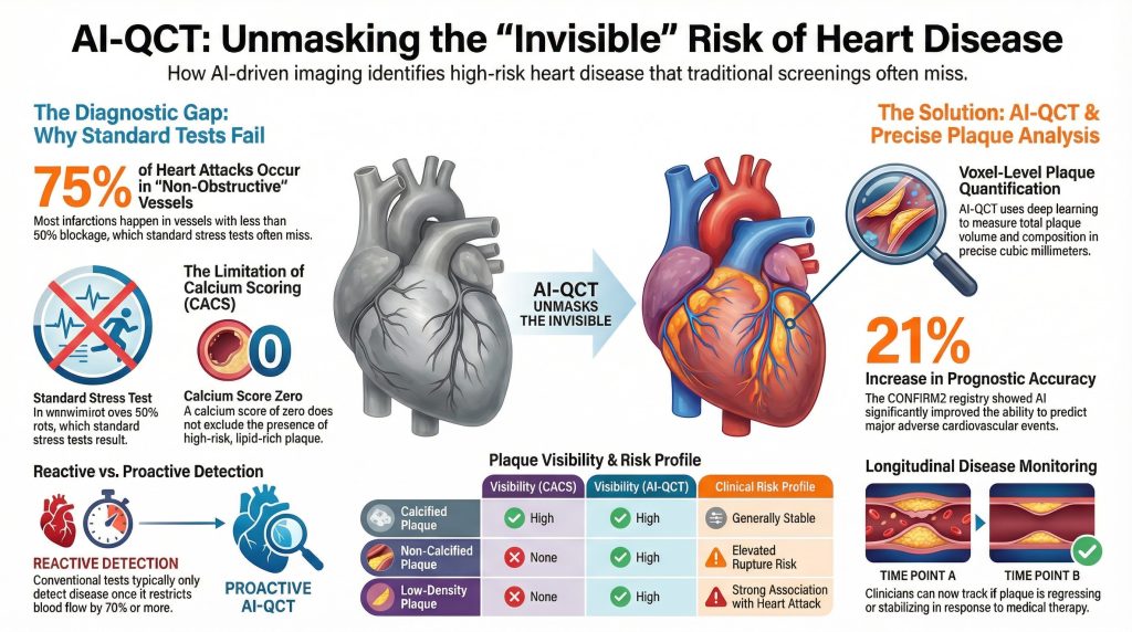
Insight 1: Autopsy and angiographic studies demonstrate that approximately 70–75% of myocardial infarctions occur in vessels with less than 50% stenosisStenosis is narrowing — usually described as a percentage, like a 70 percent blockage. prior to the index event, indicating that most patients who experience acute coronary syndromes would not have been identified as high risk by standard stress testing alone (2).
Insight 2: Coronary arteryAn artery is a blood vessel that carries blood away from the heart to the rest of the body. calcium scoring (CACS) was developed to improve detection of subclinical disease by identifying calcified plaqueCalcified plaque is the hardened, calcium-filled part of a plaque. It shows up brightly on a CT scan, which is what a calcium scan measures.. However, CACS does not detect non-calcified plaqueNon-calcified plaque is the soft, fatty portion of a plaque that has not hardened with calcium. It shows up dark on a CT scan., and a calcium scoreA calcium score (coronary artery calcium score) is a number derived from a CT scan that quantifies the total amount of calcified plaque in the coronary arteries; a score of zero indicates no detectable calcified plaque, while higher scores reflect greater plaque burden and elevated cardiovascular risk. of zero does not exclude the presence of lipid-rich, rupture-prone lesions (6).
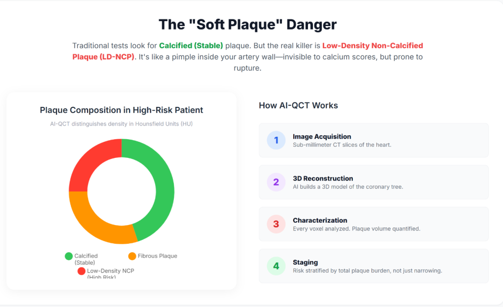
Insight 3: AI-QCT enables non-invasive quantification of total coronary plaque burdenPlaque burden is the total amount of plaque in your arteries, everywhere — not just at the single worst spot., including low-density non-calcified plaqueA type of coronary plaque with low X-ray attenuation on CT, corresponding to a lipid-rich, soft core that is not yet calcified; this morphology is associated with a higher risk of rupture and acute coronary syndromes than either fibrous or calcified plaque., which has been shown to carry a higher association with future cardiovascular events than stenosis severity alone (7).
| Plaque Characteristic | Visibility (CACS) | Visibility (Standard CCTA) | Visibility (AI-QCT) | Clinical Risk Profile |
| Calcified plaque | High | High | High | Generally stable; marker of chronic disease |
| Non-calcified plaque | None | Qualitative / limited | Quantitative / high | Elevated rupture risk |
| Low-density plaque | None | Variable | High | Strong association with ACS |
| Total plaque volumePlaque volume is the total physical amount of plaque in a stretch of artery, measured in cubic millimeters. | None | Indirect estimate | Precise (mm³) | Strong predictor of future events |
Technical Architecture of AI-Enabled Coronary Analysis
AI-QCT platforms perform voxel-level analysis of the coronary tree rather than relying on visual interpretation alone. These systems employ deep-learning architectures—including convolutional neural networks (CNNs), three-dimensional U-Net segmentation models, and VGG-derived classifiers—trained on large, curated datasets that link imaging features with invasive validation and long-term clinical outcomes (8).
Insight 4: AI-QCT begins with acquisition of high-resolution CCTA. Contemporary multi-detector CT systems, including 64- and 640-slice scanners, provide the temporal and spatial resolutionSpatial resolution in medical imaging refers to the smallest structure a scanner can distinguish as a separate object; coronary CT angiography has a practical resolution limit of about 0.4–0.5 mm, meaning early atherosclerotic lesions thinner than a human hair—typically 100–300 micrometers—are invisible to the scan even when biologically present. necessary to minimize motion artifact and enable accurate segmentation of coronary anatomy (5).
Insight 5: Automated algorithms identify the coronary lumenThe lumen is the open channel inside a blood vessel where blood actually flows. and vessel wall and classify plaque components according to Hounsfield unit attenuation values, allowing differentiation of lipid-rich, fibrous, and calcified tissue (9).
Insight 6: AI-derived ischemiaIschemia is when a tissue is not getting enough blood and oxygen for what it is being asked to do. indices integrate plaque morphology, lesion length, vessel remodeling, and luminal geometry to estimate flow limitation, providing a non-invasive correlate of invasive fractional flow reserveFractional flow reserve measures the pressure drop across a narrowing to find out whether it is actually restricting blood flow. (FFR) (10).
Clinical Validation and Peer-Reviewed Evidence
AI-QCT has been evaluated in multiple prospective trials, registries, and comparative studies published in leading cardiovascular and imaging journals, including Journal of the American College of Cardiology, Circulation: Cardiovascular Imaging, and European Heart Journal – Cardiovascular Imaging (11).
Landmark Trials and Registries
Insight 7: The CREDENCE trialA prospective trial of 513 patients that compared AI-assisted CCTA analysis with invasive quantitative coronary angiography and fractional flow reserve, demonstrating high concordance and supporting AI-QCT as a viable non-invasive substitute for catheter-based ischemia assessment. demonstrated close agreement between AI-assisted CCTA analysis and invasive quantitative coronary angiographyQuantitative coronary angiography is a way of measuring artery narrowing precisely from angiogram images, rather than eyeballing it. and FFR, supporting the diagnostic accuracy of AI-QCT for identifying hemodynamically significant disease (12).
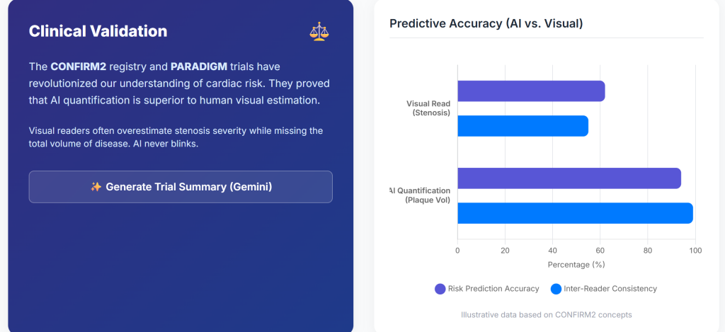
Insight 8: The CONFIRM2 registryA large, multinational registry of over 6,000 patients in which AI-quantified coronary plaque features were evaluated for their ability to predict major adverse cardiovascular events; the registry showed that AI-derived plaque analysis improved the area under the ROC curve for event prediction from 0.62 to 0.75 compared with standard methods., encompassing tens of thousands of patients across multiple countries, showed that AI-quantified plaque features identify a wide gradient of cardiovascular risk among patients with non-obstructive coronary artery diseaseCoronary artery disease is plaque buildup in the arteries feeding the heart muscle. (7).
Insight 9: The CERTAIN studyA clinical study evaluating the impact of AI-QCT coronary plaque analysis on physician treatment decisions; it found that AI-derived results changed management in 57.1% of patients, increasing statin use by 28.1%, aspirin prescribing by 23%, and reducing unnecessary downstream testing by 37%. demonstrated that AI-QCT significantly improved diagnostic certainty compared with standard interpretation, resulting in more appropriate medical therapy and fewer unnecessary downstream tests (1).
| Study | N | Key Finding | P-valueA p-value estimates how likely you would be to see a result at least this striking if the treatment did nothing at all. | Clinical Implication |
| CONFIRM2 | 6,550 | AUC for MACE improved from 0.62 to 0.75 | <0.001 | Improved prognostic accuracy |
| CREDENCE | 513 | High concordance with invasive FFR | <0.01 | Viable non-invasive ischemia assessment |
| PACIFIC-1A clinical study in which AI-assisted quantitative CT coronary angiography (AI-QCT) was compared head-to-head with human expert readers; AI-QCT achieved an AUC of 0.91 for detecting obstructive stenosis, outperforming level-3 expert cardiologists who scored 0.77. | 208 | AI exceeded expert readers | <0.05 | Reduced inter-observer variability |
| PROMISE (subset) | 4,347 | 41% stenoses reclassified | — | Reduced false positives |
Preventive Cardiology Implementation and Longitudinal Disease Monitoring
Integration of AI-QCT into preventive cardiology workflows shifts the clinical focus from episodic evaluation toward longitudinal monitoring of atherosclerotic disease.
Insight 10: Quantification of total plaque burden enables objective assessment of disease progression or regression over time, facilitating a treat-to-target approach guided by serial imaging (7).
Insight 11: AI-based comparison tools permit evaluation of changes in plaque composition, including stabilization or regression of lipid-rich plaqueAn atherosclerotic lesion whose core is dominated by cholesterol esters and inflammatory lipids rather than calcium or fibrous tissue; the article notes that such plaques are highly responsive to intensive treatment and that the dramatic 65-percentage-point regression at the diagonal branch origin is consistent with reversal of a lipid-rich lesion. in response to medical therapy (13).
Hardware Advances: Photon-Counting Computed Tomography
The performance of AI-QCT is dependent on the quality of input imaging data, which continues to improve with advances in CT hardware.
Insight 12: Photon-counting CT (PCCT)A next-generation CT detector technology that registers individual X-ray photons rather than averaging their energy, yielding higher spatial resolution and reduced blooming artifact compared with conventional detectors; it is under evaluation for improving both calcium quantification and non-calcified plaque characterization. systems directly convert X-ray photons into electrical signals, reducing electronic noise and calcium blooming artifactsA CT imaging artifact in which a dense calcified deposit spreads or 'blooms' beyond its true boundaries due to detector limitations, obscuring adjacent soft tissue and making it impossible to assess non-calcified plaque sitting next to the calcium. compared with conventional energy-integrating detectors (14).
Insight 13: PCCT provides higher spatial resolution with lower radiation and contrast dose, expanding eligibility for coronary CT imaging to patients with high heart ratesHeart rate is how many times your heart beats per minute., elevated body mass indexBody mass index, or BMI, is a number calculated from your height and weight, used as a rough measure of body size., or extensive coronary calcificationCalcification is when calcium gets deposited into a plaque, turning part of it hard and bony. (15).
Economic and Health System Implications
The adoption of AI-enabled cardiac imaging aligns with broader healthcare trends toward value-based care and prevention.
Insight 14: Market analyses project rapid growth of AI applications in cardiology, driven by improved diagnostic efficiency and potential reductions in downstream costs associated with acute coronary events (16).
Insight 15: Early identification of high-risk plaque phenotypesPlaque phenotype refers to the biological and structural characteristics of an atherosclerotic lesion — including the size of its lipid-rich necrotic core, fibrous cap thickness, degree of calcification, and inflammatory cell content — which together determine whether a plaque is stable or at high risk of rupturing. may reduce myocardial infarction rates and associated healthcare expenditures by enabling earlier and more targeted intervention (17).
Conclusions and Future Directions
Preventive cardiology is increasingly transitioning toward a biologically informed model that emphasizes plaque burden and composition rather than stenosis severity alone. AI-QCT provides a reproducible, quantitative framework for assessing coronary atherosclerosis before clinical events occur.
Insight 16: Future developments are likely to include integration of agentic AI systems, longitudinal digital heart models, and multimodal data streams incorporating genomic and metabolic information (18).
Insight 17: As AI assumes a greater role in image interpretation and quantification, clinicians may increasingly focus on higher-level clinical decision-making, risk communication, and individualized therapy optimization (19).
References
- Nurmohamed NS, Cole JH, Budoff MJ, et al. Impact of atherosclerosis imaging-quantitative computed tomography on diagnostic certainty, downstream testing, coronary revascularization, and medical therapy: the CERTAIN study. Eur Heart J Cardiovasc Imaging. 2024;25(6):857-866. doi:10.1093/ehjci/jeae029
- Falk E, Shah PK, Fuster V. Coronary plaque disruption. Circulation. 1995;92(3):657-671. doi:10.1161/01.cir.92.3.657
- Global Burden of Cardiovascular Diseases and Risks 2023 Collaborators. Global, Regional, and National Burden of Cardiovascular Diseases and Risk Factors in 204 Countries and Territories, 1990-2023. J Am Coll Cardiol. 2025;86(22):2167-2243. doi:10.1016/j.jacc.2025.08.015
- Lloyd-Jones DM. Cardiovascular risk prediction: basic concepts, current status, and future directions. Circulation. 2010;121(15):1768-1777. doi:10.1161/CIRCULATIONAHA.109.849166
- Douglas PS, Hoffmann U, Patel MR, et al. Outcomes of anatomical versus functional testing for coronary artery disease. N Engl J Med. 2015;372(14):1291-1300. doi:10.1056/NEJMoa1415516
- Greenland P, Blaha MJ, Budoff MJ, Erbel R, Watson KE. Coronary Calcium Score and Cardiovascular Risk. J Am Coll Cardiol. 2018;72(4):434-447. doi:10.1016/j.jacc.2018.05.027
- Feuchtner GM, Lacaita PG, Bax JJ, et al. AI-Quantitative CT Coronary Plaque Features Associate With a Higher Relative Risk in Women: CONFIRM2 Registry. Circ Cardiovasc Imaging. 2025;18(6):e018235. doi:10.1161/CIRCIMAGING.125.018235
- Oikonomou EK, Khera R. Machine learning in precision diabetes care and cardiovascular risk prediction. Cardiovasc Diabetol. 2023;22(1):259. Published 2023 Sep 25. doi:10.1186/s12933-023-01985-3
- Koo BK, Yang S, Jung JW, et al. Artificial Intelligence-Enabled Quantitative Coronary Plaque and Hemodynamic Analysis for Predicting Acute Coronary Syndrome. JACC Cardiovasc Imaging. 2024;17(9):1062-1076. doi:10.1016/j.jcmg.2024.03.015
- Nørgaard BL, Leipsic J, Gaur S, et al. Diagnostic performance of noninvasive fractional flow reserve derived from coronary computed tomography angiography in suspected coronary artery disease: the NXT trial (Analysis of Coronary Blood Flow Using CT Angiography: Next Steps). J Am Coll Cardiol. 2014;63(12):1145-1155. doi:10.1016/j.jacc.2013.11.043
- Beerkens FJ, Tang GHL, Kini AS, et al. Transcatheter Aortic Valve Replacement Beyond Severe Aortic Stenosis: JACC State-of-the-Art Review. J Am Coll Cardiol. 2025;85(9):944-964. doi:10.1016/j.jacc.2024.11.051
- Griffin WF, Choi AD, Riess JS, et al. AI Evaluation of Stenosis on Coronary CTA, Comparison With Quantitative Coronary Angiography and Fractional Flow Reserve: A CREDENCE Trial Substudy. JACC Cardiovasc Imaging. 2023;16(2):193-205. doi:10.1016/j.jcmg.2021.10.020
- Zaman S, Wasfy JH, Kapil V, et al. The Lancet Commission on rethinking coronary artery disease: moving from ischaemia to atheroma. Lancet. 2025;405(10486):1264-1312. doi:10.1016/S0140-6736(25)00055-8
- Willemink MJ, Persson M, Pourmorteza A, Pelc NJ, Fleischmann D. Photon-counting CT: Technical Principles and Clinical Prospects. Radiology. 2018;289(2):293-312. doi:10.1148/radiol.2018172656
- Schiebler ML, Jinzaki M, Yanagawa M, et al. Future Applications of Cardiothoracic CT. Radiology. 2025;315(3):e240085. doi:10.1148/radiol.240085
- Windecker S, Gilard M, Achenbach S, et al. Device innovation in cardiovascular medicine: a report from the European Society of Cardiology Cardiovascular Round Table. Eur Heart J. 2024;45(13):1104-1115. doi:10.1093/eurheartj/ehae069
- Undas A, Musiał J, Ząbczyk M. Antiphospholipid antibodies and atherosclerotic vascular disease: recent advances. Rheumatol Int. 2025;45(12):279. Published 2025 Nov 29. doi:10.1007/s00296-025-06050-8
- Topol EJ. High-performance medicine: the convergence of human and artificial intelligence. Nat Med. 2019;25(1):44-56. doi:10.1038/s41591-018-0300-7
- Shetty NS, Gaonkar M, Pampana A, et al. Association of Pathogenic/Likely Pathogenic Inherited Cardiomyopathy Variants With Heart Failure: A TOPMed Multiancestry Analysis. Mayo Clin Proc. 2025;100(11):1948-1955. doi:10.1016/j.mayocp.2025.01.021

