
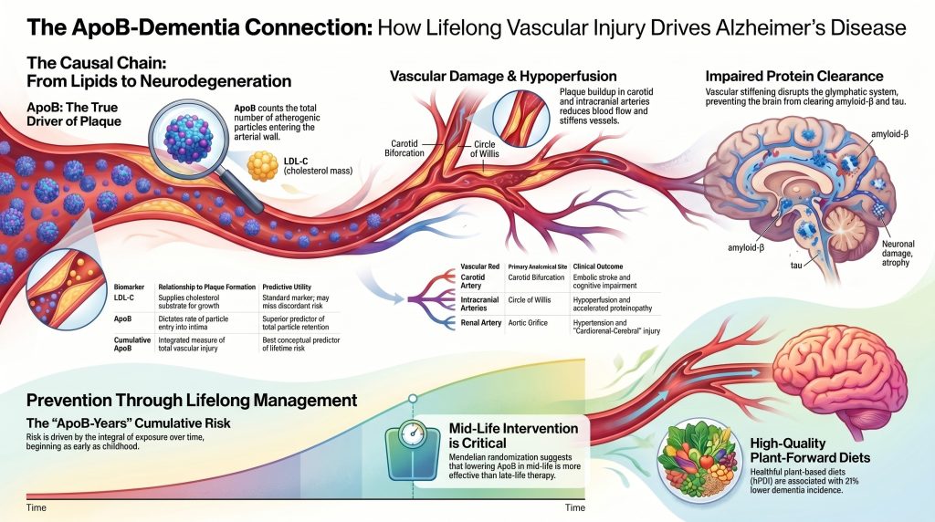
تصل الشرايين الناجم عن بروتين الشحم المرتبط بالورم القلبي البشري (أبوليبوبروتين ب) كأساس مسبب لمرض الزهايمر
منظور متعدد التخصصات حول المحور القلبي الكلوي الدماغي
ملخص
خلفية. يُعرّف الإطار العصبي المهيمن مرض ألزهايمر بيولوجياً من خلال الأميلويد والتاو المؤشرات الحيوية وتعامل اعتلال الأوعية الدموية باعتباره متغيراً متكرراً ولكنه ثانوي. تدعم الأدلة الجينية والوبائية التصويرية بشكل متزايد قراءة بديلة يكون فيها التعرض طوال الحياة لـ أبروليپوبروتين بأبوبروتين ب)-يحتوي على البروتينات الدهنية هو محرك سببي علوي للإصابة الدماغية والأوعية الدموية الدقيقة والتي، لدى فرع حيوي من المرضى، تسبق وتسرع علم الأمراض المحدد لمرض الزهايمر.
الهدف. لتركيب الأدلة الحالية التي تربط التعرض مدى الحياة لبروتين الأبوليبيوبروتين ب (ApoB) بالإصابة تصلب الشرايين في السرير الوعائي السباتي، داخل الجمجمة، والكلوي، وبالتالي، متلازمة الإدراك المشخصة سريرياً بمرض الزهايمر.
الطرق. منظم مراجعة سردية من الدراسات الأولية المفهرسة في PubMed،, الوراثة العشوائية المندلية التحليلات، ومجموعات التصوير طولية المدى، وتجارب الوقاية القلبية الوعائية وتجارب الوقاية من الخرف العشوائية، وبيانات الإجماع الحالية الصادرة عن جمعية القلب الأمريكية، والجمعية الوطنية للدهون، وجمعية ألزهايمر. جرى تقييم ادعاءات السببية مقارنة بمعايير إعداد التقارير الخاصة بـ STROBE-MR.29]
الاستنتاجات. التعرض طوال العمر للبروتينات الدهنية التي تحتوي على الأبوليپوبروتين ب (ApoB) مثبت مسبباً للإصابة بأمراض الأوعية الدموية تصلب الشرايين. وتتفق الأدلة المتقاربة من دراسات Mendelian الموجهة بالأدوية عشوائية في أكثر من مليون فرد،7تصوير مقطعي بالإصدار البوزيتروني الطولي داخل القحف تضيق,[13] وتجارب عشوائية لتقليل المخاطر الوعائية[14تدعم مساهمة سببية لهذا الإصابة الوعائية في التراجع المعرفي، لـ الضعف الإدراكي والخرف الوعائي, ، وإلى أنماط الخرف ذات الأمراض المختلطة التي هيمن على سلسلة تشريح الجثث لدى كبار السن.2,32] الامتداد من إصابة الأوعية الدماغية إلى مرض الزهايمر المؤكد بالمؤشرات الحيوية كما هو محدد بموجب معايير المعهد الوطني للشيخوخة وجمعية الزهايمر (NIA-AA) المحدثة لعام 2024[40] مدعومة ولكن بثقة أضعف بشكل مناسب، وقد تم تخفيف لغة هذه المخطوطة وفقاً لذلك عند نقطة النهاية تلك. إن معايير المعهد الوطني للشيخوخة وجمعية الزهايمر (NIA-AA) لعام 2024 هي إطار تشخيصي وليست ادعاءً مسبباً للمرض؛ فهي متوافقة تماماً مع كل من الدور الإمراضين المتسلط والمستقل لبروتين أميلويد-بيتا (والذي يتم إثباته بشكل مباشر عن طريق مرض الزهايمر العائلي السائد الصبغي الجسدي)[41] ودور سببي محتمل من المنبع للإصابة الدماغية الوعائية المدفوعة بـ (ApoB) في المرض المتقطع متأخر البدء. إن خفض بروتين (ApoB) طوال العمر -من خلال أنماط غذائية نباتية عالية الجودة، وعند الإشارة إلى ذلك، العلاج الدوائي الذي يبدأ في منتصف العمر- هو استراتيجية وقائية يمكن الدفاع عنها ضد التدهور المعرفي ومكون معقول للوقاية من الخرف ومرض الزهايمر.
مقدمة
يتعرض النموذج العصبي المركزي السائد لمرض الزهايمر، والذي يمنح الأولوية لسلسلة أميلويد-بيتا كمسبب رئيسي التنكس العصبي، لتحدٍ متزايد من قبل الأدلة الجينية والوبائية والتصويرية التي تشير إلى أن صحة الأوعية الدموية ليست مجرد عامل مصاحب بل هي محرك أساسي ومنبعث مبكر لعملية المرض.1,5,17] يبحث هذا التحليل في الفرضية القائلة بأن التعرض طوال العمر للبروتينات الدهنية المحتوية على الأبوبروتين B (ApoB) المرتفعة يحفز تصلب الشرايين التدريجي لوحة الترسب في الشرايين السباتية، داخل الجمجمة، والكلوية، مما يطلق سلسلة من الإصابات الجهازية والموضعية التي تبلغ ذروتها في الإصابة بالخرف لدى نسبة كبيرة من المرضى.4,7,20,22من خلال examining الآليات المتداخلة لتكوين اللويحات عبر الأوعية الدموية والاستنتاجات السببية التي تتيحها التداخلات المندلية، يبرز إطار عمل صارم لفهم مرض ألزهايمر باعتباره المظهر العصبي في المرحلة المتأخرة لإصابة وعائية دهنية التهابية استمرت لعقود.2,3,9]
تُستخدم لغة السببية بشكل متعمد ولكن يتم معايرتها وفقاً لقوة الأدلة عند كل رابط في السلسلة. لقد ثبت أن بروتين أوبو بي (ApoB) مسبب لتصلب الشرايين أمراض القلب والأوعية الدموية من خلال التثليث عبر التوزيع الحر المندلي متعدد المتغيرات،21,25] تحليلات التعرض التراكمي [22,33] والتجارب العشوائية لخفض الدهون.[26إن امتداد هذا المسار السببي إلى الاعتلال المعرفي الوعائي والخرف ذو الأمراض المختلطة مدعوم جيداً العشوائية Мендель الموجهة لهدف الدواء لبروتين دهني منخفض الكثافة غير مرتفع الكثافة والخرف في أكثر من مليون فرد7] ومن خلال التصوير المقطعي بالإصدار البوزيتروني الطولي الذي يربط بين تيقن الجمجمة الأساسي وتراكم الأميلويد والتاو اللاحق.13الامتداد الأبعد لمرض الزهايمر المؤكد بالعلامات الحيوية تحديداً - أي مرض الزهايمر كما هو محدد بالمعايير المعدلة للعهد الوطني للaging والجمعية الأمريكية لمرض الزهايمر (NIA-AA) لعام 202440— يدعمها تحليل العندليف المندلي النوعي لـ ApoB المتسق في الاتجاه4وبالتوافق الآلي للإصابة الوعائية مع بيولوجيا الأميلويد والتاو،16,17,42] ولكن بقوة أضعف بشكل مناسب من الارتباط القلب والأوعية الدموية. وعليه، تتبنى المخطوطة لغة متحفظة عند نقطة نهاية مرض الزهايمر وتُميز بوضوح طوال النص بين خرف الأسباب المشتركة، والضعف الإدراكي الوعي، والخرف المختلط الأمراض، ومرض الزهايمر المؤكد بال المؤشرات الحيوية.
الفرضية الوعائية تصلب الشرايين لمرض ألزهايمر والخرف
تعترض الفرضية الوعائية تصلب الشرايين بأن المتلازمة السريرية لمرض ألبرت (ألزهايمر)، في مجموعة فرعية كبيرة من المرضى، هي النتيجة النهائية لنقص التروية المخية المزمن واختلال وظائف الوحدة العصبية الوعائية التي تبدأ بها الشرايين الكبيرة تصلب الشرايين وانتشرت عبر شبكة الأوعية الدموية الدقيقة في الدماغ.1,5,17في هذا الإطار، يُحدث التضييق البؤري للشرايين السباتية داخل الجمجمة، إلى جانب فقدان مرونة الشرايين، بيئة ديناميكية دموية معادية لبقاء الخلايا العصبية و بروتين تصريح.12,18]
تُظهر الأدلة العصبيّة المرضيّة المعاصرة أن التمييز التصنيفي التقليدي بين الخرف الوعائي البحت ومرض ألزهايمر البحت غير قابل للدعم تجريبياً لدى كبار السن.3,32تفيد دراسات التشريح المرضي المجتمعية بأن الاعتلالات العصبية المختلطة — أي مجموعة مزيجة من نوع ألزهايمر، وأجسام لوي، وبروتين TDP-43، والاعتلال الوعائي آفات — موجودة في أغلبية أδماغ المتقدمين في السن، وإن الاعتلال البروتيني النقي من دون أمراض وعائية دماغية هو الاستثناء وليس القاعدة.3,32تصلب الشرايين الكبيرة، بما في ذلك تضيق السباتي و مرض تصلب الشرايين داخل الجمجمة, commonly coexists with cerebral small-vessel disease characterized by white-matter hyperintensities, lacunar infarcts, and microbleeds.[3,8,9]
تصلب الشرايين — لا سيما في الناحية القريبة الوريد الأبهر والشرايين السباتية — تنقل النبض عالي الضغط إلى الشعيرات الدموية الدقيقة في الدماغ.12يؤدي هذا التدفق شديد النبض إلى إتلاف حاجز الدم في الدماغ وتعطيل الجهاز اللمفاوي الجانبي, ، وهي شبكة حول الأوعية الدموية مسؤولة عن التخلص من الفضلات الأيضية، بما في ذلك أميلويد-بيتا وتاو.15,18في هذا الإطار، يُعد تراكم هذه البروتينات، جزئياً، نتيجة لضعف التخلص منها وليس فقط نتيجة لفرط إنتاجها.16,17,18]
هذه الفرضية لا تستبعد خطر الإصابة الوراثي بمرض الزهايمر الكلاسيكي. إن أبوليبوبروتين E ε4 (أبوليبيوبروتين إي أليل ε4 - العامل الوراثي الأكثر شيوعاً والأكبر تأثيرًا عامل خطر بالنسبة لمرض الزهايمر متأخر البدء - فهو يعمل بشكل أساسي من خلال المسارات الدماغية الوعائية. تُظهر الأدلة التصويرية المباشرة للبشر أن حاملي جينات APOE ε4 يُظهرون انهياراً في حاجز الدم في الدماغ يسبق التدهور المعرفي ويتنبأ به بشكل مستقل عن الأميلويد والتاو،42توفير جسر ميكانيكي بين بيولوجيا البروتينات الدهنية الجهازية، وإصابة الأوعية الدموية الدماغية، والنمط الظاهري السريري لمرض الزهايمر.5,16,42]
نحن نتفاعل بشكل صريح مع المعايير المُعدلة لجمعية مرض الزهايمر لعام 2024،40] التي تعرّف مرض الزهايمر بيولوجياً بواسطة المؤشرات الحيوية الأساسية للأميلويد والتاو، بغض النظر عن المتلازمة السريرية. هذه المعايير هي إطار تشخيصي وليست ادعاءً سببياً، وتدرك مجموعة العمل أن الأمراض الوعائية المصاحبة وغيرها هي عوامل معدلة شائعة لمسار المرض. لذلك فإن موقفنا ليس أن معايير عام 2024 خاطئة أو أنها تنكر المساهمات الوعائية - فهي لا تفعل شيئاً من ذلك. موقفنا هو أن بصمة المؤشرات الحيوية التي تفعّلها لها، بالنسبة لمجموعة فرعية كبيرة من المرضى، سببٌ أولي: عقود من إصابات الأوعية الدموية الدماغية المدفوعة بـ ApoB والتي تتفاعل مع بيولوجيا الأملويد والتاو الذاتية. إن تعريف مرض الزهايمر من خلال بصمة المؤشرات الحيوية اللاحقة يتوافق تماماً مع عزو تلك البصمة، جزئياً، إلى الأسباب الوعائية الأولية.
الجدول 1. السمات المرضية والديناميكية الدموية المتداخلة في أنواع الخرف الفرعية
| ميزة | مرض ألزهايمر | الخرف الوعائي | خرف مختلط |
| اعتلال بروتيني أولي | لويحات الأميلويد-بيتا؛ تشابكات تاو | أقل ما يمكن أو غائب | لوحات أميلويد بيتا وبروتين تاو واسعة النطاق |
| آفات وعائية | غالباً ما يتواجد (اعتلال الأوعية الدموية النخاعي المرتبط بالأميلويد، النزف الدقيق) | احtкаркarات كbيرة، فجوات، فرط إشارe المادe البيضاe | وعائي وبروتيني مترافق |
| الديناميكا الدموية | انخفاض تدفق الدم الدماغي؛ فشل الجهاز الجليمفاوي | بؤري أو عام نقص الدم | قصور التروية المزمن + التيبس |
| محرك دهني | الرابط الخاص ببروتين الببتيد الدهني أوبوليبيوبروتين ب عبر إصابة الأوعية الدموية الدماغية | ارتباط قوي بين الكوليسترول الضار (LDL-C) وبروتين الببتيد الدهني (ApoB) | عبء البروتين الدهني (ب) المرتفع مدى الحياة |
| الخصائص التصويرية | ضمور الحصين؛ إيجابية التصوير المقطعي بالإصدار البوزيتروني للأميلويد-بيتا | احتشاءات متعددة؛ تضيق السباتي | ضمور، فرط إشارة المادة البيضاء،, عبء اللويحات |
مقتبس من المراجع [3,5,9,32,40]. CAA = اعتلال الأوعية الدمازية النشواني؛ WMH = فرط كثافة المادة البيضاء؛ CBF = تدفق الدم الدماغي.
ديناميكيات البروتينات الدهنية والتعرض السببي مدى الحياة
يُعد تركيز البروتينات الدهنية المحتوية على أوبو-بي في الدورة الدموية أمراً أساسياً في تطور تصلب الشرايين.10,19,20[ البروتين الدهني بي (ApoB) هو بروتين هيكلي يوجد على سطح كل جزيء بروتين دهني محتمل تصلب الشرايين. البروتينات الدهنية منخفضة الكثافة جداً والمشتقة من الكبد (بروتين دهني منخفض الكثافة جداً), البروتينات الدهنية متوسطة الكثافة (لغة IDL)، والبروتينات الدهنية منخفضة الكثافة (البروتين الدهني منخفض الكثافة)، و بروتين دهني (أ) يحمل كل منها جزيئاً واحداً من أبو بروتين-100 لكل جسيم، بينما الكيلومكرونات وتحمل بقايا الكيلومكرون أبو بروتين ب-48 منشؤه من الأمعاء.20في البالغين في حالة ما بعد الامتصاص، يتكون عبء تصلب الشرايين السائد الذي يعكسه قياس الأبوبروتين ب (ApoB) في البلازما بشكل ساحق من الجزيئات الحاملة للأبوبروتين ب-100 (apoB-100).20] بصرف النظر عن التمييز في العدد، فإن جزيئات البروتين الدهني منخفض الكثافة (apoB-100) لها مدة بقاء داخلية أطول بكثير من مخلفات الكيلومايكرون الأكبر حجمًا والأكثر عابرة في الدوران (apoB-48)، لذا فإن جزء apoB-100 يساهم بشكل غير متناسب في الإصابة التراكمية لجدار الشريان ويهيمن على “سنوات الأبوبروتين ب”أجزاء أساسية في السرير الدماغي الوعائي.
في حين أن الكوليسترول الضار يقيس (LDL-C) كوليسترول الكتلة داخل جزيئات الدهون البروتينية منخفضة الكثافة (LDL)، يوفر بروتين بي (ApoB) عدداً مباشراً للإجمالي جزيئات مُتصلبة العصيد.[20هذا التمييز ذو أهمية حاسمة من الناحية الآلية: احتمال دخول جزيء بروتين دهني الشريان بطانة وكونها محتجزة بواسطة السكريات البروتينية خارج الخلايا هي وظيفة لتركيز الجسيمات بدلاً من محتوى الكوليسترول لكل جسيم.19أثبتت تحليلات التراكم Mendelian المتعددة المتغيرات أن ApoB هو سمة الدهون المهيمنة في العلاقة بين البروتينات الدهنية و مرض الشريان التاجي; عند ضبط تأثير بروتين أوبو بي (ApoB)، لا يُظهر كل من الكوليسترول المرتبط بالبروتينات الدهنية منخفضة الكثافة (LDL-C) ولا الدهون الثلاثية الاحتفاظ بمعلومات سببية مستقلة لأمراض القلب والأوعية الدموية تصلب الشرايين.21,25]
المخاطر التي يفرزها بروتين أوبوليبروتين ب (ApoB) هي مخاطر تراكمية - وهو مفهوم تم إضفاء الطابع الرسمي عليه الآن باسم LDL/ApoB التعرض التراكمي فرضية.22] تصلب الشرايين هي عملية بطيئة تبدأ عادة في مرحلة الطفولة بتشكل النواة خطوط دهنية.[19] كما الجسيمات المحتوية على أبوليبوبروتين ب تظل مرتفعة لعقود، وتتراكم عبء اللويحات العصيدية التصلبية تدريجياً حتى يتم تجاوز عتبات حرجة، مما يؤدي إلى تضيق اللمعة أو تمزق اللويحة.[22,33] التعرض التراكمي مدى الحياة (غالباً ما يُوصَف بأنه “سنوات الكوليسترول”أو “سنوات-ApoB”) هو مؤشر بيولوجي أكثر ملائمة للتنبؤ بخطر الإصابة بأمراض القلب والأوعية الدموية، ومن المحتمل الخرف، مقارنة بأي قياس منفرد في منتصف العمر أو متأخره، والذي قد يتأثر بـ السببية العكسية لدى كبار السن الذين يصابون بالضعف و فقدان الوزن في المرحلة ما قبل السريرية للخرف.22,24,28]
الجدول 2. القيمة التنبؤية المقارنة للحيوانات الحيوية الدهنية لخطر تصلب الشرايين
| مؤشر حيوي | تعريف | العلاقة بتكوين اللويحات | منفعَة |
| البروتين الدهني منخفض الكثافة (LDL-C) | كتلة الكوليسترول في جزيئات البروتين الدهني منخفض الكثافة | يوفر ركيزة الكوليسترول لنمو اللويحات | علامة قياسية؛ تفوت المخاطر في عدم توافق |
| أبوبروتين ب | العدد الإجمالي للجزيئات المسببة تصلب الشرايين (جزيء واحد لكل جزيء) | يُحِدِّدُ مُعَدَّلَ دُخُولِ الدَّقِيقَاتِ إِلَى الطَّبَقَةِ البَاطِنَةِ | مؤنب متفوق للاحتفاظ بالجسيمات |
| الكوليسترول غير عالي الكثافة | إجمالي الكوليسترول ناقص الكوليسترول المرتبط بالبروتينات الدهنية عالية الكثافة | يلتقط الكوليسترول من نوع LDL و VLDL و IDL و Lp(a) | بديل عملي للعبء العصيدي |
| بروتين دهني (أ) | جسيم شبيه بالبروتين الدهني منخفض الكثافة مع بروتين دهني (أ) | مخثر ومنهب للالتهاب | عامل خطر جينيني مستقل |
| التعرض التراكمي لبروتين البميم (أ) أو التعرض التراكمي لبروتين الدهون القابلة للاختزان (أبوB) | تكامل الأبوبروتين ب بمرور الوقت (“سنوات-ApoB”) | مقياس متكامل لإجمالي الإصابة الوعائية | أفضل مؤشر تصوري لخطر مدى الحياة |
مقتبس من المراجع [19,20,21,22,25,33].
مراجعة أدلة التخمين المندلي في الخرف
الت عشوائية Mendelian randomization (MR) على التوزيع العشوائي للأليلات الوراثية أثناء تكوين الأمشاج لتقدير التأثير السببي للتعرضات القابلة للتعديل على النتائج، محاكاةً لتصميم تجربة معشاة ذات شواهد مع تجنب العديد من مصادر الملاحظة ربط مربك.[29تم تقييم جميع مطالبات التنجيم المندلي الملخصة أدناه مقابل بيان STROBE-MR بشأن الإبلاغ الشفاف عن تحقيقات التنجيم المندلي.29]
تحليلات التندب الجيني لأهداف أدوية خفض الدهون الشائعة — HMGCR (ستاتين الهدف, PCSK9 (مثبط بروكاتين كونسيرتيز سابتايليسين/كيكسين من النوع 9 الهدف, NPC1L1 (إزيتيمايب الهدف) و CETP — توفير بعض أقوى الأدلة السببية الحالية التي تربط انخفاض الكوليسترول المرتبط بالبروتينات الدهنية غير عالية الكثافة طوال العمر بانخفاض خطر الإصابة بالخرف. في دراسة العشوائية المندلية عينة واحدة تحليل تلوي من بين 1,091,775 فردًا، بلغت نسب الاحتمالات لكل انخفاض بمقدار 1 مليمول/لتر (≈39 ملغم/ديسيلتر) في مستوى الكوليسترول غير عالي الكثافة (non-HDL-C) فيما يتعلق بخطر الإصابة بالخرف لأي سبب كان 0.24 (نطاق الثقة 95% 0.18–0.31) بالنسبة لـ HMGCR، و0.18 (0.12–0.25) بالنسبة لـ NPC1L1، و0.30 (0.26–0.34) بالنسبة لـ CETP، و0.97 (0.70–1.35) بالنسبة لـ PCSK9.[7] أفادت دراسة دنماركية سابقة للتحليل الجيني المندلعي (MR) بوجود نسب خطورة لكل انخفاض بمقدار 1 ملمول/لتر في الكوليسترول المرتبط بالبروتينات الدهنية منخفضة الكثافة (LDL-C) بلغت 0.57 (0.27–1.17) لمرض ألزهايمر و0.81 (0.34–1.89) للخرف الوعائي — وهي نتائج تتسق في اتجاهها مع الحماية ولكنها غير كافية إحصائياً لنتائج فردية.[27]
تجدر الإشارة بوضوح إلى نقطتين. أولاً، النتيجة الأساسية في دراسة نوردستغارد وآخرين هي الخرف بجميع أسبابه وليس مرض ألزهايمر المؤكد بالعلامات الحيوية بموجب معايير المعهد الوطني للaging وجمعية ألزهايمر لعام 2024.40ثانياً، يتوافق تقدير PCSK9 مع فرضية العدم، على عكس الإشارات القوية لكل من HMGCR، وNPC1L1، وCETP.7تعارض هذه الغيرية الخاصة بالهدف نموذجاً بسيطاً تفترض فيه أي آلية دووائية تخفض البروتينات الدهنية المحتوية على الأبوليبيوبروتين (بي) أنها ستخفض خطر الإصابة بالخرف بشكل مكافئ، وتشير إلى بيولوجيا دماغية وعيائية خاصة بالهدف يجب أن تعالجها التجارب المستقبلية.
أدلة التراث الجيني (MR) الخاصة بـ ApoB ومرض ألزهايمر أحدث عهداً. باستخدام التراث الجيني الثنائي العينة أحادي المتغير البنك الحيوي البريطاني تدل أدوات الأبوبروتين ب (ApoB) وإحصائيات الملخص الخاصة بمرض الزهايمر (IGAP AD) على أن زيادة مستوى الأبوبروتين ب (ApoB) المتوقع جينياً ترتبط بقصر فترة الصحة وقد — وفقاً للغة الحذرة التي استخدمها المؤلفون أنفسهم — قد تزيد المحتمل أن تزيد من خطر الإصابة بمرض الزهايمر.4يشير التحليل المندلي متعدد المتغيرات المشروط على الكوليسترول المرتبط بالبروتينات الدهنية منخفضة الكثافة (LDL-C) إلى أن بروتين أبوليبوبروتين ب (ApoB) يحتفظ بارتباطه بقصر فترة الصحة حتى عندما يكون الكوليسترول (LDL-C) معدوماً في النموذج المشترك.4نحن نتبنى وصف المؤلفين أنفسهم: هذا دليل سببي داعم ولكنه أولي عند نقطة النهاية لمرض الزهايمر، والذي ينبغي تكراره مقابل نتائج مرض الزهايمر المؤكدة المؤشرات الحيوية مع توفر هذه البيانات.
قيود التنديل العشوائي
تنطبق عدة قيود على جميع أدلة التمفصل المندلي (MR) المذكورة أعلاه ويجب أن تؤخذ في الاعتبار عند تفسير العلاقة السببية:
- تحيز البقاء قد يموت الأفراد الذين يعانون من ارتفاع الجينات في بروتين أبوب (ApoB) بسبب حوادث قلبية وعascularية مبكرة قبل الوصول إلى السن الذي تتجلى فيه الخرف سريرياً، مما قد يضعف الارتباط الملاحظ بين الدهون ومرض الزهايمر.4,7]
- خطر الموت التنافسي: بشكل أكثر تحديداً من تحيز البقاء العام، فإن ApoB يعد مسبباً قوياً لـ احشاء عضلة القلب و سكتة دماغية إن الأفراد الذين لديهم مستويات مرتفعة الوراثة من بروتين أوبو بي (ApoB) والذين كانوا سيصابون لولا ذلك بمرض الزهايمر، يتم إزالتهم بشكل غير متناسب من السكان المؤهلين للإصابة بمرض الزهايمر بسبب الوفيات القلبية الوعائية المبكرة. يؤدي هيكل المخاطر المتنافسة هذا إلى خفض الارتباط الملاحظ بين الدهون ومرض الزهايمر بشكل منهجي في تصميمات التوزيع العشوائي Мендель (Mendelian randomization) القياسية، وهو أحد الأسباب المحددة وراء كون إشارة نقطة نهاية مرض الزهايمر في التوزيع العشوائي Мендель الحالي أضعف من إشارة القلب والأوعية الدموية - وليس بالضرورة لأن المسار السببي ضعيف.4,27]
- تعدد التأثير الجينات: قد تؤثر المتغيرات الجينية على النتيجة من خلال مسارات أخرى غير التعرض المقصود. استخدمت الدراسات المذكورة مق تقديرات الانحدار العرضي لـ مندل (MR-Egger)، والمتوسط المرجح، ومق تقديرات الانحدار العرضي المتعددة لتقييم المتانة؛ بينما تحتفظ تحليلات ApoB المشروطة بإشارة متسقة في الاتجاه.4,21,25]
- تباين النتائج غالبًا ما تتضمن مرض ألزهايمر المشخص سريرياً في مجموعات البيانات الإدارية أمراضاً وعائية مرافقَة كبيرة؛3,32] نقاط end-points لمرض الزهايمر المؤكدة المؤشرات الحيوية بموجب إطار عمل NIA-AA لعام 2024 [40] لكان من شأن ذلك أن يعزز أبحاث التنميش المندلي المستقبلية. إن أدلة التنميش المندلي المستهدفة بالعقاقير لخرف الأسباب كافة [7أقوى من الناحية المنهجية ولكنها تنطبق على الخرف بشكل عام، وليس على مرض الزهايمر المؤكد بالمؤشرات الحيوية بشكل خاص.
- النسب وقابلية التعميم: أجريت معظم دراسات الترافق الجيني (MR) حول الدهون والخرف في مجموعات سكانية من أصل أوروبي؛ وقد تختلف التقديرات السببية عبر المجموعات العرقية المختلفة.7]
- تفسير التعرض مدى الحياة: يُقدّر التنميط العشوائي المندلي (MR) التأثير التراكمي للتعرض الجيني طوال العمر، والذي يتجاوز عادةً التأثير الممكن تحقيقه عن طريق العلاج الدوائي المبدأ في مرحلة متأخرة من العمر. هذا الاختلاف مفيد لتوقيت الوقاية ولكنه لا يترجم مباشرة إلى حجم التأثيرات المتوقعة من تجارب الخرف في مرحلة متأخرة من العمر.22,26]
الفيزيولوجيا المرضية الإقليمية لتكون اللويحات العصيدية
تصلب الشرايين هو مرض التهابي مزمن تبدأ الإصابة به باحتباس الدهون في مناطق بؤرية من الشرايين، لا سيما مناطق التدفق المضطرب وغير الصفائحي.10,19تتشابه الآليات الجزيئية والخلوية لتكوين اللويحات بشكل عام عبر الأوعية الدموية الرئيسية، إلا أن التشريح المحلي وديناميكيات الدم تضفي خصائص مميزة على المرض في كل موقع.10,11,31]
تصلب الشرايين السباتية
تعتبر تشعبات الشريان السباتي عرضة بشكل خاص لتشكل اللويحات نظراً لهندستها المعقدة وتدفق الدم فيها عكس خلال الدورة القلبية.10,11منخفض ومتذبذب إجهاد قص الجدار في أصل الداخلي الשريان السباتي يعزز اختلال وظيفة بطانة الأوعية الدموية ويزيد من نفاذية جدار الأوعية الدموية للبروتينات الدهنية المحتوية على الأبوبروتين ب.10,11,19ميزة مميزة لويحات السباتي هي ميلها إلى إحداث صمات: التدفق عالي السرعة يزيل الخثرات التي تتكون على أسطح اللويحات التقرحية، مماولّد صمات مجهرية متكررة بدلاً من صمات دانية كاملة انسداد.[11,34الاصابة الميكروصماتية التراكمية، والاحتشاء الجريبي، والنزيف الدقيق هي الركائز الأساسية للاختلال المعرفي الوعائي.2,9,34]
مرض تصلب الشرايين داخل الجمجمة
تشير تضيق الشرايين داخل الجمجمة إلى تكوين اللويحات في الشرايين الكبيرة في قاعدة الدماغ - بما في ذلك الشريان الدماغي الأوسط شريان, ، الشريان القاعدي، ودائرة ويليس.11,13على عكس مرض الشبات الجانبي خارج الجمجمة، تم ربط مرض الشبات الجانبي داخل الجمجمة مباشرة، في التصوير الطولي المستقبلي، بالترسب اللاحق لأميلويد-بيتا وتاو في متماسك الدماغ. في أ دراسة أترابية استباقية باستخدام تصوير بيتا أميلويد (PiB-PET) وتصوير تاو (AV-1451-PET) التسلسلي، وُجد أن وجود أي تضيق داخل القحف في بداية الدراسة ارتبط بشكل ملحوظ بتراكم أكبر لأميلويد-بيتا على مدى أربع سنوات، كما أن التضيق في شريانين أو أكثر تنبأ بترسب أكبر لتاو على مدى عامين.13هذه الاتجاهية الطولية — الإصابة الوعائية التي تسبق اعتلال البروتينات المتسارع — تعد من أقوى أدلة التصوير البشري الحالية على أن صحة الأوعية الدموية داخل الجمجمة تقع سببياً في مرتبة تسبق علم الأمراض المُميّز لمرض الزهايمر بدلاً من مجرد التزامن معه.
تصلب الشريان الكلوي ومحور القلب والكلى والدماغ
تضيق الشريان الكلوي (RAS) يعود سببه في الغالب إلى تصلب الشرايين، ويتركز عند الفوهة الأبهريّة أو ضمن الثلث القريب من الشريان الكلوي الرئيسي.31,37الكلية، مثل الدماغ، عبارة عن سرير وعائي منخفض المقاومة حسّاس للتدفق مفرط النبض ونقص التروية.6,12يتفاقم تطور اللويحات في الشريان الكلوي بسبب الخلل الوظيفي في الأنسجة الدهنية حول الأوعية الدموية وتوسع الأوعية الغاذية, التي تطلق السيتوكينات المؤيدة للالتهابات والحويصلات خارج الخلية.31]
تضيق الشريان الكلوي العصيدي ينشط نظام الرينين-أنجيوتنسين-ألدوستيرون، مما ينتج عنه ثانوي ارتفاع ضغط الدم هذا يسرع بشكل أكبر الضرر الذي يلحق بالأوعية الدقيقة الدوائرية الدماغية ويؤسس لعملية تغذية مرتدة إيجابية تربط بين الكلى، القلب، والدماغ.6,31,36,39] مرض الكلى المزمن يتميز بدوره بنظام مؤسسي التهاب, الإجهاد التأكسدي, ، وتسمم بطانة الأوعية الدموية البولي، وجميعها مرتبطة بشكل مستقل بالتدهور المعرفي عبر المحور القلبي الكلوي الدماغي.6,38,39]
الجدول 3. مقارنة خصائص اللويحات عبر الأسرّة الوعائية
| السرير الوعائي | الموقع التشريعي الأساسي | محرك ديناميكي دموي فريد | النتيجة السريرية الأساسية |
| الशريان السباتي | تفرع السباتي؛ منشأ الشريان السباتي الباطن | انعكاس التدفق والاضطراب؛ إجهاد قص منخفض/متذبذب | السكتة الدماغية الصصية والضعف الإدراكي |
| الشرايين داخل القحف | الشريان المخي الأوسط، الشريان القاعدي، دائرة ويليس | تفرع معقد؛ فقدان التنظيم التلقائي | قصور التروية وتسرع اعتلال البروتين |
| الشريان الكلوي | فوهة الأبهر / الثلث الداني | الإجهاد القصي المرتبط بالفتحة | تضيق الشريان الكلوي، ارتفاع ضغط الدم، مرض الكلى المزمن |
| شريان تاجي | الشريان الأمامي النازل الأقصى والتفرعات | إجهاد ضغطي دوري؛ انحناء | احتشاء عضلة القلب و ذبحة صدرية |
مقتبس من المراجع [10,11,13,31,37].
المسار الآلي: من الدهون إلى التنكس العصبي
الإنتقال من ارتفاع مستوى البروتين الدهني (أبو بي) في الدم إلى الخرف السريري يتضمن سلسلة متعددة المراحل فيها الاحتباس تحت البطانة من البروتينات الدهنية المحتوية على أبوليبوبروتين بي (ApoB) بمثابة الحدث البادئ.19تخضع الجسيمات المحتجزة لتعديلات تأكسدية وأنزيمية، مما يولد إشارات قوية محفزة للالتهاب تعمل على تنشيط الخلايا البطانية, ، وتجذب الخلايا الوحيدة، وتقود بالعمى تكون الخلايا الرغوية.10,19,53]
احتباس الدهون، والتنشيط البطاني، ونقص التروية
تستجيب الخلايا البطانية للبروتينات الدهنية المؤكسدة عن طريق التعبير عن جزيئات الالتصاقجزيء التصاق الخلايا الوعائية-1, الكاكام-1وإطلاق الكيموكينات (MCP-1، CXCL1) والسيتوكينات (IL-1β،, إنترلوكين-6).[10,19,53يؤدي نمو اللويحات التدريجي إلى تضيق الشرايين وتصلبها،10,12] وهو ما يتجلى في شجرة الأوعية الدموية الدماغية في شكل انخفاض تدفق الدم الدماغي ونقص تמיيز مزمن.12,17يؤدي نقص التروية المزمن إلى إجهاد استقلابي في الخلايا العصبية والخلايا الدبقية، ويُنشِّط الجسد الالتهابي NLRP3, ويُهِّئ الدماغ لتنكس عصبي متسارع.17,53]
أميلويد-بيتا في المسار وعائي المنشأ
العنصر المركزي في الإطار الوعائي هو أن تراكم الأميلويد-بيتا ($A\beta$) يُعد، جزئياً، سبباً ونتيجةً في آنٍ واحد للإصابة الدماغية الوعائية.16,17يؤدي نقص الإحترار والإجهاد التأكسدي إلى تنظيم تصاعدي لتعبير بروتين سلائف الأميلويد وتحويل معالجته نحو المسار الأميلويدي،16,17] بينما يضعف ترسب الأميلويد بيتا الوعائي (اعتلال الأوعية الدموية الدماغي الأميلويدي) انقباض جدار الوعاء الدموي ويقلل من تدفق البروتين خارج الدماغ. [16تؤدي هذه العملية إلى إنشاء حلقة تغذية مرتدة: الإصابة الأوعية الدموية تحفز تراكم الأميلويد بيتا (Aβ)، مما يسبب ضرراً إضافياً للأوعية الدموية والتهاباً عصبياً من خلال تنشيط المناعة الفطرية.16,17,53]
نحن نوضح، مع ذلك، أن الأميلويد ليس مجرد حطام تالي للإصابة الوعائية. إن مرض الزهايمر العائلي السائد الصبغي الجسدي الناجم عن طفرات في الجينات APP أو PSEN1 أو PSEN2 يثبت أن الأميلويد عامل ممرض كافٍ في حد ذاته - فهذه الطفرات تؤدي إلى الإصابة بمرض الزهايمر من خلال بيولوجيا الأميلويد المتغيرة دون الحاجة إلى ضرر وعائي سابق.41] وتُثبت التجارب التي أجريت على الأجسام المضادة أحادية النسيلة المضادة للأميلويد في حالات مرض الزهايمر المتقطع — والتي ستتم مناقشتها أدناه في قسم «الحجج المضادة» — أن إزالة الأميلويد تحقق فائدة سريرية قوية إحصائيًا، وهو ما يتعارض مع النموذج الذي يعتبر الأميلويد ظاهرة ثانوية. وبالتالي، فإن ادعاءنا أضيق نطاقًا وأكثر قابلية للدفاع عنه: في مرض الزهايمر المتقطع المتأخر الظهور — الذي يمثل الغالبية العظمى من الحالات — تُعد عقودًا من الإصابة الوعائية الدماغية الناتجة عن بروتين ApoB عاملاً محددًا رئيسيًّا في المراحل المبكرة للبيئة الدماغية التي تصبح فيها بيولوجيا الأميلويد والتاو الذاتية مسببةً للأمراض. فالمسارات الوعائية والأميلويدية تتفاعل مع بعضها البعض وتتعزز بشكل ثنائي الاتجاه، بدلاً من أن تكون متنافية.
خلل الجهاز الغليمفاوي والتخلص من البروتينات
يتم التخلص من الفضلات الاستقلابية من الدماغ بوساطة الجهاز الجليمفاوي جزئياً، وهو شبكة تبادل بين السائل الدماغي الشوكي والسائل الخلالي حول الأوعية الدموية تعتمد قوة دافعها على نبض الشرايين والتدفق الوريدي المستقر.15,18صلابة الشرايين — نتيجة ميكانيكية مباشرة لتصلب الشرايين و شيخوخة الأوعية الدموية — يضعف قوة الدفع النابضة،12وأظهرت الدراسات التي أجريت على القوارض والبشر أن ارتفاع ضغط الدم والشيخوخة يقللان من تدفق السائل النخاعي على طول الفراغ حول الأوعية.18عندما يُضعف التطهير اللمفاوي العصبي، تتراكم البروتينات السامة للأعصاب بما في ذلك بيتا أميلويد وتاو، مما يساهم في خلل الوظيفة المشبكية والتدهور المعرفي.12,15,18نحن نتعامل مع سلسلة تصلب الشرايين - ضعف التصفية الجمفية - مرض الزهايمر فرضية معقولة من الناحية الميكانيكية ومدعومة جزئياً وليست مساراً سريرياً مؤكداً. لا تزال وظائف الفسيولوجيا الجمفية البشرية غير مصدقة بالكامل، ومؤشر تحليل صور موتر الانتشار على طول الفراغ حول الأوعية الدموية (DTI-ALPS) هو مؤشر تصويري للانتشارية حول الأوعية بدلاً من قياس مباشر للتدفق الجمفي؛ وتُعد قابليته للتكرار وخصوصيته البيولوجية موضوعاً لعمل منهجي نشط.35]
اختبار فرضية الحماية مدى الحياة
مبكرة، ومستمرة خافض للدهون أمر بالغ الأهمية لمنع التنكس العصبي غير القابل للعكس.22,26توفر دراسات التراخي Мендель (MR) بشكل فعّال نموذجاً طبيعياً لانخفاض مدى الحياة لكل من الكوليسترول منخفض الكثافة (LDL-C) وبروتين الأبوبروتين ب (ApoB)، مما يظهر أن الأفراد الذين لديهم تعرض منخفض جينياً يحملون خطراً قلباً عائياً مدى الحياة أقل بكثير مقارنة أولئك الذين يبدأون العلاج الخافض للدهون فقط في أواخر الحياة.22,25]
كمياً, التجارب السريرية تُظهر الدراسات التي أُجريت في إطار ’تعاونية الباحثين في علاج الكوليسترول» انخفاضًا نسبيًا يبلغ حوالي 22% في الأحداث الوعائية الكبرى لكل انخفاض بمقدار 1 مليمول/لتر (≈39 ملغ/ديسيلتر) في مستوى الكوليسترول الضار (LDL-C) الذي يتم تحقيقه بفضل العلاج بالستاتينات، بغض النظر عن مستوى الخطر الأساسي.[26في المقابل، تُقدِّر دراسات الت عيني العشوائي المندلي انخفاضاً أكبر بشكل ملحوظ في أمراض الشريان التاجي لكل 1 ميمول/لتر من الكوليسترول منخفض الكثافة (LDL-C) المنخفض جينياً مدى الحياة - أي ما يقريب ثلاثة أضعاف الفائدة لكل ميمول/لتر التي تُلاحظ مع علاج الستاتين في أواخر العمر.22,33نحن نُصنف هذا الاختلاف بوضوح على أنه استنتاج حول قيمة المدة: فهو يجادل بأنه من أجل الوقاية من الخرف، ينبغي أن تحدث التدخلات بشكل مثالي في منتصف العمر، قبل تطور اللويحات الناضجة والركيزة المخية العازلة للتراجع المعرفي.22,40لا يُبرر الاستقراء المباشر لتقدير القلب والأوعية الدموية لكل ميمول/لتر على نتائج الخرف في غياب بيانات التجربة، فنحن نعرض الاستدلال الزمني بدلاً من التنبؤ الرقمي بانخفاض أحداث الخرف.
تقدم التجارب العشوائية لخفض المخاطر الوعائية أدلة تكاملية، وإن كانت ذات تأثير أقل، في سبرنت MIND، أدى التحكم المكثف في ضغط الدم (الضغط الانقباضي المستهدف <120 ملم زئبق) مقارنة بالتحكم القياسي (<140 ملم زئبق) إلى تقليل العامل المركب المتمثل في ضعف الإدراك الخفيف أو الخرف المحتمل (نسبة الخطر 0.85، 95% CI 0.74–0.97) وانخفاض معدل الإصابة بالضعف الإدراكي المعتدل (MCI) على وجه التحديد (HR 0.81، 95% CI 0.69–0.94)، على الرغم من أن النقطة النهائية الأولية المتمثلة في «الخرف المحتمل» لم تصل إلى دلالة إحصائية.[14في تجربة FINGER متعددة المجالات للوقاية، أدى تدخل مشترك لمدة عامين يتضمن النظام الغذائي، والتمارين الرياضية، والتدريب المعرفي، ومراقبة المخاطر الأوعية الدموية إلى تحسين درجات التركيب المعرفي مقارنة بالمجموعة الضابطة لدى كبار السن المعرضين للخطر.50تُظهر هذه التجارب أن التعديل العشوائي لعوامل الخطر الأوعية الدموية يمكن أن ينتج عنه فائدة معرفية قابلة للقياس، مما يدعم المنطق الأوسع لتقليل مخاطر الأوعية الدموية مدى الحياة للوقاية من الخرف.
دور الأنظمة الغذائية القائمة على النباتات
الأنماط الغذائية النباتية عالية الجودة هي استراتيجية قوية غير دوائية لتقليل التعرض لبروتين أبوليبيوبروتين ب (ApoB) مدى الحياة والالتهابات الوعائية الجهازية.23,46,49] جودة النظام الغذائي مهمة: أ مؤشر النظام الغذائي الصحي القائم على النباتات يرتبط نمط التغذية (hPDI) الذي يركز على الحبوب الكاملة والفواكه والخضروات والبقوليات والمكسرات بانخفاض خطر الإصابة بالخرف، في حين يرتبط نمط التغذية النباتي غير الصحي (uPDI) الذي يحتوي على نسبة عالية من الحبوب المكررة والمشروبات المحلاة بالسكر بارتفاع هذا الخطر.[43,46]
تُظهر الأدلة التحليلية التلوية-تُظهر الأدلة التحليلية المستمدة من دراسات الأتراب المستقبلية (n≈208,000) أن الشريحة الثلثية العليا من مؤشر hPDI ترتبط بانخفاض معدل الإصابة بالخرف بنحو 21% مقارنة بالشريحة الثلثية الدنيا، في حين ترتبط الشريحة الثلثية العليا من مؤشر uPDI بارتفاع معدل الإصابة بالخرف بنحو 26%.[43] في البالغين المصابين بأمراض قلبية-أيضية مؤكدة — وهي فئة من السكان يكون المحور القلبي-الكلوي-الدماغي لديهم نشطًا بالفعل — فإن اتباع نمط حياة صحي نظام غذائي نباتي يؤدي هذا النمط، مقترناً بعوامل أوسع نطاقاً تتعلق بنمط الحياة الصحي، إلى تقليل مخاطر الإصابة بالخرف بشكل كبير في دراسة المتابعة التي أجراها «بنك البيانات الحيوية البريطاني».[45من بين الأنظمة الغذائية التي تركز على النباتات، يعتبر النظام الغذائي المتوسطي و حمية ميند لديها حالياً أقوى قاعدة أدلة تراكمية، بما في ذلك بيانات نتائج القلب والأوعية الدموية العشوائيةPREDIMED)[49وździer ارتباطات رصدية متسقة بانخفاض خطر الإصابة بالخرف؛44] تفتقر أنماط الأكل النباتي الصارم الكامل أو النباتي الصرف إلى أدلة مباشرة أضعف فيما يتعلق بنقاط النهاية للخرف أو مرض الزهايمر على وجه تحديد. ولذلك، فإننا نؤطر الأنماط الغذائية المعتمدة على النباتات بشكل عام باعتبارها استراتيجية وقائية مدعومة جيداً من الناحية البيولوجية، ومدعومة ببيانات رصدية قوية و(من أجل حمية البحر الأبيض المتوسط) الأدلة القلبية الوعائية العشوائية، ولا نؤكد تفوق الأنظمة الغذائية النباتية الصارمة الكاملة أو النباتية الصرفة على نمط البحر الأبيض المتوسط أو نظام MIND بناءً على بيانات الخرف المتوفرة حالياً.
إلى جانب خفض بروتين الببتيد الدهني (أبو بي) بحد ذاته، فإن التأثيرات الوقائية للأعصاب الناتجة عن النظم الغذائية النباتية عالية الجودة يتم بوساطة آليات تكميلية: تحسين الوظيفة البطانية و قلل من الإجهاد التأكسدي؛23,46التعديل الإيجابي لـ ميكروبيوم الأمعاء مع انخفاض إنتاج أكسيد ثلاثي الميثيل أمين (تريتيلمين N-أكسيد)، وهو مستقلب مرتبط تصلب الشرايين والضعف الإدراكي؛[47,48والمحسنة ضغط الدم, حساسية الأنسولين, ، ونبرة تحريضية.23,49,50يتشارك نظاما البحر الأبيض المتوسط ونظام MIND في العديد من هذه الآليات، وهما مدعومان بأدلة قائمة على الملاحظة وعشوائية للنتائج القلبية الوعائية والنقاط النهائية الإدراكية.44,49,50]
الجدول 4. تأثير الأنماط الغذائية على عوامل الخطر الوعائية والمعرفية
| نمط غذائي | المكونات الرئيسية | التأثير على بروتين الأبوليبيوبروتين بي | التأثير على الالتهاب | خطر الإصابة بالخرف |
| مؤشر النظام الغذائي النباتي الصحي | النباتات الكاملة، البقوليات، المكسرات | انخفض | انخفض | أقل43,45] |
| البحر الأبيض المتوسط | زيت الزيتون, ، نباتات، أسماك، مكسرات | منخفض بشكل معتدل | منخفض بشكل معتدل | أقل44,49] |
| عقل | نباتات، توت، سمك، زيت زيتون | منخفض بشكل معتدل | انخفض | أقل44] |
| النظام الغذائي النباتي غير الصحي | الحبوب المكررة، والحلويات، والبطاطس المقلية | متغير / محايد | زادت | أعلى43] |
| غربي | اللحوم الحمراء والمعالجة، والسكريات المكررة | زادت | زادت | أعلى23,46] |
مقتبس من المراجع [23,43,44,45,46,49].
الدفوع المعارضة والمواءمة مع التعريف البيولوجي لمرض الزهايمر
يجب على فرضية أن تصلب الشرايين المدفوع بـ ApoB هو مساهم سببي منبعي في مرض آلزهايمر أن تتناول ثلاثة حجج مضادة مشروعة: (1) مفارقة انخفاض الكوليسترول في متأخرات العمر، (2) حالات التنكس العصبي التي لا تخابرها أمراض وعائية واضحة، و(3) النجاح العلاجي الجزئي للأجسام المضادة أحادية النسيلة المضادة للأميلويد، والتي تبدو داعمة لنموذج سببية الأميلويد.
مفارقة انخفاض الكوليسترول في متأخر العمر
تُظهر الدراسات الرصدية غالباً أن انخفاض الكوليسترول لدى البالغين الذين تبلغ أعمارهم 75 عاماً أو أكثر يرتبط بزيادة خطر الإصابة بالخرف.24,28التفسير الأكثر اختصاراً هو العكس السببيةيسبق الخرف قبل السريري سنوات من فقدان الوزن غير التعمد والوهاب، وفي المراحل المتأخرة من التنكس العصبي أحياناً باختلال وظيفي اصطناعي كبدي، وكلها أمور تخفض الكوليسترول الدوّار بشكل مستقل عن أي تأثير وقائي لانخفاض التعرض لـ ApoB طوال الحياة.24,28تؤكد هذه المفارقة سبب كون قياسات الكوليسترول في منتصف العمر والأسوار الجينية طوال الحياة — وليس مستويات الكوليسترول في الشيخوخة — هي الأساس المناسب للاستدلال السببي حول العلاقة بين بروتين الأبوليبيوبروتين ب (ApoB) والخرف.22,29]
مرض الزهايمر غير الوعائي والبنية الوراثية
مرض الزهايمر التنكسي العصبي بشكل أساسي يحدث من خلال طفرات محددة سائدة جسمية في جينات APP و PSEN1 و PSEN2،41] وتشمل المساهمات الإضافية العدوى المزمنة، واختلال وظائف الميتوكوندريا، واضطرابات التنفس أثناء النوم، والحرمان المزمن من النوم.[41هذه ليست دحضاً لنموذج وعائي سببي؛ بل هي ببساطة تذكير بأن مرض الزهايمر متلازمة غير متجانسة يشكل فيها تصلب الشرايين سبباً رئيسياً مساهماً في مجموعة فرعية كبيرة من المرضى، وليس السبب الحصري في جميع الحالات.3,32,41] ال النسخة الجينية APOE ε4 يزيد من خطر الإصابة بمرض الزهايمر بشكل كبير من خلال آليات وعائية دماغية — بما في ذلك انهيار حاجز الدم في الدماغ الذي يسبق التدهور المعرفي،42] ضعف تصفية بيتا أميلويد، وزيادة الاعتلال العصبي الأميلويدي الوعائي [16] — مما يوضح أن الخطر الجيني والأوعية الدموية مترابطان وليسا متنافسين.
تجارب الأجسام المضادة أحادية النسيلة المضادة للأميلويد
ليكانيماب و دونانيماب يوفر الاختبار الأكثر مباشرة لمسار الأميلويد في المرض المصحوب بأعراض. في تجربة CLARITY AD، أبطأ ليكانيماب انخفاض مقياس CDR-SB بمقدار 0.45 نقطة مقارنة بـ دواء موهم على مدى 18 شهراً على مقياس من 18 نقطة (المتوسط 1.21 مقابل 1.66؛ تباطؤ نسبي بنسبة 27%)، مع ظهور تشوهات تصويرية مرتبطة بالأميلويد (ARIA-E) في 12.6% و ARIA-E مصحوبة بأعراض في 2.8%.[51] في دراسة TRAILBLAZER-ALZ 2، أدى دونانيماب إلى إبطاء تدهور مقياس iADRS بمقدار 35% وتدهور مقياس CDR-SB بمقدار 36% على مدار 76 أسبوعًا؛ وقد حدثت ARIA-E في 24.0% (6.1% منها مصحوبة بأعراض)، مع ثلاث حالات وفاة مرتبطة بعقار دونانيماب تعزى إلى ARIA.[52]
نحن نقبل هذه التجارب كدليل موضوعي على أن الأميلويد مهم ميكانيكياً في مرض الزهايمر المصحوب بأعراض: إزالة الأميلويد تنتج تباطؤاً قابلاً للقياس، وقوياً إحصائياً، ومتسقاً في التراجع المعرفي عبر نقاط النهاية الأولية والثانوية. هذه النتيجة لا تتوافق مع نموذج يكون فيه الأميلويد حطاماً وعائياً ظاهرياً ثانوياً، ونحن لا نتبنى مثل هذا النمط. وسواء كان الحجم المطلق للتباطؤ يبلغ الحد الأدنى من الفرق ذي الأهمية السريرية (MCID) فهذا أمر متنازع عليه حقاً وليس محسوماً، حيث تتراوح قيم MCID المقترحة لمقياس CDR-SB لمرض الزهايمر المبكر تقريباً بين 0.5 و2.0 نقطة، وتستقر مجموعات عمل مختلفة في أماكن مختلفة على طول هذا النطاق. والأشخاص العقلاء يختلفون حول ما إذا كانت تأثيرات ليكانيماب ودونانيماب تتجاوز هذه العتبة.
ما لا تظهره التجارب هو أن إزالة الأميلويد في مرحلة متأخرة من المرض المصحوب بأعراض كافية لوقف أو عكس تطور مرض الزهايمر: فالآثار تدريجية، وملف السلامة الخاص بـ ARIA ليس بسيئ الذكر ويتركز في متماثلي الزيجوت APOE ε4، وتختبر التجارب إزالة الأميلويد في مرحلة متأخرة بدلاً من الوقاية المبكرة. تتوافق هذه النتائج تماماً مع إطارنا - فالأميلويد مهم من الناحية الآلية وهدف علاجي مشروع؛ وإن عقوداً من إصابة الأوعية الدموية الدماغية المدفوعة بـ ApoB تشكل محددًا رئيسيًا إضافيًا وقابلًا للتعديل للركيزة التي تعمل عليها أمراض الأميلويد؛ وإن استراتيجية الوقاية الكاملة من الخرف تستثمر بشكل معقول في كلا المسارين.
التفاعل مع التعريف البيولوجي لمرض الزهايمر الصادر عن المعهد الوطني للشيخوخة وجمعية الزهايمر لعام 2024
تعمل المعايير المحدثة لجمعية ألزهايمر لعام 2024 على تفعيل مرض ألزهايمر بيولوجياً من خلال المؤشرات الحيوية الأساسية للأميلويد والتاو، بشكل مستقل عن المتلازمة السريرية.40] هذه المعايير هي إطار تشخيصي وليست نظرية مسببة للأمراض. لا تدعي مجموعة العمل أن الأميلويد والتاو هما السببان الأساسيان الوحيدان لمرض الزهايمر ولا تنكر المساهمات الوعائية — في الواقع، يتم الاعتراف صراحة بالعوامل المرضية المصاحبة الوعائية وغيرها باعتبارها عوامل تعديل شائعة لمسار المرض. لذلك فإننا لا نجادل ضد معايير عام 2024؛ بل نقدم حجة مسببة تكميلية متوافقة معها. بالنسبة لقطاع كبير من المرضى الذين يستوفون المعايير البيولوجية لمرض الزهايمر، فإن السبب الرئيسي لبصمة المؤشرات الحيوية للأميلويد/التاو هو، حسب قراءتنا، تصلب الشرايين الدماغية المدفوع ببروتين الأبوليبيوبروتين بي (ApoB) طوال الحياة والذي يتفاعل مع بيولوجيا الأميلويد الذاتية. الدليل الطولي على أن تصلب الشرايين داخل الجمجمة يتنبأ بتراكم الأميلويد والتاو لاحقًا [13] متوافق مباشرةً مع هذه القراءة.
تصميمات البحث المقترحة لاختبار الفرضية
يتطلب الاختبار الدقيق للفرضية السببية الوعائية تصلب الشرايين إجراء دراسات طولية تدمج مؤشرات القلب والأوعية الدموية والتنكس العصبي عبر مسار الحياة.9,40يجب أن تتابع الدراسات الأفراد بدءاً من سن الثلاثينيات والأربعينيات مع قياسات متسلسلة لبروتين أبوليبيوبروتين ب (ApoB)، وبروتين الدهن (أ) [Lp(a)]، والعلامات الالتهابية.20,22ينبغي أن تراقب التصوير عالي الدقة، بما في ذلك التصوير بالموجات فوق الصوتية مع استخدام عوامل التباين والتصوير بالرنين المغناطيسي لجدار الأوعية الدموية، نمو اللويحات وتراجعها في الشرايين السباتية داخل الجمجمة.11,13] من شأن تقنيات التصوير العصبي المتقدمة، بما في ذلك التصوير المقطعي بالإصدار البوزيتروني (PET) للكشف عن الأميلويد وتاو، والمؤشرات الناشئة للانتشار حول الأوعية الدموية التي تعكس وظيفة الجهاز اللمفاوي الدماغي، أن تحدد كيف تسبق الإصابة الوعائية تراكم البروتينات.[13,15,18,35]
ت, [ينبغي أن تفصل دراسات التصوير بالرنين المغناطيسي الجديدة بروتين بارو أبوليبيو (ApoB) عن سمات الدهون الأخرى باستخدام أدوات متعددة المتغيرات، وفحص تأثيره المستقل مقابل نقاط نهاية مرض الزهايمر المؤكدة بالمؤشرات الحيوية بموجب معايير المعهد الوطني للشيخوخة وجمعية الزهايمر (NIA-AA) لعام 2024.4,40] هناك حاجة إلى تجارب وقائية عشوائية لاختبار ما إذا كان خفض أوبو بّي المبكر والعدواني - الذي يبدأ في منتصف العمر وليس في أواخر العمر - يقلل من التدهور المعرفي اللاحق عبء البروتينات التنكسية العصبيّة. [7,22,26] يجب أن تُصنف التجارب السريرية بناءً على حالة الجينات APOE ε4 وعبء الأوعية الدموية الدماغية الأساسي، [16,42] ويجب أن تمتثل لمعايير إعداد التقارير STROBE-MR أو CONSORT حسب الاصول [29]
الرسالة النهائية والاتفاقية
الالتضعضع المعرفي، والاضطراب المعرفي الوعائي والخرف، والخرف المختلط الأمراض، وفي مجموعة فرعية كبيرة من المرضى، مرض ألبرهايمر المحدد بيولوجياً، يمكن فهمها بشكل مثمر على أنها مظاهر عصبي متأخرة لإصابة وعائية دهنية التهابية استمرت لعقود من الزمن وتعمل جنباً إلى جنب مع بيولوجيا الأميلويد والتاو الذاتية.1,2,3,17,40المحرك الرئيسي القابل للتعديل لهذا المسار الوعائي هو التعرض مدى الحياة للبروتينات الدهنية التي تحتوي على الأبوبروتين ب (ApoB)، والتي تبدأ وتدفع تكوين اللويحات العصيدية في الشرايين الحيوية المغذية للدماغ والكلى.7,20,22,31تسبب هذه اللويحات نقص تروية مزمن في الدماغ، وتصلب الشرايين، واختلال وظيفة حاجز الدم في الدماغ، وتضعف التصفية الجليمفية، مما يسهم في تراكم ترسبات الأميلويد وبروتين تاو، فضلاً عن التدهور المعرفي السريري نتيجة للتفاعل مع بيولوجيا التنكس العصبي الذاتية.12,13,16,17,18,42]
ولis therefore not that Alzheimer’s disease should be reclassified as vascular disease or that amyloid is mechanistically unimportant. Familial AD,[41] نتائج تجربة علاجية مضادة للأميلويد في حالات مرض الزهايمر المتفرقة،[51,52] والمعايير البيولوجية لمعهد المعهد الوطني للشيخوخة وجمعية الزهايمر (NIA-AA) لعام 2024[40تثبت كل منها أن بيولوجيا الأميلويد هي مكون مشروع وضروري لأي نموذج متكامل لمرض الزهايمر. إن حجتنا أكثر تركيزاً: بالنسبة لقطاع عريض من المرضى الذين يستوفون المعايير البيولوجية لمرض الزهايمر، فإن السبب الكامن وراء بصمة الأميلويد/تاو يعود، في جزء هام منه، إلى عقود من إصابة الأوعية الدموية الدماغية المدفوعة ببروتين "أبو بي" (ApoB). يتوافق هذا الإطار تماماً مع المعايير البيولوجية للمعهد الوطني للشيخوخة وجمعية الزهايمر (NIA-AA) لعام 2024، والتي تُعرّف مرض الزهايمر تشخيصياً من خلال المؤشرات الحيوية للأميلويد والتاو دون تقديم ادعاءات مسببة حول السبب الجذري لتلك المؤشرات الحيوية.
النتيجة العملية هي أن استراتيجية الوقاية من مرض الزهايمر والخرف المرتبط به يجب أن تتوسع لتمنح وزناً أكبر بكثير للحفاظ على سلامة الأوعية الدموية طوال الحياة - من خلال أنماط غذائية نباتية عالية الجودة،43,44,45] استراتيجيات عشوائية للحد من المخاطر الوعائية مثل تلك التي تم التحقق من صحتها في دراستي SPRINT MIND وFINGER، [14,50]، وحيثما يُشار إلى ذلك، بدء العلاج الدوائي لخفض مستويات الدهون في منتصف العمر بدلاً من أواخر العمر[7,22,26] - إلى جانب الاستثمار المستمر في العلاجات الموجهة للبروتينات في المراحل المتأخرة. كلا مساري التدخل مبرران بالأدلة الحالية؛ إذ يقدم مسار الأوعية الدموية المبكر المكسب الأكبر المحتمل للصحة العامة على أطول المديات الزمنية، وهو محور هذه المخطوطة.
المراجع
- Iadecola C. The pathobiology of vascular dementia. Neuron. 2013;80(4):844-866. doi:10.1016/j.neuron.2013.10.008
- Smith EE, Aparicio HJ, Gottesman RF, et al. Vascular Contributions to Cognitive Impairment and Dementia in the United States: Prevalence and Incidence: A Scientific Statement From the American Heart Association. Stroke. 2025;56(10):e317-e330. doi:10.1161/STR.0000000000000494
- Kapasi A, DeCarli C, Schneider JA. Impact of multiple pathologies on the threshold for clinically overt dementia. Acta Neuropathol. 2017;134(2):171-186. doi:10.1007/s00401-017-1717-7
- Martin L, Boutwell BB, Messerlian C, Adams CD. Mendelian randomization reveals apolipoprotein B shortens healthspan and possibly increases risk for Alzheimer’s disease. Commun Biol. 2024;7(1):230. Published 2024 Feb 24. doi:10.1038/s42003-024-05887-2
- Sweeney MD, Sagare AP, Zlokovic BV. Blood-brain barrier breakdown in Alzheimer disease and other neurodegenerative disorders. Nat Rev Neurol. 2018;14(3):133-150. doi:10.1038/nrneurol.2017.188
- Bugnicourt JM, Godefroy O, Chillon JM, Choukroun G, Massy ZA. Cognitive disorders and dementia in CKD: the neglected kidney-brain axis. J Am Soc Nephrol. 2013;24(3):353-363. doi:10.1681/ASN.2012050536
- Nordestgaard LT, Hanson A, Sanderson E, et al. Cholesterol-lowering drug targets reduce risk of dementia: Mendelian randomization and meta-analyses of 1 million individuals. Alzheimers Dement. 2025;21(10):e70638. doi:10.1002/alz.70638
- Bos D, Vernooij MW, de Bruijn RF, et al. Atherosclerotic calcification is related to a higher risk of dementia and cognitive decline. Alzheimers Dement. 2015;11(6):639-47.e1. doi:10.1016/j.jalz.2014.05.1758
- Wardlaw JM, Smith C, Dichgans M. Small vessel disease: mechanisms and clinical implications. Lancet Neurol. 2019;18(7):684-696. doi:10.1016/S1474-4422(19)30079-1
- Libby P, Buring JE, Badimon L, et al. Atherosclerosis. Nat Rev Dis Primers. 2019;5(1):56. Published 2019 Aug 16. doi:10.1038/s41572-019-0106-z
- Yahagi K, Kolodgie FD, Lutter C, et al. Pathology of Human Coronary and Carotid Artery Atherosclerosis and Vascular Calcification in Diabetes Mellitus. Arterioscler Thromb Vasc Biol. 2017;37(2):191-204. doi:10.1161/ATVBAHA.116.306256
- Mitchell GF, van Buchem MA, Sigurdsson S, et al. Arterial stiffness, pressure and flow pulsatility and brain structure and function: the Age, Gene/Environment Susceptibility–Reykjavik study. Brain. 2011;134(Pt 11):3398-3407. doi:10.1093/brain/awr253
- Kang KM, Park C, Nam H, et al. Intracranial stenosis and longitudinal progression of Alzheimer’s disease pathologies. Alzheimers Dement. 2025;21(12):e71010. doi:10.1002/alz.71010
- SPRINT MIND Investigators for the SPRINT Research Group, Williamson JD, Pajewski NM, et al. Effect of Intensive vs Standard Blood Pressure Control on Probable Dementia: A Randomized Clinical Trial. JAMA. 2019;321(6):553-561. doi:10.1001/jama.2018.21442
- Iliff JJ, Wang M, Liao Y, et al. A paravascular pathway facilitates CSF flow through the brain parenchyma and the clearance of interstitial solutes, including amyloid β. Sci Transl Med. 2012;4(147):147ra111. doi:10.1126/scitranslmed.3003748
- Greenberg SM, Bacskai BJ, Hernandez-Guillamon M, Pruzin J, Sperling R, van Veluw SJ. Cerebral amyloid angiopathy and Alzheimer disease – one peptide, two pathways. Nat Rev Neurol. 2020;16(1):30-42. doi:10.1038/s41582-019-0281-2
- Iadecola C. The Neurovascular Unit Coming of Age: A Journey through Neurovascular Coupling in Health and Disease. Neuron. 2017;96(1):17-42. doi:10.1016/j.neuron.2017.07.030
- Mestre H, Tithof J, Du T, et al. Flow of cerebrospinal fluid is driven by arterial pulsations and is reduced in hypertension. Nat Commun. 2018;9(1):4878. Published 2018 Nov 19. doi:10.1038/s41467-018-07318-3
- Tabas I, Williams KJ, Borén J. Subendothelial lipoprotein retention as the initiating process in atherosclerosis: update and therapeutic implications. Circulation. 2007;116(16):1832-1844. doi:10.1161/CIRCULATIONAHA.106.676890
- Soffer DE, Marston NA, Maki KC, et al. Role of apolipoprotein B in the clinical management of cardiovascular risk in adults: An Expert Clinical Consensus from the National Lipid Association. J Clin Lipidol. 2024;18(5):e647-e663. doi:10.1016/j.jacl.2024.08.013
- Richardson TG, Sanderson E, Palmer TM, et al. Evaluating the relationship between circulating lipoprotein lipids and apolipoproteins with risk of coronary heart disease: A multivariable Mendelian randomisation analysis. PLoS Med. 2020;17(3):e1003062. Published 2020 Mar 23. doi:10.1371/journal.pmed.1003062
- Ference BA, Braunwald E, Catapano AL. The LDL cumulative exposure hypothesis: evidence and practical applications. Nat Rev Cardiol. 2024;21(10):701-716. doi:10.1038/s41569-024-01039-5
- Sacks FM, Lichtenstein AH, Wu JHY, et al. Dietary Fats and Cardiovascular Disease: A Presidential Advisory From the American Heart Association. Circulation. 2017;136(3):e1-e23. doi:10.1161/CIR.0000000000000510
- Mielke MM, Zandi PP, Sjögren M, et al. High total cholesterol levels in late life associated with a reduced risk of dementia. Neurology. 2005;64(10):1689-1695. doi:10.1212/01.WNL.0000161870.78572.A5
- Zuber V, Gill D, Ala-Korpela M, et al. High-throughput multivariable Mendelian randomization analysis prioritizes apolipoprotein B as key lipid risk factor for coronary artery disease. Int J Epidemiol. 2021;50(3):893-901. doi:10.1093/ije/dyaa216
- Cholesterol Treatment Trialists’ (CTT) Collaboration, Baigent C, Blackwell L, et al. Efficacy and safety of more intensive lowering of LDL cholesterol: a meta-analysis of data from 170,000 participants in 26 randomised trials. Lancet. 2010;376(9753):1670-1681. doi:10.1016/S0140-6736(10)61350-5
- Benn M, Nordestgaard BG, Frikke-Schmidt R, Tybjærg-Hansen A. Low LDL cholesterol, PCSK9 and HMGCR genetic variation, and risk of Alzheimer’s disease and Parkinson’s disease: Mendelian randomisation study. BMJ. 2017;357:j1648. Published 2017 Apr 24. doi:10.1136/bmj.j1648
- Iwagami M, Qizilbash N, Gregson J, et al. Blood cholesterol and risk of dementia in more than 1·8 million people over two decades: a retrospective cohort study. Lancet Healthy Longev. 2021;2(8):e498-e506. doi:10.1016/S2666-7568(21)00150-1
- Skrivankova VW, Richmond RC, Woolf BAR, et al. Strengthening the Reporting of Observational Studies in Epidemiology Using Mendelian Randomization: The STROBE-MR Statement. JAMA. 2021;326(16):1614-1621. doi:10.1001/jama.2021.18236
- Schwartz GG, Steg PG, Szarek M, et al. Alirocumab and Cardiovascular Outcomes after Acute Coronary Syndrome. N Engl J Med. 2018;379(22):2097-2107. doi:10.1056/NEJMoa1801174
- Safian RD. Renal artery stenosis. Prog Cardiovasc Dis. 2021;65:60-70. doi:10.1016/j.pcad.2021.03.003
- Schneider JA, Arvanitakis Z, Bang W, Bennett DA. Mixed brain pathologies account for most dementia cases in community-dwelling older persons. Neurology. 2007;69(24):2197-2204. doi:10.1212/01.wnl.0000271090.28148.24
- Domanski MJ, Tian X, Wu CO, et al. Time Course of LDL Cholesterol Exposure and Cardiovascular Disease Event Risk. J Am Coll Cardiol. 2020;76(13):1507-1516. doi:10.1016/j.jacc.2020.07.059
- Bos D, Arshi B, van den Bouwhuijsen QJA, et al. Atherosclerotic Carotid Plaque Composition and Incident Stroke and Coronary Events. J Am Coll Cardiol. 2021;77(11):1426-1435. doi:10.1016/j.jacc.2021.01.038
- Taoka T, Ito R, Nakamichi R, et al. Reproducibility of diffusion tensor image analysis along the perivascular space (DTI-ALPS) for evaluating interstitial fluid diffusivity and glymphatic function: CHanges in Alps index on Multiple conditiON acquIsition eXperiment (CHAMONIX) study. Jpn J Radiol. 2022;40(2):147-158. doi:10.1007/s11604-021-01187-5
- Toyoda K. Cerebral small vessel disease and chronic kidney disease. J Stroke. 2015;17(1):31-37. doi:10.5853/jos.2015.17.1.31
- Hansen KJ, Edwards MS, Craven TE, et al. Prevalence of renovascular disease in the elderly: a population-based study. J Vasc Surg. 2002;36(3):443-451. doi:10.1067/mva.2002.127351
- Sedaghat S, Sorond F, Yaffe K, et al. Decline in kidney function over the course of adulthood and cognitive function in midlife. Neurology. 2020;95(17):e2389-e2397. doi:10.1212/WNL.0000000000010631
- Drew DA, Weiner DE, Sarnak MJ. Cognitive Impairment in CKD: Pathophysiology, Management, and Prevention. Am J Kidney Dis. 2019;74(6):782-790. doi:10.1053/j.ajkd.2019.05.017
- Jack CR Jr, Andrews JS, Beach TG, et al. Revised criteria for diagnosis and staging of Alzheimer’s disease: Alzheimer’s Association Workgroup. Alzheimers Dement. 2024;20(8):5143-5169. doi:10.1002/alz.13859
- Knopman DS, Amieva H, Petersen RC, et al. Alzheimer disease. Nat Rev Dis Primers. 2021;7(1):33. Published 2021 May 13. doi:10.1038/s41572-021-00269-y
- Montagne A, Nation DA, Sagare AP, et al. APOE4 leads to blood-brain barrier dysfunction predicting cognitive decline. Nature. 2020;581(7806):71-76. doi:10.1038/s41586-020-2247-3
- Shen J, Chen H, Gong Y, et al. Association between plant-based diets and incident dementia: results from prospective cohort studies and a meta-analysis. J Prev Alzheimers Dis. 2026;13(2):100457. doi:10.1016/j.tjpad.2025.100457
- de Crom TOE, Mooldijk SS, Ikram MK, Ikram MA, Voortman T. MIND diet and the risk of dementia: a population-based study. Alzheimers Res Ther. 2022;14(1):8. Published 2022 Jan 12. doi:10.1186/s13195-022-00957-1
- Dunk MM, Dove A, Wang J, Sakakibara S, Carballo-Casla A, Xu W. Plant-Based Diet Quality, Healthy Lifestyle, and Dementia Risk in Older Adults With Cardiometabolic Diseases. JACC Adv. 2025;4(11 Pt 1):102229. doi:10.1016/j.jacadv.2025.102229
- Satija A, Bhupathiraju SN, Spiegelman D, et al. Healthful and Unhealthful Plant-Based Diets and the Risk of Coronary Heart Disease in U.S. Adults. J Am Coll Cardiol. 2017;70(4):411-422. doi:10.1016/j.jacc.2017.05.047
- Tang WH, Wang Z, Levison BS, et al. Intestinal microbial metabolism of phosphatidylcholine and cardiovascular risk. N Engl J Med. 2013;368(17):1575-1584. doi:10.1056/NEJMoa1109400
- Vogt NM, Romano KA, Darst BF, et al. The gut microbiota-derived metabolite trimethylamine N-oxide is elevated in Alzheimer’s disease. Alzheimers Res Ther. 2018;10(1):124. Published 2018 Dec 22. doi:10.1186/s13195-018-0451-2
- Estruch R, Ros E, Salas-Salvadó J, et al. Primary Prevention of Cardiovascular Disease with a Mediterranean Diet Supplemented with Extra-Virgin Olive Oil or Nuts. N Engl J Med. 2018;378(25):e34. doi:10.1056/NEJMoa1800389
- Ngandu T, Lehtisalo J, Solomon A, et al. A 2 year multidomain intervention of diet, exercise, cognitive training, and vascular risk monitoring versus control to prevent cognitive decline in at-risk elderly people (FINGER): a randomised controlled trial. Lancet. 2015;385(9984):2255-2263. doi:10.1016/S0140-6736(15)60461-5
- van Dyck CH, Swanson CJ, Aisen P, et al. Lecanemab in Early Alzheimer’s Disease. N Engl J Med. 2023;388(1):9-21. doi:10.1056/NEJMoa2212948
- Sims JR, Zimmer JA, Evans CD, et al. Donanemab in Early Symptomatic Alzheimer Disease: The TRAILBLAZER-ALZ 2 Randomized Clinical Trial. JAMA. 2023;330(6):512-527. doi:10.1001/jama.2023.13239
- Heneka MT, Carson MJ, El Khoury J, et al. Neuroinflammation in Alzheimer’s disease. Lancet Neurol. 2015;14(4):388-405. doi:10.1016/S1474-4422(15)70016-5

