
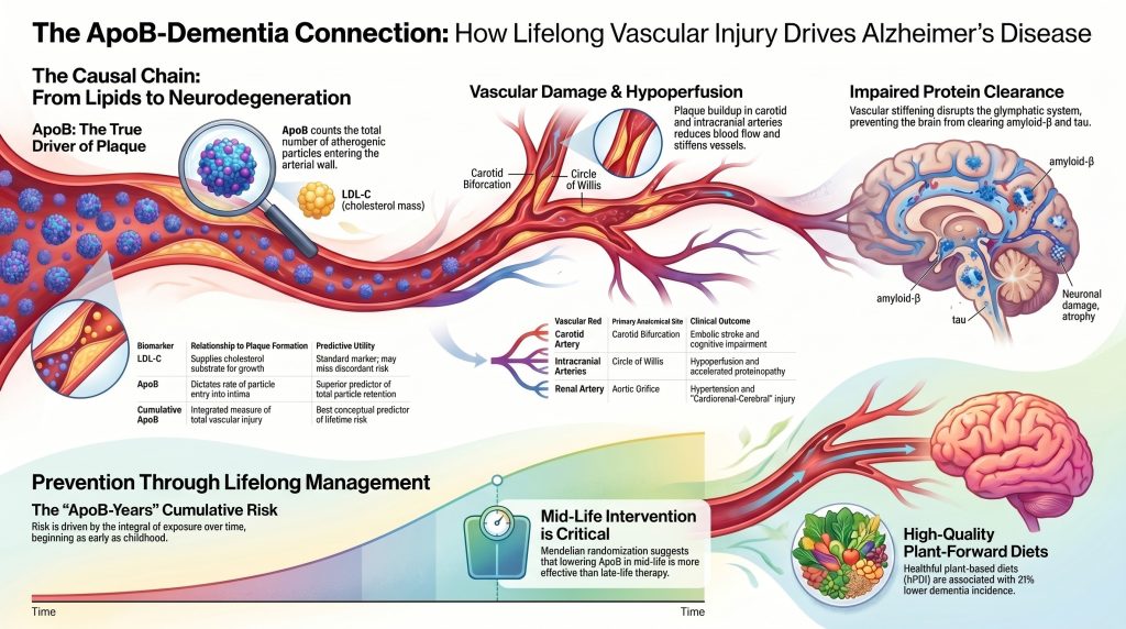
Athérosclérose induite par l'apolipoprotéine B comme base causale de la maladie d'Alzheimer
Une perspective multidisciplinaire sur l'axe cardiorénal-cérébral
Résumé
Contexte. Le cadre neurocentrique dominant définit la maladie d'Alzheimer (MA) sur le plan biologique par l'amyloïde et la tau biomarqueurs et traite la pathologie vasculaire comme un modificateur fréquent mais secondaire. Les données génétiques, épidémiologiques et d'imagerie étayent de plus en plus une autre lecture dans laquelle l'exposition tout au long de la vie à apolipoprotéine BApoB)-contenant lipoprotéines est un facteur causal en amont de l'atteinte cérébro-vasculaire et des petits vaisseaux qui, dans un sous-groupe substantiel de patients, précède et accélère la pathologie caractéristique de la MA.
Objectif. Synthétiser les données probantes actuelles reliant l'exposition à vie à l'apoB aux lésions athérosclérotiques dans les lits vasculaires carotidien, intracrânien et rénal et, par conséquent, au syndrome cognitif cliniquement diagnostiqué comme la maladie d'Alzheimer.
Méthodes. Structuré revue narrative d'études primaires indexées sur PubMed, Randomisation mendélienne analyses, des cohortes d'imagerie longitudinale, des essais randomisés de prévention cardiovasculaire et de la démence, ainsi que des déclarations de consensus actuelles de l'American Heart Association, de la National Lipid Association et de l'Alzheimer’s Association. Les allégations de causalité ont été évaluées par rapport aux normes de reporting STROBE-MR.29]
Conclusions. L'exposition tout au long de la vie aux lipoprotéines contenant l'ApoB est établie de manière causale comme un facteur déterminant de la maladie vasculaire athérogène. Des données convergentes issues de la randomisation mendélienne ciblant les médicaments randomisation chez plus d'un million d'individus,7] imagerie TEP longitudinale intracrânienne sténose,[13] et des essais randomisés de réduction du risque vasculaire [14] soutient une contribution causale de cette lésion vasculaire au déclin cognitif, à troubles cognitifs vasculaires et démence, et aux phénotypes de démence à pathologie mixte qui dominent dans les séries d'autopsies de sujets âgés.2,32L'extension d'une lésion cérébrovasculaire à la maladie d'Alzheimer confirmée par biomarqueurs telle qu'définie sous le Critères révisés 2024 du NIA-AA[40] est pris en charge, mais avec une confiance proportionnellement plus faible, et la langue de ce manuscrit est nuancée en conséquence à ce point final. Les critères NIA-AA de 2024 sont un cadre diagnostique, et non une affirmation étiologique ; ils sont entièrement compatibles avec un rôle pathogène indépendant établi pour l'amyloïde-β (démontré le plus directement par la MA familiale autosomique dominante)[41] et un rôle causal en amont de la lésion cérébrovasculaire induite par l'ApoB dans la maladie sporadique à début tardif. La réduction à vie de l'ApoB — par des régimes alimentaires de haute qualité privilégiant les végétaux et, le cas échéant, une pharmacothérapie initiée au milieu de la vie — est une stratégie de prévention défendable contre le déclin cognitif et un composant plausible de la prévention de la démence et de la MA.
Introduction
Le modèle neurocentrique dominant de la maladie d'Alzheimer, qui privilégie la cascade amyloïde-β comme initiateur principal de la neurodégénérescence, est de plus en plus contesté par des données génétiques, épidémiologiques et d'imagerie indiquant que la santé vasculaire n'est pas seulement un facteur comorbide mais un moteur amont fondamental du processus pathologique.1,5,17Cette analyse étudie l'hypothèse selon laquelle l'exposition à long terme à des taux élevés de lipoprotéines contenant de l'apolipoprotéine B (ApoB) entraîne une athérosclérose progressive plaque accumulation dans les artères carotides, intracrâniennes et rénales, déclenchant une cascade de lésions systémiques et locales qui aboutissent à une démence chez un sous-ensemble substantiel de patients.4,7,20,22En examinant les mécanismes qui se chevauchent de la formation de plaques dans tous les lits vasculaires et les inférences causales offertes par la randomisation mendélienne, un cadre rigoureux émerge pour comprendre la maladie d'Alzheimer comme la manifestation neurologique tardive de décennies de lésions vasculaires, lipidiques et inflammatoires.2,3,9]
Le langage causal est utilisé délibérément mais est calibré en fonction de la force des preuves à chaque maillon de la chaîne. L'ApoB est établie comme causale pour l'athérosclérose maladie cardiovasculaire par triangulation via la randomisation mendélienne multivariée,21,25analyses d'exposition cumulée,22,33] et des essais randomisés de diminution des lipides.[26L'extension de cette voie causale aux troubles cognitifs vasculaires et à la démence à pathologie mixte est largement étayée par la méthode de randomisation mendélienne ciblée par médicament pour le non-HDL-C et la démence chez plus d'un million d'individus7et par l'imagerie TEP longitudinale reliant la sténose intracrânienne initiale à l'accumulation ultérieure d'amyloïde et de tau.13] L'extension supplémentaire à la maladie d'Alzheimer confirmée par biomarqueurs spécifiquement — la MA telle que définie par les critères révisés 2024 du NIA-AA [40— est étayé par une randomisation mendélienne spécifique à l'ApoB et orientée de manière cohérente [4] et par la compatibilité mécanistique de la lésion vasculaire avec la biologie de l'amyloïde et de la tau, [16,17,42] mais avec une intensité proportionnellement plus faible que le lien cardiovasculaire. Le manuscrit adopte en conséquence un langage nuancé concernant le critère d'évaluation de la MA et distingue explicitement la démence toutes causes confondues, les troubles cognitifs vasculaires, la démence à pathologie mixte et la MA confirmée par biomarqueurs tout au long du texte.
L'hypothèse vasculo-athérosclérotique de la maladie d'Alzheimer et de la démence
L'hypothèse vasculo-athérosclérotique postule que le syndrome clinique de la maladie d'Alzheimer est, chez un sous-ensemble substantiel de patients, la conséquence en aval d'une hypoperfusion cérébrale chronique et d'un dysfonctionnement de l'unité neurovasculaire initiés par la grande artère athérosclérose et propagées à travers le réseau des petits vaisseaux cérébraux.1,5,17Dans ce cadre, le rétrécissement focal des artères carotides et intracrâniennes, associé à une perte d'élasticité artérielle, crée un environnement hémodynamique hostile à la survie neuronale et protéine dégagement.12,18]
Les preuves neuropathologiques contemporaines démontrent que la distinction catégorielle classique entre la démence vasculaire pure et la maladie d'Alzheimer pure est empiriquement intenable chez les sujets âgés.3,32Des études d'autopsie en communauté rapportent que des neuropathologies mixtes — toute combinaison de types Alzheimer, corps de Lewy, TDP-43 et vasculaires lésions — sont présentes dans la majorité des cerveaux âgés, et la protéinopathie pure sans pathologie cérébrovasculaire est l'exception plutôt que la règle.3,32Athérosclérose des gros vaisseaux, y compris la sténose carotidienne et maladie athéosclérotique intracrânienne, commonly coexists with cerebral small-vessel disease characterized by white-matter hyperintensities, lacunar infarcts, and microbleeds.[3,8,9]
Rigidité artérielle — en particulier dans le proximal aorte et les artères carotides — transmettent la pulsatilité à haute pression dans la microvasculature délicate du cerveau.12Ce flux hyperpulsatile endommage la barrière hémato-encéphalique et perturbe la système glymphatique, un réseau périvasculaire responsable de l'élimination des déchets métaboliques, notamment l'amyloïde-β et la tau.15,18Dans ce cadre, l'accumulation de ces protéines est, en partie, la conséquence d'une élimination altérée plutôt que la seule conséquence d'une surproduction.16,17,18]
Cette hypothèse n'est pas exclusive du risque génétique canonique de la MA. L'apolipoprotéine E ε4 (APOE allèle ε4 — le plus grand facteur génétique commun facteur de risque pour la MA à début tardif — agit principalement par des voies cérébrovasculaires. Des preuves directes d'imagerie chez l'homme démontrent que les porteurs d'APOE ε4 présentent une rupture de la barrière hémato-encéphalique qui précède et prédit le déclin cognitif indépendamment de l'amyloïde et du tau,42fournissant un pont mécaniste entre la biologie des lipoprotéines systémiques, la lésion cérébrovasculaire et le phénotype clinique de la MA.5,16,42]
Nous nous engageons explicitement avec les critères révisés de l'Alzheimer’s Association de 2024,40] qui définissent biologiquement la MA par des biomarqueurs centraux de l'amyloïde et du tau, indépendamment du syndrome clinique. Ces critères constituent un cadre diagnostique et non une affirmation étiologique, et le groupe de travail reconnaît les co-pathologies vasculaires et autres comme des modificateurs courants de l'évolution de la maladie. Notre position n'est donc pas que les critères de 2024 sont erronés ou qu'ils nient les contributions vasculaires — ils ne font ni l'un ni l'autre. Notre position est que l'empreinte biomarqueur qu'ils opérationnalisent a, pour un sous-ensemble substantiel de patients, une cause amont : des décennies de lésions cérébrovasculaires induites par l'ApoB interagissant avec la biologie intrinsèque de l'amyloïde et du tau. Définir la MA par son empreinte biomarqueur aval est parfaitement compatible avec l'attribution de cette empreinte, en partie, à des causes vasculaires amont.
Tableau 1. Caractéristiques pathologiques et hémodynamiques qui se chevauchent dans les sous-types de démence
| Fonctionnalité | Maladie d'Alzheimer (MA) | Démence vasculaire (DV) | Démence mixte |
| Protéinopathie primaire | plaques d'amyloïde-β ; enchevêtrements de tau | Minimal ou absent | Dépôts importants de Aβ et de tau |
| Lésions vasculaires | Souvent présent (CAA, micro-saignements) | Gros infarctus, lacunes, HBM | Vasculaire et protéinopathique combinés |
| Hémodynamique | Débit sanguin cérébral réduit ; défaillance glymphatique | Focal ou global ischémie | Hypoperfusion chronique + rigidité |
| Moteur lipidique | Lien de l'ApoB via la lésion cérébrovasculaire | Forte association LDL-C / ApoB | Charge élevée en ApoB tout au long de la vie |
| Caractéristiques d'imagerie | Atrophie hippocampique ; TEP-Aβ (+) | Infarctus multiples ; sténose carotidienne | Atrophie, HVB, charge athéromateuse |
Adapté des références [3,5,9,32,40]. AAC = angiopathie amyloïde cérébrale ; LBM = hyperintensités de la substance blanche ; FDC = débit sanguin cérébral.
Dynamique des lipoprotéines et exposition causale tout au long de la vie
La concentration des lipoprotéines contenant de l'ApoB dans la circulation est au cœur du développement de l'athérosclérose.10,19,20] L'ApoB est une protéine structurelle présente à la surface de chaque particule de lipoprotéine athérogène. Les lipoprotéines de très basse densité d'origine hépatique (VLDL), des lipoprotéines de densité intermédiaire (IDL), les lipoprotéines de basse densité (LDL), et lipoprotéine(a) chacun transporte une molécule de apoB-100 par particule, tandis que chylomicrons et les restes de chylomicrons transportent apoB-48 provenant de l'intestin.20Chez les adultes en état post-absorptif, la charge athérogène dominante reflétée par la mesure de l'apoB plasmatique est principalement composée de particules porteuses d'apoB-100.20] Au-delà de la distinction numérique, les particules de LDL (apoB-100) ont des temps de séjour intimaux sensiblement plus longs que les restes de chylomicrons (apoB-48), qui circulent de manière plus transitoire ; la fraction d'apoB-100 contribue donc de manière disproportionnée aux lésions cumulées de la paroi artérielle et domine le “Années-ApoB”intègre dans le lit cérébro-vasculaire.
Alors que cholestérol LDL (LDL-C) mesure le cholestérol masse au sein des particules de LDL, l'ApoB fournit un décompte direct du total particules athérogènes.[20] Cette distinction est cruciale sur le plan mécanistique : la probabilité qu'une particule de lipoprotéine pénètre dans l'artère intima et le fait d'être retenu par les protéoglycanes extracellulaires relève de la concentration des particules plutôt que de la teneur en cholestérol par particule.19Les analyses de mendélisation mendélienne multivariables ont établi que l'ApoB est le paramètre lipidique dominant dans la relation entre les lipoprotéines et maladie coronarienne; lorsque l'ApoB est contrôlée, ni le LDL-C ni triglycérides conserver des informations causales indépendantes pour la maladie cardiovasculaire athéosclérotique.21,25]
Le risque conféré par l'ApoB est cumulatif, un concept désormais formalisé sous le nom de LDL/ApoB exposition cumulative hypothèse.22] athérogénèse est un processus lent qui commence généralement dans l'enfance par la formation de naissants stries lipidiques.[19] Comme particules contenant de l'ApoB restent élevées sur plusieurs décennies, la charge de plaque athérosclérotique s'accumule progressivement jusqu'à ce que des seuils critiques soient franchis, entraînant un rétrécissement luminal ou plaque de rupture.[22,33] Exposition cumulée tout au long de la vie (souvent décrite comme “années-cholestérol”ou “ ApoB-années ” est un prédicteur biologiquement plus approprié du risque cardiovasculaire et, plausiblement, de démence que toute mesure unique au milieu ou à la fin de la vie, qui peut être faussée par causalité inverse chez les personnes âgées développant une fragilité et perte de poids dans la phase préclinique de la démence.22,24,28]
Tableau 2. Valeur prédictive comparative des biomarqueurs lipidiques pour le risque athérogène
| Biomarqueur | Définition | Relation avec la formation de plaque | Utilité |
| LDL-C | Masse de cholestérol dans les particules de LDL | Fournit un substrat de cholestérol pour la croissance de la plaque | Marqueur standard ; omet le risque dans discordance |
| ApoB | Nombre total de particules athérogènes (une par particule) | Détermine le taux de pénétration des particules dans l'intima | Meilleur prédicateur de la rétention des particules |
| Cholestérol non-HDL | Cholestérol total HDL-C bas | Capture le cholestérol LDL, VLDL, IDL et Lp(a) | Substitut pratique de la charge athérogène |
| Lp(a) | particule de type LDL avec apolipoprotéine(a) | Pro-thrombotique et pro-inflammatoire | Facteur de risque génétique indépendant |
| Exposition cumulée à l'apoB | Intégrale de l'ApoB au fil du temps (“ ApoB-années ”) | Mesure intégrée de la lésion vasculaire totale | Meilleur prédicteur conceptuel du risque à vie |
Adapté des références [19,20,21,22,25,33].
Revue des données de randomisation mendélienne dans la démence
La randomisation mendélienne (RM) met à profit la répartition aléatoire des allèles génétiques lors de la gamétogenèse pour estimer l'effet causal d'expositions modifiables sur des devenirs de santé, imitant ainsi la conception d'un essai contrôlé randomisé tout en évitant de nombreuses sources d'observation facteur de confusion.[29] Toutes les revendications RM résumées ci-dessous ont été évaluées par rapport à la déclaration STROBE-MR sur la communication transparente des enquêtes de randomisation mendélienne.29]
Analyses mendéliennes de la randomisation des instruments génétiques pour les cibles de médicaments hypolipémiants courants — HMGCR (statine cible), PCSK9 (Inhibiteur de PCSK9 cible), NPC1L1 (ézétimibe cible), et CETP — fournissent certaines des preuves causales les plus solides à ce jour établissant un lien entre un faible taux de cholestérol non-HDL tout au long de la vie et un risque moindre de démence. Dans une analyse MR à échantillon unique méta-analyse Sur un échantillon de 1 091 775 personnes, les rapports de cotes associés à une baisse de 1 mmol/L (≈39 mg/dL) du cholestérol non-HDL pour la démence toutes causes confondues étaient de 0,24 (IC à 95 % : 0,18–0,31) pour le gène HMGCR, de 0,18 (0,12–0,25) pour le gène NPC1L1, de 0,30 (0,26–0,34) pour le gène CETP et de 0,97 (0,70–1,35) pour le gène PCSK9.[7Une analyse danoise antérieure par mendelian randomization a rapporté des rapports de risque par baisse de 1 mmol/L du LDL-C de 0,57 (0,27–1,17) pour la maladie d'Alzheimer et de 0,81 (0,34–1,89) pour la démence vasculaire — ce qui est directionnellement cohérent avec une protection mais manque de puissance statistique pour les critères de jugement individuels.27]
Deux points méritent d'être explicitement reconnus. Premièrement, le critère d'évaluation principal de l'étude de Nordestgaard et al. est la démence toutes causes confondues plutôt que la maladie d'Alzheimer confirmée par des biomarqueurs selon les critères NIA-AA de 2024.40Deuxièmement, l'estimation de PCSK9 est compatible avec l'hypothèse nulle, contrairement aux signaux robustes pour HMGCR, NPC1L1 et CETP.7Cette hétérogénéité spécifique à la cible plaide contre un modèle simple dans lequel tout mécanisme pharmacologique qui abaisse les lipoprotéines contenant de l'apoB réduirait de manière équivalente le risque de démence, et pointe vers une biologie cérébrovasculaire spécifique à la cible que les futurs essais devront aborder.
Les preuves MR spécifiques concernant l'ApoB et la maladie d'Alzheimer sont plus récentes. La MR univariée à deux échantillons utilisant UK Biobank Les instruments ApoB et les statistiques sommaires de la MA d'IGAP montrent qu'un taux d'ApoB prédit génétiquement plus élevé est associé à un raccourcissement espérance de santé et — selon les termes prudents des auteurs eux-mêmes — pourraient éventuellement augmenter le risque de maladie d'Alzheimer.4La MR multivariée conditionnée sur le LDL-C indique que l'ApoB conserve son association avec une espérance de vie en bonne santé raccourcie même lorsque le LDL-C est nul dans le modèle conjoint.4Nous adoptons la propre caractérisation des auteurs : il s'agit de preuves causales favorables mais préliminaires au critère d'évaluation de la MA, qui devraient être répliquées par rapport à des résultats de la MA confirmés par biomarqueurs au fur et à mesure que de telles données seront disponibles.
Limites de la randomisation mendélienne
Plusieurs limites s'appliquent à toutes les données de RM présentées ci-dessus et doivent guider l'interprétation causale :
- Biais du survivant les personnes présentant un taux d'ApoB génétiquement élevé peuvent mourir d'événements cardiovasculaires prématurés avant d'atteindre l'âge auquel la démence se manifesterait cliniquement, atténuant potentiellement l'association observée entre les lipides et la maladie d'Alzheimer.4,7]
- Risque de décès compétitif : plus spécifiquement que le biais de survie général, l'ApoB est si fortement causale pour infarctus du myocarde et accident vasculaire cérébral que les individus présentant un taux d’ApoB génétiquement élevé, qui auraient autrement développé la maladie d’Alzheimer, sont retirés de manière disproportionnée de la population éligible à la maladie d’Alzheimer en raison d’une mortalité cardiovasculaire précoce. Cette structure de risques concurrents atténue systématiquement l’association observable entre les lipides et la MA dans les plans d’étude standard de randomisation mendélienne, et constitue l’une des raisons spécifiques pour lesquelles le signal concernant le critère d’évaluation de la MA dans les études actuelles de randomisation mendélienne est plus faible que le signal cardiovasculaire — ce qui n’est pas nécessairement dû à la faiblesse du mécanisme causal.[4,27]
- Pléiotropie : Les variantes génétiques peuvent influencer le résultat par des mécanismes autres que l'exposition visée. Les études citées ont utilisé les estimateurs MR-Egger, la médiane pondérée et les estimateurs MR multivariables pour évaluer la robustesse ; les analyses conditionnelles à l'ApoB conservent un signal cohérent sur le plan directionnel.[4,21,25]
- Hétérogénéité des résultats : la MA diagnostiquée cliniquement dans les bases de données administratives inclut fréquemment une copathologie vasculaire substantielle ;3,32] critères d'évaluation de la MA confirmés par biomarqueurs selon le cadre NIA-AA de 2024 [40] renforcerait les futurs travaux de MR. Les preuves de MR ciblées par les médicaments pour la démence de toutes causes[7] est méthodologiquement plus solide, mais s'applique à la démence en général, et non spécifiquement à la MA confirmée par biomarqueurs.
- Ascendance et généralisabilité : La plupart des études d'IRM portant sur les lipides et la démence ont été menées auprès de populations d'ascendance européenne ; les estimations causales peuvent varier selon les groupes d'ascendance.[7]
- Interprétation de l'exposition à vie : L'IRM estime l'effet cumulatif d'une exposition génétique tout au long de la vie, qui dépasse généralement l'effet pouvant être atteint par une pharmacothérapie initiée tard dans la vie. Cette différence est informative quant au moment de la prévention, mais ne se traduit pas directement par les tailles d'effet attendues des essais sur la démence menés tard dans la vie.22,26]
Physiopathologie régionale de la formation de la plaque athéromateuse
L'athérosclérose est une maladie inflammatoire chronique amorcée par la rétention de lipides dans des zones focales des artères, en particulier les régions soumises à un flux perturbé et non laminaire.10,19Les mécanismes moléculaires et cellulaires de la formation des plaques sont largement conservés dans les principaux lits vasculaires, mais l'anatomie locale et l'hémodynamique confèrent des caractéristiques distinctives à la maladie dans chaque localisation.10,11,31]
Athérosclérose carotidienne
La bifurcation carotide est particulièrement vulnérable à la formation de plaques en raison de sa géométrie et de son flux complexes. inversion pendant le cycle cardiaque.10,11] Faible et oscillatoire contrainte de cisaillement à la paroi à l'origine de l'interne artère carotide promeut dysfonction endothéliale et augmente la perméabilité de la paroi vasculaire aux lipoprotéines contenant de l'ApoB.10,11,19Une caractéristique distinctive de la plaque carotidienne est sa tendance à emboliser : un flux à haute vitesse déloge les thrombus se formant sur les surfaces de plaques ulcérées, générant des micro-emboles récurrents plutôt qu'une occlusion proximale complète occlusion.[11,34Lésion microembolique cumulative, infarctus lacunaires et microhémorragies sont les substrats fondamentaux des troubles cognitifs vasculaires.2,9,34]
Maladie athérosclérotique intracrânienne
L'ICAD désigne la formation de plaques dans les grandes artères à la base du cerveau, y compris l'artère cérébrale moyenne artère, artère basilaire et polygone de Willis.11,13Contrairement à la maladie carotidienne extracrânienne, l'ICAD a été directement liée, dans l'imagerie longitudinale prospective, à un dépôt ultérieur d'amyloïde-β et de tau dans le parenchyme cérébral. Dans un cohorte prospective avec l'imagerie TEP au PiB (amyloïde) et à l'AV-1451 (tau) en série, la présence d'une sténose intracrânienne quelconque à l'inclusion était significativement associée à une plus grande accumulation d'amyloïde-β sur quatre ans, et la sténose dans deux artères ou plus prédisait une plus grande déposition de tau sur deux ans.13Cette directionnalité longitudinale — une lésion vasculaire précédant une protéinopathie accélérée — constitue l'une des preuves d'imagerie humaine actuelles les plus solides indiquant que la santé vasculaire intracrânienne est causalement en amont de la pathologie déterminante de la maladie d'Alzheimer, plutôt que de simplement coïncider avec elle.
Athérosclérose de l'artère rénale et axe cardiorénal-cérébral
La sténose de l'artère rénale (SAR) est d'origine essentiellement athéroscléreuse et se concentre au niveau de l'orifice aortique ou dans le tiers proximal de l'artère rénale principale.31,37Le rein, tout comme le cerveau, est un lit vasculaire à faible résistance sensible au flux hyperpulsatile et à l'ischémie.6,12] La progression de la plaque dans l'artère rénale est accentuée par le tissu adipeux périvasculaire dysfonctionnel et l'expansion du vasa vasorum, qui libèrent des cytokines pro-inflammatoires et des vésicules extracellulaires.31]
La sténose de l'artère rénale athéromateuse active le système rénine-angiotensine-aldostérone, produisant une seconde hypertension cela accélère davantage les dommages causés à la microvasculature cérébrale et établit une boucle de rétroaction positive reliant les reins, le cœur et le cerveau.6,31,36,39] Maladie rénale chronique est lui-même caractérisé par une dimension systémique inflammation, stress oxydatif, et la toxicité endothéliale urémique, qui sont toutes indépendamment associées au déclin cognitif par le biais de l'axe cardiorénal-cérébral.6,38,39]
Tableau 3. Comparaison des caractéristiques des plaques selon les lit vasculaires
| Lit vasculaire | Site anatomique primitif | Déterminant hémodynamique unique | Critère de jugement clinique principal |
| Artère carotide | Bifurcation carotidienne ; origine de la CCl | Inversion du flux et turbulence ; cisaillement faible/oscillatoire | Accident vasculaire cérébral embolique et troubles cognitifs |
| Artères intracrâniennes | ACM, basilaire, polygone de Willis | Ramification complexe ; perte d'autorégulation | Hypoperfusion et protéinopathie accélérée |
| Artère rénale | Orifice aortique / tiers proximal | Contrainte de cisaillement liée à l'orifice | RAS, hypertension, MRC |
| Artère coronaire | IVA proximale et bifurcations | Contrainte de compression cyclique ; flexion | Infarctus du myocarde et angine |
Adapté des références [10,11,13,31,37].
La voie mécaniste : des lipides à la neurodégénérescence
La transition entre une élévation de l'ApoB circulante et la démence clinique implique une cascade à plusieurs étapes dans laquelle rétention sous-endothéliale des lipoprotéines contenant l'ApoB sert d'événement initiateur.19] Les particules retenues subissent des modifications oxydatives et enzymatiques, générant de puissants signaux pro-inflammatoires qui activent cellules endothéliales, mobilisent les monocytes et stimulent macrophage formation de cellules de mousse.[10,19,53]
Rétention lipidique, activation endothéliale et hypoperfusion
Les cellules endothéliales réagissent à la présence de lipoprotéines oxydées en exprimant des molécules d'adhésion (VCAM-1, ICAM-1) et en libérant des chimiokines (MCP-1, CXCL1) et des cytokines (IL-1β, IL-6).[10,19,53] La progression de la plaque entraîne un rétrécissement et un durcissement des artères,[10,12] ce qui, au niveau de l'arbre vasculaire cérébral, se traduit par une diminution du débit sanguin cérébral et une ischémie chronique.[12,17] L'hypoperfusion chronique provoque un stress métabolique chez les neurones et les cellules gliales, et active le inflammasome NLRP3, et prédispose le cerveau à une neurodégénérescence accélérée.[17,53]
L'amyloïde-β dans la voie vasculaire
Un élément central du modèle vasculaire réside dans le fait que l'accumulation d'amyloïde-β (Aβ) est, en partie, à la fois une cause et une conséquence des lésions cérébrovasculaires.[16,17] L'ischémie et le stress oxydatif entraînent une augmentation de l'expression de la protéine précurseur amyloïde et orientent son traitement vers la voie amyloïdogène, [16,17] tandis que les dépôts vasculaires d'Aβ (angiopathie amyloïde cérébrale, CAA) altèrent la contractilité de la paroi vasculaire et réduisent l'efflux de protéines hors du cerveau.[16] Cela engendre une boucle de rétroaction positive : les lésions vasculaires favorisent l'accumulation d'Aβ, ce qui entraîne des lésions vasculaires supplémentaires et une neuroinflammation par le biais de l'activation du système immunitaire inné.[16,17,53]
Nous précisons toutefois que l’amyloïde n’est pas simplement un débris résultant d’une lésion vasculaire. La maladie d’Alzheimer familiale autosomique dominante, causée par des mutations des gènes APP, PSEN1 ou PSEN2, établit que l’amyloïde est un facteur pathogène suffisant en soi : ces mutations provoquent la maladie d’Alzheimer par le biais d’une altération de la biologie de l’amyloïde, sans qu’une atteinte vasculaire en amont soit nécessaire.[41] Les essais cliniques sur les anticorps monoclonaux anti-amyloïdes dans la MA sporadique, abordés ci-dessous dans la section « Contre-arguments », démontrent en outre que l’élimination de l’amyloïde produit un bénéfice clinique statistiquement significatif, ce qui est incompatible avec un modèle dans lequel l’amyloïde serait un épiphénomène. Notre affirmation est donc plus restreinte et plus défendable : dans la MA sporadique à début tardif — qui représente la grande majorité des cas —, des décennies de lésions cérébrovasculaires induites par l’ApoB constituent un déterminant en amont majeur de l’environnement cérébral dans lequel la biologie intrinsèque de l’amyloïde et de la protéine tau devient pathogène. Les voies vasculaires et amyloïdes interagissent et s’amplifient de manière bidirectionnelle, plutôt que de s’exclure mutuellement.
Dysfonctionnement glymphatique et élimination des protéines
L'élimination des déchets métaboliques du cerveau est assurée en partie par le système glymphatique, un réseau périvasculaire d'échange entre le LCR et le liquide interstitiel dont le fonctionnement dépend de la pulsatilité artérielle et d'un écoulement veineux stable.[15,18] Le durcissement artériel — une conséquence mécanique directe de l'athérosclérose et vieillissement vasculaire — altère la force motrice pulsatile, [12] Des études menées chez les rongeurs et chez l'homme ont montré que l'hypertension et le vieillissement réduisent l'afflux de LCR dans l'espace périvasculaire.[18] Lorsque la clairance glymphatique est altérée, des protéines neurotoxiques, notamment l'Aβ et la protéine tau, s'accumulent, ce qui contribue à un dysfonctionnement synaptique et à un déclin cognitif.[12,15,18] Nous considérons la chaîne « rigidité artérielle → altération de la clairance glymphatique → MA » comme une hypothèse plausible sur le plan mécanistique et partiellement étayée, plutôt que comme un mécanisme clinique établi. La physiologie glymphatique humaine n’est pas encore entièrement validée, et l’indice d’analyse par imagerie par tenseur de diffusion le long de l’espace périvasculaire (DTI-ALPS) constitue un indicateur d’imagerie de la diffusivité périvasculaire plutôt qu’une mesure directe du flux glymphatique ; sa reproductibilité et sa spécificité biologique font l’objet de travaux méthodologiques actifs.[35]
Vérification de l'hypothèse de la protection à vie
Précoce, durable hypolipémiant est essentiel pour prévenir une neurodégénérescence irréversible.[22,26] Les études de génomique associative (MR) constituent en effet un modèle naturel de faibles taux de LDL-C et d’ApoB tout au long de la vie, démontrant que les personnes présentant une exposition génétiquement plus faible ont un risque cardiovasculaire à vie nettement inférieur à celui des personnes qui ne commencent un traitement hypolipidémiant qu’à un âge avancé.[22,25]
D'un point de vue quantitatif, essais cliniques Les études menées dans le cadre de la Cholesterol Treatment Trialists’ Collaboration montrent une réduction relative d’environ 22% des événements vasculaires majeurs pour chaque baisse de 1 mmol/L (≈39 mg/dL) du taux de LDL-C obtenue grâce à un traitement aux statines, quel que soit le risque initial.[26] En revanche, l'étude MR estime que la réduction du risque de maladie coronarienne est nettement plus importante pour chaque mmol/L de LDL-C génétiquement plus bas tout au long de la vie — soit environ trois fois le bénéfice par mmol/L observé avec un traitement aux statines à un âge avancé.[22,33] Nous qualifions clairement cette différence d’inférence concernant l’importance de la durée : elle suggère que, pour prévenir la démence, les interventions devraient idéalement avoir lieu à l’âge mûr, avant que ne se développent des plaques matures et le substrat cérébrovasculaire à l’origine du déclin cognitif.[22,40] Une extrapolation directe de l'estimation cardiovasculaire par mmol/L aux résultats relatifs à la démence n'est pas justifiée en l'absence de données issues d'essais cliniques ; nous présentons donc une estimation temporelle plutôt qu'une prévision chiffrée de la réduction des cas de démence.
Les essais randomisés portant sur la réduction du risque vasculaire apportent des données complémentaires, bien que d'une ampleur moindre. Dans SPRINT Dans l'étude MIND, un contrôle intensif de la tension artérielle (objectif : TAS < 120 mmHg) par rapport à un contrôle standard (< 140 mmHg) a réduit le risque composite de troubles cognitifs légers ou de démence probable (rapport des cotes 0,85, IC 95 % 0,74–0,97) et une réduction spécifique de la déficience cognitive légère (MCI) (HR 0,81, IC 95 % 0,69–0,94), bien que le critère d'évaluation principal, à savoir la démence probable, n'ait pas atteint le seuil de signification statistique.[14] Dans l'essai clinique FINGER sur la prévention multidomaine, une intervention combinée de deux ans associant régime alimentaire, activité physique, entraînement cognitif et suivi des risques vasculaires a permis d'améliorer les scores cognitifs composites par rapport au groupe témoin chez les personnes âgées à risque.[50] Ces essais démontrent que la modification aléatoire des facteurs de risque vasculaires peut entraîner des bénéfices cognitifs mesurables, ce qui vient étayer l'idée générale selon laquelle la réduction des risques vasculaires tout au long de la vie contribue à la prévention de la démence.
Le rôle des régimes alimentaires à base de végétaux
Un régime alimentaire à base de végétaux de haute qualité constitue une stratégie non pharmacologique efficace pour réduire l'exposition à l'ApoB tout au long de la vie et l'inflammation vasculaire systémique.[23,46,49] La qualité de l'alimentation est importante : a indice d'une alimentation saine à base de végétaux (hPDI), qui privilégie les céréales complètes, les fruits, les légumes, les légumineuses et les fruits à coque, est associé à un risque moindre de démence, tandis qu’un régime végétal malsain (uPDI), riche en céréales raffinées et en boissons sucrées, est associé à un risque plus élevé.[43,46]
Les données méta--analyses issues de cohortes prospectives (n ≈ 208 000) montrent que le tertile supérieur de l’hPDI est associé à une incidence de démence inférieure d’environ 21% par rapport au tertile inférieur, tandis que le tertile supérieur de l’uPDI est associé à une incidence de démence supérieure d’environ 26%.[43] Chez les adultes atteints d'une maladie cardiométabolique avérée — une population chez laquelle l'axe cardiorénal-cérébral est déjà activé —, une alimentation saine alimentation à base de plantes Ce profil, associé à des facteurs plus généraux liés à un mode de vie sain, réduit considérablement le risque de démence dans le cadre du suivi de la UK Biobank.[45] Parmi les régimes alimentaires à prédominance végétale, le régime méditerranéen et Les régimes MIND disposent actuellement des données probantes cumulées les plus solides, notamment des données issues d'essais randomisés sur les résultats cardiovasculaires (PREDIMED)[49] ainsi que des associations observées de manière constante avec un risque moindre de démence ;[44] Les régimes strictement à base d’aliments végétaux non transformés ou végétaliens s’appuient sur des preuves directes moins solides en ce qui concerne spécifiquement les critères d’évaluation de la démence ou de la maladie d’Alzheimer. Nous considérons donc les régimes alimentaires privilégiant les végétaux, au sens large, comme une stratégie de prévention biologiquement bien fondée, étayée par des données observationnelles solides et (pour régime méditerranéen) issues d’études cardiovasculaires randomisées, et nous n’affirmons pas la supériorité des régimes strictement à base d’aliments végétaux non transformés ou végétaliens par rapport aux régimes méditerranéen ou MIND, sur la base des données actuellement disponibles concernant la démence.
Au-delà de la réduction de l'ApoB en soi, les effets neuroprotecteurs des régimes alimentaires végétaux de haute qualité s'exercent par le biais de mécanismes complémentaires : une amélioration fonction endothéliale et une diminution du stress oxydatif ;[23,46] une modulation favorable de la microbiote intestinal avec une production réduite de N-oxyde de triméthylamine (TMAO), un métabolite associé à l'athérosclérose et aux troubles cognitifs ;[47,48] et amélioré tension artérielle, sensibilité à l'insuline, et le niveau d’inflammation.[23,49,50] Les régimes méditerranéen et MIND partagent bon nombre de ces mécanismes et sont étayés par des données issues à la fois d'études observationnelles et d'essais randomisés concernant les résultats cardiovasculaires et les critères d'évaluation cognitifs.[44,49,50]
Tableau 4. Impact des habitudes alimentaires sur les facteurs de risque vasculaires et cognitifs
| Habitudes alimentaires | Éléments clés | Effet sur l'ApoB | Effet sur l'inflammation | Risque de démence |
| Alimentation saine à base de végétaux (hPDI) | Céréales complètes, légumineuses, fruits à coque | En baisse | En baisse | Inférieur [43,45] |
| Méditerranée | Huile d'olive, plantes, poissons, fruits à coque | Légère baisse | Légère baisse | Inférieur [44,49] |
| MIND | Légumes, baies, poisson, huile d'olive | Légère baisse | En baisse | Inférieur [44] |
| Aliments d'origine végétale malsains (uPDI) | Céréales raffinées, sucreries, frites | Variable / neutre | Augmenté | Plus haut43] |
| Occidental | Viande rouge et viande transformée, sucres raffinés | Augmenté | Augmenté | Plus haut23,46] |
Adapté des références [23,43,44,45,46,49].
Contre-arguments et réconciliation avec la définition biologique de la maladie d'Alzheimer
L'hypothèse selon laquelle l'athérosclérose induite par l'ApoB serait un facteur causal en amont de la maladie d'Alzheimer doit tenir compte de trois contre-arguments valables : (1) le paradoxe du faible taux de cholestérol chez les personnes âgées, (2) les cas de neurodégénérescence sans maladie vasculaire manifeste, et (3) le succès thérapeutique partiel des anticorps monoclonaux anti-amyloïdes, qui semblent étayer un modèle attribuant la cause à l’amyloïde.
Le paradoxe du faible taux de cholestérol chez les personnes âgées
Des études observationnelles montrent fréquemment qu'un faible taux de cholestérol chez les adultes âgés de 75 ans et plus est associé à un risque accru de démence.[24,28] L'explication la plus simple est l'inverse causalité: la démence préclinique est précédée de plusieurs années de perte de poids involontaire et de fragilité, et, aux stades avancés de la neurodégénérescence, parfois d'un dysfonctionnement de la synthèse hépatique, tous ces facteurs réduisant le taux de cholestérol circulant indépendamment de tout effet protecteur lié à une faible exposition à l'ApoB tout au long de la vie.[24,28] Ce paradoxe met en évidence pourquoi les mesures du cholestérol à l'âge mûr et les marqueurs génétiques observés tout au long de la vie — et non les taux de cholestérol chez les personnes âgées — constituent la base appropriée pour établir un lien de causalité entre l'ApoB et la démence.[22,29]
La MA non vasculaire et l'architecture génétique
La maladie d'Alzheimer, de nature essentiellement neurodégénérative, résulte de mutations autosomiques dominantes spécifiques des gènes APP, PSEN1 et PSEN2, [41] Parmi les autres facteurs contributifs, on peut citer les infections chroniques, le dysfonctionnement mitochondrial, les troubles respiratoires du sommeil et la privation chronique de sommeil.[41] Il ne s'agit pas là de réfutations d'un modèle causal vasculaire ; ce sont simplement des rappels du fait que la maladie d'Alzheimer est un syndrome hétérogène dans lequel l'athérosclérose est un facteur causal majeur chez une partie importante des patients, et non la cause exclusive chez tous.[3,32,41] Le Allèle APOE ε4 augmente le risque de MA en grande partie par le biais de mécanismes cérébrovasculaires — notamment la rupture de la barrière hémato-encéphalique qui précède le déclin cognitif, [42] une altération de l'élimination de l'Aβ et une augmentation de la CAA[16] — ce qui montre que les risques génétiques et vasculaires sont étroitement liés plutôt que concurrents.
Essais cliniques sur les anticorps monoclonaux anti-amyloïdes
Lecanemab et donanemab constituent l'essai le plus direct portant sur la voie amyloïde dans la maladie symptomatique. Dans l'étude CLARITY AD, le lécanemab a ralenti le déclin du score CDR-SB de 0,45 point par rapport à placebo sur une période de 18 mois, sur une échelle de 18 points (moyenne de 1,21 contre 1,66 ; ralentissement relatif de 27%), avec des anomalies d'imagerie liées à l'amyloïde (ARIA-E) chez 12,6% et des ARIA-E symptomatiques chez 2,8%.[51] Dans l'étude TRAILBLAZER-ALZ 2, le donanemab a ralenti la progression de l'iADRS de 35% et celle du CDR-SB de 36% sur une période de 76 semaines ; une ARIA-E est survenue chez 24,0% patients (dont 6,1% présentaient des symptômes), avec trois décès liés au donanemab et imputables à une ARIA.[52]
Nous considérons ces essais comme des preuves solides démontrant que l’amyloïde joue un rôle mécanistique important dans la MA symptomatique : l’élimination de l’amyloïde entraîne un ralentissement mesurable, statistiquement significatif et constant du déclin cognitif, tant au niveau des critères d’évaluation primaires que secondaires. Ce résultat n’est pas compatible avec un modèle dans lequel l’amyloïde serait un débris vasculaire épiphénoménal, et nous n’adoptons pas un tel modèle. La question de savoir si l’ampleur absolue de ce ralentissement atteint une différence minimale cliniquement significative (MCID) fait véritablement l’objet d’un débat plutôt que d’un consensus, les MCID proposées par le CDR-SB pour la MA précoce s’étendant approximativement de 0,5 à 2,0 points, et différents groupes de travail se situant à des niveaux différents de cette fourchette. Des personnes raisonnables divergent quant à savoir si les effets du lécanemab et du donanemab franchissent ce seuil.
Ce que ces essais ne démontrent pas, c’est que l’élimination de l’amyloïde à un stade avancé et symptomatique de la maladie soit suffisante pour stopper ou inverser la progression de la MA : les effets sont progressifs, le profil de sécurité lié à l’ARIA n’est pas négligeable et se concentre chez les homozygotes APOE ε4, et les essais portent sur l’élimination de l’amyloïde à un stade avancé plutôt que sur une prévention en amont. Ces résultats sont tout à fait compatibles avec notre approche : l’amyloïde revêt une importance mécanistique et constitue une cible thérapeutique légitime ; des décennies de lésions cérébrovasculaires en amont induites par l’ApoB constituent un déterminant majeur supplémentaire et modifiable du substrat sur lequel opère la pathologie amyloïde ; et une stratégie complète de prévention de la démence doit raisonnablement miser sur ces deux voies.
Participation aux travaux de la NIA-AA sur la définition biologique de la maladie d'Alzheimer pour 2024
Les critères révisés de l’Association Alzheimer pour 2024 définissent la maladie d’Alzheimer sur le plan biologique à partir de biomarqueurs clés liés à l’amyloïde et à la protéine tau, indépendamment du syndrome clinique.[40] Ces critères constituent un cadre diagnostique, et non une théorie étiologique. Le groupe de travail ne prétend pas que l’amyloïde et la protéine tau soient les seules causes en amont de la MA et ne nie pas l’implication vasculaire — en effet, les pathologies vasculaires et autres comorbidités sont explicitement reconnues comme des facteurs communs modifiant l’évolution de la maladie. Nous ne contestons donc pas les critères de 2024 ; nous avançons un argument étiologique complémentaire compatible avec ceux-ci. Pour une partie importante des patients répondant aux critères biologiques de la MA, la cause en amont de l’empreinte biomarqueur amyloïde/tau est, selon notre interprétation, une athérogenèse cérébrovasculaire induite par l’ApoB tout au long de la vie, interagissant avec la biologie intrinsèque de l’amyloïde. Des données longitudinales montrent que l’athérosclérose intracrânienne prédit une accumulation ultérieure d’amyloïde et de protéine tau[13] est tout à fait compatible avec cette interprétation.
Modèles de recherche proposés pour tester l'hypothèse
Pour valider rigoureusement l’hypothèse causale vasculo-athéroscléreuse, il est nécessaire de mener des études longitudinales intégrant des marqueurs cardiovasculaires et neurodégénératifs tout au long de la vie.[9,40] Les études devraient suivre des personnes âgées de 30 à 40 ans et effectuer des mesures répétées des taux d'ApoB, de Lp(a) et des marqueurs inflammatoires.[20,22] L'imagerie haute résolution — notamment l'échographie avec produit de contraste et l'IRM de la paroi vasculaire — devrait permettre de suivre la progression et la régression des plaques dans les artères carotides et intracrâniennes.[11,13] La neuroimagerie de pointe, notamment la TEP à l’amyloïde et à la protéine tau, ainsi que les nouveaux indicateurs de la diffusivité périvasculaire reflétant la fonction glymphatique, permettrait de mettre en évidence comment les lésions vasculaires précèdent l’accumulation de protéines.[13,15,18,35]
Les nouvelles études de recherche clinique devraient isoler l'ApoB des autres paramètres lipidiques à l'aide d'outils multivariables et examiner son effet indépendant sur les critères d'évaluation de la MA confirmés par des biomarqueurs, conformément aux critères NIA-AA de 2024.[4,40] Des essais cliniques randomisés axés sur la prévention sont nécessaires pour déterminer si une réduction précoce et intensive du taux d’ApoB — mise en place à l’âge mûr plutôt qu’à un âge avancé — permet de réduire le déclin cognitif ultérieur et la charge en protéines neurodégénératives.[7,22,26] Les essais devraient être stratifiés en fonction du statut APOE ε4 et de la charge cérébrovasculaire initiale,[16,42] et doivent respecter les principes de publication STROBE-MR ou CONSORT, selon le cas.[29]
Thèse finale et conclusion
Le déclin cognitif, les troubles cognitifs d’origine vasculaire, la démence, la démence à pathologies mixtes et — chez une partie importante des patients — la maladie d’Alzheimer définie sur le plan biologique peuvent être considérés, de manière constructive, comme des manifestations neurologiques tardives d’une atteinte vasculaire, lipidique et inflammatoire s’étendant sur plusieurs décennies, agissant de concert avec la biologie intrinsèque de l’amyloïde et de la protéine tau.[1,2,3,17,40] Le facteur déterminant central et modifiable de cette voie vasculaire est l’exposition tout au long de la vie aux lipoprotéines contenant de l’ApoB, qui déclenche et favorise la formation de plaques athéroscléreuses dans les artères rénales et celles qui irriguent le cerveau.[7,20,22,31] Ces plaques entraînent une hypoperfusion cérébrale chronique, un durcissement artériel, un dysfonctionnement de la barrière hémato-encéphalique et une altération de la clairance glymphatique, ce qui contribue à l’accumulation d’amyloïde et de protéine tau ainsi qu’au déclin cognitif clinique, en interaction avec les mécanismes intrinsèques de la neurodégénérescence.[12,13,16,17,18,42]
Notre argument n’est donc pas que la maladie d’Alzheimer doive être reclassée comme une maladie vasculaire, ni que l’amyloïde soit sans importance sur le plan mécanistique. La MA familiale, [41] résultats d’un essai clinique sur les traitements anti-amyloïdes dans la MA sporadique, [51,52] et les critères biologiques de la NIA-AA pour 2024[40] établissent toutes que la biologie de l’amyloïde est une composante légitime et nécessaire de tout modèle complet de la MA. Notre argumentation est plus ciblée : pour un sous-groupe important de patients répondant aux critères biologiques de la MA, la cause en amont de l’empreinte amyloïde/tau réside, en grande partie, dans des décennies de lésions cérébrovasculaires induites par l’ApoB. Ce cadre est pleinement compatible avec les critères biologiques 2024 de la NIA-AA, qui définissent la MA sur le plan diagnostique par les biomarqueurs amyloïdes et tau sans émettre d’hypothèses étiologiques sur la cause en amont de ces biomarqueurs.
En pratique, cela signifie que la stratégie de prévention de la maladie d’Alzheimer et des démences associées devrait être élargie afin d’accorder une importance nettement plus grande à la préservation de l’intégrité vasculaire tout au long de la vie — grâce à des habitudes alimentaires de haute qualité, privilégiant les végétaux, [43,44,45] des stratégies randomisées de réduction du risque vasculaire, telles que celles validées dans les études SPRINT MIND et FINGER,[14,50] et, le cas échéant, un traitement médicamenteux hypolipidémiant instauré à l'âge mûr plutôt qu'à un âge avancé[7,22,26] — parallèlement à la poursuite des investissements dans les thérapies ciblant les protéines à un stade avancé. Ces deux axes d’intervention sont justifiés par les données scientifiques actuelles ; l’approche vasculaire en amont offre le bénéfice le plus important en matière de santé publique à long terme et constitue le sujet central de cet article.
Références
- Iadecola C. The pathobiology of vascular dementia. Neuron. 2013;80(4):844-866. doi:10.1016/j.neuron.2013.10.008
- Smith EE, Aparicio HJ, Gottesman RF, et al. Vascular Contributions to Cognitive Impairment and Dementia in the United States: Prevalence and Incidence: A Scientific Statement From the American Heart Association. Stroke. 2025;56(10):e317-e330. doi:10.1161/STR.0000000000000494
- Kapasi A, DeCarli C, Schneider JA. Impact of multiple pathologies on the threshold for clinically overt dementia. Acta Neuropathol. 2017;134(2):171-186. doi:10.1007/s00401-017-1717-7
- Martin L, Boutwell BB, Messerlian C, Adams CD. Mendelian randomization reveals apolipoprotein B shortens healthspan and possibly increases risk for Alzheimer’s disease. Commun Biol. 2024;7(1):230. Published 2024 Feb 24. doi:10.1038/s42003-024-05887-2
- Sweeney MD, Sagare AP, Zlokovic BV. Blood-brain barrier breakdown in Alzheimer disease and other neurodegenerative disorders. Nat Rev Neurol. 2018;14(3):133-150. doi:10.1038/nrneurol.2017.188
- Bugnicourt JM, Godefroy O, Chillon JM, Choukroun G, Massy ZA. Cognitive disorders and dementia in CKD: the neglected kidney-brain axis. J Am Soc Nephrol. 2013;24(3):353-363. doi:10.1681/ASN.2012050536
- Nordestgaard LT, Hanson A, Sanderson E, et al. Cholesterol-lowering drug targets reduce risk of dementia: Mendelian randomization and meta-analyses of 1 million individuals. Alzheimers Dement. 2025;21(10):e70638. doi:10.1002/alz.70638
- Bos D, Vernooij MW, de Bruijn RF, et al. Atherosclerotic calcification is related to a higher risk of dementia and cognitive decline. Alzheimers Dement. 2015;11(6):639-47.e1. doi:10.1016/j.jalz.2014.05.1758
- Wardlaw JM, Smith C, Dichgans M. Small vessel disease: mechanisms and clinical implications. Lancet Neurol. 2019;18(7):684-696. doi:10.1016/S1474-4422(19)30079-1
- Libby P, Buring JE, Badimon L, et al. Atherosclerosis. Nat Rev Dis Primers. 2019;5(1):56. Published 2019 Aug 16. doi:10.1038/s41572-019-0106-z
- Yahagi K, Kolodgie FD, Lutter C, et al. Pathology of Human Coronary and Carotid Artery Atherosclerosis and Vascular Calcification in Diabetes Mellitus. Arterioscler Thromb Vasc Biol. 2017;37(2):191-204. doi:10.1161/ATVBAHA.116.306256
- Mitchell GF, van Buchem MA, Sigurdsson S, et al. Arterial stiffness, pressure and flow pulsatility and brain structure and function: the Age, Gene/Environment Susceptibility–Reykjavik study. Brain. 2011;134(Pt 11):3398-3407. doi:10.1093/brain/awr253
- Kang KM, Park C, Nam H, et al. Intracranial stenosis and longitudinal progression of Alzheimer’s disease pathologies. Alzheimers Dement. 2025;21(12):e71010. doi:10.1002/alz.71010
- SPRINT MIND Investigators for the SPRINT Research Group, Williamson JD, Pajewski NM, et al. Effect of Intensive vs Standard Blood Pressure Control on Probable Dementia: A Randomized Clinical Trial. JAMA. 2019;321(6):553-561. doi:10.1001/jama.2018.21442
- Iliff JJ, Wang M, Liao Y, et al. A paravascular pathway facilitates CSF flow through the brain parenchyma and the clearance of interstitial solutes, including amyloid β. Sci Transl Med. 2012;4(147):147ra111. doi:10.1126/scitranslmed.3003748
- Greenberg SM, Bacskai BJ, Hernandez-Guillamon M, Pruzin J, Sperling R, van Veluw SJ. Cerebral amyloid angiopathy and Alzheimer disease – one peptide, two pathways. Nat Rev Neurol. 2020;16(1):30-42. doi:10.1038/s41582-019-0281-2
- Iadecola C. The Neurovascular Unit Coming of Age: A Journey through Neurovascular Coupling in Health and Disease. Neuron. 2017;96(1):17-42. doi:10.1016/j.neuron.2017.07.030
- Mestre H, Tithof J, Du T, et al. Flow of cerebrospinal fluid is driven by arterial pulsations and is reduced in hypertension. Nat Commun. 2018;9(1):4878. Published 2018 Nov 19. doi:10.1038/s41467-018-07318-3
- Tabas I, Williams KJ, Borén J. Subendothelial lipoprotein retention as the initiating process in atherosclerosis: update and therapeutic implications. Circulation. 2007;116(16):1832-1844. doi:10.1161/CIRCULATIONAHA.106.676890
- Soffer DE, Marston NA, Maki KC, et al. Role of apolipoprotein B in the clinical management of cardiovascular risk in adults: An Expert Clinical Consensus from the National Lipid Association. J Clin Lipidol. 2024;18(5):e647-e663. doi:10.1016/j.jacl.2024.08.013
- Richardson TG, Sanderson E, Palmer TM, et al. Evaluating the relationship between circulating lipoprotein lipids and apolipoproteins with risk of coronary heart disease: A multivariable Mendelian randomisation analysis. PLoS Med. 2020;17(3):e1003062. Published 2020 Mar 23. doi:10.1371/journal.pmed.1003062
- Ference BA, Braunwald E, Catapano AL. The LDL cumulative exposure hypothesis: evidence and practical applications. Nat Rev Cardiol. 2024;21(10):701-716. doi:10.1038/s41569-024-01039-5
- Sacks FM, Lichtenstein AH, Wu JHY, et al. Dietary Fats and Cardiovascular Disease: A Presidential Advisory From the American Heart Association. Circulation. 2017;136(3):e1-e23. doi:10.1161/CIR.0000000000000510
- Mielke MM, Zandi PP, Sjögren M, et al. High total cholesterol levels in late life associated with a reduced risk of dementia. Neurology. 2005;64(10):1689-1695. doi:10.1212/01.WNL.0000161870.78572.A5
- Zuber V, Gill D, Ala-Korpela M, et al. High-throughput multivariable Mendelian randomization analysis prioritizes apolipoprotein B as key lipid risk factor for coronary artery disease. Int J Epidemiol. 2021;50(3):893-901. doi:10.1093/ije/dyaa216
- Cholesterol Treatment Trialists’ (CTT) Collaboration, Baigent C, Blackwell L, et al. Efficacy and safety of more intensive lowering of LDL cholesterol: a meta-analysis of data from 170,000 participants in 26 randomised trials. Lancet. 2010;376(9753):1670-1681. doi:10.1016/S0140-6736(10)61350-5
- Benn M, Nordestgaard BG, Frikke-Schmidt R, Tybjærg-Hansen A. Low LDL cholesterol, PCSK9 and HMGCR genetic variation, and risk of Alzheimer’s disease and Parkinson’s disease: Mendelian randomisation study. BMJ. 2017;357:j1648. Published 2017 Apr 24. doi:10.1136/bmj.j1648
- Iwagami M, Qizilbash N, Gregson J, et al. Blood cholesterol and risk of dementia in more than 1·8 million people over two decades: a retrospective cohort study. Lancet Healthy Longev. 2021;2(8):e498-e506. doi:10.1016/S2666-7568(21)00150-1
- Skrivankova VW, Richmond RC, Woolf BAR, et al. Strengthening the Reporting of Observational Studies in Epidemiology Using Mendelian Randomization: The STROBE-MR Statement. JAMA. 2021;326(16):1614-1621. doi:10.1001/jama.2021.18236
- Schwartz GG, Steg PG, Szarek M, et al. Alirocumab and Cardiovascular Outcomes after Acute Coronary Syndrome. N Engl J Med. 2018;379(22):2097-2107. doi:10.1056/NEJMoa1801174
- Safian RD. Renal artery stenosis. Prog Cardiovasc Dis. 2021;65:60-70. doi:10.1016/j.pcad.2021.03.003
- Schneider JA, Arvanitakis Z, Bang W, Bennett DA. Mixed brain pathologies account for most dementia cases in community-dwelling older persons. Neurology. 2007;69(24):2197-2204. doi:10.1212/01.wnl.0000271090.28148.24
- Domanski MJ, Tian X, Wu CO, et al. Time Course of LDL Cholesterol Exposure and Cardiovascular Disease Event Risk. J Am Coll Cardiol. 2020;76(13):1507-1516. doi:10.1016/j.jacc.2020.07.059
- Bos D, Arshi B, van den Bouwhuijsen QJA, et al. Atherosclerotic Carotid Plaque Composition and Incident Stroke and Coronary Events. J Am Coll Cardiol. 2021;77(11):1426-1435. doi:10.1016/j.jacc.2021.01.038
- Taoka T, Ito R, Nakamichi R, et al. Reproducibility of diffusion tensor image analysis along the perivascular space (DTI-ALPS) for evaluating interstitial fluid diffusivity and glymphatic function: CHanges in Alps index on Multiple conditiON acquIsition eXperiment (CHAMONIX) study. Jpn J Radiol. 2022;40(2):147-158. doi:10.1007/s11604-021-01187-5
- Toyoda K. Cerebral small vessel disease and chronic kidney disease. J Stroke. 2015;17(1):31-37. doi:10.5853/jos.2015.17.1.31
- Hansen KJ, Edwards MS, Craven TE, et al. Prevalence of renovascular disease in the elderly: a population-based study. J Vasc Surg. 2002;36(3):443-451. doi:10.1067/mva.2002.127351
- Sedaghat S, Sorond F, Yaffe K, et al. Decline in kidney function over the course of adulthood and cognitive function in midlife. Neurology. 2020;95(17):e2389-e2397. doi:10.1212/WNL.0000000000010631
- Drew DA, Weiner DE, Sarnak MJ. Cognitive Impairment in CKD: Pathophysiology, Management, and Prevention. Am J Kidney Dis. 2019;74(6):782-790. doi:10.1053/j.ajkd.2019.05.017
- Jack CR Jr, Andrews JS, Beach TG, et al. Revised criteria for diagnosis and staging of Alzheimer’s disease: Alzheimer’s Association Workgroup. Alzheimers Dement. 2024;20(8):5143-5169. doi:10.1002/alz.13859
- Knopman DS, Amieva H, Petersen RC, et al. Alzheimer disease. Nat Rev Dis Primers. 2021;7(1):33. Published 2021 May 13. doi:10.1038/s41572-021-00269-y
- Montagne A, Nation DA, Sagare AP, et al. APOE4 leads to blood-brain barrier dysfunction predicting cognitive decline. Nature. 2020;581(7806):71-76. doi:10.1038/s41586-020-2247-3
- Shen J, Chen H, Gong Y, et al. Association between plant-based diets and incident dementia: results from prospective cohort studies and a meta-analysis. J Prev Alzheimers Dis. 2026;13(2):100457. doi:10.1016/j.tjpad.2025.100457
- de Crom TOE, Mooldijk SS, Ikram MK, Ikram MA, Voortman T. MIND diet and the risk of dementia: a population-based study. Alzheimers Res Ther. 2022;14(1):8. Published 2022 Jan 12. doi:10.1186/s13195-022-00957-1
- Dunk MM, Dove A, Wang J, Sakakibara S, Carballo-Casla A, Xu W. Plant-Based Diet Quality, Healthy Lifestyle, and Dementia Risk in Older Adults With Cardiometabolic Diseases. JACC Adv. 2025;4(11 Pt 1):102229. doi:10.1016/j.jacadv.2025.102229
- Satija A, Bhupathiraju SN, Spiegelman D, et al. Healthful and Unhealthful Plant-Based Diets and the Risk of Coronary Heart Disease in U.S. Adults. J Am Coll Cardiol. 2017;70(4):411-422. doi:10.1016/j.jacc.2017.05.047
- Tang WH, Wang Z, Levison BS, et al. Intestinal microbial metabolism of phosphatidylcholine and cardiovascular risk. N Engl J Med. 2013;368(17):1575-1584. doi:10.1056/NEJMoa1109400
- Vogt NM, Romano KA, Darst BF, et al. The gut microbiota-derived metabolite trimethylamine N-oxide is elevated in Alzheimer’s disease. Alzheimers Res Ther. 2018;10(1):124. Published 2018 Dec 22. doi:10.1186/s13195-018-0451-2
- Estruch R, Ros E, Salas-Salvadó J, et al. Primary Prevention of Cardiovascular Disease with a Mediterranean Diet Supplemented with Extra-Virgin Olive Oil or Nuts. N Engl J Med. 2018;378(25):e34. doi:10.1056/NEJMoa1800389
- Ngandu T, Lehtisalo J, Solomon A, et al. A 2 year multidomain intervention of diet, exercise, cognitive training, and vascular risk monitoring versus control to prevent cognitive decline in at-risk elderly people (FINGER): a randomised controlled trial. Lancet. 2015;385(9984):2255-2263. doi:10.1016/S0140-6736(15)60461-5
- van Dyck CH, Swanson CJ, Aisen P, et al. Lecanemab in Early Alzheimer’s Disease. N Engl J Med. 2023;388(1):9-21. doi:10.1056/NEJMoa2212948
- Sims JR, Zimmer JA, Evans CD, et al. Donanemab in Early Symptomatic Alzheimer Disease: The TRAILBLAZER-ALZ 2 Randomized Clinical Trial. JAMA. 2023;330(6):512-527. doi:10.1001/jama.2023.13239
- Heneka MT, Carson MJ, El Khoury J, et al. Neuroinflammation in Alzheimer’s disease. Lancet Neurol. 2015;14(4):388-405. doi:10.1016/S1474-4422(15)70016-5

