
L'athérosclérose est-elle biologiquement éliminée à des taux de LDL extrêmement bas tout au long de la vie ?
Une évaluation critique de l'hypothèse du “ risque zéro ”
athéromateux maladie cardiovasculaire (MCV) a longtemps été présentée comme une conséquence inévitable de la sénescence humaine, bien que la biologie moléculaire contemporaine et la génétique épidémiologie convergent de plus en plus vers un modèle dépendant du substrat dans lequel la maladie est, en principe, biologiquement évitable [1], [2]. La prémisse de l'hypothèse du “ risque zéro ” est que athérosclérose est principalement un apolipoprotéine BApoBtrouble piloté par2], [3]. En maintenant une exposition tout au long de la vie à des niveaux ultra-faibles de basse densité lipoprotéine cholestérol (LDL-C) et sa concentration de particules d'ApoB correspondante — environ 10 à 20 mg/dL (0,26 à 0,52 mmol/L), proche de la valeur de base physiologique du nouveau-né — la probabilité d'initier le pathognomonique lésion d'athérosclérose (rétention sous-endothéliale des lipoprotéines athérogènes) peut être abaissé à un niveau biologiquement négligeable [2], [4]. Ce rapport évalue si une exposition aussi ultra-faible élimine effectivement l'initiation de la maladie, ou si des voies résiduelles impliquant inflammation, lipoprotéine(a) [Lp(a)] et dysfonction endothéliale conserver la capacité de disséminer plaque artérielle indépendamment de l'ApoB [5], [6].
Le modèle de réponse à la rétention : un cadre déterministe pour l'initiation
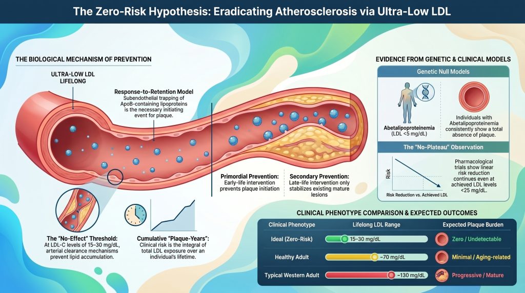
La base théorique de l'élimination biologique de l'athérosclérose repose sur hypothèse de réponse à la rétention, qui identifie la rétention sous-endothéliale de lipoprotéines contenant de l'ApoB comme l'événement initiateur nécessaire et suffisant pour athérogenèse [1], [7]. Alors que les modèles traditionnels mettaient l'accent sur une lésion endothéliale franche comme déclencheur principal, les données actuelles indiquent qu'un endothélium intact, bien que dysfonctionnel, endothélium recouvre généralement les lésions précoces et intermédiaires1], [8]. L'initiation de la maladie se produit lorsque des lipoprotéines d'un diamètre inférieur à environ 70 nm — notamment LDL, les résidus riches en triglycérides et la Lp(a) traversent la barrière endothéliale et pénètrent dans la tunique intima [2], [7].
Interactions moléculaires au sein de la matrice intimale
Au sein de l'intima, les particules retenues interagissent avec la matrice extracellulaire (ECM), en particulier avec des protéoglycanes chargés négativement tels que versican, le perlecan, biglycane, et la décorine [1], [7]. Les régions chargées positivement de l'ApoB, en particulier les séquences riches en lysine et arginine les résidus se lient par interaction électrostatique à la glycosaminoglycane chaînes (GAG) de ces protéoglycanes [1]. Il ne s'agit pas d'un piégeage passif, mais d'un événement biochimique crucial qui prolonge le temps de séjour de la lipoprotéine dans la paroi artérielle d'un ordre de grandeur par rapport à son transit plasmatique [9].
| Protéoglycane | Interaction avec les lipoprotéines ApoB | Rôle dans l'athérogenèse |
| Versican | Protéoglycane de grande taille, capable de s'agréger et d'augmenter le volume de l'intima | Favorise le piégeage des lipoprotéines et cellule musculaire lisse migration [7] |
| Perlecan | Membrane basale héparane sulfate protéoglycane | Structure de support facilitant la rétention précoce des particules [7] |
| Biglycane | Petit protéoglycane riche en leucine présentant une forte affinité pour l'ApoB | Est lié au développement de cœur nécrotique riche en lipides [7] |
| Décorin | Interagit avec le collagène et les particules de LDL | Module la réponse fibrotique et l'agrégation des lipoprotéines [7] |
Une présence prolongée au niveau de l'intima rend les lipoprotéines retenues très sensibles aux modifications chimiques, notamment l'oxydation, la glycation, le clivage enzymatique et l'agrégation [1], [9]. Les particules modifiées déclenchent une réponse immunitaire inappropriée [1], [7]. Cellules endothéliales activées par des lipides modifiés et des facteurs de stress mécaniques, expriment des molécules d'adhésion (VCAM-1, ICAM-1) et sécrètent des chimiokines qui recrutent des monocytes dans le espace sous-endothélial [1], [10]. Ces monocytes se différencient en macrophages qui internalisent les lipoprotéines modifiées via des récepteurs « scavenger » et se transforment en particules chargées de lipides cellules spumeuses—la caractéristique morphologique distinctive de la strie lipidique [10].
Limites thermodynamiques et cinétiques de la rétention
La probabilité de rétention des particules dépend de la concentration locale en particules d'ApoB et de la disponibilité de liaison aux protéoglycanes sites situés dans la matrice [7]. Dans un modèle stochastique de la paroi artérielle, le taux d'accumulation des lipides est déterminé par le flux de lipoprotéines vers l'intima et par la cinétique de liaison des protéoglycanes [9]. À des concentrations circulantes extrêmement faibles (par exemple, LDL-C ≈ 15 mg/dL), le nombre de particules disponibles pour transcytose est considérablement réduite [2], [11].
Si le débit d'entrée des particules est suffisamment faible pour que les mécanismes endogènes de clairance de la paroi artérielle — la phagocytose médiée par les macrophages et celle médiée par les HDL — transport inverse du cholestérol—peut éliminer les particules avant qu'elles ne subissent une modification pro-inflammatoire, la cascade inflammatoire n'est donc jamais déclenchée [1], [12]. Il s'agit là du mécanisme à l'origine d'un seuil biologique “ sans effet ” pour la concentration en ApoB, en dessous duquel la probabilité d'apparition d'une lésion tend vers zéro [7].
La relation quantitative entre l'exposition cumulative et les « années-plaque »
Le risque clinique d'ASCVD ne reflète pas le taux instantané de LDL-C, mais l'intégrale de l'exposition au fil du temps, un concept désormais formalisé sous le nom d“” exposition cumulative au LDL “ ou «années-plaques” [13], [14]. Dans ce cadre, l’athérosclérose est une maladie caractérisée par une accumulation progressive de substrat, et le délai avant la survenue d’un événement clinique dépend de la rapidité avec laquelle un individu franchit un seuil personnel seuil de plaque dentaire [13]. Domanski et ses collègues ont formellement démontré, dans le cadre d'une analyse groupée portant sur 4 958 participants issus de ARIC, CARDIA, MESA, ainsi que l'étude Framingham Offspring Study, ont montré que la charge cumulative liée à l'exposition au LDL-C, la durée de cette exposition et la pente de l'évolution du LDL-C au fil du temps permettaient chacune, de manière indépendante, de prédire la survenue d'événements cardiovasculaires [14].
Définition des seuils « plaque-année »
Analyse des données épidémiologiques et Randomisation mendélienne les données permettent une quantification provisoire des seuils d'exposition cumulative [13], [15]. Les seuils publiés restent approximatifs et dépendent de la cohorte concernée, sans avoir été formellement validés sur l'ensemble des populations ; il convient donc de considérer les valeurs ci-dessous comme des estimations heuristiques issues d'études de cohortes à long terme et d'études mendéliennes randomisation données [13]–[15].
| Indicateur de risque (hommes) | Exposition cumulative approximative au LDL (g·an/dL) | Risque lié aux événements majeurs au cours de la vie (environ) |
| Seuil de faible risque | ~ 5,000 | < 10% probabilité de l'événement [13], [14] |
| Seuil intermédiaire | ~ 8,000 | Âge médian approximatif pour un CAC non nul (~55-60 ans) [13], [16] |
| Seuil de risque élevé | ~ 11,000 | >20% : probabilité de l'événement [13], [14] |
| Seuil de risque très élevé | ~ 14,000 | Âge médian approximatif pour un CAC ≥ 100 [13], [16] |
Remarque : Les seuils chez les femmes sont généralement plus élevés (estimés entre environ 20 et 301 TP9T pour des strates de risque comparables), ce qui concorde avec l'apparition plus tardive observée de la maladie cardiovasculaire atheroscléreuse (ASCVD) chez les femmes et l'atténuation, avant la ménopause, de la transcytose des LDL et de la biologie de la paroi artérielle [13].
Influence des facteurs de risque secondaires sur le seuil de rétention
L'exposition cumulative au LDL nécessaire pour déclencher des événements n'est pas fixe ; elle est modulée par l'environnement biologique du artère [13]. Hypertension, diabète, et tabagisme abaisser efficacement le seuil en augmentant la rétention des particules ou en accélérant la réponse inflammatoire aux lipides retenus [2], [17].
- Hypertension : Surélevé tension artérielle augmente la transcytose du LDL à travers l'endothélium et favorise la synthèse de protéoglycanes présentant une affinité plus élevée pour l'ApoB [7]. Contrainte de cisaillement sur les éléments existants plaques augmente le risque de rupture [13].
- Diabète de type 2 / résistance à l'insuline: La dysglycémie chronique et l'hyperinsulinémie endommagent la paroi artérielle, favorisent un remodelage pathologique et rétrécissent les artères coronaires lumens, de sorte que même les petites plaques acquièrent une importance hémodynamique et que les thrombus sont plus susceptibles de provoquer une occlusion [14].
- Tabagisme : L'exposition au tabac provoque des lésions endothéliales directes et stress oxydatif, ce qui accélère la modification des lipoprotéines au niveau de l'intima [2].
- Abus d'androgènes : Chez les jeunes hommes consommant des stéroïdes anabolo-androgènes, volume de la plaque dentaire et score calcique coronaire ces scores présentent une forte corrélation avec l'exposition au cours de la vie, ce qui corrobore l'hypothèse d'une accélération indépendante de l'athérogenèse [18].
Chez une personne en bonne santé sur le plan métabolique, dont la tension artérielle est bien contrôlée et qui ne présente aucune comorbidité inflammatoire, la paroi artérielle peut supporter une charge cumulative plus importante avant l'apparition de manifestations cliniques [13]. En vertu de la hypothèse du risque nul, le fait de maintenir le taux de LDL-C à environ 15 mg/dL sur une durée de vie de 80 ans ne génère qu'environ 1 200 g·an/dL de exposition cumulative— bien en deçà du seuil empirique de faible risque d’environ 5 000 g·an/dL et d’environ un ordre de grandeur en dessous des seuils associés à des probabilités d’événements cliniquement significatifs [13].
Modèles génétiques nuls : les expériences de la nature
Le test le plus rigoureux permettant de déterminer si l'athérosclérose peut être éliminée repose sur des modèles génétiques humains présentant une exposition extrêmement faible à l'ApoB tout au long de la vie [2], [3]. Ces “ expériences de la nature ” constituent la preuve la plus solide dont on dispose pour affirmer que, en l'absence quasi totale de particules athérogènes, l'apparition de la maladie est considérablement atténuée [19], [20]. Le nombre de cas rapportés reste faible et les données d'autopsie sont limitées ; par conséquent, les affirmations faisant état d'une absence “ totale ” doivent être interprétées comme une “ réduction marquée de la prévalence, au-delà de ce qui peut raisonnablement être attribué au seul hasard ”, plutôt que comme un zéro mathématique.
Abétalipoprotéinémie (ABL) et déficit en MTTP
L'abétalipoprotéinémie est une maladie autosomique récessive causée par des mutations bialléliques entraînant une perte de fonction du gène MTTP, qui joue un rôle essentiel dans l'assemblage et la sécrétion des lipoprotéines contenant l'ApoB au niveau du foie et de l'intestin [19], [21]. Chez les personnes atteintes, on observe une absence quasi totale de lipoprotéines contenant de l'ApoB dans la circulation ; cholestérol total est généralement inférieur à 30 mg/dL et le LDL-C inférieur à 5 mg/dL (souvent indétectable) [19].
Malgré la présence de pathologies non cardiaques graves (malabsorption des graisses, acanthocytose, dégénérescence spinocérébelleuse, rétinite pigmentaire), les données cliniques et les résultats d'autopsie, bien que limités, indiquent une absence frappante de lésions athéroscléreuses chez les patients atteints d'ABL [19], [21]. Cardiomyopathie et les arythmies, lorsqu'elles surviennent, sont principalement attribuées à une carence en vitamines liposolubles (vitamine E) plutôt qu'à ischémie [19]. L'ABL constitue donc le modèle « null » humain le plus proche dont on dispose pour l'athérosclérose induite par l'ApoB.
Hypobétalipoprotéinémie familiale (FHBL)
La FHBL résulte de mutations hétérozygotes ou bialléliques du gène APOB entraînant la formation de protéines tronquées, incapables d'être sécrétées protéines [22].
- FHBL hétérozygote : Taux de LDL-C généralement compris entre 20 et 50 mg/dL, associé à une réduction marquée du risque d'ASCVD à vie [22], [23].
- Homozygote / hétérozygote composé FHBL : Le taux de LDL-C est souvent inférieur à 10 mg/dl ; le phénotype s'apparente à celui de l'ABL et présente une protection tout aussi marquée contre les maladies athéroscléreuses, tant à l'imagerie que d'après les rapports d'autopsie disponibles [22], [23].
| Maladie génétique | Mécanisme | LDL-C (mg/dl) | Phénotype de la maladie coronarienne atheroscléreuse (ASCVD) |
| Abétalipoprotéinémie | MTTP LOF ; pas d'assemblage de particules | < 5 | Plaque nettement réduite / absente [19] |
| FHBL homozygote | APOB LOF ; protéines tronquées | < 10 | Plaque nettement réduite / absente [22], [23] |
| PCSK9 LOF (hétérozygote composé) | Amélioré LDLR recyclage ; élimination rapide des LDL | ~ 14-15 | Phénotype sain ; aucune ASCVD avérée chez le cas index [24] |
| ANGPTL3 Carence | Augmentation de l'activité des LPL et des EL ; faibles taux de triglycérides et de LDL | ~ 30–40 | Réduction significative de la MC (rapport de cotes inférieur d'environ 34 à 41%) [25], [26] |
PCSK9 et ANGPTL3 : le phénotype “ sain ” à très faible taux
Contrairement à l'ABL, qui s'accompagne d'une morbidité non cardiaque grave, les variants avec perte de fonction des gènes PCSK9 et ANGPTL3 entraînent un taux de LDL-C extrêmement bas sans entraîner de malabsorption des lipides ni de stéatose hépatique [3], [27]. Le premier cas rapporté d’hétérozygotie composée pour une perte de fonction (LOF) du gène PCSK9, celui d’une femme afro-américaine en bonne santé et fertile décrite par Zhao et ses collègues, présentait un taux de LDL-C d’environ 14 mg/dL (0,36 mmol/L) et ne présentait aucun signe clinique particulier, ce qui démontre que l’absence quasi totale de PCSK9 circulante tout au long de la vie est compatible avec une physiologie humaine normale [24]. Dans la cohorte ARIC (Atherosclerosis Risk in Communities), les variants hétérozygotes de perte de fonction (LOF) du gène PCSK9 (PCSK9 Y142X et C679X) ont été associés à une baisse du taux de LDL-C de 28% et à une réduction de 88% de maladie coronarienne incidence sur une période d'environ 15 ans chez les participants noirs, avec une réduction plus modeste d'environ 471 TP9T chez les porteurs de la mutation R46L du gène PCSK9 parmi les participants blancs [28]. Sur l'ensemble d'une vie d'exposition, la réduction du risque se rapproche de la protection quasi totale observée chez les personnes atteintes d'ABL et d'FHBL homozygotes [2].
Un déficit en ANGPTL3 entraîne une “ hypolipidémie combinée ” caractérisée par de faibles taux de LDL-C, de HDL-C et de triglycérides [27]. Dans l'analyse de Stitziel publiée en 2017, trois individus hétérozygotes composés ne présentaient aucune plaque coronarienne à l'angiographie par tomodensitométrie, contre une moyenne de 39% charge athéromateuse chez des parents appariés ; les porteurs hétérozygotes de la mutation LOF présentaient un risque environ 34% plus faible de maladie coronarienne (OR = 0,66 ; IC à 95 % : 0,44–0,98) [25]. Dewey et ses collègues ont reproduit ce résultat chez 58 335 participants à l’étude DiscovEHR, faisant état d’une réduction de 41% du risque de MC (OR 0,59 ; IC 95% 0,41–0,85 ; p = 0,004) [26]. Il est important de noter que l’imagerie quantitative confirme que les porteurs d’une perte de fonction (LOF) de l’ANGPTL3 ne présentent pas d’augmentation de la graisse hépatique, ce qui fait de cette voie une cible prometteuse pour une mimique pharmacologique à long terme [27].
Données pharmacologiques : le paradigme “ moins, c'est mieux ”
Les modèles génétiques portent sur une exposition tout au long de la vie ; les essais pharmacologiques évaluent l'effet d'une réduction du LDL-C à un âge plus avancé, généralement chez des personnes présentant déjà une pathologie subclinique ou clinique [2].
La méta-régression et la linéarité des bénéfices
Données issues de la collaboration « Cholesterol Treatment Trialists » (CTT) et d'essais cliniques ultérieurs portant sur des traitements autres que les statines (ézétimibe, Inhibiteurs de PCSK9) font preuve d'une cohérence remarquable relation dose-effet: chaque réduction de 1 mmol/L (38,7 mg/dL) du LDL-C correspond à une diminution d'environ 22% des événements vasculaires majeurs par année de traitement, cette relation restant linéaire même aux niveaux les plus bas atteints parmi ceux examinés [29], [30]. Cette relation est en effet indépendante du mécanisme par lequel le taux de LDL-C est réduit [15], [31].
| Essai clinique / Analyse | Taux de LDL-C atteint (mg/dL) | Principal constat |
| CTT Méta-analyse | Plage : 60–180 | ~22% Réduction de la RR par mmol/L (38,7 mg/dL) [29] |
| IMPROVE-IT (ézétimibe + simvastatine) | 53,7 contre 69,5 (moyenne pondérée dans le temps) | HR : 0,936 (0,89–0,99) pour le critère d'évaluation principal ; bénéfice au-delà de statine seul [30] |
| FOURIER (évocumab) | Médiane : environ 30 (sous-groupe : < 20) | Un bénéfice linéaire a été observé pour un taux de LDL-C inférieur à 20 mg/dl [32], [33] |
| ODYSSEY RÉSULTATS (alirocumab) | ~53 (moyenne sur 48 semaines) | HR 0,85 MACE ; premier signe de mortalité pour les inhibiteurs de la PCSK9 (HR 0,85) [34] |
| PROLONG-ANG3 (solbinsiran) | Phase 2 : réduction durable | siRNA ciblant ANGPTL3 ; réduction durable des taux d'ApoB et de TG [35] |
| Lipides CORALreef (enlicitide) | ~60 sous traitement de fond par statine | Inhibiteur oral de la PCSK9 ; étude de phase III sur la réduction du LDL-C [36] |
L'intérêt de ces données pharmacologiques réside dans l'observation d'une “ absence de plateau ” [32], [37]. La réduction du nombre d'événements se poursuit de manière linéaire même lorsque le taux de LDL-C est inférieur à 20 mg/dL, ce qui suggère que les facteurs biologiques à l'origine de l'athérosclérose restent limités par le substrat aux extrémités du spectre lipidique [2], [32].
Maladie résiduelle ou nouvelle apparition de la maladie
Il convient d’établir une distinction fondamentale entre le primaire et prévention secondaire [13]. Parmi les participants à l'essai ayant atteint un taux de LDL-C très bas, une proportion non négligeable continue de subir des événements [38]. Ceci risque résiduel ce n'est pas un échec de l'hypothèse du risque zéro, mais plutôt la conséquence prévisible des caractéristiques structurelles irréversibles de la plaque avancée [2]. Une fois qu'une lésion s'est développée, une cœur nécrotique et un dilué chape fibreux, la rupture peut être provoquée par des forces mécaniques locales ou par une inflammation systémique, de manière largement indépendante du taux actuel de LDL-C [2], [39]. L'hypothèse du risque nul porte sur l'apparition de la maladie, et non sur la rupture terminale d'une plaque préexistante [1]. Si les taux de lipoprotéines ultra-basses étaient maintenus dès la naissance, le substrat à l'origine d'événements aigus (matures, plaque instable) ne parviendrait tout simplement pas à se former [2], [14].
Voies de risque résiduel : l'inflammation ou la Lp(a) peuvent-elles à elles seules déclencher la maladie ?
Pour réfuter l'hypothèse du risque zéro, il faut identifier une voie autre que celle de l'ApoB capable de déclencher de manière indépendante l'athérosclérose en l'absence de lipoprotéines athérogènes [5], [6].
La lipoprotéine (a) et la nécessité structurelle de l'ApoB
La Lp(a) est une particule complexe composée d'une protéine de type LDL ApoB-100 noyau lié de manière covalente à l'apolipoprotéine (a) [40]. Il est indépendamment associé au risque d'ASCVD, même lorsque le taux de LDL-C est contrôlé de manière optimale [5]. D'un point de vue mécanistique, la Lp(a) est plus athérogène que le LDL à l'échelle de chaque particule en raison de sa propension à se lier à la matrice et de sa charge en phospholipides oxydés (OxPL), qui déclenchent une inflammation importante [40], [41]. Il est toutefois essentiel de noter que chaque particule de Lp(a) nécessite une structure portante constituée d’ApoB-100 ; dans les cas d’ABL et de FHBL homozygote, la Lp(a) ne peut pas être synthétisée en raison de l’absence de sécrétion d’ApoB [19], [22]. Ainsi, la Lp(a) ne correspond pas à une voie de synthèse indépendante de l’ApoB, mais à une sous-classe quantitativement plus athérogène de la famille des lipoprotéines contenant de l’ApoB.
L'inflammation : un facteur amplificateur, et non un facteur déclencheur
Le rôle de l'inflammation (IL-6, IL-1β, hsCRP) dans l'ASCVD est bien établie, et le Essai CANTOS ont démontré que le blocage de l'IL-1β induit par le canakinumab réduit les événements cardiovasculaires sans modifier les taux de lipides [42]. Néanmoins, le consensus actuel est que l'inflammation agit comme une réponse physiologique de la paroi artérielle à la rétention de lipoprotéines plutôt que comme un facteur déclencheur autonome [7]. Dans les modèles animaux de type sauvage, l'inflammation systémique ne provoque pas d'athérosclérose en l'absence d'hyperlipidémie [43]. L'inflammation de la paroi artérielle, le recrutement des monocytes et la formation de cellules spumeuses sont des conséquences en aval de la rétention des lipoprotéines [1]. Maladies inflammatoires chroniques (par exemple, polyarthrite rhumatoïde, SLE) abaissent le seuil de formation de lésions induites par la rétention, mais elles ne déclenchent pas l'athérosclérose en l'absence du substrat ApoB [7].
Imagerie du segment ultra-bas de gamme : données subcliniques
Score calcique coronaire, échographie intravasculaire (IVUS), tomographie par cohérence optique (OCT), et angioscanner coronaire (CCTA) offrent des aperçus complémentaires de la présence et de la composition de la plaque subclinique au niveau des couches lipidiques extrêmes [16], [44].
Le pouvoir d'un CAC nul
Un score CAC égal à zéro constitue un indicateur fiable d'un faible risque d'événement à court terme, mais son interprétation dépend de l'âge [16], [45]. Chez les populations plus jeunes (âgées de moins de 45 ans), il est fréquent d'observer un CAC nul, même chez les personnes présentant une exposition importante au LDL, car calcification est un marqueur de stabilisation à un stade avancé [45]. À l'inverse, un CAC nul chez une personne âgée est rare et témoigne d'une résistance exceptionnelle à l'athérogenèse, souvent associée à un faible taux d'ApoB d'origine génétique ou à une biologie de la matrice favorable [16], [46].
| Catégorie CAC | Unités Agatston | Signification clinique |
| Zéro | 0 | Aucune calcification détectable ; risque très faible d'ASCVD à court terme [16] |
| Léger | 1–99 | Charge athéroscléreuse précoce ; justifie un reclassement dans une catégorie de risque supérieure [16] |
| Modéré | 100–399 | Plaque importante ; le risque d'événement à 10 ans dépasse généralement 7,51 TP9T [16] |
| Grave | ≥ 400 | Risque très élevé ; souvent considéré comme équivalent à une ASCVD [16] |
L'IVUS et le seuil de régression
Les essais cliniques d'imagerie intravasculaire démontrent systématiquement qu'une réduction importante du taux de LDL-C peut inverser la charge globale de plaque [47], [48]. Le essai ASTEROID ont atteint un taux moyen de LDL-C de 60,8 mg/dL avec rosuvastatine 40 mg et ont montré une régression de volume d'athérome en pourcentage (PAV) pour plusieurs paramètres d'imagerie [47]. SATURNE a confirmé une diminution du taux de LDL-C dans la fourchette de 60 à 70 mg/dL [48]. GLAGOV ont prolongé cette relation jusqu’à l’ère des inhibiteurs de la PCSK9, montrant une régression linéaire continue avec l’évolocumab jusqu’à un taux de LDL-C inférieur à 40 mg/dL, sans plateau apparent [49].
- Régression : On observe systématiquement une diminution de la PAV lorsque le taux de LDL-C passe en dessous d’environ 70 mg/dL, l’ampleur de cette diminution étant proportionnelle à l’importance de la baisse du taux de LDL-C [47]–[49].
- Stabilisation : À des taux extrêmement faibles, les plaques subissent un remodelage bénéfique : le noyau riche en lipides rétrécit et la couche fibreuse s'épaissit et s'enrichit en collagène, transformant ainsi une lésion “ vulnérable ” en une lésion structurellement stable [49], [50].
Selon l'hypothèse du risque nul, le maintien d'un taux de LDL-C ≈ 15 mg/dL dès le plus jeune âge empêcherait totalement la formation du noyau riche en lipides ; la régression et la stabilisation n'ont alors plus lieu d'être, puisqu'il n'y a pas de lésion à faire régresser [2].
Considérations temporelles : exposition précoce vs intervention à l’âge adulte
Le modèle d'exposition cumulative souligne que l'âge auquel commence la réduction du taux de LDL est tout aussi important que l'intensité de cette réduction [13].
La fenêtre d'opportunité
L’analyse historique de randomisation mendélienne menée en 2012 par Ference et ses collègues, qui a regroupé neuf SNP issus de six gènes régulant le LDL, a révélé que chaque unité standard de LDL-C génétiquement plus faible entraînait une réduction proportionnelle du risque de maladie coronarienne environ trois fois supérieure à celle obtenue par une baisse de même ampleur du LDL-C induite par les statines à un âge avancé [15]. Plus précisément, une exposition tout au long de la vie à un taux de LDL-C inférieur d’environ 1 mmol/L a été associée à une réduction du risque de maladie coronarienne de l’ordre de 54–55%, contre environ 22% risque relatif réduction par mmol/L grâce à un traitement aux statines commencé à l'âge mûr [15], [29]. Cette divergence s'explique par le fait qu'une intervention précoce empêche l'apparition des lésions, tandis qu'une intervention tardive ne fait que ralentir la progression des plaques déjà formées [2], [14].
Pour une personne en bonne santé métabolique :
- Maintenir un taux de LDL-C d’environ 15 mg/dL tout au long de la vie : Empêche la formation de la plaque dentaire ; le risque à vie est quasi nul [2], [15].
- Réduire le taux de LDL-C à environ 15 mg/dL à l'âge de 40 ans : Il freine la progression de la maladie, stabilise les plaques subcliniques existantes et retarde l'apparition d'événements cliniques d'environ 10 à 20 ans, selon la charge plaque initiale [13], [14].
- Réduction du taux de LDL-C à environ 15 mg/dL après un événement clinique : Réduit le risque de récidive en stabilisant plaques vulnérables; n'élimine pas le risque résiduel lié à des dommages structurels irréversibles [34], [50].
La primauté biologique de la prévention primaire — et, idéalement, de la prévention primordiale — en découle directement : le maintien de taux physiologiques de lipoprotéines dès les tout premiers stades de la vie permet d’éviter le passage d’une charge subclinique à une charge cliniquement dangereuse [3].
Limites théoriques et mécanistiques : aléatoire contre risque nul
L'élimination de l'athérosclérose est-elle totale, ou existe-t-il un seuil biologique minimum ?
Rétention stochastique à des niveaux extrêmement bas
L'apparition de l'athérosclérose est, par essence, un processus probabiliste [7]. Même avec un taux de LDL-C ≈ 15 mg/dL, une particule d'ApoB pourrait en principe pénétrer dans la paroi artérielle, se lier à un protéoglycane et subir une oxydation [7]. Cependant, la probabilité qu’un nombre suffisant de ces particules soit retenu dans une seule zone focale pour déclencher une réaction inflammatoire qui s’autoalimente devient infinitésimale à des concentrations ultra-faibles [7], [9].
La biologie de la paroi artérielle intègre plusieurs mécanismes de sécurité :
- Élimination des déchets par les résidents : Les macrophages intimaaux sains sont capables d'éliminer de petites quantités de lipoprotéines modifiées sans se transformer en cellules spumeuses ni sécréter de cytokines pro-inflammatoires [1].
- HDL/Efflux de l'ApoA-I : Le HDL agit comme un vecteur lipidique bidirectionnel capable d'éliminer l'excès de cholestérol de l'intima ; lorsque l'apport en ApoB est faible, sa capacité d'efflux permet de maintenir facilement l'équilibre homéostatique [8], [51].
- Intégrité de la matrice : En l'absence de rétention lipidique persistante, l'intima reste fine et structurellement intacte, avec une élasticité préservée fibres et une expansion minimale des protéoglycanes [9].
À l'objectif de risque zéro correspondant à un taux de LDL-C ≈ 15 mg/dL, la probabilité d'apparition d'une plaque n'est peut-être pas mathématiquement nulle, mais elle est effectivement nulle d'un point de vue biologique et clinique : l'accumulation n'atteindrait jamais la masse critique nécessaire à l'apparition de la maladie au cours d'une vie humaine [2], [14]. Le Étude Horus Une étude portant sur 137 momies couvrant 4 000 ans d’histoire — y compris des peuples préindustriels et anciens apparentés aux Tsimane — met en évidence une calcification vasculaire à toutes les époques examinées, mais il est à noter que les populations préindustrielles présentant un taux de LDL-C très faible (par exemple, les Tsimane, avec un taux moyen de LDL-C d’environ 91 mg/dL et la prévalence d’athérosclérose coronarienne la plus faible jamais observée dans une population humaine) ne présentent que de faibles taux de CAC subclinique, même chez les personnes âgées [52], [53]. Si l'on extrapole à un taux de LDL-C de 15 mg/dL tout au long de la vie, cela implique une charge bien inférieure même à cette valeur de référence.
Synthèse des résultats de l'hypothèse du risque zéro
Trois variantes de l'hypothèse peuvent être testées à la lumière des éléments probants examinés ci-dessus.
Forme forte : hypothèse du risque nul
Un taux de LDL-C ≈ 15 mg/dL tout au long de la vie chez un individu en bonne santé métabolique réduit pratiquement à zéro la probabilité d’une athérosclérose cliniquement significative. Évaluation : étayée par des modèles génétiques humains « null » (ABL, FHBL homozygote) dans lesquels la charge athéroscléreuse est nettement réduite, voire absente [19], [22], par le taux de référence physiologique de LDL-C chez le nouveau-né, et par l'absence constante d'événements dans les cohortes génétiques à exposition ultra-faible [2], [24].
Forme faible : hypothèse asymptotique
Le risque tend vers zéro mais ne l'atteint jamais complètement en raison de la rétention stochastique ou des voies non liées à l'ApoB. Évaluation : étayée par la nature fondamentalement probabiliste des interactions entre les particules et la paroi [7]. Un âge très avancé ou une inflammation systémique sévère pourraient, en principe, entraîner des modifications minimes de l'intima, bien que de telles lésions resteraient probablement subcliniques tout au long de la vie d'un individu [7].
Hypothèse nulle
L'athérosclérose continue de se manifester à des taux significatifs, indépendamment du taux de LDL, même lorsque celui-ci est très faible. Évaluation : réfutée pour le phénotype idéal [2], [15]. Le risque résiduel observé chez les populations traitées dans le cadre de la prévention secondaire est systématiquement imputable à la plaque préexistante et aux particules non-LDL particules contenant de l'ApoB (résidus riches en triglycérides et Lp(a)) plutôt qu’à un mécanisme d’initiation non lié à l’ApoB [5], [40].
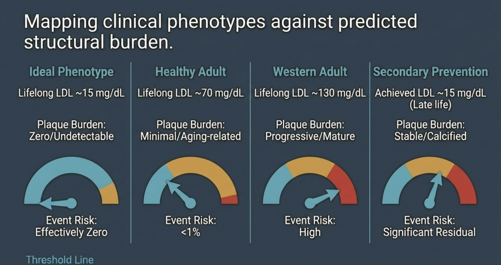
| Phénotype clinique | Charge de plaque estimée | Probabilité de survenue d'un événement |
| Phénotype idéal (taux de LDL-C stable à environ 15 mg/dL tout au long de la vie) | Zéro / indétectable | Pratiquement nul [15] |
| Adulte en bonne santé (taux de LDL-C stable tout au long de la vie, d'environ 70 mg/dL) | Minime / lié au vieillissement | Faible (~5–10%) [14] |
| Adulte occidental (taux de LDL-C stable tout au long de la vie ≈ 130 mg/dL) | Progressiste / mûr(e) | Élevé [2], [14] |
| Prévention secondaire (taux de LDL-C atteint d’environ 15 mg/dL) | Stable / calcifié | Résidu significatif [34] |
Conclusion : redéfinir le seuil de la santé humaine
La vérification de l'hypothèse du risque nul vient étayer l'idée selon laquelle l'athérosclérose est une maladie dépendante du substrat, dont l'apparition nécessite une exposition cumulative minimale, tout au long de la vie, à des lipoprotéines contenant de l'ApoB [2], [14]. La relation quantitative entre l’exposition cumulative et la formation de plaque suggère que le maintien du taux de LDL-C dans une fourchette comprise entre 10 et 20 mg/dL dès la naissance repousse l’apparition de la maladie au-delà des limites biologiques de la longévité humaine [13], [15].
Modèles génétiques (abétalipoprotéinémie, FHBL homozygote et phénotypes « ultra-bas » chez des sujets sains résultant d’une perte de fonction des gènes PCSK9 et ANGPTL3) démontrent que de tels taux sont à la fois sans danger et compatibles avec une réduction marquée — voire, dans certains cas, une absence apparente — de plaque athéroscléreuse, tout en reconnaissant que le nombre de cas reste limité [19], [22], [24], [25]. Bien que des risques résiduels liés à la Lp(a), aux particules résiduelles et à l'inflammation persistent chez les patients présentant une maladie à un stade avancé, ces facteurs ne suffisent pas à déclencher l'athérogenèse en l'absence quasi totale de particules d'ApoB [2], [40].
Les implications pour la cardiologie préventive sont considérables [3]. Plutôt que de considérer le risque comme une conséquence inévitable du vieillissement, l'objectif à viser est prévention primordiale: maintenir des taux physiologiques de lipoprotéines tout au long des premières étapes de la vie afin d'éliminer le substrat de l'athérogenèse [2], [15]. Ce paradigme suggère que les maladies cardiaques — première cause de mortalité dans le monde — peuvent être éradiquées sur le plan biologique si l'on empêche la rétention des particules d'ApoB tout au long de la vie humaine [2], [14].
Références
- J. Borén, M. J. Chapman, R. M. Krauss, C. J. Packard, J. F. Bentzon, C. J. Binder, M. J. Daemen, L. L. Demer, R. A. Hegele, S. J. Nicholls, B. G. Nordestgaard, G. F. Watts, E. Bruckert, S. Fazio, B. A. Ference, I. Graham, J. D. Horton, U. Landmesser, U. Laufs, L. Masana, G. Pasterkamp, F. J. Raal, K. K. Ray, H. Schunkert, M.-R. Taskinen, B. van de Sluis, O. Wiklund, L. Tokgözoğlu, A. L. Catapano, and H. N. Ginsberg, “Low-density lipoproteins cause atherosclerotic cardiovascular disease: pathophysiological, genetic, and therapeutic insights—a consensus statement from the European Atherosclerosis Society Consensus Panel,” Eur. Heart J., vol. 41, no. 24, pp. 2313–2330, Jun. 2020, doi: 10.1093/eurheartj/ehz962.
- B. A. Ference, H. N. Ginsberg, I. Graham, K. K. Ray, C. J. Packard, E. Bruckert, R. A. Hegele, R. M. Krauss, F. J. Raal, H. Schunkert, G. F. Watts, J. Borén, S. Fazio, J. D. Horton, L. Masana, S. J. Nicholls, B. G. Nordestgaard, B. van de Sluis, M.-R. Taskinen, L. Tokgözoğlu, U. Landmesser, U. Laufs, O. Wiklund, J. K. Stock, M. J. Chapman, and A. L. Catapano, “Low-density lipoproteins cause atherosclerotic cardiovascular disease. 1. Evidence from genetic, epidemiologic, and clinical studies,” Eur. Heart J., vol. 38, no. 32, pp. 2459–2472, Aug. 2017, doi: 10.1093/eurheartj/ehx144.
- A. L. Catapano, I. Graham, G. De Backer, O. Wiklund, M. J. Chapman, H. Drexel, A. W. Hoes, C. S. Jennings, U. Landmesser, T. R. Pedersen, Ž. Reiner, G. Riccardi, M.-R. Taskinen, L. Tokgözoğlu, W. M. M. Verschuren, P. Vlachopoulos, D. A. Wood, and J. L. Zamorano, “2016 ESC/EAS guidelines for the management of dyslipidaemias,” Eur. Heart J., vol. 37, no. 39, pp. 2999–3058, Oct. 2016, doi: 10.1093/eurheartj/ehw272.
- J. H. O’Keefe, L. Cordain, W. H. Harris, R. M. Moe, and R. Vogel, “Optimal low-density lipoprotein is 50 to 70 mg/dl: lower is better and physiologically normal,” J. Am. Coll. Cardiol., vol. 43, no. 11, pp. 2142–2146, Jun. 2004, doi: 10.1016/j.jacc.2004.03.046.
- B. G. Nordestgaard, M. J. Chapman, K. Ray, J. Borén, F. Andreotti, G. F. Watts, H. Ginsberg, P. Amarenco, A. Catapano, O. S. Descamps, E. Fisher, P. T. Kovanen, J. A. Kuivenhoven, P. Lesnik, L. Masana, Z. Reiner, M. R. Taskinen, L. Tokgözoğlu, and A. Tybjærg-Hansen, “Lipoprotein(a) as a cardiovascular risk factor: current status,” Eur. Heart J., vol. 31, no. 23, pp. 2844–2853, Dec. 2010, doi: 10.1093/eurheartj/ehq386.
- B. G. Nordestgaard and A. Varbo, “Triglycerides and cardiovascular disease,” Lancet, vol. 384, no. 9943, pp. 626–635, Aug. 2014, doi: 10.1016/S0140-6736(14)61177-6.
- I. Tabas, K. J. Williams, and J. Borén, “Subendothelial lipoprotein retention as the initiating process in atherosclerosis: update and therapeutic implications,” Circulation, vol. 116, no. 16, pp. 1832–1844, Oct. 2007, doi: 10.1161/CIRCULATIONAHA.106.676890.
- P. Libby, “The changing landscape of atherosclerosis,” Nature, vol. 592, no. 7855, pp. 524–533, Apr. 2021, doi: 10.1038/s41586-021-03392-8.
- D. C. Schwenke and T. E. Carew, “Initiation of atherosclerotic lesions in cholesterol-fed rabbits. II. Selective retention of LDL vs. selective increases in LDL permeability in susceptible sites of arteries,” Arteriosclerosis, vol. 9, no. 6, pp. 908–918, Nov./Dec. 1989, doi: 10.1161/01.atv.9.6.908.
- R. Ross, “Atherosclerosis—an inflammatory disease,” N. Engl. J. Med., vol. 340, no. 2, pp. 115–126, Jan. 1999, doi: 10.1056/NEJM199901143400207.
- F. Mach, C. Baigent, A. L. Catapano, K. C. Koskinas, M. Casula, L. Badimon, M. J. Chapman, G. G. De Backer, V. Delgado, B. A. Ference, I. M. Graham, A. Halliday, U. Landmesser, B. Mihaylova, T. R. Pedersen, G. Riccardi, D. J. Richter, M. S. Sabatine, M.-R. Taskinen, L. Tokgözoğlu, and O. Wiklund, “2019 ESC/EAS guidelines for the management of dyslipidaemias: lipid modification to reduce cardiovascular risk,” Eur. Heart J., vol. 41, no. 1, pp. 111–188, Jan. 2020, doi: 10.1093/eurheartj/ehz455.
- A. R. Tall and L. Yvan-Charvet, “Cholesterol, inflammation and innate immunity,” Nat. Rev. Immunol., vol. 15, no. 2, pp. 104–116, Feb. 2015, doi: 10.1038/nri3793.
- J. Brandts and K. K. Ray, “The LDL cumulative exposure hypothesis: evidence and practical applications,” Nat. Rev. Cardiol., vol. 21, no. 9, pp. 600–608, Sep. 2024, doi: 10.1038/s41569-024-01039-5.
- M. J. Domanski, X. Tian, C. O. Wu, J. P. Reis, A. K. Dey, Y. Gu, L. Zhao, S. Bae, K. Liu, A. A. Hasan, D. Zimrin, M. R. Farkouh, A. R. Hong, D. M. Lloyd-Jones, and V. Fuster, “Time course of LDL cholesterol exposure and cardiovascular disease event risk,” J. Am. Coll. Cardiol., vol. 76, no. 13, pp. 1507–1516, Sep. 2020, doi: 10.1016/j.jacc.2020.07.059.
- B. A. Ference, W. Yoo, I. Alesh, N. Mahajan, K. K. Mirowska, A. Mewada, J. Kahn, L. Afonso, K. A. Williams, and J. M. Flack, “Effect of long-term exposure to lower low-density lipoprotein cholesterol beginning early in life on the risk of coronary heart disease: a Mendelian randomization analysis,” J. Am. Coll. Cardiol., vol. 60, no. 25, pp. 2631–2639, Dec. 2012, doi: 10.1016/j.jacc.2012.09.017.
- R. Detrano, A. D. Guerci, J. J. Carr, D. E. Bild, G. Burke, A. R. Folsom, K. Liu, S. Shea, M. Szklo, D. A. Bluemke, D. H. O’Leary, R. Tracy, K. Watson, N. D. Wong, and R. A. Kronmal, “Coronary calcium as a predictor of coronary events in four racial or ethnic groups,” N. Engl. J. Med., vol. 358, no. 13, pp. 1336–1345, Mar. 2008, doi: 10.1056/NEJMoa072100.
- S. Yusuf, S. Hawken, S. Ôunpuu, T. Dans, A. Avezum, F. Lanas, M. McQueen, A. Budaj, P. Pais, J. Varigos, and L. Lisheng, “Effect of potentially modifiable risk factors associated with myocardial infarction in 52 countries (the INTERHEART study): case-control study,” Lancet, vol. 364, no. 9438, pp. 937–952, Sep. 2004, doi: 10.1016/S0140-6736(04)17018-9.
- A. L. Baggish, R. B. Weiner, G. Kanayama, J. I. Hudson, M. H. Picard, A. M. Hutter Jr., and H. G. Pope Jr., “Cardiovascular toxicity of illicit anabolic-androgenic steroid use,” Circulation, vol. 135, no. 21, pp. 1991–2002, May 2017, doi: 10.1161/CIRCULATIONAHA.116.026945.
- A. J. Hooper, B. A. Burnett, and J. R. Burnett, “Abetalipoproteinemia,” GeneReviews® [Internet], Univ. of Washington, Seattle, updated Jul. 2023. [Online]. Available: https://www.ncbi.nlm.nih.gov/books/NBK532447/.
- J. C. Cohen, E. Boerwinkle, T. H. Mosley Jr., and H. H. Hobbs, “Sequence variations in PCSK9, low LDL, and protection against coronary heart disease,” N. Engl. J. Med., vol. 354, no. 12, pp. 1264–1272, Mar. 2006, doi: 10.1056/NEJMoa054013.
- M. M. Berriot-Varoqueaux, L. P. Aggerbeck, M. Samson-Bouma, and J. R. Wetterau, “The role of the microsomal triglyceride transfer protein in abetalipoproteinemia,” Annu. Rev. Nutr., vol. 20, pp. 663–697, 2000, doi: 10.1146/annurev.nutr.20.1.663.
- S. G. Young and A. J. Hooper, “APOB-related familial hypobetalipoproteinemia,” GeneReviews® [Internet], Univ. of Washington, Seattle, updated Feb. 2021. [Online]. Available: https://www.ncbi.nlm.nih.gov/books/NBK570370/.
- L. Tarugi, E. Averna, E. Di Leo, A. Cefalù, D. Noto, S. Magnolo, M. Cattin, S. Bertolini, and M. Calandra, “Molecular diagnosis of hypobetalipoproteinemia: an ENID review,” Atherosclerosis, vol. 195, no. 2, pp. e19–e27, Dec. 2007, doi: 10.1016/j.atherosclerosis.2007.05.003.
- Z. Zhao, Y. Tuakli-Wosornu, T. A. Lagace, L. Kinch, N. V. Grishin, J. D. Horton, J. C. Cohen, and H. H. Hobbs, “Molecular characterization of loss-of-function mutations in PCSK9 and identification of a compound heterozygote,” Am. J. Hum. Genet., vol. 79, no. 3, pp. 514–523, Sep. 2006, doi: 10.1086/507488.
- N. O. Stitziel, A. V. Khera, X. Wang, A. J. Bierhals, A. C. Vourakis, A. E. Sperry, P. Natarajan, D. Klarin, C. A. Emdin, S. M. Zekavat, A. Nomura, J. Erdmann, H. Schunkert, N. J. Samani, W. E. Kraus, S. H. Shah, B. Yu, E. Boerwinkle, D. J. Rader, N. Gupta, P. M. Frossard, A. Rasheed, J. Danesh, E. S. Lander, S. Gabriel, D. Saleheen, K. Musunuru, and S. Kathiresan, “ANGPTL3 deficiency and protection against coronary artery disease,” J. Am. Coll. Cardiol., vol. 69, no. 16, pp. 2054–2063, Apr. 2017, doi: 10.1016/j.jacc.2017.02.030.
- F. E. Dewey, V. Gusarova, R. L. Dunbar, C. O’Dushlaine, C. Schurmann, O. Gottesman, S. McCarthy, C. V. Van Hout, S. Bruse, H. M. Dansky, J. B. Leader, M. F. Murray, M. D. Ritchie, H. L. Kirchner, D. H. Ledbetter, J. Penn, A. Lopez, I. B. Borecki, J. D. Overton, J. G. Reid, D. J. Carey, A. J. Murphy, G. D. Yancopoulos, A. Baras, J. Gromada, and A. R. Shuldiner, “Genetic and pharmacologic inactivation of ANGPTL3 and cardiovascular disease,” N. Engl. J. Med., vol. 377, no. 3, pp. 211–221, Jul. 2017, doi: 10.1056/NEJMoa1612790.
- K. Musunuru, J. P. Pirruccello, R. Do, G. M. Peloso, C. Guiducci, C. Sougnez, K. V. Garimella, S. Fisher, J. Abreu, A. J. Barry, T. Fennell, E. Banks, L. Ambrogio, K. Cibulskis, A. Kernytsky, E. Gonzalez, N. Rudzicz, J. C. Engert, M. A. DePristo, M. J. Daly, J. C. Cohen, H. H. Hobbs, D. Altshuler, G. Schonfeld, S. B. Gabriel, P. Yue, and S. Kathiresan, “Exome sequencing, ANGPTL3 mutations, and familial combined hypolipidemia,” N. Engl. J. Med., vol. 363, no. 23, pp. 2220–2227, Dec. 2010, doi: 10.1056/NEJMoa1002926.
- C. Baigent, L. Blackwell, J. Emberson, L. E. Holland, C. Reith, N. Bhala, R. Peto, E. H. Barnes, A. Keech, J. Simes, and R. Collins (Cholesterol Treatment Trialists’ Collaboration), “Efficacy and safety of more intensive lowering of LDL cholesterol: a meta-analysis of data from 170,000 participants in 26 randomised trials,” Lancet, vol. 376, no. 9753, pp. 1670–1681, Nov. 2010, doi: 10.1016/S0140-6736(10)61350-5.
- Cholesterol Treatment Trialists’ (CTT) Collaboration, “Efficacy and safety of LDL-lowering therapy among men and women: meta-analysis of individual data from 174 000 participants in 27 randomised trials,” Lancet, vol. 385, no. 9976, pp. 1397–1405, Apr. 2015, doi: 10.1016/S0140-6736(14)61368-4.
- C. P. Cannon, M. A. Blazing, R. P. Giugliano, A. McCagg, J. A. White, P. Theroux, H. Darius, B. S. Lewis, T. O. Ophuis, J. W. Jukema, G. M. De Ferrari, W. Ruzyllo, P. De Lucca, K. Im, E. A. Bohula, C. Reist, S. D. Wiviott, A. M. Tershakovec, T. A. Musliner, E. Braunwald, and R. M. Califf, “Ezetimibe added to statin therapy after acute coronary syndromes,” N. Engl. J. Med., vol. 372, no. 25, pp. 2387–2397, Jun. 2015, doi: 10.1056/NEJMoa1410489.
- J. Silverman, L. A. Ballantyne, D. A. Bhatt, S. D. Wiviott, M. S. Sabatine, and Cholesterol Treatment Trialists, “Association between lowering LDL-C and cardiovascular risk reduction among different therapeutic interventions: a systematic review and meta-analysis,” JAMA, vol. 316, no. 12, pp. 1289–1297, Sep. 2016, doi: 10.1001/jama.2016.13985.
- M. S. Sabatine, R. P. Giugliano, A. C. Keech, N. Honarpour, S. D. Wiviott, S. A. Murphy, J. F. Kuder, H. Wang, T. Liu, S. M. Wasserman, P. S. Sever, and T. R. Pedersen, “Evolocumab and clinical outcomes in patients with cardiovascular disease,” N. Engl. J. Med., vol. 376, no. 18, pp. 1713–1722, May 2017, doi: 10.1056/NEJMoa1615664.
- R. P. Giugliano, T. R. Pedersen, A. C. Keech, A. Park, S. D. Wiviott, S. A. Murphy, J. F. Kuder, H. Wang, T. Liu, J. Lopez, and M. S. Sabatine, “Clinical efficacy and safety of achieving very low LDL-cholesterol concentrations with the PCSK9 inhibitor evolocumab: a prespecified secondary analysis of the FOURIER trial,” Lancet, vol. 390, no. 10106, pp. 1962–1971, Oct. 2017, doi: 10.1016/S0140-6736(17)32290-0.
- G. G. Schwartz, P. G. Steg, M. Szarek, D. L. Bhatt, V. A. Bittner, R. Diaz, J. M. Edelberg, S. G. Goodman, C. Hanotin, R. A. Harrington, J. W. Jukema, G. Lecorps, K. W. Mahaffey, A. Moryusef, R. Pordy, K. Quintero, M. T. Roe, W. J. Sasiela, J.-F. Tamby, P. Tricoci, H. D. White, A. M. Zeiher, and ODYSSEY OUTCOMES Committees and Investigators, “Alirocumab and cardiovascular outcomes after acute coronary syndrome,” N. Engl. J. Med., vol. 379, no. 22, pp. 2097–2107, Nov. 2018, doi: 10.1056/NEJMoa1801174.
- C. M. Ballantyne, J. P. Kastelein, L. A. Leiter, S. J. Nicholls, and colleagues, “Durability and efficacy of solbinsiran, a GalNAc-conjugated siRNA targeting ANGPTL3, in adults with mixed dyslipidaemia (PROLONG-ANG3): a double-blind, randomised, placebo-controlled, phase 2 trial,” Lancet, 2025 (advance online publication).
- S. J. Nicholls, C. M. Ballantyne, and colleagues (CORALreef Lipids Investigators), “Efficacy and safety of enlicitide, an oral PCSK9 inhibitor, in adults with hypercholesterolemia: the Phase 3 CORALreef Lipids trial,” N. Engl. J. Med., 2025 (publication pending).
- M. Bhandari, S. M. Nissen, and colleagues, “How low can you go? New evidence supports no lower bound to low-density lipoprotein cholesterol level in secondary prevention,” Circulation, vol. 147, no. 19, pp. 1433–1435, May 2023, doi: 10.1161/CIRCULATIONAHA.123.064041.
- P. M. Ridker, N. Mora, L. Rose, and JUPITER Trial Study Group, “Percent reduction in LDL cholesterol following high-intensity statin therapy: potential implications for guidelines and for the prescription of emerging lipid-lowering agents,” Eur. Heart J., vol. 37, no. 17, pp. 1373–1379, May 2016, doi: 10.1093/eurheartj/ehw046.
- P. Libby, P. M. Ridker, and G. K. Hansson, “Progress and challenges in translating the biology of atherosclerosis,” Nature, vol. 473, no. 7347, pp. 317–325, May 2011, doi: 10.1038/nature10146.
- S. Tsimikas, “A test in context: lipoprotein(a): diagnosis, prognosis, controversies, and emerging therapies,” J. Am. Coll. Cardiol., vol. 69, no. 6, pp. 692–711, Feb. 2017, doi: 10.1016/j.jacc.2016.11.042.
- S. Tsimikas, H.-K. Gordts, C. Nora, C. Yeang, and J. L. Witztum, “Statin therapy increases lipoprotein(a) levels,” Eur. Heart J., vol. 41, no. 24, pp. 2275–2284, Jun. 2020, doi: 10.1093/eurheartj/ehz310.
- P. M. Ridker, B. M. Everett, T. Thuren, J. G. MacFadyen, W. H. Chang, C. Ballantyne, F. Fonseca, J. Nicolau, W. Koenig, S. D. Anker, J. J. P. Kastelein, J. H. Cornel, P. Pais, D. Pella, J. Genest, R. Cifkova, A. Lorenzatti, T. Forster, Z. Kobalava, L. Vida-Simiti, M. Flather, H. Shimokawa, H. Ogawa, M. Dellborg, P. R. F. Rossi, R. P. T. Troquay, P. Libby, R. J. Glynn, and CANTOS Trial Group, “Antiinflammatory therapy with canakinumab for atherosclerotic disease,” N. Engl. J. Med., vol. 377, no. 12, pp. 1119–1131, Sep. 2017, doi: 10.1056/NEJMoa1707914.
- A. Daugherty, S. E. Tall, M. J. Daemen, E. Falk, E. M. Fisher, E. Garcia-Cardena, A. R. Lusis, A. P. Owens III, M. E. Rosenfeld, and I. Tabas, “Recommendation on design, execution, and reporting of animal atherosclerosis studies: a scientific statement from the American Heart Association,” Arterioscler. Thromb. Vasc. Biol., vol. 37, no. 9, pp. e131–e157, Sep. 2017, doi: 10.1161/ATV.0000000000000062.
- M. J. Budoff, S. Achenbach, R. S. Blumenthal, J. J. Carr, J. G. Goldin, P. Greenland, A. D. Guerci, J. A. Lima, D. J. Rader, G. D. Rubin, L. J. Shaw, S. E. Wiegers, American Heart Association Committee on Cardiovascular Imaging and Intervention, American Heart Association Council on Cardiovascular Radiology and Intervention, American Heart Association Committee on Cardiac Imaging, “Assessment of coronary artery disease by cardiac computed tomography: a scientific statement from the AHA,” Circulation, vol. 114, no. 16, pp. 1761–1791, Oct. 2006, doi: 10.1161/CIRCULATIONAHA.106.178458.
- H. S. Hecht, “Coronary artery calcium scanning: past, present, and future,” JACC Cardiovasc. Imaging, vol. 8, no. 5, pp. 579–596, May 2015, doi: 10.1016/j.jcmg.2015.02.006.
- K. Nasir, M. B. Rivera, R. Blankstein, M. J. Blaha, A. D. Choi, R. S. Blumenthal, M. G. Silverman, A. Dardari, J. Berman, J. Yeboah, M. J. Budoff, J. J. Carr, and M. Cainzos-Achirica, “Implications of coronary artery calcium testing for primary prevention,” J. Am. Coll. Cardiol., vol. 66, no. 15, pp. 1657–1668, Oct. 2015, doi: 10.1016/j.jacc.2015.07.066.
- S. E. Nissen, S. J. Nicholls, I. Sipahi, P. Libby, J. S. Raichlen, C. M. Ballantyne, J. Davignon, R. Erbel, J. C. Fruchart, J.-C. Tardif, P. Schoenhagen, T. Crowe, V. Cain, K. Wolski, M. Goormastic, and E. M. Tuzcu, “Effect of very high-intensity statin therapy on regression of coronary atherosclerosis: the ASTEROID trial,” JAMA, vol. 295, no. 13, pp. 1556–1565, Apr. 2006, doi: 10.1001/jama.295.13.jpc60002.
- S. E. Nissen, S. J. Nicholls, K. Wolski, R. Nesto, S. Kupfer, A. Perez, H. Jure, R. De Larochellière, C. S. Staniloae, K. Mavromatis, J. Saw, B. Hu, A. M. Lincoff, E. M. Tuzcu, and PERISCOPE Investigators, “Effect of rosuvastatin versus atorvastatin on progression of coronary atherosclerosis in patients with coronary artery disease (SATURN),” N. Engl. J. Med., vol. 365, no. 22, pp. 2078–2087, Dec. 2011, doi: 10.1056/NEJMoa1110874.
- S. J. Nicholls, R. Puri, T. Anderson, C. M. Ballantyne, L. Cho, J. J. P. Kastelein, W. Koenig, R. Somaratne, H. Kassahun, J. Yang, S. M. Wasserman, R. Scott, I. Ungi, J. Podolec, A. O. Ophuis, J. H. Cornel, M. Borgman, D. M. Brennan, and S. E. Nissen, “Effect of evolocumab on progression of coronary disease in statin-treated patients: the GLAGOV randomized clinical trial,” JAMA, vol. 316, no. 22, pp. 2373–2384, Dec. 2016, doi: 10.1001/jama.2016.16951.
- A. V. Khera, E. S. Everett, M. G. Caulfield, F. E. Hantgan, J. Deasy, R. K. Scott, P. S. Sever, J. Barton, L. Heller, A. Nambi, and M. S. Sabatine, “Evolocumab, plaque regression, and clinical outcomes,” J. Am. Coll. Cardiol., vol. 75, no. 9, pp. 1037–1050, Mar. 2020, doi: 10.1016/j.jacc.2019.12.052.
- D. J. Rader and G. K. Hovingh, “HDL and cardiovascular disease,” Lancet, vol. 384, no. 9943, pp. 618–625, Aug. 2014, doi: 10.1016/S0140-6736(14)61217-4.
- R. C. Thompson, A. H. Allam, G. P. Lombardi, L. S. Wann, M. L. Sutherland, J. D. Sutherland, M. A. Soliman, B. Frohlich, D. T. Mininberg, J. M. Monge, C. M. Vallodolid, S. L. Cox, G. A. El-Maksoud, I. Badr, M. I. Miyamoto, A. el-Halim Nur el-Din, J. Narula, C. E. Finch, and G. S. Thomas, “Atherosclerosis across 4000 years of human history: the Horus study of four ancient populations,” Lancet, vol. 381, no. 9873, pp. 1211–1222, Apr. 2013, doi: 10.1016/S0140-6736(13)60598-X.
- H. Kaplan, R. C. Thompson, B. C. Trumble, L. S. Wann, A. H. Allam, B. Beheim, B. Frohlich, M. L. Sutherland, J. D. Sutherland, J. Stieglitz, D. E. Rodriguez, D. E. Michalik, C. J. Rowan, G. P. Lombardi, R. Bedi, C. E. Garcia, J. K. Min, J. Narula, C. E. Finch, M. Gurven, and G. S. Thomas, “Coronary atherosclerosis in indigenous South American Tsimane: a cross-sectional cohort study,” Lancet, vol. 389, no. 10080, pp. 1730–1739, Apr. 2017, doi: 10.1016/S0140-6736(17)30752-3.

