
Die genetische Architektur des kardiovaskulären Schutzes: Validierte Zielstrukturen, kumulative Exposition und die Biologie des lebenslangen Risikos
Herz-Kreislauf-ErkrankungHerz-Kreislauf-Erkrankungen sind der Oberbegriff für Probleme mit dem Herzen und den Blutgefäßen, einschließlich Herzinfarkten, Schlaganfällen und verstopften Beinarterien. bleibt die weltweit führende Ursache von Morbidität und Mortalität, die aus einem komplexen Zusammenspiel von Umwelteinflüssen, metabolischer Dysregulation und Gefäßschäden resultiert, ThromboseEine Thrombose ist ein Blutgerinnsel, das sich in einem Blutgefäß bildet., und vererbter Anfälligkeit1Trotz großer Fortschritte in Prävention und Behandlung ist die atherosklerotische Herz-Kreislauf-Erkrankung (ASCVD) nach wie vor für einen unverhältnismäßig großen Anteil an vorzeitigen Todesfällen und Behinderungen verantwortlich. Traditionelle RisikofaktorenEin Risikofaktor ist etwas, das Ihre Wahrscheinlichkeit erhöht, an einer Krankheit zu erkranken — Partikel mit hohem Cholesterinspiegel, Bluthochdruck, Rauchen, Diabetes, familiäre Vorbelastung.—erhöhtes LDL-Cholesterin LipoproteinEin Lipoprotein ist ein winziges Paket, das Fett und Cholesterin durch Ihren Blutkreislauf transportiert. Da sich Fett nicht in Wasser löst, benötigt es eine Proteinhülle, um zu reisen. CholesterinCholesterin ist eine wachsartige Substanz, die Ihr Körper braucht. Es fließt in Zellwände, Hormone, Vitamin D und die Galle ein, die Ihre Nahrung verdaut. Ohne es würden Sie sterben. (LDL-C), BluthochdruckBluthochdruck ist der medizinische Begriff für hohe Blutdruckwerte., DiabetesDiabetes ist eine Erkrankung, bei der der Blutzucker zu hoch bleibt, entweder weil der Körper zu wenig Insulin produziert oder weil er auf das produzierte Insulin nicht mehr reagiert. Mellitus, RauchenRauchen schädigt die Innenauskleidung der Blutgefäße, erhöht den Blutdruck, lässt das Blut leichter gerinnen und beschleunigt das Plaque-Wachstum., AdipositasFettleibigkeit bedeutet, so viel überschüssiges Körperfett mit sich zu tragen, dass die Gesundheit beeinträchtigt wird., und die sitzende Lebensweise – bleiben zentrale Bestimmtheiten der Krankheitslast. Doch das moderne Verständnis von AtherogeneseAtherogenese ist der schrittweise Prozess der Plaquebildung. wurde vom Menschen grundlegend umgestaltet GenetikGenetik ist die Lehre davon, was man von seinen Eltern erbt. [2], [3]. In den letzten zwei Jahrzehnten hat sich das Feld weit über Familienstudien und Beobachtungen hinausentwickelt EpidemiologieEpidemiologie ist die Erforschung von Gesundheitstrends in großen Bevölkerungsgruppen – wer erkrankt, wo und was diese Personen gemeinsam hatten. in die Ära der genomweiten Assoziationsstudien (GWAS), ExomsequenzierungDie Exom-Sequenzierung ist eine genomische Technik, die die proteinkodierenden Abschnitte des Genoms liest. Sie ermöglicht es Forschern, seltene kodierende Varianten – einschließlich Loss-of-Function-Mutationen – zu identifizieren, die erhebliche Auswirkungen auf das Krankheitsrisiko haben können; sie wurde eingesetzt, um schützende Varianten in Genen wie CIDEB und ZNRF3 zu entdecken, die bei standardmäßigen GWAS übersehen würden., groß angelegte Biobanken und Mendelsche RandomisierungDie Mendelsche Randomisierung ist eine intelligente Forschungsmethode, die die Gene, mit denen Menschen geboren wurden, als natürliches Experiment nutzt.. Gemeinsam haben diese Ansätze nicht nur verändert, wie AtheroskleroseAtherosklerose ist die Erkrankung, die den meisten Herzinfarkten und vielen Schlaganfällen zugrunde liegt. Cholesterinteilchen bleiben in der Wand einer Arterie stecken, der Körper schickt Immunzellen zur Beseitigung, und über Jahre hinweg verhärtet sich dieses Chaos zu Plaque. wird untersucht, wie jedoch Kausalität zugewiesen und wie therapeutische Ziele priorisiert werden [3], [4].
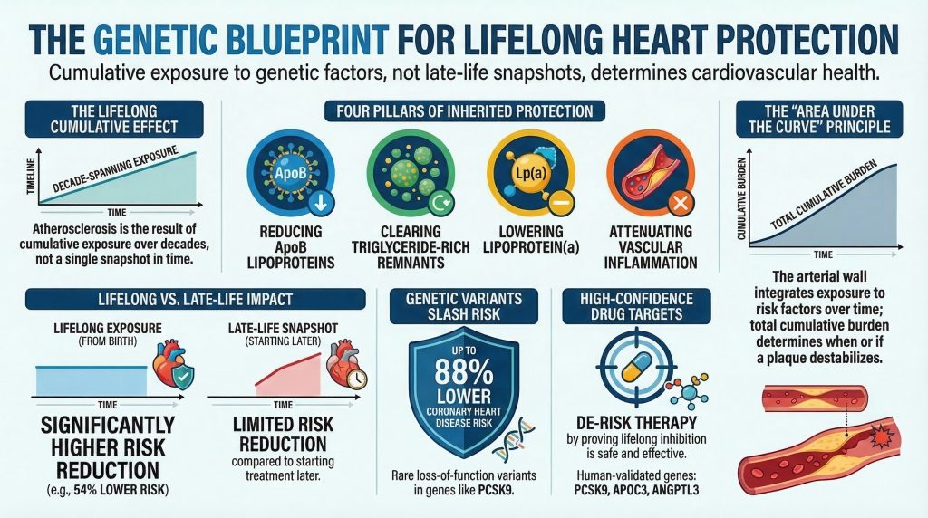
Eine der folgenschwersten Lektionen aus diesem Zeitalter der Genomforschung ist, dass von Natur vorkommende kardioprotektive Varianten unverhältnismäßig aussagekräftig sind. Diese Varianten, oft selten Loss-of-Function-AlleleEin Loss-of-Function-Allel ist eine DNA-Variante, die die normale Aktivität des durch dieses Gen kodierten Proteins teilweise oder vollständig aufhebt; in der kardiovaskulären Genetik haben Individuen, die mit Loss-of-Function-Allelen in Genen wie PCSK9 oder APOC3 geboren wurden, lebenslang von Natur aus niedrigere Spiegel atherogener Lipoproteine, was jahrzehntelange Evidenz zur Sicherheit und Wirksamkeit der Blockierung dieser... oder Funktionsmindernde Missense-Mutationen wirken als Experimente der Natur. Durch teilweises oder vollständiges Reduzieren der Aktivität eines ProteinProtein ist der Nährstoff, den Ihr Körper zum Aufbau und zur Reparatur von Muskeln und Gewebe verwendet. von Geburt an approximieren sie eine lebenslange Zielinhibition und liefern dadurch ungewöhnlich starke Beweise für Kausalität, das Ausmaß des Nutzens und die langfristige Verträglichkeit beim Menschen5], [6]. Die Humangenetik hat gezeigt, dass eine anhaltende Senkung von ApoB-haltigen Lipoproteinen, eine Beschleunigung der Clearance triglyceridreicher Lipoproteine, eine Reduktion von Lipoprotein(a)Lipoprotein(a), geschrieben Lp(a) und "L-P-klein-a" ausgesprochen, ist ein LDL-ähnliches Partikel mit einem extra klebrigen Protein daran. Belastung und Dämpfung von Entzündungssignalen können jeweils das Risiko verringern von Koronare HerzkrankheitKoronare Herzkrankheit ist eine Plaque-Ansammlung in den Arterien, die den Herzmuskel versorgen., MyokardinfarktSiehe Herzinfarkt für den vollständigen Eintrag., ischämischer SchlaganfallEin ischämischer Schlaganfall tritt auf, wenn die Blutzufuhr zu einem Teil des Gehirns unterbrochen wird und das Gehirngewebe abstirbt., und damit verbundene vaskuläre Ereignisse4], [7], [13], [17], [39], [40].
Die Bedeutung dieser Varianten ist zweifach. Erstens liefern sie kausale Beweise, die spezifische Signalwege mit Atherosklerose verknüpfen, oft überzeugender als die konventionelle Epidemiologie. Da genetische Varianten bei der Zeugung zufällig verteilt und über die gesamte Lebensdauer hinweg unveränderlich sind, sind sie weniger anfällig für umgekehrte KausalitätUmgekehrte Kausalität liegt vor, wenn der Pfeil in die andere Richtung zeigt – die Krankheit hat die Exposition verursacht, anstatt dass die Exposition die Krankheit verursacht hat. und viele traditionelle Formen von verwirrendEine Störgröße liegt vor, wenn ein versteckter dritter Faktor zwei ansonsten unverbundene Dinge miteinander verknüpft erscheinen lässt. [8], [35Zweitens minimieren sie das Risiko bei der Arzneimittelentwicklung. Wenn eine lebenslange teilweise Hemmung eines Proteins das vaskuläre Risiko senkt, ohne nennenswerte Toxizität beim Menschen zu verursachen, wird dieses Protein zu einem ungewöhnlich attraktiven therapeutischen Ziel. PCSK9PCSK9 ist ein von der Leber produziertes Protein, das die Andockstellen zerstört, die Ihre Leber nutzt, um Cholesterin aus Ihrem Blut zu entfernen. ist das deutlichste Beispiel für dieses Modell, aber es ist nicht mehr einzigartig. APOC3APOC3 ist das Gen für ein Protein, das den Abbau von Triglycerid-reichen Partikeln aus Ihrem Blut blockiert., ANGPTL3ANGPTL3 ist ein Protein, das den Abbau von Triglycerid-reichen Partikeln im Blut verlangsamt., NPC1L1NPC1L1 ist der Transporter in Ihrem Darm, der Cholesterin aus der Nahrung und der Galle aufnimmt. Ezetimib blockiert ihn., LPA und IL6R haben sich jeweils von humangenetischen Erkenntnissen zur pharmakologischen Entwicklung, zur therapeutischen Validierung oder zu beidem entwickelt [5], [6], [16], [17], [21], [26], [39], [40].
Gleichzeitig ist die Evidenzbasis nicht für alle vorgeschlagenen kardioprotektiven Gene einheitlich. Einige Locusse verfügen über eine direkte Validierung auf der Ebene menschlicher Endpunkte und einen klaren mechanistischen Pfad für Interventionen. Andere zeigen überzeugende BiomarkerEin Biomarker ist etwas im Körper Messbarem, das Aufschluss über Gesundheit oder Krankheit gibt – ein Laborwert, ein Scanergebnis, ein Blutdruckwert. Auswirkungen, aber weniger reife kardiovaskuläre Endpunktevidenz. Wieder andere gehen hervor aus GründerpopulationenEine Gründerpopulation ist eine Gruppe, die von einer kleinen Anzahl gemeinsamer Vorfahren abstammt, was zu einer verringerten genetischen Vielfalt und einer erhöhten Häufigkeit bestimmter seltenen Varianten führt; in der kardiovaskulären Genetik wurden Gründerpopulationen wie bestimmte isländische oder finnische Kohorten genutzt, um kardioprotektive Varianten zu entdecken, die zu selten sind, um in heterogenen globalen Stichproben effizient nachgewiesen zu werden., Exom-Scans oder pathway-basierter Inferenz, ohne bisher einen vollständig geklärten, therapeutisch validierten Mechanismus zu liefern [4], [11Aus diesem Grund ist eine strenge Evidenzhierarchie unerlässlich. Niedriger TriglycerideTriglyceride sind die Hauptform von Fett in Ihrem Blut und in den Fettspeichern Ihres Körpers., höhere HDL-C- oder niedrigere LDL-C-Werte bei Variantenträgern sind für sich genommen nicht ausreichend. Die klinisch aussagekräftigsten kardioprotektiven Zielgrößen sind diejenigen, die durch konvergente Evidenz gestützt werden, welche Genotyp mit Mechanismus, Mechanismus mit Biomarker und Biomarker mit harten kardiovaskulären Endpunkten verknüpft.
Diese Übersicht bietet eine evidenzbasierte Analyse der genetischen Architektur des kardiovaskulären Schutzes. Zunächst werden die wichtigsten mechanistischen Domänen kartiert, durch die ein vererbter Schutz vermittelt wird. Anschließend werden die kardioprotektiven Signalwege und Gene mit der höchsten Evidenz untersucht – solche, die durch humane Ergebnisevidenz und in mehreren Fällen durch therapeutische Translation gestützt werden –, darunter PCSK9, APOC3, ANGPTL3, LPL, NPC1L1, LPA und IL6R. Als Nächstes werden biologisch überzeugende, aber weniger vollständig validierte Zielstrukturen evaluiert, darunter ANGPTL4, ASGR1, CETPCETP ist ein Protein, das Cholesterin und Triglyceride zwischen HDL und den schädlichen ApoB-Partikeln austauscht., HMGCRHMGCR ist das Gen für die HMG-CoA-Reduktase, das Enzym, das den geschwindigkeitsbestimmenden Schritt bei der Cholesterinsynthese katalysiert. Es ist das genaue Ziel aller Statine., günstig LDLRLDLR ist das Gen, das den LDL-Rezeptor bildet, die Andockstelle, die Ihre Leber nutzt, um Cholesterinteilchen aus dem Blutkreislauf zu ziehen. und APOBApoB ist ein Protein, das sich auf der Außenseite jedes Cholesterinteilchens befindet, das in Ihrer Arterienwand stecken bleiben und Plaque verursachen kann. Jedes dieser Teilchen trägt genau ein ApoB. Variation, SORT1 und neu auftretende Exomsignale wie CIDEB und ZNRF3 [4], [9], [11], [31], [38Es berücksichtigt auch die besondere Rolle von Gründerpopulationen als Entdeckungsplattformen, während es hypothesengenerierende Isolate von etablierten kardioprotektiven Mechanismen unterscheidet. Schließlich wird argumentiert, dass die wichtigste Lehre aus der Humangenetik eher konzeptueller als taxonomischer Natur sein könnte: Das lebenslange vaskuläre Risiko lässt sich durch die kumulative vererbte Exposition besser erklären als durch eine einzelne klinische Momentaufnahme zu einem späteren Zeitpunkt im Leben.8], [12], [34].
Mechanistischer Signalweg-Plan: Vier Säulen des vererbten kardiovaskulären Schutzes
Die Architektur des vererbten Herzschutzes lässt sich am sinnvollsten nicht als Liste isolierter Gene verstehen, sondern als eine Reihe wiederkehrender mechanistischer Domänen. Die aktuellen Belege legen nahe, dass sich ein robuster genetischer Schutz vor atherosklerotischer kardiovaskulärer Erkrankung (ASCVD) in vier Hauptachsen konzentriert: (1) lebenslange Reduktion der Last an apoB-haltigen Lipoproteinen, (2) verbesserte Hydrolyse und Clearance von triglyceridreichen Lipoproteinen und deren Remnants, (3) Reduktion der durch Lipoprotein(a) vermittelten atherothrombotischen Last und (4) Abschwächung der vaskulären EntzündungEntzündungen sind die Reaktion Ihres Immunsystems auf eine Verletzung oder etwas, das es als Eindringling behandelt. Sie verursachen Schwellungen, Hitze und Aufräumzellen. [3], [7], [13], [15], [17], [39].
Die erste Säule ist die lebenslange Reduktion von apoB-haltigen Lipoproteinen, insbesondere LDLLDL oder Low-Density-Lipoprotein ist das Hauptpartikel, das Cholesterin durch Ihr Blut transportiert – und das Hauptpartikel, das in den Arterienwänden stecken bleibt. Teilchen. ApoB-haltige PartikelLipoproteine – einschließlich LDL, IDL, VLDL und deren Remnants –, die jeweils ein Molekül Apolipoprotein B an ihrer Oberfläche tragen; die Partikelanzahl (und nicht allein die Cholesterinmasse) ist ein Haupttreiber der Atherosklerose, da jedes Partikel in der Arterienwand retiniert werden kann. sind die notwendigen ursächlichen Partikel bei Atherosklerose. Ihr Eintritt in und ihre Retention innerhalb der arteriellen IntimaDie Intima ist die innerste Schicht einer Arterienwand und liegt direkt unter der glatten Auskleidung. leitet die Kaskade der Oxidation, endothelialen Aktivierung, Monozytenrekrutierung, Schaumzellbildung und PlaquePlaque ist die Ansammlung von Cholesterin, Immunzellen, Narbengewebe und Kalzium in einer Arterienwand. Das Fortschreiten, das letztendlich den meisten Myokardinfarkten und ischämischen [Zuständen/Erkrankungen] zugrunde liegt SchlaganfallEin Schlaganfall tritt auf, wenn die Blutversorgung eines Teils des Gehirns unterbrochen wird, entweder durch einen Verschluss oder durch eine Blutung. [13]. Zu den in diesem Bereich wirkenden Genen gehören PCSK9, NPC1L1, LDLR, APOB, HMGCR und im weiteren Sinne SORT1 [6], [9], [13], [38]. Schutzvarianten in diesen Genen senken die Konzentration von atherogene PartikelAtherogene Partikel sind die ApoB-haltigen Lipoproteine – einschließlich LDL, IDL, VLDL und Lipoprotein(a) –, die in die Arterienwand eindringen und dort zurückgehalten werden können, um das Plaque-Wachstum einzuleiten und aufrechtzuerhalten; der Artikel verwendet den Begriff, um zu beschreiben, was wesentlich und nachhaltig gesenkt werden muss, um eine Plaque-Rückbildung zu erreichen. oder die kumulative lebenslange Exposition gegenüber diesen zu verringern, wodurch die Wahrscheinlichkeit verringert wird, dass sich Plaque bildet, vergrößert und destabilisiert.
Die zweite Säule ist der Stoffwechsel von triglyceridreiche Lipoproteine (TRLs)Triglyzeridreiche Lipoproteine sind eine Klasse ApoB-haltiger Partikel – vor allem VLDL und IDL –, die große Mengen an Triglyzeriden transportieren und im Übermaß produziert werden, wenn die Leber überlastet ist, wie bei Insulinresistenz oder Adipositas; ihre Remnants sind direkt atherogen und können in die arterielle Intima vordringen, was zu einem Risiko beiträgt, das bei der Standard-LDL-C-Messung übersehen wird., einschließlich Lipoproteinen sehr niedriger Dichte (VLDLVLDL oder Lipoprotein sehr niedriger Dichte ist das Partikel, das Ihre Leber herstellt, um Triglyceride in den Rest des Körpers zu transportieren.), ChylomikroneEin Chylomikron ist ein sehr großes Partikel, das Fett aus einer Mahlzeit aus dem Darm in den Blutkreislauf transportiert., und ihre cholesterinreichen Überreste. Obwohl LDL das kanonische atherogene Partikel bleibt, belegen humangenetische Daten nun stark eine kausale Rolle von TRL-Überresten bei ASCVD [7], [15APOC3, ANGPTL3, ANGPTL4 und LPL sind Schlüsselregulatoren dieser Domäne. Diese Gene verändern die Hydrolyse und Clearance von TRLs durch die Modulation LipoproteinlipaseLipoproteinlipase ist ein Enzym, das an den Wänden kleiner Blutgefäße verankert ist, Triglyceride aus vorbeiziehenden Partikeln herauslöst und das Fett an Muskel- und Fettgewebe übergibt. Aktivität, Aktivität der endothelialen Lipase und Aufnahme von hepatischen Remnants15], [16], [22], [27]. Varianten, die den TRL-Katabolismus verstärken, senken Remnant-CholesterinRemnant-Cholesterin ist das Cholesterin, das in den Überbleibseln triglyceridreicher Partikel transportiert wird, nachdem diese den Großteil ihres Fettes abgegeben haben. und das vaskuläre Risiko zu verringern.
Eine dritte Säule, die in älteren lipidzentrierten Modellen oft zu wenig betont wird, ist Lipoprotein(a) oder Lp(a). Lp(a) ist ein apoB-haltiges Partikel, das kovalent verbunden ist mit ApolipoproteinEin Apolipoprotein ist ein Protein, das an ein fetttransportierendes Partikel in Ihrem Blut gebunden ist. Da Fett und Wasser sich nicht mischen, wirken diese Proteine wie eine Hülle, die es dem Fett ermöglicht, sicher durch den Blutkreislauf zu reisen.(a), deren Größe und Konzentration größtenteils durch genetische Variation am LPA-Lokus bestimmt werden [39], [40]. Im Gegensatz zu LDL allein scheint Lp(a) den atherogenen Cholesterintransport mit proinflammatorischen und prothrombotischen Eigenschaften zu verbinden. Die Humangenetik hat einige der stärksten kausalen Beweise in der Herz-Kreislauf-Biologie geliefert, dass erhöhtes Lp(a) das Koronarrisiko erhöht, während genetisch niedrigeres Lp(a) Schutz gewährt [39], [40].
Die vierte Säule ist Entzündung. Atherosklerose ist nicht nur eine Lipid-Speicherkrankheit, sondern eine chronische Entzündungserkrankung der Arterienwand. Angeborene und adaptive Immun-Signale beeinflussen die Entstehung und das Wachstum von Plaques, nekrotischer KernDer nekrotische Kern ist das tote, breiige Zentrum einer fortgeschrittenen Plaque, das aus Immunzellen besteht, die eingeschlossenes Cholesterin gefressen haben und dann an Ort und Stelle starben. Formation, faserige KappeDie fibröse Kappe ist die zähe Gewebeschicht, die eine Plaque bedeckt und ihren fettigen Kern vom Blutstrom trennt. Stabilität und Thrombogenität. Der klarste genetisch unterstützte entzündungshemmende kardioprotektive Signalweg ist IL-6Interleukin-6 oder IL-6 ist ein Signalmolekül, das das Immunsystem nutzt, um eine entzündliche Botschaft im Körper zu verbreiten. Signalisierung, insbesondere durch funktionelle Variation in IL6R [17Obwohl dieser Signalweg noch nicht den gleichen Grad an pharmakologischer Validierung kardiovaskulärer Endpunkte aufweist wie PCSK9, haben die Kombination aus humangenetischen Evidenzen und Daten aus entzündungshemmenden Studien Entzündungen als kausalen Bereich bei ASCVD fest etabliert [17], [18].
Diese vier Säulen sind voneinander abhängig und nicht autonom. ApoB-Partikel rufen vaskuläre Entzündungen hervor. Remnants tragen zu Endotheliale DysfunktionEndotheliale Dysfunktion liegt vor, wenn diese dünne Auskleidung ihre Aufgabe nicht mehr gut erfüllt. Die Blutgefäße weiten sich nicht richtig und die Barriere wird durchlässiger. und der Schaumzellenbildung. Lp(a) verbindet die Lipidbiologie mit Thrombose und Entzündung. Entzündliche Zytokine beeinflussen die hepatische Lipoproteinproduktion, die endotheliale Aktivierung und die Plaque-Instabilität. Dennoch bietet dieses Rahmenwerk eine nützliche Landkarte zur Evaluierung kardioprotektiver Zielstrukturen und zum Verständnis, warum bestimmte Gene wiederholt als beim Menschen validierte Schutzmechanismen hervorgehen.
Von der Lokalisierungsentdeckung zur kausalen Biologie: Warum Genetik wichtig ist
Die Stärke der Humangenetik liegt nicht nur in ihrer Fähigkeit, Assoziationen zu erkennen, sondern in ihrer Kapazität, kausale Mechanismen zu priorisieren. Studien zu Mendelschen Erkrankungen haben zuerst gezeigt, dass seltene Mutationen mit großer Wirkung allgemein relevante Biologie offenbaren können. Familiäre HypercholesterinämieFamiliäre Hypercholesterinämie, kurz FH, ist eine vererbte Erkrankung, bei der die Leber Cholesterin nicht richtig aus dem Blut entfernen kann. Die Werte sind von Geburt an sehr hoch. begründete die Zentralität von LDL-RezeptorDer LDL-Rezeptor ist eine Andockstelle auf Leberzellen, die LDL-Partikel aus dem Blut greift und sie zur Zersetzung hineinzieht. Biologie; seltene PCSK9-Mutationen zeigten später, dass die Störung desselben Signalwegs je nach Richtung schädlich oder schützend sein kann2], [5], [20. Ähnlich verhalten sich seltene Mendelsche DyslipämienDyslipidemia is the medical word for an unhealthy pattern of fats in the blood. It can mean high LDL, high triglycerides, low HDL, or some combination. anticipated later population-level findings showing that common and rare variants often converge on the same pathways [2].
GWAS added another dimension by revealing that common variants of small effect could still identify therapeutically decisive biology. Indeed, one of the most important lessons of cardiovascular genetics is that the variance explained by a variant is not the same as the biological or therapeutic importance of its gene [2], [3]. Common variants near HMGCR or NPC1L1 have small effects on LDL-C, but the proteins they implicate became targets of effective therapies [6], [9], [14], [30]. Conversely, some large-effect loci have remained mechanistically opaque for years, particularly when the causal signal resides in non-coding regulatory sequence rather than protein-coding variation [2], [3].
A canonical example of successful movement from association to mechanism is the chromosome 1p13 locus near SORT1. Initial GWAS linked the region to LDL-C and myocardial infarction risk, but the biology was uncertain. Subsequent functional work identified regulatory variants that altered SORT1 expression in the liver, thereby affecting lipoprotein secretion and LDL metabolism [38]. This case established that non-coding association signals can indeed be translated into specific genes and specific mechanisms, though such successes remain the exception rather than the rule. By contrast, loci such as 9p21 illustrate that a strong and reproducible association with coronary ArterieEine Arterie ist ein Blutgefäß, das Blut vom Herzen weg in den Rest des Körpers transportiert. disease does not necessarily yield rapid mechanistic clarity [2], [3].
For cardioprotective genetics, then, the highest-confidence targets tend to be those where association, coding perturbation, biologic mechanism, biomarker shift, and outcome reduction all align. That is the standard used below.
Tier 1: High-Confidence Cardioprotective Targets with Human Outcome Support
The most rigorous definition of a cardioprotective gene requires more than a favorable biomarker profile. It requires human evidence that function-altering variants in the gene are associated with lower incidence of cardiovascular events, ideally reinforced by mechanistic plausibility and therapeutic translation. On that standard, a limited number of pathways stand out.
PCSK9 (Proprotein Convertase Subtilisin/Kexin Type 9)
PCSK9 is the archetypal success story of cardiovascular genetics and remains the clearest demonstration of the path from human loss-of-function genetics to precision therapy. PCSK9 is synthesized predominantly in the liver as a secreted SerinproteaseA class of enzymes that cleave protein bonds using a serine residue in their active site; PCSK9 belongs to this class and uses this activity to target LDL receptors for degradation. that regulates the abundance of the LDL receptor (LDLR) on hepatocytes [19]. Its essential action is to bind LDLR and redirect it toward lysosomal degradation rather than recycling. Under normal conditions, LDLR internalizes circulating LDL particles, releases cargo in the acidic endosomal compartment, and then returns to the cell surface. PCSK9 disrupts this recycling process and therefore reduces hepatocyte LDLR density [19], [20].
The importance of PCSK9 first became evident when gain-of-function mutations were identified as a cause of autosomal dominant HypercholesterinämieHypercholesterinämie ist ein abnormal erhöhter Spiegel an cholesterintragenden Partikeln im Blut, der in Primatenexperimenten typischerweise durch die Fütterung einer an Nahrungscholesterin und gesättigten Fetten reichen Diät verursacht wird und mit einer beschleunigten Plaquebildung in den Arterienwänden einhergeht. [20]. The field changed when naturally occurring loss-of-function variants, including Y142X, C679X, and R46L, were identified in population cohorts [5]. Carriers had increased hepatic LDL receptor density, enhanced LDL clearance, and lifelong lower LDL-C levels. In the landmark study by Cohen and colleagues, Black carriers of nonsense variants in PCSK9 had an approximately 28% reduction in LDL-C and an 88% lower risk of koronare HerzkrankheitDie koronare Herzkrankheit ist die Verengung oder Verstopfung der Arterien, die den Herzmuskel mit Blut versorgen, verursacht durch die Anlagerung von atherosklerotischer Plaque; sie ist weltweit die häufigste Ursache für Herzinfarkt und Herztod., while White carriers of R46L had more modest LDL-C lowering but still significant protection [5]. The finding was transformative because it showed that lifelong LDL lowering from birth produces much larger proportional risk reductions than short-term treatment initiated later in life.
The translational consequences were immediate. Monoclonal antibodies such as EvolocumabEvolocumab ist ein injizierbares Cholesterinmedikament aus der Familie der PCSK9-Hemmer, das normalerweise alle zwei bis vier Wochen verabreicht wird. und AlirocumabAlirocumab, sold as Praluent, is an injectable antibody that blocks PCSK9, given every two to four weeks., and later silencing approaches such as InclisiranInclisiran ist eine cholesterinsenkende Injektion, die nach den ersten beiden Dosen nur zweimal im Jahr verabreicht wird., were developed to mimic the protective genetic phenotype. Outcome trials demonstrated that pharmacologic PCSK9 inhibition markedly lowers LDL-C on top of StatineEin Statin verlangsamt das Enzym, das Ihre Leber zur Bildung von Cholesterin nutzt. Ihre Leber reagiert darauf, indem sie mehr Cholesterin aus Ihrem Blut zieht, und genau daraus ergibt sich der eigentliche Nutzen. and reduces major cardiovascular events [21]. PCSK9 is therefore not just a well-supported target; it is the paradigm by which contemporary cardioprotective genetics is judged.
NPC1L1 (Niemann-Pick C1-Like 1)
NPC1L1 is the principal transporter mediating intestinal cholesterol absorption. It is expressed in the brush border of enterocytes and facilitates uptake of both dietary and biliary SterineEin Sterol ist eine Familie wachsartiger Moleküle, die auf derselben Vier-Ring-Struktur aufgebaut sind. Cholesterin ist dasjenige, das Tiere herstellen; Pflanzen stellen ihre eigenen Versionen her. [6]. Rare inactivating mutations in NPC1L1 were identified in large exome sequencing datasets and were associated with modest lifelong reductions in LDL-C but disproportionately large reductions in coronary heart disease risk [6]. In the study by Stitziel and colleagues, carriers had approximately 12 mg/dL lower LDL-C and about 53% lower risk of coronary disease [6]. Although the magnitude of effect in rare variant carriers should not be extrapolated uncritically to general-population treatment effects, the result strongly reinforced a key principle of cardiovascular biology: modest LDL differences, when sustained over decades, can yield very large cumulative risk differences.
NPC1L1 also exemplifies the convergence of genetics and therapeutics. EzetimibEzetimib ist eine Tablette, die die Aufnahme von Cholesterin im Darm blockiert. inhibits NPC1L1 pharmacologically, and the IMPROVE-ITIMPROVE-IT added ezetimibe to a statin after a heart attack, testing whether lowering LDL by a non-statin mechanism would help. trial showed that adding ezetimibe to statin therapy lowers cardiovascular events [30]. Thus, as with PCSK9, human genetics anticipated clinical benefit and strengthened causal confidence in the target.
APOC3 (Apolipoprotein C-III)
APOC3 is one of the major human regulators of triglyceride-rich lipoprotein metabolism. Synthesized largely by the liver and intestine, apolipoprotein C-III resides on the surface of chylomicrons, VLDL, and HDLHDL oder Lipoprotein hoher Dichte ist das Partikel, das oft als "gutes Cholesterin" bezeichnet wird. Es nimmt Cholesterin aus dem Gewebe auf und transportiert es zurück zur Leber. particles, where it inhibits lipoprotein lipase, delays lipolysis of triglyceride-rich particles, and impairs hepatic remnant uptake [15], [22]. Elevated APOC3 therefore promotes accumulation of triglycerides and remnant cholesterol, both increasingly recognized as causal mediators of ASCVD.
Exome sequencing studies identified rare loss-of-function mutations in APOC3, including R19X and splice-disrupting alleles, that lower APOC3 levels and reduce plasma triglycerides by roughly 40% [23], [24]. More importantly, these variants were associated with lower risk of ischemic vascular disease and coronary disease [23], [24]. These observations strengthened the case that triglyceride-rich remnants are not merely correlated with risk, but are themselves causally involved in atherosclerosis. The likely benefit appears to be mediated primarily through improved remnant clearance rather than major LDL reduction.
The APOC3 story has also translated rapidly. Antisense oligonucleotide and RNA-targeted approaches that suppress APOC3 have shown marked triglyceride lowering in severe hypertriglyceridemia. Cardiovascular outcomes data are still maturing, but genetically APOC3 already belongs among the strongest validated targets for RestrisikoResidual risk is the risk that remains after you have done the obvious things — cholesterol treated, blood pressure controlled, not smoking. beyond LDL.
ANGPTL3 (Angiopoietin-Like 3)
ANGPTL3 is a liver-derived secretory protein that inhibits both lipoprotein lipase and endothelial lipase, placing it at a central junction in systemic lipid trafficking [16], [25]. Homozygous loss-of-function mutations in ANGPTL3 cause familial combined hypolipidemia, characterized by low LDL-C, low triglycerides, and low HDL-C [25]. This phenotype is notable because it simultaneously alters several lipid compartments relevant to vascular disease.
Population-based sequencing studies showed that heterozygous carriers of inactivating ANGPTL3 variants have lower plasma lipid levels and approximately 34% lower risk of coronary artery disease [16]. The protective signature is broader than in a purely LDL-centered pathway: lower triglycerides, lower remnant cholesterol, lower apoB, and lower LDL-C all contribute. Some LDL lowering appears partly independent of canonical LDL receptor biology, increasing the therapeutic interest of ANGPTL3 in disorders such as homozygot familiäre HypercholesterinämieHomozygous familial hypercholesterolemia, or HoFH, is the rare and severe form of inherited high cholesterol, where a child inherits the faulty gene from both parents instead of one. [16], [25].
This promise has been realized in part through evinacumabEvinacumab is an injectable antibody that blocks ANGPTL3, used for the most severe inherited cholesterol disorders., a monoclonal antibody against ANGPTL3, which lowers LDL-C substantially even in settings where LDLR-dependent therapies are limited [26]. ANGPTL3 therefore stands with PCSK9 and NPC1L1 as one of the clearest examples of protective human genetics guiding successful translational development.
LPL (Lipoprotein Lipase)
Lipoprotein lipase is the key effector enzyme for hydrolysis of triglycerides within chylomicrons and VLDL, thereby facilitating tissue fatty acid uptake and remnant clearance [15], [28]. If APOC3 and ANGPTL proteins act as brakes on triglyceride clearance, LPL is the engine that confers protection when activated or disinhibited.
The best-known naturally occurring protective variant in LPL is S447X (Ser447Ter, S447*), a near-terminal truncating variant associated with enhanced lipolytic efficiency, lower triglycerides, higher HDL-C, and lower coronary risk [29]. Although the exact biochemical mechanism has been debated, the epidemiologic signal has been consistent. More broadly, coding variation in and around the LPL pathway has repeatedly shown that greater lipolytic clearance of TRLs and remnants is cardioprotective [4].
LPL is conceptually important because it provides reciprocal validation of the remnant hypothesis. If inhibiting APOC3 or ANGPTL proteins lowers cardiovascular risk by releasing the LPL brake, then enhancing LPL itself should be protective. Human genetics confirms exactly that.
LPA and Lipoprotein(a)
Any contemporary discussion of inherited cardioprotection is incomplete without lipoprotein(a). Lp(a) levels are determined predominantly by variation at the LPA locus, particularly by apo(a) isoform size and related genetic structure [39], [40]. Elevated Lp(a) is now among the most strongly genetically supported causal risk factors for coronary disease. Conversely, inherited reductions in Lp(a) confer protection [39], [40].
The key conceptual importance of LPA is that it broadens the field beyond standard LDL biology. Lp(a) appears to promote atherosclerosis through apoB-mediated arterial retention while also exerting proinflammatory and prothrombotic effects. Genetic studies have shown that variants associated with elevated Lp(a) increase myocardial infarction risk, while Mendelian-randomization analyses support causality rather than mere correlation [39], [40]. Although naturally occurring protective LPA variants are less neatly framed as classic loss-of-function alleles than PCSK9 or APOC3, the pathway is sufficiently validated to merit inclusion among high-confidence cardioprotective mechanisms. Therapeutic programs targeting Lp(a) now represent one of the most promising frontiers in preventive cardiology.
IL6R and IL-6 Signaling
IL6R occupies a special place in the cardioprotective landscape because it extends validated protection beyond lipoprotein metabolism. Atherosclerosis is a chronic inflammatory disease, and IL-6 is a central mediator of acute-phase and vascular inflammatory signaling [17], [18]. The common Asp358Ala variant in IL6R increases receptor shedding and attenuates classical IL-6 signaling. Carriers have lower CRP and fibrinogen levels and lower coronary heart disease risk [17].
The magnitude of vascular protection is smaller than that seen with some lipid pathways, and IL6R has not yet reached the same level of definitive pharmacologic cardiovascular outcome validation as PCSK9. Nevertheless, the mechanistic significance is profound. It shows that genetically reduced inflammatory signaling can reduce ASCVD risk independently of lipid levels. This genetic result complements interventional evidence from anti-inflammatory trials such as GesängeCANTOS (Canakinumab Anti-inflammatory Thrombosis Outcomes Study) was a large randomized trial that tested canakinumab, a drug blocking the inflammatory signal IL-1β, against placebo; at its prespecified 150 mg dose it reduced major cardiovascular events by roughly 15% without lowering LDL cholesterol, providing direct human evidence that inflammation drives heart attacks through a pathway indepen…, even though CANTOS targeted IL-1β rather than IL-6 directly [18]. IL6R is therefore best viewed as a genetically validated protective pathway with strong translational promise.
Tier 2: Biologically Compelling but Less Fully Validated Targets
A second tier includes genes and pathways with substantial biologic credibility and often convincing biomarker effects, but with less complete human outcome evidence, less certain mechanism, or less mature therapeutic translation.
ANGPTL4
ANGPTL4, like ANGPTL3, inhibits lipoprotein lipase and influences fasting-state lipid partitioning [27]. The E40K missense variantA missense variant is a single nucleotide change in a gene that causes one amino acid to be substituted for another in the resulting protein, potentially altering its function; in cardioprotective genetics, function-reducing missense variants—such as those in PCSK9—can lower atherogenic lipoprotein levels from birth without fully abolishing the protein. reduces inhibitory activity and is associated with lower triglycerides, higher HDL-C, and reduced coronary disease risk [4], [28]. These observations strongly support the general principle that LPL disinhibition is cardioprotective. However, ANGPTL4 does not yet have the same breadth of outcome validation or therapeutic maturity as PCSK9, ANGPTL3, or APOC3. In addition, animal studies raised safety concerns with more complete pathway disruption under certain dietary conditions [27]. Human genetics supports ANGPTL4 as an important pathway-supported target, but not yet as a top-tier clinically validated one.
ASGR1
ASGR1 is a hepatic receptor involved in clearance of desialylated glycoproteins. Rare loss-of-function variants were associated with lower Nicht-HDL-CholesterinNon-HDL-Cholesterin ist eine einfache Berechnung: Ihr Gesamtcholesterin minus Ihr HDL. Was übrig bleibt, ist das Cholesterin in all den Partikeln, die Ihren Arterien schaden können. and reduced coronary artery disease risk in Icelandic sequencing studies [31]. The signal is intriguing and potentially important, but the mechanism remains incompletely resolved and therapeutic translation is still early. ASGR1 is therefore best placed among promising cardioprotective loci rather than among the most validated targets.
CETP
CETP facilitates exchange of CholesterinesterCholesterinester sind Speicherformen des Cholesterins, bei denen eine Fettsäure an Cholesterin gebunden ist; sie akkumulieren in großen Mengen in Schaumzellen und den extrazellulären Räumen von Plaques, und ihre Depletion – gemessen als Regression des lipidreichen Pools – ist ein primärer Marker für eine Plaqueverbesserung in Primaten-Regressionsstudien. and triglycerides between HDL and apoB-containing lipoproteins. Variants that reduce CETP activity substantially raise HDL-C and may modestly lower apoB and LDL-C [9], [32]. Human genetics and the long arc of CETP inhibitor development have clarified an important point: whatever cardiovascular benefit CETP inhibition may confer is likely mediated by lowering apoB-containing particles, not by raising HDL per se [9], [32]. That distinction is crucial, because CETP was long misinterpreted as an HDL story when it is more properly an apoB story. Clinical trialEine klinische Studie ist eine Untersuchung, bei der Forscher einer Gruppe eine Behandlung und einer anderen Gruppe ein Placebo oder eine Standardbehandlung verabreichen und dann die Ergebnisse vergleichen. results have been heterogeneous, reflecting both off-target toxicity and varying efficacy across compounds. CETP therefore remains pathway-supported but not definitively validated at the level of the strongest cardioprotective genes.
HMGCR, LDLR, and APOB Favorable Variation
These genes reinforce the centrality of cumulative LDL exposure. HMGCR variation has been especially informative because common variants such as rs12916 genetically mimic partial statin exposure over the lifespan [9]. Carriers have lower LDL-C and lower coronary risk, but also slightly increased diabetes risk, mirroring a known tradeoff of statin therapy. This is a powerful example of how genetics can predict both therapeutic benefit and mechanism-based adverse effects. Favorable regulatory variation in LDLR and APOB likewise supports the centrality of lifelong apoB lowering, even though the most dramatic pathogenic consequences of these genes are seen when function is impaired in the opposite direction [13], [14].
SORT1 and the 1p13 Locus
SORT1 merits discussion because it represents one of the best examples of successful translation from GWAS signal to specific mechanism. Variants at 1p13 are associated with LDL-C and myocardial infarction risk; functional studies showed that regulatory changes at the locus alter hepatic SORT1 expression and lipoprotein metabolism [38]. The locus is therefore highly informative biologically, but its role is more as a mechanistically illuminating regulatory node than as a classical human loss-of-function protective target with direct therapeutic analogy.
CIDEB, ZNRF3, SVEP1, KLF14, and Emerging Exome Signals
Large-scale sequencing continues to identify loci that may inform future therapeutic development. CIDEB appears relevant to hepatic lipid droplet biology and may influence both liver disease and cardiometabolic phenotypes [11]. ZNRF3 has emerged as a possible LDL-lowering target without obvious adverse glycemic or hepatic signatures, though the evidence remains early [11]. SVEP1 is notable because coding variation has been associated with coronary risk [4], but its directionality implicates vascular biology more than protective loss-of-function. KLF14 is metabolically interesting, especially in adipose biology, yet remains less directly validated as a cardiovascular protection target. These loci are best viewed as discovery-stage or pathway-informative rather than therapeutically settled.
Evidence Grading: A Practical Hierarchy of Cardioprotective Targets
A useful synthesis integrates four dimensions: (1) nature of genetic perturbation, (2) biomarker effect, (3) cardiovascular outcome evidence, and (4) degree of therapeutic translation.
Tier 1: highest-confidence cardioprotective targets
- PCSK9: major lifelong LDL lowering, strong human outcome evidence, approved therapies [5], [21]
- NPC1L1: modest lifelong LDL lowering with large rare-carrier outcome effects, approved therapy [6], [30]
- APOC3: major triglyceride/remnant lowering with strong human outcome support, advanced targeted therapies [23], [24]
- ANGPTL3: broad lipidsenkendLipidsenkung bedeutet die Reduzierung der schädlichen, ApoB-haltigen Partikel in Ihrem Blut – durch Ernährung, Medikamente oder beides. with human outcome support and approved therapy [16], [26]
- LPL: direct effector of remnant clearance with consistent human protective evidence [4], [29]
- LPA pathway: strong causal evidence that genetically lower Lp(a) reduces ASCVD risk, active therapeutic translation [39], [40]
- IL6R pathway: strong human genetic support for inflammatory causality, translational development underway [17], [18]
Tier 2: biologically compelling, but less fully validated
- ANGPTL4 [4], [27], [28]
- ASGR1 [31]
- CETP [9], [32]
- HMGCR [9], [14]
- Favorable LDLR/APOB variation [13], [14]
- SORT1 [38]
- CIDEB, ZNRF3, KLF14, and related emerging exome signals [11]
This hierarchy emphasizes a central principle: the most useful cardioprotective genes are not necessarily those with the largest biomarker effects, but those for which genetics, mechanism, outcomes, and therapeutic tractability all align.
Founder Populations and Isolates: Discovery Platforms, Not Evidence Substitutes
Founder populations occupy a privileged place in human genetics because isolation, bottlenecks, and endogamy can amplify otherwise rare variants, thereby increasing power to detect disease-modifying alleles. This logic has already yielded important discoveries in cardiometabolic biology, including protective lipid variants in relatively isolated populations [23], [24]. Such populations are especially valuable for hypothesis generation and rare variant discovery.
However, founder status is not itself evidence of cardioprotection. Claims regarding unusual vascular protection in specific isolates should be evaluated with the same rigor as any other proposed mechanism: the variant must be identified, its functional direction demonstrated, and its relationship to hard cardiovascular outcomes established. In the absence of that sequence of evidence, such populations remain discovery opportunities rather than validated examples of inherited protection. This distinction is particularly important when media narratives outpace peer-reviewed mechanistic literature.
Why Inherited Exposure Outperforms Late-Life Snapshots
The most important conceptual lesson from cardioprotective genetics is that atherosclerosis is fundamentally a disease of kumulative ExpositionKumulative Exposition ist die Gesamtmenge an schädlichen Cholesterinpartikeln, denen Ihre Arterien im Laufe Ihres gesamten Lebens ausgesetzt waren – wie hoch, multipliziert mit der Dauer.. Clinical risk assessment traditionally relies on measurements taken in midlife: an LDL-C value, a BlutdruckBlutdruck ist die Kraft, mit der das Blut gegen Ihre Arterienwände drückt. Er wird als zwei Zahlen geschrieben, wie 120/80. Die obere Zahl ist der Druck, wenn sich Ihr Herz zusammenzieht, die untere, wenn es sich entspannt. reading, an HbA1c, a high-sensitivity CRP. These are clinically useful, but biologically they are snapshots [12]. The arterial wall, by contrast, integrates exposure over decades. What matters mechanistically is the area under the curve of apoB burden, remnant cholesterol, Lp(a), blood pressure, glycemic injury, and inflammatory signaling [8], [13], [34].
Mendelian-randomization studies have shown that lifelong genetically mediated LDL lowering produces much larger reductions in coronary risk than the same absolute LDL reduction achieved pharmacologically for only a few years in adulthood [8]. Ference and colleagues demonstrated that a 1 mmol/L lower LDL-C level sustained from early life is associated with an approximately 54% lower risk of coronary heart disease [8]. This is far greater than the relatives RisikoDas relative Risiko vergleicht zwei Gruppen: Diese Gruppe hatte 30 Prozent weniger Herzinfarkte als jene Gruppe. reduction usually observed when the same LDL difference is achieved later in life with treatment [8], [14]. The explanation is straightforward: lifelong protection prevents plaque from forming in the first place, whereas later therapy often modifies an already established disease process.
The same principle almost certainly extends beyond LDL. Persistently lower remnant burden, lower Lp(a), lower blood pressure, and lower inflammatory signaling from early life should all be expected to produce larger lifetime benefits than late correction alone. Human genetics is uniquely valuable here because it measures stable inherited predisposition rather than transient physiology. Genetic variants are fixed at conception, unaffected by most short-term environmental noise, and less prone to reverse causality [35]. In this sense, genetics allows researchers to look through day-to-day biologic fluctuation and infer a person’s long-term exposure architecture.
Polygenic Risk and the Continuum Between Monogenic and Common Variation
Rare monogenic protective variants are highly informative but uncommon. At the population level, inherited risk and protection are often polygenic. Polygenic risk scoresA polygenic risk score adds up the effects of many small genetic variants to estimate your inherited risk of a disease. aggregate the effects of many common variants across the genome to estimate baseline susceptibility to coronary disease [10], [36], [37]. These tools have shown that a subset of individuals carry polygenic risk approaching that seen in some monogenic disorders, particularly when interpreted together with conventional clinical information [36], [37].
The key contribution of polygenic models is not that they replace causal pathway biology, but that they complement it. Rare variants reveal mechanisms. Polygenic scores capture background burden. Together they suggest that inherited cardiovascular protection exists on a continuum: from rare, high-impact perturbations such as PCSK9 or APOC3 loss-of-function, to common regulatory variation in pathways such as HMGCR or CETP, to broad genome-wide architectures that influence lifelong arterial injury in aggregate.
PRS still requires caution. Performance varies across ancestry groups, calibration remains an active area of research, and most claims about event prevention remain inferential rather than trial-proven [36], [37]. Still, the broader message is clear: inherited biology can identify risk and protection much earlier than clinical disease becomes apparent.
Fazit
Human genetics has fundamentally altered both the biology and strategy of cardiovascular prevention. It has shown that inherited cardiovascular protection is not diffuse or random, but concentrated in a limited number of causal domains: lifelong lowering of apoB-containing lipoproteins, more efficient clearance of triglyceride-rich remnants, reduction of Lp(a)-mediated atherothrombotic burden, and attenuation of vascular inflammation. Within these domains, a relatively small set of genes and pathways—especially PCSK9, NPC1L1, APOC3, ANGPTL3, LPL, LPA, and IL6R—now carry the strongest evidence as high-confidence cardioprotective mechanisms [5], [6], [16], [17], [21], [23], [24], [39], [40].
These pathways have done more than explain disease. They have provided a roadmap for therapy. PCSK9 inhibition is now standard clinical practice for selected patients. NPC1L1 genetics anticipated the benefit of ezetimibe. ANGPTL3 inhibition is clinically useful in severe dyslipidemia. APOC3 and Lp(a)-directed strategies continue to extend the frontier of precision prevention. IL6R has strengthened the case that inflammatory signaling is not merely associated with atherosclerosis, but causally involved in it.
Yet the most important lesson may be even broader. Cardiovascular risk is best understood not as a static trait measured in middle age, but as the cumulative consequence of decades of exposure. Human genetics provides a uniquely powerful lens on that cumulative process. Whether through rare cardioprotective alleles, regulatory variation in key pathways, or polygenic background burden, inherited variation reveals the tempo of arterial injury long before disease is clinically visible. The future of preventive cardiology will therefore depend not only on identifying more loci, but on learning to mimic nature’s most protective perturbations earlier, more precisely, and with greater mechanistic confidence.
Referenzen
-
- World Health Organization, “Cardiovascular diseases (CVDs),” WHO, 2025. https://www.who.int/news-room/fact-sheets/detail/cardiovascular-diseases-(cvds)
- Kathiresan S, Srivastava D. Genetics of human cardiovascular disease. Cell. 2012;148(6):1242-1257. doi:10.1016/j.cell.2012.03.001
- Khera AV, Kathiresan S. Genetics of coronary artery disease: discovery, biology and clinical translation. Nat Rev Genet. 2017;18(6):331-344. doi:10.1038/nrg.2016.160
- Myocardial Infarction Genetics and CARDIoGRAM Exome Consortia Investigators, Stitziel NO, Stirrups KE, et al. Coding Variation in ANGPTL4, LPL, and SVEP1 and the Risk of Coronary Disease. N Engl J Med. 2016;374(12):1134-1144. doi:10.1056/NEJMoa1507652
- Cohen JC, Boerwinkle E, Mosley TH Jr, Hobbs HH. Sequence variations in PCSK9, low LDL, and protection against coronary heart disease. N Engl J Med. 2006;354(12):1264-1272. doi:10.1056/NEJMoa054013.
- Myocardial Infarction Genetics Consortium Investigators, Stitziel NO, Won HH, et al. Inactivating mutations in NPC1L1 and protection from coronary heart disease.
- Budoff M. Triglycerides and Triglyceride-Rich Lipoproteins in the Causal Pathway of Cardiovascular Disease. Am J Cardiol. 2016;118(1):138-145. doi:10.1016/j.amjcard.2016.04.004
- Ference BA, Yoo W, Alesh I, et al. Effect of long-term exposure to lower low-density lipoprotein cholesterol beginning early in life on the risk of coronary heart disease: a Mendelian randomization analysis. J Am Coll Cardiol. 2012;60(25):2631-2639. doi:10.1016/j.jacc.2012.09.017.
- Ference BA, Kastelein JJP, Ginsberg HN, et al. Association of Genetic Variants Related to CETP Inhibitors and Statins With Lipoprotein Levels and Cardiovascular Risk. JAMA. 2017;318(10):947-956. doi:10.1001/jama.2017.11467.
- Inouye M, Abraham G, Nelson CP, et al. Genomic Risk Prediction of Coronary Artery Disease in 480,000 Adults: Implications for Primary Prevention. J Am Coll Cardiol. 2018;72(16):1883-1893. doi:10.1016/j.jacc.2018.07.079
- Nielsen JB, Rom O, Surakka I, et al. Loss-of-function genomic variants highlight potential therapeutic targets for cardiovascular disease. Nat Commun. 2020;11(1):6417. Published 2020 Dec 18. doi:10.1038/s41467-020-20086-3.
- Lloyd-Jones DM, Braun LT, Ndumele CE, et al. Use of Risk Assessment Tools to Guide Decision-Making in the Primary Prevention of Atherosclerotic Cardiovascular Disease: A Special Report From the American Heart Association and American College of Cardiology.
- Ference BA, Ginsberg HN, Graham I, et al. Low-density lipoproteins cause atherosclerotic cardiovascular disease. 1. Evidence from genetic, epidemiologic, and clinical studies. A consensus statement from the European Atherosclerosis Society Consensus Panel.
- Baigent C, Keech A, Kearney PM, et al. Efficacy and safety of cholesterol-lowering treatment: prospective meta-analysis of data from 90,056 participants in 14 randomised trials of statins. Lancet. 2005;366(9493):1267-1278. doi:10.1016/S0140-6736(05)67394-1.
- Ginsberg HN, Packard CJ, Chapman MJ, et al. Triglyceride-rich lipoproteins and their remnants: metabolic insights, role in atherosclerotic cardiovascular disease, and emerging therapeutic strategies-a consensus statement from the European Atherosclerosis Society.
- Dewey FE, Gusarova V, Dunbar RL, et al. Genetic and Pharmacologic Inactivation of ANGPTL3 and Cardiovascular Disease. N Engl J Med. 2017;377(3):211-221. doi:10.1056/NEJMoa1612790.
- IL6R Genetics Consortium Emerging Risk Factors Collaboration, Sarwar N, Butterworth AS, et al. Interleukin-6 receptor pathways in coronary heart disease: a collaborative meta-analysis of 82 studies. Lancet. 2012;379(9822):1205-1213. doi:10.1016/S0140-6736(11)61931-4.
- Ridker PM, Everett BM, Thuren T, et al. Antiinflammatory Therapy with Canakinumab for Atherosclerotic Disease.
- Horton JD, Cohen JC, Hobbs HH. PCSK9: a convertase that coordinates LDL catabolism.
- Abifadel M, Varret M, Rabès JP, et al. Mutations in PCSK9 cause autosomal dominant hypercholesterolemia.
- Sabatine MS, Giugliano RP, Keech AC, et al. Evolocumab and Clinical Outcomes in Patients with Cardiovascular Disease.
- Ramms B, Gordts PLSM. Apolipoprotein C-III in triglyceride-rich lipoprotein metabolism.
- Jørgensen AB, Frikke-Schmidt R, Nordestgaard BG, Tybjærg-Hansen A. Loss-of-function mutations in APOC3 and risk of ischemic vascular disease.
- TG and HDL Working Group of the Exome Sequencing Project, National Heart, Lung, and Blood Institute, Crosby J, Peloso GM, et al. Loss-of-function mutations in APOC3, triglycerides, and coronary disease.
- Musunuru K, Kathiresan S. Surprises From Genetic Analyses of Lipid Risk Factors for Atherosclerosis.
- Raal FJ, Rosenson RS, Reeskamp LF, et al. Evinacumab for Homozygous Familial Hypercholesterolemia.
- Aryal B, Price NL, Suarez Y, Fernández-Hernando C. ANGPTL4 in Metabolic and Cardiovascular Disease.
- Romeo S, Yin W, Kozlitina J, et al. Rare loss-of-function mutations in ANGPTL family members contribute to plasma triglyceride levels in humans.
- Sun W, Wu Y, Wen Y, Guo M, Zhang H. The association of the S447X mutation in LPL with Coronary artery disease: a meta-analysis. Minerva Cardioangiol. 2019;67(3):246-253. doi:10.23736/S0026-4725.18.04668-6.
- Cannon CP, Blazing MA, Giugliano RP, et al. Ezetimibe Added to Statin Therapy after Acute Coronary Syndromes.
- Nioi P, Sigurdsson A, Thorleifsson G, et al. Variant ASGR1 Associated with a Reduced Risk of Coronary Artery Disease.
- Nelson AJ, Sniderman AD, Ditmarsch M, et al. Cholesteryl Ester Transfer Protein Inhibition Reduces Major Adverse Cardiovascular Events by Lowering Apolipoprotein B Levels.
- Vassy JL, Christensen KD, Schonman EF, et al. The Impact of Whole-Genome Sequencing on the Primary Care and Outcomes of Healthy Adult Patients: A Pilot Randomized Trial.
- Ference BA, Braunwald E, Catapano AL. The LDL cumulative exposure hypothesis: evidence and practical applications.
- Burgess S, Butterworth A, Thompson SG. Mendelian randomization analysis with multiple genetic variants using summarized data.
- Khera AV, Chaffin M, Aragam KG, et al. Genome-wide polygenic scores for common diseases identify individuals with risk equivalent to monogenic mutations. Nat Genet. 2018;50(9):1219-1224. doi:10.1038/s41588-018-0183-z
- Inouye M, Abraham G, Nelson CP, et al. Genomic Risk Prediction of Coronary Artery Disease in 480,000 Adults: Implications for Primary Prevention. J Am Coll Cardiol. 2018;72(16):1883-1893. doi:10.1016/j.jacc.2018.07.079
- Musunuru K, Strong A, Frank-Kamenetsky M, et al. From noncoding variant to phenotype via SORT1 at the 1p13 cholesterol locus. Nature. 2010;466(7307):714-719. doi:10.1038/nature09266
- Clarke R, Peden JF, Hopewell JC, et al. Genetic variants associated with Lp(a) lipoprotein level and coronary disease. N Engl J Med. 2009;361(26):2518-2528. doi:10.1056/NEJMoa0902604.
- Kamstrup PR, Tybjaerg-Hansen A, Steffensen R, Nordestgaard BG. Genetically elevated lipoprotein(a) and increased risk of myocardial infarction. JAMA. 2009;301(22):2331-2339. doi:10.1001/jama.2009.801

