
Traffico Molecolare e Malattia Arteriosa
In che modo le lipoproteine ApoB e la segnalazione della Rho GEF aiutano a spiegare l'aterosclerosi
Aterosclerosi è spesso spiegato in modo troppo semplice. Alla maggior parte delle persone viene detto che “l'alto colesterolo” ostruisce le arterie, e sebbene questa idea punti nella giusta direzione, tralascia la biologia più profonda che guida effettivamente la malattia. Il colesterolo in sé non è l'intera storia. Il quadro più accurato è che l'aterosclerosi si sviluppa quando le molecole aterogene lipoproteina le particelle entrano nel arteria parete, rimangono intrappolate e innescano una lunga cascata infiammatoria. Che ciò avvenga dipende non solo da quante particelle circolano nel sangue, ma anche da come si comporta la parete arteriosa a livello molecolare1-8].
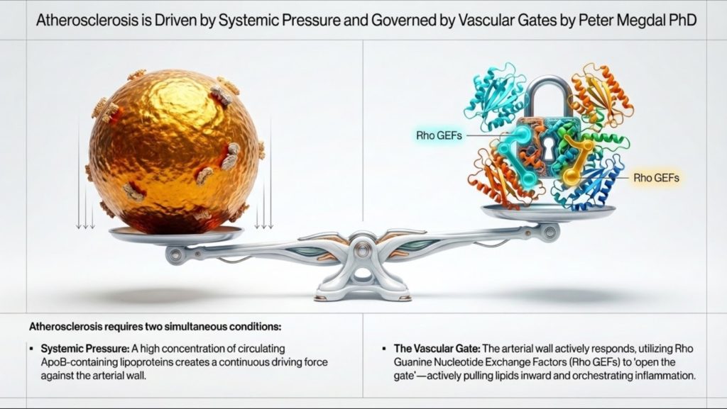
Quella seconda parte conta più di quanto la maggior parte delle discussioni riconosca. La parete arteriosa non è un tubo passivo. È un tessuto attivo e reattivo. Cellule endoteliali regolano quali particelle attraversano la parete del vaso. Le cellule immunitarie decidono come reagire una volta che tali particelle vengono trattenute. Cellule muscolari lisce quindi aiuta a determinare se un placca si stabilizza o diventa pericoloso. gran parte di questa regolazione locale è controllata da una famiglia di interruttori molecolari nota come fattori di scambio dei nucleotidi guaninici di Rho, oppure GEF di Rho, lavorando attraverso GTPasi Rho come RhoA, Rac1 e Cdc42 [4].
Questo offre un modo più completo per pensare a malattia cardiovascolare. Carico lipidico sistemico, in particolare il numero di particelle contenenti ApoB, regola la pressione nel sistema. Ma la segnalazione locale all'interno della parete arteriosa determina se a quelle particelle è consentito entrare, se vengono trattenute e trasformate in placca. In termini pratici, ApoB misura il numero di veicoli sulla strada, mentre la segnalazione Rho GEF aiuta a decidere quanto sia aperto il cancello e quanto distruttiva sarà la risposta locale1-4].
La comprensione di questa relazione aiuta a collegare la lipidologia clinica di alto livello e la moderna biologia vascolare. Rende inoltre la malattia più facile da spiegare al pubblico. L'aterosclerosi non è solo “grasso nel sangue”. È un problema di traffico di particelle, ingresso arterioso, ritenzione, attivazione immunitaria e rimodellamento tissutale.
Perché le lipoproteine esistono anzitutto
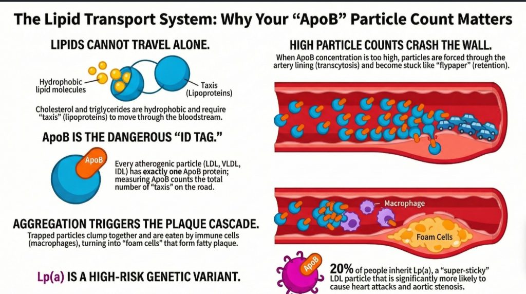
Il corpo umano ha un problema ingegneristico di fondo. Deve trasportare lipidi come trigliceridi e colesterolo, ma il plasma sanguigno è costituito principalmente da acqua. I lipidi sono idrofobi, il che significa che non si dissolvono bene in un ambiente acquoso. Se il corpo rilasciasse semplicemente grasso grezzo nel flusso sanguigno, questo si separerebbe e si agglomererebbe, un po' come l'olio nell'acqua [1,2].
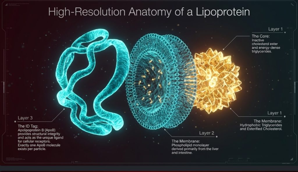
La soluzione è la lipoproteina. Le lipoproteine sono particelle di trasporto costruite per trasportare carico idrofobico attraverso il plasma acquoso. Hanno un nucleo idrofobico, dove sono immagazzinati trigliceridi ed esteri del colesterolo, e una superficie più compatibile con l'acqua fatta di fosfolipidi, colesterolo libero e proteine chiamato apolipoproteine [1,2,5].
Questo è importante perché le lipoproteine sono spesso trattate come valori di laboratorio astratti, ma sono meglio comprese come veicoli di trasporto fisico. Muovono energia e materiali strutturali attraverso il corpo. I trigliceridi sono il carico energetico principale. Il colesterolo fornisce supporto strutturale per le membrane, la sintesi degli ormoni steroidei e acido biliare produzione. Il flusso sanguigno è l'autostrada e le lipoproteine sono la flotta1-3,5].
Da quel punto di vista, il corpo non ha evoluto le lipoproteine principalmente per trasportare il colesterolo. Le ha evolute per trasportare energia. Il colesterolo è necessario, ma il sistema di trasporto è profondamente legato alla consegna di carburante. Questa distinzione è importante, perché aiuta a chiarire uno dei problemi più fraintesi nelle malattie cardiovascolari: il corpo ha bisogno di colesterolo, ma non ha bisogno di grandi quantità di colesterolo in movimento nel plasma per mantenere i tessuti vivi e funzionanti [2,11-15].
Omeostasi del colesterolo: quanto ne serve realmente
Un fatto centrale ma spesso trascurato nella biologia dei lipidi è che l'organismo richiede solo una quantità molto ridotta di colesterolo a livello cellulare. Ogni membrana cellulare necessita di colesterolo per la struttura e la fluidità, e alcuni tessuti specializzati ne hanno bisogno per la sintesi degli ormoni steroidei. Ma tale fabbisogno è straordinariamente ridotto, strettamente regolato e soddisfatto per lo più localmente anziché attraverso un apporto massiccio da parte della circolazione LDL [2,11-17].
L'omeostasi del colesterolo è regolata da vie di segnalazione intracellulare altamente regolate che mantengono la sufficienza prevenendo al contempo la tossicità. Contrariamente alle ipotesi comuni, praticamente tutte le cellule nucleate possiedono la capacità di sintetizzare il proprio colesterolo. Non aspettano semplicemente che le lipoproteine lo consegnino. Sono geneticamente attrezzate per produrre ciò di cui hanno bisogno [11,12].
Il sistema di regolazione centrale è il asse SREBP-2–SCAP–INSIG. Quando i livelli di colesterolo intracellulare diminuiscono, sterolo proteina legante l'elemento regolatore 2 (SREBP-2) è scortato da Proteina attivatrice del taglio di SREBP (SCAP) dal reticolo endoplasmatico all'apparato di Golgi. Lì, SREBP-2 subisce un'attivazione proteolitica. Il fattore di trascrizione attivo entra quindi nel nucleo e regola positivamente i geni coinvolti in sintesi del colesterolo e assorbimento, incluso Idrossi-metilglutaril-CoA reduttasi (HMGCR) e il Recettore delle LDL (LDLR) [11,12].
Questo guida il via del mevalonato, iniziando dall'acetil-CoA e procedendo attraverso l'HMG-CoA e il mevalonato verso la sintesi del colesterolo. Poiché questo macchinario esiste in tutto il corpo, le cellule non dipendono da grandi pool plasmatici di colesterolo per preservare l'integrità della membrana o la sopravvivenza di base [11,12].
Quando il colesterolo intracellulare aumenta, il sistema si spegne. Il colesterolo e ossisteroli promuovere ritenzione mediata da INSIG del complesso SCAP-SREBP nel reticolo endoplasmatico, riducendo ulteriormente l'attivazione trascrizionale. Allo stesso tempo, Idrossi-metilglutaril-CoA reduttasi è destinato alla degradazione attraverso vie ubiquitina-proteasoma. In altre parole, il metabolismo del colesterolo è controllato da un anello di feedback incorporato progettato per evitare un accumulo eccessivo [11,12].
Quell'eccesso è importante, perché il colesterolo libero non è benigno quando si accumula all'interno delle cellule. Troppo colesterolo intracellulare può destabilizzare le membrane, cristallizzare nei lisosomi, scatenare segnalazioni infiammatorie come il inflammasoma NLRP3, e promuovere l'apoptosi o la necrosi [13,14Questo è uno dei motivi per cui il corpo non è predisposto per accumularne scorte.
Le cellule pertanto non fanno affidamento solo sulla sintesi e sull'inibizione da feedback, ma anche sull'esportazione attiva. Attraverso Recettore X del Fegato (LXR) segnalazione, gli ossisteroli inducono trasportatori come ABCA1 e ABCG1, che rimuovono il colesterolo in eccesso dalle cellule e lo indirizzano verso la mediazione delle HDL trasporto inverso del colesterolo [15].
Prese nel loro insieme, queste vie metaboliche dimostrano qualcosa di importante: le cellule hanno bisogno di colesterolo, ma solo in quantità piccole e controllate. Il principale problema biologico non è acquisire più colesterolo. Si tratta di bilanciare sintesi, utilizzo, accumulo ed efflusso affinché l'eccesso non diventi tossico11-15].
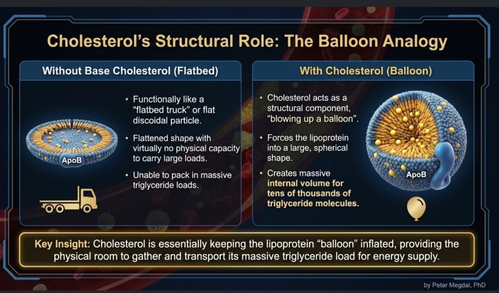
Indipendenza dal colesterolo specifica per tessuto
Questo principio diventa ancora più chiaro se si osservano organi specifici. Molti tessuti sintetizzano il colesterolo localmente e non dipendono in modo significativo dalle LDL circolanti.
Il sistema nervoso centrale è l'esempio più eclatante. Poiché le lipoproteine contenenti apoB non attraversano in modo significativo la barriera emato-encefalica, il cervello deve produrre in gran parte il proprio colesterolo. Astrociti sintetizzare il colesterolo e distribuirlo localmente attraverso lipoproteine contenenti apoE, un processo legato ai percorsi regolati da LXR [16].
Lo stesso vale in linea di massima per tessuti steroidogenici, comprese le ghiandole surrenali e le gonadi. Questi tessuti possono sintetizzare il colesterolo de novo dall'acetato. Sebbene esprimano recettori come LDLR e SR-B1, la loro dipendenza dalle LDL circolanti in condizioni ordinarie appare limitata, con l'uptake che diventa più rilevante in stati specializzati o ad alta richiesta come la stimolazione acuta da ACTH [2,17].
Questo supporta un importante punto fisiologico. Il corpo non dipende da alti livelli circolanti di LDL-C per la normale funzione endocrina o cellulare. In effetti, le discussioni meccanicistiche e cliniche nella lipidologia hanno enfatizzato che livelli molto bassi di LDL-C, persino intorno a 10 mg/dL, non implicano necessariamente una ridotta steroidogenesi o un cedimento della membrana quando la sintesi intracellulare rimane intatta2,11,17].
Quindi, sebbene il colesterolo sia essenziale, la quantità effettivamente richiesta in circolazione per la sopravvivenza cellulare è molto inferiore a quanto molte persone suppongano. Questa realtà rafforza l'opinione che le particelle di ApoB in circolazione elevate non siano una necessità biologica; sono un'esposizione al rischio.
La flotta ApoB: perché il numero di particelle è importante
Tra le numerose lipoproteine in circolazione, le più rilevanti per l'aterosclerosi sono le particelle contenenti ApoB. Queste includono chilomicroni e i loro residui, le lipoproteine a densità molto bassa (VLDL), lipoproteine a densità intermedia (IDL), lipoproteine a bassa densità (LDL) e lipoproteina(a), o Lp(a) [1,2].
La principale intuizione clinica è che ciascuno particella aterogenica contiene esattamente una molecola di ApoB. Ciò significa che l'ApoB funge da conteggio diretto del numero di particelle aterogene in circolazione. Al contrario, l'LDL-C misura solo la massa di colesterolo contenuta all'interno delle particelle LDL. Una persona può avere un LDL-C relativamente normale ma avere comunque un numero elevato di particelle se queste sono piccole e povere di colesterolo. In tale contesto, il rischio può essere sottostimato se i clinici guardano solo all'LDL-C [1-3].
Ecco perché molti esperti di lipidi considerano ora l'ApoB come il parametro biologicamente più significativo. Riflette quante opportunità ci sono per le particelle di penetrare nella parete arteriosa. In parole semplici, se ogni particella di ApoB è una potenziale collisione arteriosa, allora l'ApoB ci dice la densità del traffico [1-3].
Quella metafora del traffico diventa particolarmente utile quando si spiega il rischio ai non specialisti. Il problema non è solo quanta merce c'è in ogni camion. Il problema è quanti camion sono sulla strada, quanto tempo ci restano e quanto è probabile che si schiantino contro il muro laterale.
È qui che la discussione sul colesterolo minimo diventa clinicamente rilevante. Se i tessuti possono sintetizzare il modesto colesterolo di cui hanno bisogno, allora gli alti livelli di ApoB non vanno intesi al meglio come una riserva di consegna benefica. Vanno intesi piuttosto come un maggiore carico di particelle potenzialmente in grado di penetrare nelle arterie2,11-17].
Dalla consegna del carburante alla persistenza delle LDL
La storia inizia con particelle ricche di trigliceridi. L'intestino produce chilomicroni per trasportare i grassi alimentari. Il fegato produce VLDL per trasportare i trigliceridi endogeni. Si tratta di grandi particelle il cui compito principale è la fornitura di carburante [1,2].
Mentre queste particelle circolano, interagiscono con lipoproteina lipasi (LPL), un enzima che rilascia trigliceridi per essere utilizzati dal tessuto muscolare, cardiaco e adiposo. Man mano che i trigliceridi vengono rimossi, la particella si riduce. Il materiale di superficie viene rilasciato. Ma l'impalcatura di ApoB rimane. Nel tempo, le VLDL vengono rimodellate in IDL e poi in LDL [1,2,18].
L'LDL non è quindi una particella ribelle o malvagia per sua natura. È un sottoprodotto del normale metabolismo delle lipoproteine. Ciò che rende l'LDL particolarmente importante è la sua persistenza. L'LDL rimane in circolazione molto più a lungo rispetto alle particelle più grandi ricche di trigliceridi, spesso da 2 a 5 giorni, il che aumenta la probabilità statistica che penetri nella parete arteriosa1,2].
È qui che il modello della malattia comincia a delinearsi. aterogenesi non è causato semplicemente dal fatto che esistono le LDL. È causato dal fatto che un numero sufficiente di particelle di ApoB rimane in circolazione abbastanza a lungo da penetrare nella intima e rimanere trattenuti.
Allo stesso tempo, ciò rafforza un'importante distinzione concettuale. La presenza di LDL nel sangue non è la prova che i tessuti abbiano un bisogno disperato di colesterolo. Gran parte della biologia delle LDL riflette il rimodellamento e la persistenza delle particelle di ApoB dopo il trasporto dei trigliceridi. In questo senso, le LDL non sono meramente un “camion di consegna del colesterolo”. Sono anche un residuo a lunga circolazione del trasporto lipidico sistemico con maggiori opportunità di interagire con la parete vascolare1,2,18].
L'aterosclerosi inizia con l'ingresso, non solo con la presenza
Uno dei progressi più utili nella moderna cardiologia è la consapevolezza che l'aterosclerosi non inizia semplicemente perché ci sono particelle nel sangue. Inizia quando queste attraversano la barriera endoteliale e si depositano nella spazio sottendoteliale, o intima [1,6-8].
Ciò significa che la parete dell'arteria è estremamente importante. La endotelio è spesso un solo strato cellulare, ma è biologicamente attivo. Rileva il flusso sanguigno, risponde ai segnali infiammatori, regola la funzione di barriera e controlla il passaggio dei materiali. Alcune regioni arteriose, in particolare i punti di biforcazione e le curvature, sono più propense alla formazione di placche perché il flusso sanguigno locale è disturbato. Queste zone di flusso disturbato creano un ambiente molecolare che favorisce l'aterogenesi4,6-8].
Storicamente, molte persone immaginavano che le LDL fuoriuscissero semplicemente attraverso un endotelio danneggiato. Il quadro più recente è più preciso: l'ingresso delle LDL è un processo attivo e regolato chiamato transcitosi [6-8].
Questo è importante dal punto di vista concettuale. Se l'ingresso delle particelle è regolato, l'aterosclerosi non riguarda solo ciò che è presente nel plasma. Riguarda anche come la parete arteriosa risponde a quell'ambiente plasmatico. L'esposizione sistemica e la permissività locale agiscono insieme.
DOCK4, SR-B1 e transcitosi regolata delle LDL
Una delle scoperte più intriganti degli ultimi anni è che le cellule endoteliali possono trasportare attivamente le LDL attraverso se stesse tramite un meccanismo che coinvolge SR-B1 e DOCK4 [6-8].
SR-B1, o recettore scavenger di classe B tipo 1, è stato a lungo discusso nel contesto di HDL biologia, ma sembra anche avere un ruolo nell'assorbimento e nella transcitosi delle LDL in determinate condizioni. DOCK4 è un Rho GEF che attiva Rac1, il quale contribuisce a guidare i cambiamenti citoscheletrici e il movimento vescicolare necessari per questo processo di trasporto [4,7,8].
Ciò è importante perché ridefinisce la fase iniziale dell'aterogenesi. L'ingresso delle LDL nella parete vascolare non è solo una filtrazione accidentale. È almeno in parte controllato da un sistema di segnalazione specifico. Ancora più sorprendente è il fatto che sia stato osservato un aumento dell'espressione di DOCK4 nelle regioni predisposte all'aterosclerosi prima che queste siano visibili lesioni sono presenti, suggerendo che la parete arteriosa possa diventare permissiva molto prima che la malattia sia evidente all'imaging o all'istopatologia4,8].
Clinicamente, ciò apre a una possibilità stimolante. L'ApoB ci dice quante particelle sono disponibili per entrare. Ma il sistema DOCK4/SR-B1/Rac1 può aiutare a determinare quanto facilmente possano farlo. In altre parole, l'ApoB misura la pressione al cancello; DOCK4 aiuta a regolare il cancello stesso.
Conservazione: Il punto di non ritorno
L'ingresso da solo non basta. Una volta che una particella di ApoB raggiunge l'intima, il successivo evento chiave è la ritenzione. Queste particelle si legano a molecole della matrice chiamate proteoglicani, che agiscono quasi come una carta moschicida arteriosa1,3].
Questo passaggio è cruciale perché una particella trattenuta è ora esposta a un microambiente molto diverso. All'interno della parete arteriosa, essa può subire una serie di modificazioni. Può ossidarsi, ma l'ossidazione è solo una parte della storia. Le lipasi e altri enzimi possono alterare la superficie della particella, generando lipidi come la sfingomielina e il ceramide che rendono le particelle più adesive e più propense ad aggregarsi [4].
Quel passaggio di aggregazione è particolarmente importante. Una singola particella LDL trattenuta e isolata non è biologicamente la stessa cosa di un cluster appiccicoso di particelle modificate. Una volta che le particelle si aggregano, diventano molto più propense a provocare macrofago assorbimento e cellula schiumosa formazione1,4].
Sia per i clinici che per i lettori informati, questa è una distinzione utile: il pericolo non è solo l'LDL circolante o persino l'LDL intimale. Il pericolo è la lipoproteina contenente ApoB trattenuta, modificata e aggregata.
Questo è anche il punto in cui la “tossicità del colesterolo” diventa specifica per il contesto. Il colesterolo è necessario in piccole quantità intracellulari regolate, ma una volta che le particelle di ApoB sono intrappolate e modificate nella parete arteriosa, il loro carico lipidico diventa parte di un ambiente infiammatorio altamente tossico. Lo stesso sterolo che è essenziale nelle membrane può diventare patologico quando viene rilasciato nel compartimento tissutale sbagliato nella forma sbagliata13,14].
Reclutamento leucocitario: come l'arteria si infiamma
Una volta che l'intima contiene lipoproteine ritenute e modificate, la lesione passa da un problema lipidico a uno di natura infiammatoria. In questa fase, il reclutamento di monociti e altri leucociti diventa centrale [4].
Un GEF Rho chiamato SGEF, noto anche come Arhgef26, sembra svolgere un ruolo importante in questo contesto. SGEF aiuta le cellule endoteliali a formare strutture di ancoraggio dipendenti da ICAM-1, protrusioni ricche di actina che consentono ai leucociti di aderire in modo più efficace in condizioni di flusso. Queste strutture sono particolarmente rilevanti nell'ambiente ad alto scorrimento delle arterie, dove le cellule immunitarie potrebbero altrimenti essere lavate via4].
Un'altra molecola di segnalazione correlata, Arhgef1, collega il reclutamento infiammatorio alla segnalazione dell'angiotensina II. Ciò crea un ponte meccanicistico tra ipertensione e arteriosa infiammazione. Alto pressione sanguigna non è semplicemente un carico emodinamico; può anche amplificare il reclutamento dei leucociti attraverso vie mediate da RhoA4].
Questo aiuta a spiegare perché il sistema cardiovascolare fattori di rischio spesso si raggruppano biologicamente. L'ipertensione, insulino-resistenza, e l'ApoB elevata non agiscono in modo isolato. Convergono all'interno della parete arteriosa, dove la segnalazione endoteliale, il reclutamento immunitario e la ritenzione di lipoproteine si rafforzano a vicenda.
Cellule schiumose e risposta dei macrofagi
I macrofagi sono spesso descritti come la squadra di pulizia del corpo. Nell'aterosclerosi, tuttavia, l'operazione di pulizia può diventare autodistruttiva. I macrofagi ingeriscono lipoproteine modificate e aggregate, si caricano di colesterolo e si trasformano in cellule schiumose [1,4].
Questo passaggio non è passivo. Dipende fortemente dal rimodellamento del citoscheletro e dalla segnalazione dei recettori. La famiglia Vav di Rho GEF, in particolare Vav1, Vav2 e Vav3, contribuisce a regolare questi processi. Le proteine Vav influenzano l'assorbimento mediato da CD36 delle lipoproteine ossidate e favoriscono la formazione della sinapsi lisosomiale, che consente ai macrofagi di interagire con grandi aggregati lipoproteici e digerirli4].
Man mano che le cellule schiumose si accumulano, rilasciano mediatori infiammatori, alla fine muoiono e contribuiscono al centro necrotico della placca. Questo trasforma una lesione microscopica in una placca clinicamente significativa. Nel tempo, la lesione può calcificare, rimanere stabile o diventare vulnerabile alla rottura [1,3].
Questo è un altro punto in cui l'inquadramento molecolare aggiunge valore. La malattia non riguarda solo “troppo colesterolo”. Riguarda il modo in cui le cellule immunitarie elaborano materiale lipidico anormale all'interno di un ambiente tissutale specifico.
La letteratura aggiunta sull'omeostasi del colesterolo approfondisce questo quadro. La formazione di cellule schiumose non è semplicemente un problema di accumulo passivo di lipidi. Si tratta di una compromissione della normale gestione del colesterolo. Quando l'assorbimento da parte dei macrofagi sovrasta il controllo intracellulare e i sistemi di efflusso, il colesterolo libero si accumula, i percorsi infiammatori vengono attivati e ne consegue la morte cellulare. In questo senso, la progressione della placca può essere intesa come un fallimento localizzato dell'omeostasi del colesterolo all'interno della parete arteriosa [13-15].
Cellule muscolari lisce e stabilità della placca
Con la maturazione delle placche, le cellule della muscolatura liscia vascolare (VSMC) diventano protagoniste. Queste cellule normalmente aiutano a mantenere il tono vascolare, ma nell'aterosclerosi possono cambiare fenotipo. Possono migrare dalla tonaca media all'intima, proliferare e contribuire alla struttura della placca [4].
Questo comportamento è influenzato dalla segnalazione di Rho GEF. Molecole come Kalirin e Vav3 promuovono la migrazione e la proliferazione attraverso vie correlate a Rac1 e RhoA. Parte di questo rimodellamento può essere protettivo, poiché le VSMC possono contribuire a costruire un cappuccio fibroso sopra la placca. Ma un rimodellamento eccessivo o disordinato può anche contribuire al restringimento luminale e all'instabilità della placca [4].
Per contro, Arhgef7, chiamato anche beta-PIX, sembra sostenere l'integrità della barriera e i percorsi di polarità cellulare protettiva. Ciò suggerisce che la biologia della placca non sia guidata solo da segnali pro-malattia, ma sia anche plasmata da sistemi contro-regolatori che cercano di preservare la struttura arteriosa4].
La stabilità di una placca dipende quindi da un equilibrio tra danno, riparazione, infiammazione e rimodellamento. Tale equilibrio è in parte governato dalla stessa famiglia di interruttori molecolari che ha influenzato la lesione fin dall'inizio.
Lp(a): Il moltiplicatore del rischio genetico
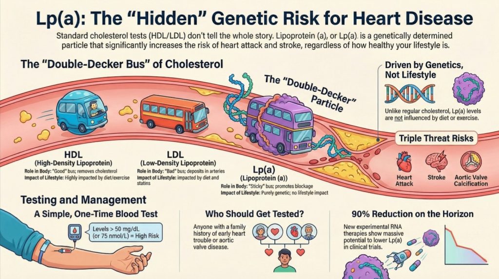
Nessuna discussione moderna sulle malattie cardiovascolari guidate dai lipidi è completa senza la lipoproteina(a), o Lp(a). Strutturalmente, la Lp(a) è una particella simile alle LDL con un'apolipoproteina(a) aggiuntiva attaccata. Clinicamente, è uno dei fattori di rischio cardiovascolare ereditari più importanti1-3,5,9].
Circa una persona su cinque ha un livello elevato di Lp(a), e i livelli sono ampiamente determinati geneticamente. A differenza delle LDL standard, la Lp(a) non migliora significativamente con la dieta o l'esercizio fisico ed è influenzata solo modestamente dalla maggior parte delle terapie lipidiche convenzionali1-3,5,9].
La Lp(a) sembra essere particolarmente pericolosa perché non è solo aterogenica ma anche pro-infiammatoria e pro-trombotica. Livelli elevati sono collegati a forme premature infarto miocardico, ictus, e valvola aortica stenosi. Per questo motivo, molti esperti raccomandano ora che a ogni paziente venga misurata la Lp(a) almeno una volta nella vita1-3,5,9].
Questo è un importante messaggio di salute pubblica. Una persona può fare molte cose giuste e tuttavia correre un rischio ereditario considerevole a causa di livelli elevati di Lp(a). Identificare quel rischio tempestivamente è importante.
Perché l'ApoB è una bussola clinica migliore rispetto all'LDL-C
Per la pratica quotidiana, la principale lezione diagnostica è semplice: l'ApoB è più vicina alla biologia della malattia rispetto all'LDL-C1-3].
L'LDL-C stima quanta massa di colesterolo è presente nelle particelle LDL. L'ApoB stima quante particelle aterogene sono in circolazione. Poiché ciascuna particella è un potenziale intruso nella parete arteriosa, l'ApoB coglie meglio la probabilità di danno arterioso.
Colesterolo non-HDL è spesso un surrogato migliore del C-LDL, poiché include il colesterolo trasportato da tutte le particelle contenenti ApoB. Ma l'ApoB rimane la misura basata sulle particelle più pulita. Il punteggio del calcio coronarico può aiutare a rilevare la malattia stabilita più avanti nella vita, e la Lp(a) aiuta a identificare il rischio ereditario. Ma per comprendere il carico causale delle particelle aterogene circolanti, l'ApoB è centrale1-3].
Questo non significa che l'LDL-C sia inutile. Rimane clinicamente utile e ampiamente disponibile. Ma quando l'obiettivo è allineare il test al meccanismo, l'ApoB è il marcatore più forte.
Si adatta anche in modo più pulito alla fisiologia descritta sopra. Se i tessuti non richiedono grandi quantità di colesterolo circolante per sopravvivere, e se il fabbisogno di colesterolo è ampiamente soddisfatto dalla sintesi locale e da vie intracellulari finemente regolate, allora la domanda clinicamente importante non diventa “Quanto colesterolo è presente nel plasma?”, bensì “Quante particelle aterogene interagiscono ripetutamente con la parete arteriosa?”. L'ApoB risponde a questo quesito meglio del C-LDL2,11-17].
Implicazioni terapeutiche: Oggi e domani
L'attuale terapia ipolipemizzante conta ancora enormemente. Statine, ezetimibe, e Inibitori di PCSK9 ridurre l'aterogenicità carico di particelle e disporre di dati attendibili sui risultati. PCSK9 gli inibitori riducono anche, seppur in misura modesta, i livelli di Lp(a), mentre le nuove terapie a base di RNA mirate all’Lp(a) stanno dimostrando riduzioni significative nei trial [1,3,9].
Lo stile di vita rimane fondamentale. Perdita di peso, migliorato sensibilità all'insulina, la ridotta produzione epatica di VLDL e il minore carico infiammatorio contribuiscono tutti a ridurre il rischio sistemico [1]. Tuttavia, le recenti scoperte nel campo della biologia suggeriscono che il futuro della prevenzione cardiovascolare potrebbe non limitarsi alla riduzione delle particelle plasmatiche, ma potrebbe anche prevedere un intervento diretto sulla parete arteriosa.
È proprio qui che la biologia delle proteine GEF della famiglia Rho diventa particolarmente interessante. Se alcune proteine GEF specifiche, come DOCK4 o SGEF, sono regolatori chiave dell’ingresso delle LDL o del reclutamento dei leucociti, allora l’inibizione diretta di tali vie potrebbe rappresentare un modo più selettivo per interferire con la formazione delle placche [4]. Anziché limitarsi a ridurre il flusso sanguigno, un giorno i medici potrebbero anche restringere il passaggio e attenuare la risposta infiammatoria all’interno della parete vascolare.
Si tratta ancora di un settore emergente, non di una strategia clinica di routine. Tuttavia, dal punto di vista concettuale, questo approccio punta verso la medicina di precisione: agire specificamente sui percorsi locali che trasformano il rischio circolante in malattia arteriosa.
Il modello integrato dell’omeostasi del colesterolo aiuta inoltre a spiegare perché una riduzione aggressiva della carica di ApoB sia biologicamente plausibile. Se il fabbisogno intracellulare può ancora essere soddisfatto attraverso la sintesi endogena e una gestione tissutale rigorosamente regolata, allora la riduzione delle particelle aterogene circolanti non deve necessariamente comportare una carenza a livello cellulare. Potrebbe semplicemente ridurre l’esposizione arteriosa non necessaria [2,11-17].
Un modo migliore per spiegare l'aterosclerosi
Per il grande pubblico, la malattia può essere descritta in modo intuitivo e preciso.
Le lipoproteine contenenti ApoB sono come camion che trasportano carburante e carico strutturale attraverso il flusso sanguigno. Se ci sono troppi camion sulla strada, alcuni finiscono per penetrare nella parete arteriosa. Una volta intrappolate lì, si danneggiano e diventano appiccicose. Il sistema immunitario invia delle cellule di pulizia, ma queste cellule vengono sovraccaricate e formano la placca. Nel frattempo, la parete arteriosa stessa non è passiva. È dotata di «guardiani» molecolari che regolano l’ingresso delle particelle, l’ancoraggio del sistema immunitario, la gestione del colesterolo e il rimodellamento dei tessuti [1-8,11-15].
Tale quadro è abbastanza semplice da poter essere utilizzato nell’educazione del pubblico, ma al contempo sufficientemente dettagliato da risultare utile ai medici. Esso evita la semplificazione eccessiva e fuorviante secondo cui il colesterolo da solo “ostruisce” le arterie, pur preservando la verità fondamentale secondo cui le particelle di ApoB contenenti colesterolo sono causalmente necessarie per l’insorgenza della malattia [1-3].
Ciò lascia spazio anche a un’importante sfumatura: l’organismo ha bisogno di colesterolo, ma solo in quantità molto ridotte e rigorosamente controllate. Il vero pericolo non è la presenza del colesterolo in sé. Il pericolo è la circolazione cronica, l’ingresso nelle arterie e la ritenzione di particelle contenenti ApoB in un ambiente tissutale che trasforma il loro contenuto in infiammazione e placca.
Conclusione
L’aterosclerosi può essere meglio compresa come l’intersezione di due sistemi. Il primo è la biologia sistemica delle lipoproteine, in cui le particelle contenenti ApoB circolano come vettori necessari dei trigliceridi e del colesterolo. Il secondo è la biologia arteriosa locale, in cui le cellule endoteliali, i leucociti, i macrofagi e le cellule muscolari lisce determinano se tali particelle si trasformino in placca.
L’ApoB rileva il numero di particelle aterogene circolanti e, di conseguenza, la probabilità statistica di insorgenza di lesioni arteriose. La via di segnalazione Rho GEF–Rho GTPasi contribuisce a spiegare perché e come tali lesioni si verifichino all’interno della parete vascolare. Nel loro insieme, questi sistemi offrono un modello della malattia più articolato rispetto al vecchio quadro di riferimento basato esclusivamente sul colesterolo LDL.
Allo stesso tempo, il colesterolo stesso è regolato da un sistema intracellulare altamente controllato. Attraverso il sistema SREBP-2–SCAP–INSIG, la via del mevalonato, la segnalazione dell’efflusso mediata dall’LXR e la sintesi specifica per tessuto, l’organismo mantiene le piccole quantità di colesterolo di cui ha bisogno, difendendosi al contempo dall’eccesso [11-17]. Ciò significa che il fabbisogno fisiologico di colesterolo è reale, ma la quantità necessaria è modesta, regolata a livello locale e fondamentalmente diversa dal carico patologico causato dall’eccesso di particelle di ApoB in circolo.
Per i medici, ciò conferma la necessità di porre maggiore enfasi sull’ApoB, sull’Lp(a) e sulla prevenzione precoce. Per il pubblico, chiarisce che le malattie cardiovascolari non dipendono semplicemente dal consumo di grassi o dall’avere il “colesterolo alto”. Si tratta piuttosto del numero di particelle aterogeniche in circolazione, della durata della loro permanenza, di come la parete arteriosa reagisce ad esse e di come le disfunzioni nella gestione locale del colesterolo trasformino le particelle trattenute in infiammazione e placca.
Il futuro della lipidologia potrebbe quindi avere un duplice orientamento: ridurre il numero di particelle pericolose nel flusso sanguigno e, al contempo, intervenire sui meccanismi molecolari che consentono a tali particelle di penetrare nella parete arteriosa e danneggiarla [1-10]. In questo contesto più ampio, una delle correzioni concettuali più importanti è la seguente: il corpo non ha bisogno di un grande eccesso di colesterolo in circolazione per sopravvivere. Ha bisogno solo di una quantità molto piccola, attentamente regolata a livello cellulare. L’aterosclerosi ha inizio quando il sistema di trasporto supera tale fabbisogno e la parete arteriosa ne paga le conseguenze.
Riferimenti
- Perone F, Bernardi M, Spadafora L, et al. Non-Traditional Cardiovascular Risk Factors: Tailored Assessment and Clinical Implications. J Cardiovasc Dev Dis. 2025;12(5):171. Published 2025 Apr 28. doi:10.3390/jcdd12050171
- Attia P, Dayspring T. #20 – Tom Dayspring, M.D., FACP, FNLA – Part I of V: an introduction to lipidology.
- Hill S. What Causes Cardiovascular Disease? Lipid Series Part 1. The Proof with Simon Hill.
- Li M, Jiao Q, Xin W, et al. The Emerging Role of Rho Guanine Nucleotide Exchange Factors in Cardiovascular Disorders: Insights Into Atherosclerosis: A Mini Review. Front Cardiovasc Med. 2022;8:782098. Published 2022 Jan 3. doi:10.3389/fcvm.2021.782098
- Heart Matters. Cholesterol Explained: HDL, LDL, and the Hidden Risk of Lp(a).
- Hackethal V. Research Suggests LDL Is Actively Transported Into Arterial Walls. NeurologyLive.
- Getz GS, Reardon CA. The “HDL receptor” scavenger receptor class B type 1 finesses the uptake of low-density lipoproteins into the subendothelial space of arteries. Biotarget.
- Huang L, Chambliss KL, Gao X, et al. SR-B1 drives endothelial cell LDL transcytosis via DOCK4 to promote atherosclerosis. Nature. 2019;569(7757):565-569. doi:10.1038/s41586-019-1140-4
- Nutrition Made Simple. Everything you need to know about Lp(a) | ft. Dr. Tom Dayspring.
- Heart Fit Clinic. Webinar on Cholesterol from a Lipidologist. Dr. Tom Dayspring.
- Brown MS, Goldstein JL. The SREBP pathway: regulation of cholesterol metabolism by proteolysis of a membrane-bound transcription factor. Cell. 1997;89(3):331-340. doi:10.1016/s0092-8674(00)80213-5
- Brown MS, Goldstein JL. Sterol regulatory element binding proteins (SREBPs): controllers of lipid synthesis and cellular uptake. Nutr Rev. 1998;56(2 Pt 2):S1-S75. doi:10.1111/j.1753-4887.1998.tb01680.x
- Muneer PMA, Alikunju S, Mishra V, et al. Activation of NLRP3 inflammasome by cholesterol crystals in alcohol consumption induces atherosclerotic lesions. Brain Behav Immun. 2017;62:291-305. doi:10.1016/j.bbi.2017.02.014
- Feng B, Yao PM, Li Y, et al. The endoplasmic reticulum is the site of cholesterol-induced cytotoxicity in macrophages. Nat Cell Biol. 2003;5(9):781-792. doi:10.1038/ncb1035
- Murphy AJ, Akhtari M, Tolani S, et al. ApoE regulates hematopoietic stem cell proliferation, monocytosis, and monocyte accumulation in atherosclerotic lesions in mice. J Clin Invest. 2011;121(10):4138-4149. doi:10.1172/JCI57559
- Dietschy JM, Turley SD. Cholesterol metabolism in the brain. Curr Opin Lipidol. 2001;12(2):105-112. doi:10.1097/00041433-200104000-00003
- Miller WL. MECHANISMS IN ENDOCRINOLOGY: Rare defects in adrenal steroidogenesis. Eur J Endocrinol. 2018;179(3):R125-R141. doi:10.1530/EJE-18-0279
- Ginsberg HN. Lipoprotein metabolism and its relationship to atherosclerosis. Med Clin North Am. 1994;78(1):1-20. doi:10.1016/s0025-7125(16)30174-2

