
Apolipoproteina B, risoluzione dell'imaging e l'illusione delle arterie coronarie “normali” all'età di 60 anni
L'affermazione clinica secondo cui gli individui che mantengono livelli marcatamente elevati di bassa densità lipoproteina colesterolo (LDL-C) e apolipoproteina B (ApoB) possono possedere anatomicamente “normali” arterie coronarie all'età di 60 anni rappresenta un paradosso diagnostico. Questo fenomeno dipende spesso dalla risoluzione dello strumento di imaging utilizzato. Sebbene gli strumenti contemporanei come il Calcio delle arterie coronarie punteggio e visivo standard coronarico Tomografia Computerizzata L'angioplastica (CCTA) offre una finestra ad alta risoluzione sulla parete arteriosa, ma esiste in tensione con le prove patologiche che suggeriscono che aterosclerosi è una condizione quasi universale dell'invecchiamento umano, e i dati clinici che mostrano che infarti può verificarsi anche in assenza di significative ostruzioni anatomiche1].

Il quadro causale dell'aterogenesi guidata dalle lipoproteine
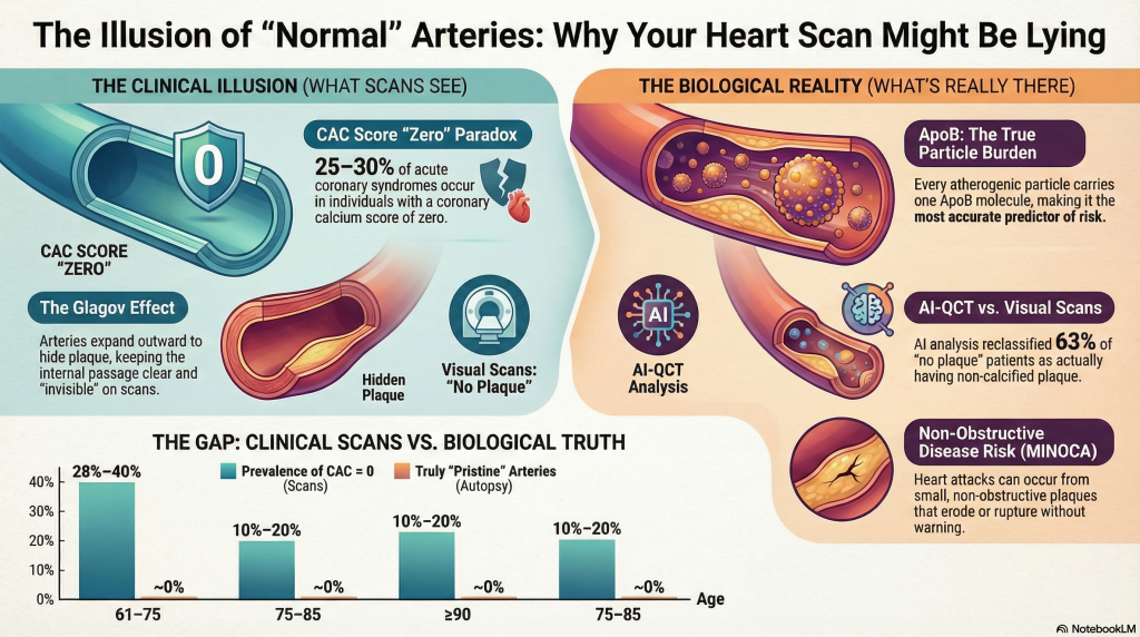
Il consenso stabilisce che l'esposizione prolungata alle lipoproteine contenenti ApoB è il principale motore dell'aterosclerosi placca [2]. Ciascuno particella aterogenicaincluso LDL, VLDL, e IDL—trasporta una singola molecola di ApoB, rendendolo una misura diretta del numero totale di particelle circolanti che possono penetrare nella parete arteriosa2]. La probabilità che una particella rimanga intrappolata nel spazio sottendoteliale è una funzione sia della concentrazione delle particelle che della durata dell'esposizione, spesso quantificata come “Anni-ApoB” [2].
Mentre l'LDL-C misura la massa del colesterolo, l'ApoB riflette il carico di particelle [3]. Nei casi di discordanza—dove l'ApoB è alta ma l'LDL-C è relativamente più basso—l'ApoB rimane il predittore più accurato del futuro infarto miocardico (MI) ed eventi clinici4].
Parametri delle lipoproteine
| Metrica delle lipoproteine | Definizione e ruolo patologico | Associazione con Carico di placca |
| Colesterolo delle lipoproteine a bassa densità (LDL-C) | La massa di colesterolo contenuta nelle particelle LDL | Casuale, ma potrebbe essere discordante con il conteggio delle particelle [3] |
| Apolipoproteina B (ApoB) | Strutturale proteina; ne esiste una su ogni particella aterogenica | Il più forte predittore di rischio; riflette il reale carico di particelle3] |
| Lipoproteina(a) [Lp(a)] | Una particella simile alle LDL con una proteina aggiuntiva [apo(a)] | Indipendente fattore di rischio; promuove calcificazione e trombosi [5] |
Realtà forense vs. clinica: L'illusione dell'arteria “incontaminata”
L'affermazione che un individuo possa raggiungere l'età di 60 anni senza “danno arterioso” è spesso il risultato dell'imaging clinico che non riesce a rilevare la malattia microscopica. Studi autoptici forensi, inclusi i Determinanti Patobiologici dell'Aterosclerosi nei Giovani (PDAY) e il Studio sul cuore di Bogalusa, dimostrare che microscopico strie lipidiche—la più precoce evidenza macroscopica di aterosclerosi—è presente in quasi tutti gli adolescenti esaminati6].
Entro la sesta decade di vita, le arterie coronarie patologicamente incontaminate sono praticamente assenti nelle popolazioni occidentalizzate. Diffuse ispessimento intimale e il deposito lipidico diventa onnipresente a metà dell'età adulta, e le coorti di autopsie contemporanee riportano aterosclerosi coronarica in quasi tutti gli adulti entro la quarta decade [6].
L'effetto Glagov: come le arterie nascondono il carico di placca
Un fattore determinante per la “normalità” imaging coronarico è fenomeno di Glagov, oppure rimodellamento estroflesso compensatorio [7]. Poiché la placca si accumula all'interno della parete arteriosa, il vaso si espande esternamente per preservare il diametro luminale e mantenere il flusso sanguigno [7]. Il restringimento luminale emodinamicamente significativo si manifesta in genere solo quando il carico di placca supera circa 40% dell’area della lamina elastica interna [7].
Poiché l'angiografia invasiva standard e la CCTA visiva convenzionale si concentrano principalmente sul lumen, un notevole carico di placca può esistere all'interno della parete del vaso rimanendo invisibile all'angiografia7]. Queste placche nascoste sono spesso ricche di lipidi e biologicamente attive, il che le rende inclini all'erosione o alla rottura in grado di precipitare l'infarto miocardico anche in assenza di preesistenti stenosi [1].
IA-QCT (Cleerly) vs. Interpretazione Visiva Standard
Le piattaforme di tomografia computerizzata quantitativa basata sull'intelligenza artificiale (AI-QCT), come Cleerly, hanno dimostrato che l'interpretazione visiva standard dell'angio-TC coronarica (CCTA) sottostima sistematicamente il carico di placca [8]. Abilitando quantificazione volumetrica della placca e analisi compositiva, l'AI-QCT raggiunge un'accuratezza vicina a quella invasiva ecografia intravascolare [8].
In una coorte di circa 750 pazienti, l'analisi AI-QCT ha portato a una modifica della diagnosi in 39% casi, mentre il numero di pazienti classificati come “privi di placca” è sceso da 159 a 58 a seguito della valutazione quantitativa [8]. Questi risultati confermano che una TC coronarica visivamente “normale” angiografie ospitano frequentemente rilevanti dal punto di vista clinico placca non calcificata.
Punteggio del calcio delle arterie coronarie e rischio di infarto miocardico
Coronarica Arteria Il punteggio di calcio è un potente strumento di stratificazione del rischio a livello di popolazione, ma la sua relazione con il rischio individuale di infarto miocardico è probabilistica anziché assoluta. Ampie coorti prospettiche, compreso il Studio Multietnico dell'Aterosclerosi (MESA), dimostrano una forte associazione graduale tra il carico di CAC e il futuro malattia coronarica eventi, con un rischio marcatamente elevato osservato a punteggi ≥300 [11].
Tuttavia, un punteggio CAC pari a zero non conferisce immunità dall'infarto miocardico. Sebbene i tassi di eventi a breve termine siano bassi, le placche non calcificate, placche ricche di lipidi—in particolare nei soggetti con ApoB elevata —rimangono suscettibili di rottura o erosione [12]. Circa 25–30% di sindromi coronariche acute si verificano in individui con calcificazione coronarica assente o minima al livello basale, riflettendo la disconnessione temporale tra formazione della placca, calcificazione e instabilità della placca [12]. La calcificazione è sempre più riconosciuta come un indicatore di cronicità della placca e guarigione piuttosto che vulnerabilità, mentre il carico di placca non calcifica si correla più strettamente al rischio a breve termine.
Pertanto, il punteggio CAC è interpretato al meglio come una misura del carico cumulativo di placca e del rischio a lungo termine piuttosto che come un rilevatore di aterosclerosi biologicamente attiva. Negli individui con alti livelli di ApoB, un punteggio CAC pari a zero riflette più probabilmente una manifestazione ritardata della malattia piuttosto che una sua vera assenza.
Rischio di infarto miocardico con un carico di placca “prossimo allo zero”: la sindrome MINOCA
L'infarto miocardico con arterie coronarie non ostruttive (MINOCA) rappresenta circa il 5–15% di tutti gli infarti miocardici acuti [1]. Anche piccoli, placche non ostruttive può subire un'erosione endoteliale, innescando una trombosi localizzata senza ostruzione visibile all'angiografia9].
È importante notare che l'ApoB rimane un forte predittore indipendente del rischio di infarto miocardico anche tra individui senza coronaropatia ostruttiva, come dimostrato in ampie coorti di popolazione e in studi randomizzati di riduzione dei lipidi [4].
Dati longitudinali: Prevalenza di assenza di placca dai 60 ai 100 anni
Nonostante l'aterosclerosi quasi universale all'esame anatomopatologico, i registri di imaging clinico identificano un sottogruppo di individui che mantengono un'assenza di calcificazione coronarica in età avanzata [10].
| Fascia d'età (Anni) | Prevalenza di CAC = 0 (Imaging Clinico) | Probabilità di arterie veramente “incontaminate” (autopsia) |
| 61–75 | 28%–40% | ~0% |
| 75–85 | 10%–20% | ~0% |
| ≥90 | ~5% | ~0% |
Nei grandi anziani, è stato osservato un cosiddetto “paradosso del colesterolo”, in cui livelli più elevati di LDL-C (≥130 mg/dL) sono talvolta associati a una maggiore sopravvivenza, riflettendo probabilmente un bias di sopravvivenza o ruoli protettivi dipendenti dall'età del colesterolo nella funzione immunitaria e nella riparazione cellulare [10].
Riferimenti
- Tamis-Holland JE, Jneid H, Reynolds HR, et al. Contemporary Diagnosis and Management of Patients With Myocardial Infarction in the Absence of Obstructive Coronary Artery Disease: A Scientific Statement From the American Heart Association. Circulation. 2019;139(18):e891-e908. doi:10.1161/CIR.0000000000000670
- Ference BA, Ginsberg HN, Graham I, et al. Low-density lipoproteins cause atherosclerotic cardiovascular disease. 1. Evidence from genetic, epidemiologic, and clinical studies. A consensus statement from the European Atherosclerosis Society Consensus Panel. Eur Heart J. 2017;38(32):2459-2472. doi:10.1093/eurheartj/ehx144
- Sniderman AD, Thanassoulis G, Glavinovic T, et al. Apolipoprotein B Particles and Cardiovascular Disease: A Narrative Review. JAMA Cardiol. 2019;4(12):1287-1295. doi:10.1001/jamacardio.2019.3780
- Marston NA, Giugliano RP, Melloni GEM, et al. Association of Apolipoprotein B-Containing Lipoproteins and Risk of Myocardial Infarction in Individuals With and Without Atherosclerosis: Distinguishing Between Particle Concentration, Type, and Content. JAMA Cardiol. 2022;7(3):250-256. doi:10.1001/jamacardio.2021.5083
- Tsimikas S. A Test in Context: Lipoprotein(a): Diagnosis, Prognosis, Controversies, and Emerging Therapies. J Am Coll Cardiol. 2017;69(6):692-711. doi:10.1016/j.jacc.2016.11.042
- Strong JP, Malcom GT, McMahan CA, et al. Prevalence and extent of atherosclerosis in adolescents and young adults: implications for prevention from the Pathobiological Determinants of Atherosclerosis in Youth Study. JAMA. 1999;281(8):727-735. doi:10.1001/jama.281.8.727
- Glagov S, Weisenberg E, Zarins CK, Stankunavicius R, Kolettis GJ. Compensatory enlargement of human atherosclerotic coronary arteries. N Engl J Med. 1987;316(22):1371-1375. doi:10.1056/NEJM198705283162204
- Nurmohamed NS, Cole JH, Budoff MJ, et al. Impact of atherosclerosis imaging-quantitative computed tomography on diagnostic certainty, downstream testing, coronary revascularization, and medical therapy: the CERTAIN study. Eur Heart J Cardiovasc Imaging. 2024;25(6):857-866. doi:10.1093/ehjci/jeae029
- Agewall S, Beltrame JF, Reynolds HR, et al. ESC working group position paper on myocardial infarction with non-obstructive coronary arteries. Eur Heart J. 2017;38(3):143-153. doi:10.1093/eurheartj/ehw149
- Mortensen MB, Nordestgaard BG. Elevated LDL cholesterol and increased risk of myocardial infarction and atherosclerotic cardiovascular disease in individuals aged 70-100 years: a contemporary primary prevention cohort. Lancet. 2020;396(10263):1644-1652. doi:10.1016/S0140-6736(20)32233-9
- Budoff MJ, Shaw LJ, Liu ST, et al. Long-term prognosis associated with coronary calcification: observations from a registry of 25,253 patients. J Am Coll Cardiol. 2007;49(18):1860-1870. doi:10.1016/j.jacc.2006.10.079
- Blaha MJ, Cainzos-Achirica M, Greenland P, et al. Role of Coronary Artery Calcium Score of Zero and Other Negative Risk Markers for Cardiovascular Disease: The Multi-Ethnic Study of Atherosclerosis (MESA). Circulation. 2016;133(9):849-858. doi:10.1161/CIRCULATIONAHA.115.018524

