
人类动脉粥样硬化的生命历程连续体:发病、患病率、载脂蛋白B的因果关系与饮食调整
摘要
人类 动脉粥样硬化 通常作为一种衰老相关疾病来管理,然而病理记录将其最早的 病变 在出生前及其在大部分中年成年人中通过影像学可检测到的表现。本文综合了胎儿、儿童、尸检和人群影像学文献,以确定该疾病何时开始、其患病率如何,以及是什么将真正的病理学与正常动脉发育区分开来。我们认为 适应性内膜增厚 必须与脂质潴留性病变进行本质上的区分,并且未能作出这种区分解释了历史尸检文献中的大部分分歧,包括年轻军人死者中冠状动脉粥样硬化表面上持续六十年的下降。我们随后检查了识别 载脂蛋白 含硼的 脂蛋白 作为必要的起始底物以及累积暴露模型的统一 孟德尔随机化, ,儿科 家族性高胆固醇血症 队列研究以及随机降脂试验。来自以谋生为基础的生活群体和前工业化时期木乃伊遗骸的比较证据被用来检验动脉粥样硬化是衰老的必然结果这一说法。我们回顾了降低终生患病风险的饮食策略 载脂蛋白B 儿童和成人的暴露情况,探讨多不饱和植物油这一备受争议的问题,并明确指出高患病率的亚临床病变意味着什么以及不意味着什么。附录记录了来源验证结果,包括对二手文献中经常误报的声称的修正数据。.
索引词
载脂蛋白B,动脉粥样硬化,, 累积暴露, ,膳食脂肪,家族性的 高胆固醇血症, ,胎儿起源,, 内膜增厚, ,低密度脂蛋白,预防心脏病学。.
I. 引言
动脉粥样硬化通常被当作一种晚年疾病来管理。病理记录并不支持这种界定。最早期的病变在出生前就已经出现,到了三十岁时,相当一部分人群就已经带有能够进展的病变[5], [6该疾病是一个终生的连续过程,而非晚期事件。.
这产生了一个概念上的问题:如果动脉病变到了晚年变得非常普遍,那么在什么意义上动脉粥样硬化还是一种疾病呢?要回答这个问题,需要两个条件:一是区分适应与病理的组织学界限,二是解释为什么这一病理过程在某些人群中进展迅速而在另一些人群中进展缓慢的因果模型。.
第二部分和第三部分确立了组织学框架和尸检证据,包括扭曲了对历史军事病例系列之解读的方法学异质性。第四部分构建了载脂蛋白B(apoB)累积暴露模型。第五部分评估了进化假说。第六部分和第七部分探讨了饮食调整。第八部分阐明了亚临床病变的普遍存在所代表和不代表的意义。各项数字主张均已与原始资料进行了核对;不一致之处记录在附录中。.
II. 区分动脉适应与疾病
A. 组织学边界
动脉壁由三层构成: 内膜, ,由单层 内皮细胞; 肌肉层;以及外层 外膜. 在出生后早期以及几乎所有成年人中,内膜在血流受到干扰的分支点和弯曲处局灶性增厚。适应性内膜增厚的特征主要表现为 平滑肌细胞 在蛋白聚糖-胶原基质内,并且与保留脂质的动脉粥样硬化病变不同,缺乏脂质积聚和炎性泡沫细胞浸润的典型特征1], [2].
Stary 为美国心脏协会制定的分类1] 并由 Virmani 及其同事 [2] 按进展排列后续病变。 内膜黄色瘤, 历史上被称为 脂纹, ,是涉及脂质的首个病变: 含载脂蛋白B的颗粒 穿过 内皮, ,保留在内皮下基质中,并被吞噬 巨噬细胞 变得充满脂质 泡沫细胞. 随后是病理性内膜增生,其特征为深部内膜出现无细胞的细胞外脂质池以及局部的平滑肌细胞丢失。 纤维粥样斑块 是第一个真正的病变吗 坏死核心 在...下方 纤维帽. 。 薄帽纤维粥样硬化斑块, ,其中炎症性蛋白水解将纤维帽厚度减少至65微米以下,是与斑块破裂和急性发作最相关的表型 冠状动脉血栓形成 [2]. 图 1 根据证据基础总结了这一进展。.
就本综述而言,其操作性标准是:当血管发现涉及含apoB脂蛋白的潴留和修饰,并伴有细胞损伤或炎症浸润时,即构成病变,无论该病变是微观的、无症状的还是可逆的。适应性内膜增厚不符合这一标准;内膜 黄瘤 这样做。.
B. 为什么该区别决定了法律解释
这条分界线不仅仅是分类学上的。病理学家如果将任何内膜增厚都判定为动脉粥样硬化,其报告的患病率将大大高于只将那些保留脂质、呈炎症性病变的病变判定为动脉粥样硬化的病理学家。由于历史上的尸检文献跨越了数十个诊断标准和制备方法不断演变的时期,如果不把定义、制备方法和分级标准上的差异考虑在内,不同时代的患病率数据就无法直接进行对比。第三节将探讨其后果。.
III. 全生命周期的尸检证据
A. 胎儿和儿童早期病变
Napoli 及其同事对 82 例胎儿进行了研究 主动脉 来自自然流产和出生后十二小时内死亡的早产儿,平均胎龄为 6.2 ± 1.3 个月 [3]。标本根据母亲进行分组 胆固醇 状态:22名来自胆固醇水平正常的母亲,33名来自高胆固醇血症母亲,27名仅在妊娠期间患有高胆固醇血症的母亲。含有天然和氧化低密度脂蛋白的脂肪条纹(低密度脂蛋白)存在于胎儿主动脉中,且高胆固醇血症母亲的胎儿病变范围更大。在小于六个月的胎儿中,胎儿血浆胆固醇与母体胆固醇相关(R = 0.86,P = 0.001),且胎儿胆固醇随胎龄下降(R = −0.88)。低密度脂蛋白(LDL)的积聚和氧化修饰先于单核细胞的招募,这与“反应性潴留”(response-to-retention)而非“反应性损伤”(response-to-injury)的起始顺序一致。3].
根据第二条A款的标准,这些是真正的病变。然而,它们并不表明胎儿脂质积聚必然会进展为临床疾病。儿童早期病变转归(FELIC)研究检查了156名1至13岁血脂正常的儿童的主动脉,发现年龄最小的儿童的主动脉病变比相应的胎儿要小,这与宫内暴露结束后产后消退的情况相符[4随后,两组暴露人群的病变大小均随着年龄增长而增加,且高胆固醇血症母亲的孩子病情进展更快,尽管这些孩子自身的胆固醇水平正常。因此,早期病变的轨迹受后续暴露的强烈影响,并非在形成时就一成不变。.
B. 青春期与青年期
青少年动脉粥样硬化的病理生物学决定因素(PDAY) 该项目检查了因外部原因死亡的 15 至 34 岁人群的动脉。报告的样本量在不同的 PDAY 出版物中有所不同,并且是反复引起混淆的根源:1993 年的自然史分析报告了 1,532 名受试者 [5],2000年地形分析 2,876 [6]。两者对于各自的分析都是正确的。.
在最年轻的 PDAY 层(15至19岁年龄组),所有胸主动脉和腹主动脉以及约一半的右侧 冠状动脉 带有肉眼可见的嗜苏丹性病变5]。病变范围随年龄呈单调增加,且病变负担与以下死后标志物相关: 非高密度脂蛋白胆固醇, 吸烟 由血清硫氰酸盐、肥胖和血糖指数表示6]。与白人受试者相比,黑人受试者表现出更广泛的脂肪条纹,而进展为隆起性病变则遵循常规 风险因素.
由这些死后关联得出的PDAY风险评分随后预测了活体队列中的临床结果。在 冠状动脉风险发展至成年研究 研究显示,得分每增加一个标准差,心血管事件的发生率就会预测有 风险比 在十五年内从 1.74 到 2.04,C 统计量为 0.77 至 0.79 [7]。该评分还预测了颈动脉 内膜中膜厚度 在“年轻芬兰人心血管风险”队列中39]。这支持了青少年尸检中观察到的病变与几十年后临床显现的疾病之间的生物学连续性。.
这 博加卢萨心脏研究 将204名年龄在2至39岁且其中93人拥有先前危险因素数据的个体的、前瞻性测量的生前危险因素与尸检病理学联系起来8]. 病变负荷随危险因素数量的增加而逐步上升。当存在 0、1、2 和 3 至 4 个危险因素时,主动脉内膜表面被脂肪条纹覆盖的面积分别为 19.1%、30.3%、37.9% 和 35.0%;冠状动脉脂肪条纹值分别为 1.3%、2.5%、7.9% 和 11.0%;冠状动脉纤维性 斑块 参与度从0.6%上升到7.2%。对该研究的简写引文经常只报告冠状动脉系列的两个端点,从而掩盖了中间层。.
C. 军事系列与可比性问题
埃诺及其同事报告称,在阵亡于朝鲜的300名美国士兵中,有77.3%存在肉眼可见的冠状动脉疾病证据,平均年龄为22.1岁 [9] 随后对105例越南战争时期伤亡人员的一系列研究报告显示,45%的人患有冠状动脉粥样硬化,其中5%为严重病变 [10]. Webber及其同事对在伊拉克和阿富汗阵亡的3,832名现役军人(平均年龄25.9岁)进行了研究,结果显示8.5%的人存在任何形式的冠状动脉粥样硬化,其中重度占2.3%,中度占4.7%,轻微占1.5% [11]. 2021年对特种作战人员的一项分析发现,17.4%的人患有冠状动脉或主动脉粥样硬化,5.1%的人患有严重的冠状动脉疾病 [12].
跨越六个年代从77.3%下降到8.5%,可能反映了暴露情况的真实变化、不同序列之间的方法学差异,或两者兼有11]。仅凭这些尸检系列无法在它们之间进行分配,因为它们的病例选择、标本制备和分级标准各不相同。Enos 系列对未固定、塌陷的动脉进行大体肉眼评估,其疾病阈值从纤维性内膜增厚延伸至闭塞性斑块;按照现代标准,该阈值可能捕捉到现在会被归类为适应性内膜增厚而非动脉粥样硬化的病变。PDAY 则使用 10% 中性缓冲福尔马林进行标准化固定,并进行中央实验室苏丹 IV 染色以及由三位病理学家进行独立分级[5],现代军事系列则再次应用了不同的严重程度阈值。韦伯(Webber)及其同事将这种下降归因于真实或人工制造的现象 [11]。关于年轻成人中冠状动脉粥样硬化几乎已经消失的结论,并不支持原始比较所显示的那么大的幅度。.
D. 中年及晚年
在 PESA 队列 对于40至54岁的无症状成年人,, 亚临床动脉粥样硬化 在所有被调查的地区中占63%13]. 在 SCAPIS, ,该研究共纳入超过25,000名年龄在50至64岁之间、无已知冠心病的瑞典成年人,通过冠状动脉计算机断层扫描血管造影术(CCTA)发现42.1%的人患有动脉粥样硬化,其中显著的 狭窄 在 5.2% 中;5.5% 的冠心病患者 钙化积分 尽管得分为零,依然检测出了冠状动脉粥样硬化 [14]. 血管内超声 对健康心脏移植供体冠状动脉的研究表明,在20岁以下的供体中,17%存在动脉粥样硬化;在40多岁(第四个十年)的供体中,这一比例为60%;而在50岁及以上的供体中,这一比例达到85% [15表I总结了主要研究。.
必须保留两个区别。首先,任何动脉粥样硬化、阻塞性狭窄和临床显现的疾病都不是可互换的终点:SCAPIS 在一个42.1%的人群检测到斑块的研究中发现有5.2%的人群存在显着狭窄 [14其次,患病率取决于检查方法,特别是钙化积分对于早期和非钙化病变具有特异性但缺乏敏感性。14] 源验证期间确定的显著较高的阻塞性狭窄患病率估计值,无法与此处回顾的人口成像文献(附录,第 10 项)相调和。.
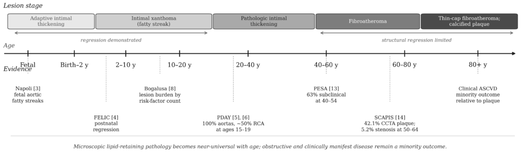
图 1. 人类生命周期中病变阶段的进展及支持证据。适应性内膜增厚(上方条带,左侧)是针对血流动力学应激的生理反应,并非疾病 [1], [2]。病理学始于内膜黄色瘤,其中含载脂蛋白B的脂蛋白被滞留并修饰2], [16]。早期脂质潴留病变已被证明具有可逆性[4]. 方括号中的数字指代参考文献列表。年龄轴经过压缩,且不是线性的。.
表 I
动脉粥样硬ification发生和患病率的主要尸检和影像学研究
| 学习 | 设计与国家 | 样本和年龄 | 方法 | 主要发现 |
| 那不勒斯等人 [3] | 胎儿尸检系列;意大利 | 82条主动脉;平均胎龄6.2个月 | 组织学;载脂蛋白B、氧化低密度脂蛋白、CD68免疫染色 | 胎儿主动脉可见脂肪纹;母体高胆固醇血症时其范围更大;脂质潴留和氧化先于单核细胞募集 |
| FELIC [4] | 儿童尸检系列;意大利 | 156名年龄在1-13岁的儿童 | 计算机辅助形态测量学 | 最小儿童的病变比对应的胎儿小,表明出生后可消退;母体高胆固醇血症会加快病变进展 |
| PDAY [5], [6] | 多中心尸检计划;美国 | 1,532 (1993年);2,876 (2000年);15–34岁 | 标准化福尔马林固定;苏丹IV;中心分级 | 在15至19岁年龄段,所有主动脉和约半数右冠状动脉均已出现病变;病变严重程度与非高密度脂蛋白胆固醇、吸烟、肥胖以及血糖水平相关。 |
| 博加卢萨8] | 与尸检关联的队列;美国 | 204例年龄在2–39岁之间的尸检;93例具有生前数据 | 大体染色;形态计量学 | 病变负担随儿童期危险因素数量的增加而逐步加重;冠状动脉纤维斑块从0.6%增加至7.2% |
| Enos 等人 [9] | 战伤尸检;朝鲜 | 300名男性;平均年龄22.1岁 | 肉眼观察;未固定动脉 | 77.3%中的冠状动脉疾病;分级标准包括纤维性内膜增厚,因此其患病率可能高于当前病变定义所显示的水平 |
| 韦伯等人 [11] | 伤亡尸检登记处;美国 | 3,832;平均年龄 25.9 岁 | 标准化尸检分级 | 任何冠状动脉粥样硬化 8.5%;重度 2.3%;历史数据中的下降趋势可能既反映了长期趋势,也反映了方法学上的差异 |
| Tuzcu 等人 [15] | 移植供体成像;美国 | 跨成年年龄段的供体心脏 | 血管内超声 | 20岁以下供体的动脉粥样硬化病例为17%,40岁左右供体为60%,50岁及以上供体为85% |
| PESA [13] | 无症状队列;西班牙 | 40至54岁成年人 | 多部位超声与钙化积分 | 63%中至少一个供血区存在亚临床动脉粥样硬化 |
| SCAPIS14] | 瑞典人口队列 | >25,000名50至64岁的成年人 | 冠状动脉CT血管成像 | 冠状动脉粥样硬化 42.1%;显著狭窄 5.2%;钙化评分均为零的患者中,5.5% 存在斑块 |
IV. 载脂蛋白 B 与累积暴露
A. 引发底物
每一个致动脉粥样硬化的脂蛋白颗粒——低密度脂蛋白及其前体 极低密度脂蛋白 以及 IDL, ,和 脂蛋白(a) —携带单个载脂蛋白 B 分子。测量 低密度脂蛋白胆固醇 定量了所输送的胆固醇质量;而apoB的测定则定量了颗粒数量。由于动脉粥样硬化是由动脉对含apoB颗粒的滞留所引发的,因此颗粒数量(而非胆固醇载荷)是具有机制相关性的暴露变量16], [41].
我们精确阐述了这一因果论断。在当前流行的动脉粥样硬化因果模型中,富含载脂蛋白 B(apoB)的脂蛋白的动脉壁滞留是必要的起始事件 [16], [41]; 局部血管生物学——包括内皮通透性、蛋白聚糖组成、血流紊乱以及炎症信号传导——决定了颗粒滞留发生的时间和位置。这种表述既保留了因果关系,同时也承认病变的定位不能仅由颗粒浓度来解释。 这与以下观察结果一致:病变优先形成于分支点和弯曲处,因为这些部位既存在适应性内膜增厚,又伴有血流紊乱。.
有三条独立的证据线索均指向这一结论:关于动脉壁内物质滞留和氧化修饰的机制研究 [41]; 遗传学研究表明,终身接触含apoB的脂蛋白水平较低,与冠心病风险显著降低有关 [42]; 以及关于药理学方法降低低密度脂蛋白(LDL)的随机试验表明,事件发生率的降低与LDL绝对降幅成正比 [43]. 这些流在 [ 中合成16] 和 [17].
B. 累积暴露模型
由于滞留的颗粒会随着时间的推移而不断积累,, 斑块负荷 反映了多年暴露期间载脂蛋白B(apoB)浓度的积分,表述为 胆固醇年 或 载脂蛋白B年 通过与……的明确类比 包-年 香烟接触的17]; 该模型的经验支持在 [ 中阐述18] 和 [19]以下。Pooled cohort analysis shows that cumulative LDL-C exposure during young adulthood and middle age predicts later cardiovascular events independently of midlife LDL-C level [18]。在CARDIA研究中,对4366名从18岁随访至40岁的参与者进行分析,累积apoB暴露量每增加一个标准差,其风险比为1.53(95%置信区间 置信区间 1.36至1.72)用于新发动脉粥样硬化 心血管疾病 40岁以后,经协变量调整后,该值降至约1.30 [19].
该模型支持基于经验的拐点,而不是固定的阈值:在同一队列中,在18至40岁期间,风险随着常规载脂蛋白B(apoB)每年约75毫克/分升的暴露量增加而上升 [19]。这是一个队列衍生值,而非生物学常数,作者将其作为需要验证的潜在临床目标。二手文献中经常引用的较大的整数——累积LDL为 5,000 至 6,000 mg/dL-年,或 apoB 为 1,300 至 1,500 mg/dL-年——是缺乏直接实证推导的建模启发式数值。相关的里程碑,即 纯合子型家族性高胆固醇血症 儿童时期跨越此类阈值、青年早期患上杂合子家族性高胆固醇血症,以及接近中年时接触西方生活方式的程度处于平均水平,这些都是该模型的典型后果,且与观察到的情况总体一致 流行病学; ;具体年龄并非实测值。.
C. 早期干预的证据
对……的20年随访显示 他汀 在儿童时期启动家族性高胆固醇血症的治疗,为人与暴露模型提供了现有的最强有力的实证之一20]. 接受治疗的患者其颈动脉内膜-中膜厚度进展情况与未患病的同胞兄弟姐妹相当。在39岁时,接受治疗的患者的心血管事件累积发生率为1%,而其患病的父母则为26%;心血管死亡率分别为0%和7%。.
这一结果常被概括为成年期冠心病事件发生率降低了25倍。这种表述夸大了研究设计推论的可靠性:该比较是将接受治疗的后代与其未接受治疗的父母进行对比,这是一种非随机化的跨代对比,且受到心血管护理水平长期改善带来的混杂因素影响。 这些发现有力地支持了早期治疗的益处,但跨代比较不应被解释为因果关系下的风险降低倍数估计值。.
五、进化假说
本节的内容属于假设而非已确立的解释,其证据等级低于第三节和第四节。将其纳入本节是因为它与“动脉粥样硬化是否不可避免”这一解释性问题有关,但因果论证的任何部分均不以此为依据。.
A. 拮抗多效性
ApoB 转运系统在胆固醇转运和肝脏脂质输出中发挥着重要作用41] 还有一个已被描述的先天宿主防御功能:apoB 能够结合并隔离金黄色葡萄球菌的群体感应肽,从而限制侵袭性感染所需的毒力基因的表达 [44]。一种针对此类系统晚年代价的进化解释认为 拮抗多效性, ,在此情况下,选择偏爱在生殖期使适应度最大化的变异,即使这些变异会在选择压力减弱的生殖后期带来代价45据假设,此类特征在传染和营养压力下具有优势,但在漫长的现代寿命中,随着载脂蛋白B(apoB)持续升高,这些特征则会变得不适应。这种解释逻辑连贯且与下面的比较数据相符,但尚未在人体中得到直接验证,不应被视为已被证实。.
B. 人口比较证据
这 齐曼人, ,一个玻利维亚亚马逊地区的狩猎采集-园艺人群,提供了惊人的证据,表明低终生暴露与几乎完全没有冠状动脉疾病并不矛盾 钙化 在老年时。在705名40岁及以上的成年人中,有85%的人拥有 冠状动脉钙化积分 为零;在75岁以上的人群中,65%的人得分为零,8%的人得分在100分或以上 [21]。这显著低于显著冠状动脉钙化患病率的五分之一 梅萨 参考人群,尽管存在大量的感染和炎症负担,但仍被观察到21].
O’Keefe 及其同事 [22] 认为,人类生理上正常的低密度脂蛋白(LDL)胆固醇水平在 50 至 70 mg/dL 之间,并将其与人类新生儿、野生非人类灵长类动物以及受环境暴露最少的人群中所报告的数值进行了比较。根据这一观点,传统的实验室参考区间描述的是一个具有病理性暴露的人群,而不是健康的参考状态。这一论点提出于 叙述性综述 这是一篇评论性文章而非原始研究,且其部分脂质对比数值的断言缺乏充分的原始文献支持。在此将其引用为一种观点立场的综合阐述,而非原始证据,其底层的对比数值需要进行独立核实。.
相反的证据来自 荷鲁斯研究, ,它执行了全身 计算机断层扫描 在跨越四千多年的四个前工业化文化的137具木乃伊遗骸上23]. 动脉钙化 在137具遗骸中的47具(34%)中被识别出来,涵盖包括乌纳根(Unangan)狩猎采集者群体在内的所有四个群体。受影响者的平均死亡年龄高于未受影响者,重现了在现存人群中观察到的年龄关联。.
这些发掘结果是可以调和的。研究的解释受到以下因素的限制:保存完好的遗骸可能存在潜在的选择性偏差、取样人群的代表性存在不确定性,以及关于历史环境暴露的信息不完整。计算机断层扫描检测到的钙化是早期疾病的不敏感的晚期标志物。合理的综合结论是,动脉粥样硬化是人类固有的潜在属性 动脉 其发病率和临床表现受多种因素强烈影响,其中终生暴露于含载脂蛋白B的脂蛋白是一个主要的、可改变的决定因素。.
VI. 终生暴露的饮食调整
A. 具有既定低密度脂蛋白(LDL)影响的模式
饮食组合方法,结合了黏性可溶性 纤维, ,植物 甾醇, ,我是 蛋白质, ,并且在植物性膳食模式中引入树坚果,能够使低密度脂蛋白(LDL)降低的幅度接近低剂量第一代他汀类药物 [24]。在地中海模式中,经测试 PREDIMED, ,一项大型饮食干预试验,其2018年的报告在记录到偏离方案的情况后重新分析了数据 随机化 某些场所的规程,心血管事件减少,尽管总脂肪摄入量很高,但仍观察到了益处 橄榄油 而不是通过限制脂肪摄入25] 在随机对照试验中,与杂食饮食相比,素食和纯素食模式可降低低密度脂蛋白胆固醇 [46].
B. 儿科安全性
两项随机试验解决了对儿童降脂饮食的主要反对意见,即对生长或神经发育的损害。儿童饮食干预研究将663名8至10岁且低密度脂蛋白(LDL)升高的儿童随机分配至低脂、低胆固醇饮食模式,平均随访七年,观察到LDL降低,且对身高、生长速率、铁状态或性发育无不良影响 [26]. 图尔库特殊冠心病危险因素干预项目在七个月大时开始进行饮食咨询,并报告了胆固醇降低且不影响生长 [27]; 在同一队列中的一项独立分析发现,在五岁时对神经发育没有不良影响 [40]。尽管如此,儿科指南仍警告在婴儿期限制脂肪摄入。当前美国综合儿科指南建议,在第一年内应喂养母乳或强化铁的配方奶,且不限制脂肪,而对于12至24个月的幼儿,则 家族史 的 肥胖, ,心血管疾病或高胆固醇血症,只有在与儿科保健提供者讨论后,并且仅在脂肪提供约百分之三十热量的整体饮食范围内,才能过渡到低脂牛奶47].
C. 极低脂和限制添加油模式
限制总脂肪并消除添加油(包括加工食品中掺入的油)的膳食模式,建立在不同的证据基础之上。 生活方式心脏试验 已举报 血管造影消退 在超低脂饮食下冠状动脉狭窄 以植物为主的饮食 [28],但该饮食方案是与运动、压力管理和戒烟一同打包提供的,因此无法单独分离出控油的作用;支持这一传统的临床研究包括诸如 [29]. 其心血管合理性主要基于对既定危险因素(包括LDL-C和apoB)的有利影响。该观点断言膳食油除了其 能量密度, 或是极低脂肪模式在硬终点指标上优于较高脂肪的地中海模式;现有的随机心血管结局证据并未确立这种优劣顺序。.
VII. 多不饱和植物油
用多不饱和脂肪酸替代 饱和脂肪 关于如何降低终生载脂蛋白B(apoB)暴露量,目前在当代饮食中仍然是一个突出的争论焦点,因此本文对其进行了探讨。支持这一论点的证据相当充分。考克兰关于减少饱和脂肪摄入的系统评价纳入了15项随机试验和56,675名参与者;在为心血管事件综合分析贡献了53,758名参与者的12项试验中,减少饱和脂肪使相对风险降低了17%(风险比为0.83;95%置信区间为0.70至0.98;; 年级 中等质量证据)。该系统评价得出结论:用……替换饱和脂肪 多不饱和脂肪 或与 碳水化合物 每一个都显得有用,该亚组分析并未表明用多不饱和脂肪替代与用碳水化合物替代之间存在显着差异,并且关于用……替代的数据 单不饱和脂肪 而且蛋白质非常有限30]。美国心脏协会总统咨询委员会在对这些试验的综合分析中估计,心血管疾病的发病率可降低约30% [31]. 对 30 项的汇总分析 前瞻性队列 发现更高的循环水平 亚油酸 与较低的发病率和死亡率相关32],且受控喂养试验并未显示 C反应蛋白, 白细胞介素-6 或肿瘤坏死因子α 与亚油酸摄入量的增加有关 [33], [34].
有两方面的证据令人担忧。一顿高脂肪餐会暂时性地损害肱动脉的血流介导的舒张功能,但这种影响会在约六小时内消失 [35]; 这种效果并非仅限于 种子油, ,因为同一组研究人员在受试者食用橄榄油餐后也观察到了这一现象 [36]. 此外,对两项历史试验中已恢复数据的重新分析发现,干预组的死亡率更高:悉尼饮食与心脏病研究报告称 全因死亡率 分别为17.6%和11.8% [37],而明尼苏达冠心病实验发现,胆固醇降得越多,死亡率就越高 [38]. 这两项恢复数据分析都存在重大的设计和数据局限性,包括原始记录未能完全恢复 [37], [38]. 反式脂肪 摄入当时常见的人造黄油可能是导致该结果的一个合理因素,但研究中并未对各组之间的摄入量进行测量,且这一观点仍存在争议,包括重新分析的作者在内;因此不应将其作为已确立的解释。悉尼干预研究使用的是红花油,其中不含 欧米伽-3脂肪酸.
这两类证据是不对称的。降低apoB的机制得到了机制研究、人类遗传学研究以及随机对照试验结果的支持 [16], [41]-[43]; 地 餐后 该信号基于一种瞬态替代指标,目前尚无证据表明该指标在由一餐诱发时能够预测确切结果。 这些试验的重新分析结果确实存在分歧,不应被忽视,但其方法学上的局限性意味着,仅凭这些结果尚不足以推翻现有的随机前瞻性证据的总体结论。综合来看,现有证据支持采用能降低终身apoB暴露水平的饮食模式,但并未证实多不饱和植物油是主要的血管风险因素 [30]-[34], [37], [38].
八、 “人人都患有动脉粥样硬化”是什么意思
人们常以“动脉粥样硬化普遍存在”这一说法为依据,据此论证其属于正常现象。本文汇集的证据支持一种更为具体的论断。.
随着年龄增长,脂质沉积性动脉病变变得非常常见。关于“几乎所有成年人最终都会出现微观脂质沉积病变”这一更强有力的说法,尚未得到本文引用的影像学队列研究的支持,需要跨越不同年龄段的直接组织病理学证据来证实。此类病变不应与阻塞性冠心病混为一谈,, 斑块破裂, ,或临床心血管事件——相对于斑块的患病率而言,这些仍属少数结果。SCAPIS研究发现,在50至64岁的成年人中,42.1%存在可检测到的冠状动脉粥样硬化,但仅有5.2%存在显著狭窄 [14]. 尽管局部血管生物学特征和其他危险因素也会影响病变的结局,但终生接触含apoB的颗粒是病变发生和进展的主要决定因素。.
因此,统计上的普遍性并不构成生理正常性的证据。. 高血压 以及 胰岛素抵抗 在老龄化的西方人群中也很常见,但这并不意味着它们就是健康的参考状态。奇马内人的研究结果表明,冠状动脉钙化并非人类衰老的必然结果 [21], 尽管目前尚无任何一项观察性队列研究能给出确凿结论。FELIC研究中观察到的产后病变消退现象表明,早期病变并非必然会进展 [4].
第九章 结论
动脉粥样硬化的倾向是人类动脉固有的特性。长期接触含有apoB的脂蛋白会随着时间的推移促进内膜中脂质的沉积 [16]; 胎儿主动脉的组织学检查表明,这一过程可能在出生前就开始了 [3],而且前工业时代的木乃伊记录表明,这种现象并非仅见于现代 [23]. 从这一有限的意义上讲,这种易感性是普遍存在的;但由此导致的脂质潴留性病变仍然属于病理现象。.
疾病进展的速度及其临床表现并非一成不变。奇马内人群的研究表明,即使冠状动脉钙化负荷极低,仍可能出现高龄现象 [21],而自幼接受治疗的家族性高胆固醇血症患者,其血管病变发展轨迹与未患病的同胞兄弟姐妹相近 [20]. 由于对含apoB颗粒的累积暴露是疾病负担的一个主要且可改变的决定因素 [16]-[19], 晚年动脉粥样硬化的高患病率表明,应当尽早且持续地降低暴露风险,而非抱持治疗宿命论。.
附录:来源核查记录
以下列出了在核查过程中发现的文件不一致之处、无法核实的主张以及夸大其词的内容。之所以保留这些内容,是因为其中若干相关主张已在二次文献中广泛流传。.
1) Napoli等人,胎儿主动脉。. 样本构成、平均胎龄以及两组相关系数均已得到证实。未得到证实的是:原始报告中是否给出了病变患病率百分比(通常引用为30%,而根据母亲状况不同最高可达90%),还是仅给出了病变面积;摘要中强调的是面积和范围。 对先前核查说明的更正:病变组成数据为17.3%(仅原生LDL)、13.3%(氧化低密度脂蛋白 (无巨噬细胞)和58.6%(两者兼有)的数据均载于已发表的摘要中,且无疑义。这些数字表示的是免疫阳性动脉粥样硬化病灶的百分比,而非胎儿的百分比——而后者正是二次引用中最常被忽略的分母。因此,本文在描述患病率时未将这些数据纳入其中。.
2) FELIC病灶消退程度。. 原始研究中提到了产后退行现象,本文对此进行了定性描述。次级文献中经常引用的“3岁以下儿童病灶缩小64%”这一具体数据,未经原始文献核实,因此本文不予确认。.
3) PDAY样本量。. 1,532和2,876这两个看似不一致的数值都是正确的,分别对应于1993年的自然史分析和2000年的地形分析。 有时被引用的确切内膜表面配对数据(白人男性为2.2%至12.2%;黑人男性为3.1%至15.4%)未与来源表格进行核对,因此未以数值形式报告。.
4) 博加卢萨风险因素梯度。. 所有四值系列均与第III-B节中的报告一致。仅报告冠状动脉脂肪条纹系列终点的简略引用省略了中间分层。.
5) 埃诺斯严重程度分类。. 77.3%这一总体数据和平均年龄均已得到证实。 有时引用的严重程度分类(35%为纤维性增厚,26%为10%至49%的狭窄,15%为重度)虽与二手资料一致,但未参照1953年的报告进行核实,且正文中未作说明。.
6) 朝鲜战争系列中的适应性内膜增厚。. 关于埃诺斯(Enos)系列研究将适应性内膜增厚归类为动脉粥样硬化的说法,是根据所报告的分级标准得出的推论,并非原始研究者本人的陈述。该说法在第三节C部分作为方法学上的注意事项予以说明。.
7) 奇马内族人群中钙沉积症的患病率。. 正确的数值应为:40岁及以上人群中,钙化评分值为零者的比例为85%;75岁及以上人群中,该比例为65%;而在老年组中,钙化评分值达到100或以上者的比例为8%。 辅助表格中报告的“钙化斑块”列中,奇马内族患病率分别为15%和35%,这些数据与钙化评分=0的数据相反,属于错误。.
8) 家族性高胆固醇血症的随访。. 已确认事件发生率分别为1%和26%,死亡率分别为0%和7%。基于上述数据推导出的“降低25倍”这一表述,并非有效的因果效应估计,原因详见第四节C部分。.
9) ApoB-years阈值。. 累积暴露的概念已得到广泛认可,CARDIA研究报告称,根据队列研究得出的结果,常规apoB暴露量在每年75 mg/dL附近存在一个转折点 [19]. 在二次文献中流传的较大整数阈值(LDL为5,000至6,000 mg/dL-年;apoB为1,300至1,500 mg/dL-年)无法追溯到原始推导来源,并在第四节B部分中被认定为启发式阈值。 本文较早的草稿曾引用该文献中每100 mg/dL-years的危险比为1.05;但该数值在任何原始文献中均无法查证,现已替换为已报告的效果估计值。.
10) 晚年患病率数据。. 已确认 PESA 和 SCAPIS 的数值。未经独立验证:ACE 1950 队列中 63 至 65 岁人群的颈动脉斑块率为 87%;汇总 ARIC 此外,数据显示:75岁及以上人群中,MESA值为11%,钙化评分均为零;90岁及以上人群中,95%存在钙化;以及11% 非钙化斑块 钙化评分均为零的症状性患者中的患病率。被夸大且未被采纳的数据包括:60至80岁人群中阻塞性狭窄的患病率为45%,80岁以上人群为65%;显微镜下病变的患病率接近100%;症状性疾病的患病率为15%至25%。.
11) 婴儿期和青春期旧病灶的发生率。. 常被引用的数据(45%的婴儿在出生后前8个月内出现孤立的巨噬细胞泡沫细胞;65%的婴儿出现脂肪条纹病变,8%的婴儿处于动脉粥样硬化前期或 动脉粥样硬化斑块 (12至14年层段中的阶段)与已发表的数据系列一致,但未与原始表格进行核对,且未以数值形式给出。.
12) 奥基夫(O’Keefe)的脂质比较值。. 参考文献 [22] 是一篇叙述性综述和观点文章,并非原创研究。文中关于狩猎采集人群和野生灵长类动物的脂质数值,所引用的原始文献有限,且未经独立验证。在第五节 B 部分中,该文被引用为一种观点的综述,而非主要证据。.
13) 新生儿适应性内膜增厚的患病率。. 关于“多达30%的新生儿”这一反复出现的数据,无法追溯到原始来源,因此未予证实。.
14) 儿童脂肪摄入限制阈值。. 早先的草案中,尚未明确脂肪限制是否在12个月以下或2岁以下儿童中属于禁忌。 这种明显的矛盾反映了同一指南中的两项规定:第一年内不得调整脂肪摄入量;仅允许特定儿童在12至24个月期间饮用低脂牛奶,且其饮食中来自脂肪的热量仍需占总热量的约30% [47]. 这两点现均载于第六节B部分。.
致谢与披露
作者是“Curing Heart Disease, LLC”公司的创始人兼唯一经营者,该公司运营一个心血管健康教育平台,并由此获得收入。本研究未获得任何外部资助,且没有任何行业赞助商参与其研究设计、数据分析或论文起草。作者声明与本研究主题相关的其他财务关系均不存在。.
本文是对已发表文献的叙述性综述。本文未涉及任何新的涉及人类或动物受试者的研究,因此无需伦理审批。本文面向科学界和专业人士,不构成医疗建议。.
参考文献
- Stary HC, Chandler AB, Dinsmore RE, et al. A definition of advanced types of atherosclerotic lesions and a histological classification of atherosclerosis. A report from the Committee on Vascular Lesions of the Council on Arteriosclerosis, American Heart Association. Circulation. 1995;92(5):1355-1374. doi:10.1161/01.cir.92.5.1355
- Virmani R, Kolodgie FD, Burke AP, Farb A, Schwartz SM. Lessons from sudden coronary death: a comprehensive morphological classification scheme for atherosclerotic lesions. Arterioscler Thromb Vasc Biol. 2000;20(5):1262-1275. doi:10.1161/01.atv.20.5.1262
- Napoli C, D’Armiento FP, Mancini FP, et al. Fatty streak formation occurs in human fetal aortas and is greatly enhanced by maternal hypercholesterolemia. Intimal accumulation of low density lipoprotein and its oxidation precede monocyte recruitment into early atherosclerotic lesions. J Clin Invest. 1997;100(11):2680-2690. doi:10.1172/JCI119813
- Napoli C, Glass CK, Witztum JL, Deutsch R, D’Armiento FP, Palinski W. Influence of maternal hypercholesterolaemia during pregnancy on progression of early atherosclerotic lesions in childhood: Fate of Early Lesions in Children (FELIC) study. Lancet. 1999;354(9186):1234-1241. doi:10.1016/S0140-6736(99)02131-5
- Natural history of aortic and coronary atherosclerotic lesions in youth. Findings from the PDAY Study. Pathobiological Determinants of Atherosclerosis in Youth (PDAY) Research Group. Arterioscler Thromb. 1993;13(9):1291-1298. doi:10.1161/01.atv.13.9.1291
- McGill HC Jr, McMahan CA, Herderick EE, et al. Effects of coronary heart disease risk factors on atherosclerosis of selected regions of the aorta and right coronary artery. PDAY Research Group. Pathobiological Determinants of Atherosclerosis in Youth. Arterioscler Thromb Vasc Biol. 2000;20(3):836-845. doi:10.1161/01.atv.20.3.836
- Gidding SS, Colangelo LA, Nwabuo CC, et al. PDAY risk score predicts cardiovascular events in young adults: the CARDIA study. Eur Heart J. 2022;43(30):2892-2900. doi:10.1093/eurheartj/ehac009
- Berenson GS, Srinivasan SR, Bao W, Newman WP 3rd, Tracy RE, Wattigney WA. Association between multiple cardiovascular risk factors and atherosclerosis in children and young adults. The Bogalusa Heart Study. N Engl J Med. 1998;338(23):1650-1656. doi:10.1056/NEJM199806043382302
- Enos WF, Holmes RH, Beyer J. Coronary disease among United States soldiers killed in action in Korea; preliminary report. J Am Med Assoc. 1953;152(12):1090-1093. doi:10.1001/jama.1953.03690120006002
- McNamara JJ, Molot MA, Stremple JF, Cutting RT. Coronary artery disease in combat casualties in Vietnam. JAMA. 1971;216(7):1185-1187. doi:10.1001/jama.1971.03180330061012
- Webber BJ, Seguin PG, Burnett DG, Clark LL, Otto JL. Prevalence of and risk factors for autopsy-determined atherosclerosis among US service members, 2001-2011. JAMA. 2012;308(24):2577-2583. doi:10.1001/jama.2012.70830
- Kotwal RS, Mazuchowski EL, Howard JT, et al. Autopsy-Determined Atherosclerosis in Elite US Military Special Operations Forces. J Spec Oper Med. 2021;21(2):19-24. doi:10.55460/RLN9-UQ2X
- Fernández-Friera L, Peñalvo JL, Fernández-Ortiz A, et al. Prevalence, Vascular Distribution, and Multiterritorial Extent of Subclinical Atherosclerosis in a Middle-Aged Cohort: The PESA (Progression of Early Subclinical Atherosclerosis) Study. Circulation. 2015;131(24):2104-2113. doi:10.1161/CIRCULATIONAHA.114.014310
- Bergström G, Persson M, Adiels M, et al. Prevalence of Subclinical Coronary Artery Atherosclerosis in the General Population. Circulation. 2021;144(12):916-929. doi:10.1161/CIRCULATIONAHA.121.055340
- Tuzcu EM, Kapadia SR, Tutar E, et al. High prevalence of coronary atherosclerosis in asymptomatic teenagers and young adults: evidence from intravascular ultrasound. Circulation. 2001;103(22):2705-2710. doi:10.1161/01.cir.103.22.2705
- Ference BA, Ginsberg HN, Graham I, et al. Low-density lipoproteins cause atherosclerotic cardiovascular disease. 1. Evidence from genetic, epidemiologic, and clinical studies. A consensus statement from the European Atherosclerosis Society Consensus Panel. Eur Heart J. 2017;38(32):2459-2472. doi:10.1093/eurheartj/ehx144
- Ference BA, Braunwald E, Catapano AL. The LDL cumulative exposure hypothesis: evidence and practical applications. Nat Rev Cardiol. 2024;21(10):701-716. doi:10.1038/s41569-024-01039-5
- Zhang Y, Pletcher MJ, Vittinghoff E, et al. Association Between Cumulative Low-Density Lipoprotein Cholesterol Exposure During Young Adulthood and Middle Age and Risk of Cardiovascular Events. JAMA Cardiol. 2021;6(12):1406-1413. doi:10.1001/jamacardio.2021.3508
- Zheutlin AR, Handoo F, Luebbe S, et al. Cumulative exposure to atherogenic lipoprotein particles in young adults and subsequent incident atherosclerotic cardiovascular disease. Eur Heart J. 2025;46(41):4302-4312. doi:10.1093/eurheartj/ehaf472
- Luirink IK, Wiegman A, Kusters DM, et al. 20-Year Follow-up of Statins in Children with Familial Hypercholesterolemia. N Engl J Med. 2019;381(16):1547-1556. doi:10.1056/NEJMoa1816454
- Kaplan H, Thompson RC, Trumble BC, et al. Coronary atherosclerosis in indigenous South American Tsimane: a cross-sectional cohort study. Lancet. 2017;389(10080):1730-1739. doi:10.1016/S0140-6736(17)30752-3
- O’Keefe JH Jr, Cordain L, Harris WH, Moe RM, Vogel R. Optimal low-density lipoprotein is 50 to 70 mg/dl: lower is better and physiologically normal. J Am Coll Cardiol. 2004;43(11):2142-2146. doi:10.1016/j.jacc.2004.03.046
- Thompson RC, Allam AH, Lombardi GP, et al. Atherosclerosis across 4000 years of human history: the Horus study of four ancient populations. Lancet. 2013;381(9873):1211-1222. doi:10.1016/S0140-6736(13)60598-X
- Jenkins DJ, Kendall CW, Marchie A, et al. Effects of a dietary portfolio of cholesterol-lowering foods vs lovastatin on serum lipids and C-reactive protein. JAMA. 2003;290(4):502-510. doi:10.1001/jama.290.4.502
- Estruch R, Ros E, Salas-Salvadó J, et al. Primary Prevention of Cardiovascular Disease with a Mediterranean Diet Supplemented with Extra-Virgin Olive Oil or Nuts. N Engl J Med. 2018;378(25):e34. doi:10.1056/NEJMoa1800389
- Obarzanek E, Kimm SY, Barton BA, et al. Long-term safety and efficacy of a cholesterol-lowering diet in children with elevated low-density lipoprotein cholesterol: seven-year results of the Dietary Intervention Study in Children (DISC). Pediatrics. 2001;107(2):256-264. doi:10.1542/peds.107.2.256
- Niinikoski H, Lagström H, Jokinen E, et al. Impact of repeated dietary counseling between infancy and 14 years of age on dietary intakes and serum lipids and lipoproteins: the STRIP study. Circulation. 2007;116(9):1032-1040. doi:10.1161/CIRCULATIONAHA.107.699447
- Ornish D, Brown SE, Scherwitz LW, et al. Can lifestyle changes reverse coronary heart disease? The Lifestyle Heart Trial. Lancet. 1990;336(8708):129-133. doi:10.1016/0140-6736(90)91656-u
- Esselstyn CB Jr, Gendy G, Doyle J, Golubic M, Roizen MF. A way to reverse CAD? J Fam Pract. 2014;63(7):356-364b.
- Hooper L, Martin N, Jimoh OF, Kirk C, Foster E, Abdelhamid AS. Reduction in saturated fat intake for cardiovascular disease. Cochrane Database Syst Rev. 2020;8(8):CD011737. Published 2020 Aug 21. doi:10.1002/14651858.CD011737.pub3
- Sacks FM, Lichtenstein AH, Wu JHY, et al. Dietary Fats and Cardiovascular Disease: A Presidential Advisory From the American Heart Association. Circulation. 2017;136(3):e1-e23. doi:10.1161/CIR.0000000000000510
- Marklund M, Wu JHY, Imamura F, et al. Biomarkers of Dietary Omega-6 Fatty Acids and Incident Cardiovascular Disease and Mortality. Circulation. 2019;139(21):2422-2436. doi:10.1161/CIRCULATIONAHA.118.038908
- Johnson GH, Fritsche K. Effect of dietary linoleic acid on markers of inflammation in healthy persons: a systematic review of randomized controlled trials. J Acad Nutr Diet. 2012;112(7):1029-1041.e10415. doi:10.1016/j.jand.2012.03.029
- Su H, Liu R, Chang M, Huang J, Wang X. Dietary linoleic acid intake and blood inflammatory markers: a systematic review and meta-analysis of randomized controlled trials. Food Funct. 2017;8(9):3091-3103. doi:10.1039/c7fo00433h
- Vogel RA, Corretti MC, Plotnick GD. Effect of a single high-fat meal on endothelial function in healthy subjects. Am J Cardiol. 1997;79(3):350-354. doi:10.1016/s0002-9149(96)00760-6
- Vogel RA, Corretti MC, Plotnick GD. The postprandial effect of components of the Mediterranean diet on endothelial function. J Am Coll Cardiol. 2000;36(5):1455-1460. doi:10.1016/s0735-1097(00)00896-2
- Ramsden CE, Zamora D, Leelarthaepin B, et al. Use of dietary linoleic acid for secondary prevention of coronary heart disease and death: evaluation of recovered data from the Sydney Diet Heart Study and updated meta-analysis. BMJ. 2013;346:e8707. Published 2013 Feb 4. doi:10.1136/bmj.e8707
- Ramsden CE, Zamora D, Majchrzak-Hong S, et al. Re-evaluation of the traditional diet-heart hypothesis: analysis of recovered data from Minnesota Coronary Experiment (1968-73). BMJ. 2016;353:i1246. Published 2016 Apr 12. doi:10.1136/bmj.i1246
- McMahan CA, Gidding SS, Viikari JS, et al. Association of Pathobiologic Determinants of Atherosclerosis in Youth risk score and 15-year change in risk score with carotid artery intima-media thickness in young adults (from the Cardiovascular Risk in Young Finns Study). Am J Cardiol. 2007;100(7):1124-1129. doi:10.1016/j.amjcard.2007.05.035
- Rask-Nissilä L, Jokinen E, Terho P, et al. Effects of diet on the neurologic development of children at 5 years of age: the STRIP project. J Pediatr. 2002;140(3):328-333. doi:10.1067/mpd.2002.122393
- Boren J, Chapman MJ, Krauss RM, et al. Low-density lipoproteins cause atherosclerotic cardiovascular disease: pathophysiological, genetic, and therapeutic insights: a consensus statement from the European Atherosclerosis Society Consensus Panel. Eur Heart J. 2020;41(24):2313-2330. doi:10.1093/eurheartj/ehz962
- Ference BA, Yoo W, Alesh I, et al. Effect of long-term exposure to lower low-density lipoprotein cholesterol beginning early in life on the risk of coronary heart disease: a Mendelian randomization analysis. J Am Coll Cardiol. 2012;60(25):2631-2639. doi:10.1016/j.jacc.2012.09.017
- Cholesterol Treatment Trialists’ (CTT) Collaboration. Efficacy and safety of more intensive lowering of LDL cholesterol: a meta-analysis of data from 170,000 participants in 26 randomised trials. Lancet. 2010;376(9753):1670-1681. doi:10.1016/S0140-6736(10)61350-5
- Peterson MM, Mack JL, Hall PR, et al. Apolipoprotein B is an innate barrier against invasive Staphylococcus aureus infection. Cell Host Microbe. 2008;4(6):555-566. doi:10.1016/j.chom.2008.10.001
- Williams GC. Pleiotropy, natural selection, and the evolution of senescence. Evolution. 1957;11(4):398-411. doi:10.1111/j.1558-5646.1957.tb02911.x
- Wang F, Zheng J, Yang B, Jiang J, Fu Y, Li D. Effects of vegetarian diets on blood lipids: a systematic review and meta-analysis of randomized controlled trials. J Am Heart Assoc. 2015;4(10):e002408. doi:10.1161/JAHA.115.002408
- Expert Panel on Integrated Guidelines for Cardiovascular Health and Risk Reduction in Children and Adolescents; National Heart, Lung, and Blood Institute. Expert panel on integrated guidelines for cardiovascular health and risk reduction in children and adolescents: summary report. Pediatrics. 2011;128(Suppl 5):S213-S256. doi:10.1542/peds.2009-2107C

